convalescence phase shedding and lung injury still present
TRANSCRIPT

Page 1/19
Intranasal exposure of African green monkeys toSARS-CoV-2 results in acute phase pneumonia withshedding and lung injury still present in the earlyconvalescence phaseRobert W Cross
University of Texas Medical Branch at GalvestonKrystle N Agans
University of Texas Medical Branch at GalvestonAbhishek N Prasad
University of Texas Medical Branch at GalvestonViktoriya Borisevich
University of Texas Medical Branch at GalvestonCourtney Woolsey
University of Texas Medical Branch at GalvestonDaniel J Deer
University of Texas Medical Branch at GalvestonNatalie S Dobias
University of Texas Medical Branch at GalvestonJoan B Geisbert
University of Texas Medical Branch at GalvestonKarla A Fenton
University of Texas Medical Branch at GalvestonThomas W Geisbert ( [email protected] )
University of Texas Medical Branch https://orcid.org/0000-0003-0858-1877
Short report
Keywords: Coronavirus, SARS-CoV-2, COVID-19, nonhuman primate, animal models
Posted Date: August 13th, 2020
DOI: https://doi.org/10.21203/rs.3.rs-50023/v2
License: This work is licensed under a Creative Commons Attribution 4.0 International License. Read Full License

Page 2/19
Version of Record: A version of this preprint was published on August 18th, 2020. See the publishedversion at https://doi.org/10.1186/s12985-020-01396-w.

Page 3/19
AbstractWe recently reported the development of the �rst African green monkey (AGM) model for COVID-19 basedon a combined liquid intranasal (i.n.) and intratracheal (i.t.) exposure to severe acute respiratorysyndrome coronavirus 2 (SARS-CoV-2). Here, we followed up on this work by assessing an i.n. particleonly route of exposure using the LMA mucosal atomization device (MAD). Six AGMs were infected withSARS-CoV-2; three animals were euthanized near the peak stage of virus replication (day 5) and threeanimals were euthanized during the early convalescence period (day 34). All six AGMs supported robustSARS-CoV-2 replication and developed respiratory disease. Evidence of coagulation dysfunction as notedby a transient increases in aPTT and circulating levels of �brinogen was observed in all AGMs. The levelof SARS-CoV-2 replication and lung pathology was not quite as pronounced as previously reported withAGMs exposed by the combined i.n. and i.t. routes; however, SARS-CoV-2 RNA was detected in nasalswabs of some animals as late as day 15 and rectal swabs as late as day 28 after virus challenge. Ofparticular importance to this study, all three AGMs that were followed until the early convalescence stageof COVID-19 showed substantial lung pathology at necropsy as evidenced by multifocal chronicinterstitial pneumonia and increased collagen deposition in alveolar walls despite the absence ofdetectable SARS-CoV-2 in any of the lungs of these animals. These �ndings are consistent with humanCOVID-19 further demonstrating that the AGM faithfully reproduces the human condition.
IntroductionThe unprecedented pandemic of COVID-19 caused by severe acute respiratory syndrome coronavirus 2(SARS-CoV-2) has had devastating effects on public health and the global economy. Considerableresources have been allocated by Governments, philanthropic organizations, and private companies in anattempt to expedite the development of vaccines and treatments to combat COVID-19. With the rapiddevelopment of 24 preventative vaccines in clinical evaluation [1], and nearly 200 more in the pipeline [2],coupled with the availability of nearly 300 candidate antivirals and disease modulators [2] it is impossibleto investigate the safety and e�cacy of all of these various interventions in humans. Both small animalmodels and nonhuman primates (NHP) may prove valuable in triaging the most promising medicalcountermeasures prior to use in humans. Hamsters and ferrets are currently being used asimmunocompetent small animal models of COVID-19 [3-5] while several NHP models have been quicklydeveloped [6-12]. Among the nonhuman primate models evaluated the African green monkey (AGM)appears to best recapitulate the most salient features of human COVID-19 [10-12].
We recently reported the development of the �rst AGM model for COVID-19 and showed that back-challenge of animals with SARS-CoV-2 �ve weeks after initial exposure resulted in protection fromreinfection [10]. In this study the AGMs were exposed to SARS-CoV-2 by a combination of the intranasal(i.n.) and intratracheal (i.t.) routes with the virus delivered in liquid media. As a natural extension of thisinitial work we sought to assess the pathogenesis of SARS-CoV-2 in AGMs exposed by the i.n. route onlyusing the LMA Mucosal Atomization Device (MAD). Previous studies with another respiratory virus, Nipahvirus, showed that there were no major differences in disease pathogenesis when virus was delivered to

Page 4/19
AGMs by a combined liquid-based i.n. and i.t. delivery [13] or by the LMA MAD system [14]. The LMAMAD was developed for the e�cient and safe delivery of test particles and is currently employed toadminister US FDA approved drugs for i.n. delivery. The LMA MAD delivers atomized particles that rangein size from 30 to 100 μm, which is highly consistent with the size of droplets exhaled by humans due tocoughing [15]. In addition, in our previous work as the AGMs were back challenged with SARS-CoV-2 itwas impossible to assess tissue pathology during convalescence after primary challenge. Here, wefocused on assessing the pathogenesis of SARS-CoV-2 infection in AGMs when administered as 30 to100 μm particles and on evaluating virus shedding and lung pathology during early convalescence.
Materials And MethodsVirus
The virus (SARS-CoV-2/INMI1-Isolate/2020/Italy) was isolated on January 30, 2020 from the sputum ofthe �rst clinical case in Italy, a tourist visiting from the Hubei province of China that developed respiratoryillness while traveling [16]. The virus was initially passaged twice (P2) on Vero E6 cells; the supernatantand cell lysate were collected and clari�ed following a freeze/thaw cycle. This isolate is certi�edmycoplasma and Foot-and-Mouth Disease virus free. The complete sequence was submitted to GenBank(MT066156) and is available on the GISAID website (BetaCoV/Italy/INMI1-isl/2020: EPI_ISL_410545)upon registration. For in vivo challenge, the P2 virus was propagated on Vero E6 cells and thesupernatant was collected and clari�ed by centrifugation making the virus used in this study a P3 stock.
Animal challenge
SARS-CoV-2 seronegative AGMs (Chlorocebus aethiops) (6 females) (St Kitts origin, Worldwide Primates,Inc.) were randomized into two cohorts where one group (n=3) was scheduled for euthanasia at 5 dpi andthe other at 34 dpi. Animals were anesthetized with ketamine and inoculated with a target dose of 3.0 x10⁶ PFU of SARS-CoV-2 (SARS-CoV-2/INMI1-Isolate/2020/Italy) using the LMA MAD, with the dose beingequally divided between each nostril. All animals were longitudinally monitored for clinical signs ofillness including temperature (measured by surgically implanted DST micro-T small implantable thermologgers (Star-Oddi, Gardabaer, Iceland)), respiration quality, and clinical pathology. All measurementsrequiring physical manipulation of the animals were performed under sedation by ketamine. Mucosalswabs were obtained using sterile swabs inserted into the mucosal cavity, gently rotated to maximizecontact with the mucosal surface, and deposited into 2.0 mL screw-top tubes containing sterile MEMmedia supplemented to 2% with FBS.
RNA isolation from SARS-CoV-2-infected AGMs
On speci�ed procedure days (days 0, 2, 3, 4, 5, 7, 12, 15, 21, 28, 34), 100 μl of blood was added to 600 μlof AVL viral lysis buffer (Qiagen) for virus inactivation and RNA extraction. Following removal from thehigh containment laboratory, RNA was isolated from blood and swabs using the QIAamp viral RNA kit(Qiagen).

Page 5/19
Detection of SARS-CoV-2 load
RNA was isolated from blood and mucosal swabs and assessed using the CDC SARS-CoV-2 N2 assayprimers/probe for reverse transcriptase quantitative PCR (RT-qPCR) [17]. SARS-CoV-2 RNA was detectedusing One-step probe RT-qPCR kits (Qiagen) run on the CFX96 detection system (Bio-Rad), with thefollowing cycle conditions: 50°C for 10 minutes, 95°C for 10 seconds, and 45 cycles of 95°C for10 seconds and 55°C for 30 seconds. Threshold cycle (CT) values representing SARS-CoV-2 genomeswere analyzed with CFX Manager Software, and data are presented as GEq. To generate the GEqstandard curve, RNA was extracted from supernatant derived from Vero E6 cells infected with SARS-CoV-2/INMI1-Isolate/2020/Italy was extracted and the number of genomes was calculated using Avogadro’snumber and the molecular weight of the SARS-CoV-2 genome.
Infectious virus was quantitated by plaque assay on Vero E6 cells (ATCC CRL-1586) from all bloodplasma and mucosal swabs, and bronchoalveolar lavage (BAL) samples. Brie�y, increasing 10-folddilutions of the samples were adsorbed to Vero E6 cell monolayers in duplicate wells (200 μl). Cells wereoverlaid with EMEM medium plus 1.25% Avicel, incubated for 2 days, and plaques were counted afterstaining with 1% crystal violet in formalin. The limit of detection for this assay is 25 PFU/ml.
Hematology and serum biochemistry
Total white blood cell counts, white blood cell differentials, red blood cell counts, platelet counts,hematocrit values, total hemoglobin concentrations, mean cell volumes, mean corpuscular volumes, andmean corpuscular hemoglobin concentrations were analyzed from blood collected in tubes containingEDTA using a Vetscan HM5 hematologic analyzer (Abaxis). Serum samples were tested forconcentrations of albumin, amylase, alanine aminotransferase (ALT), aspartate aminotransferase (AST),alkaline phosphatase (ALP), blood urea nitrogen (BUN), calcium, creatinine (CRE), C-reactive protein(CRP), gamma-glutamyltransferase (GGT), glucose, total protein, and uric acid by using a Piccolo point-of-care analyzer and Biochemistry Panel Plus analyzer discs (Abaxis). Partial pressures of CO2 and O2
were obtained using an iSTAT Alinity hematological analyzer (Abbott).
Serum neutralization assay
Neutralization titers were calculated by determining the dilution of serum that reduced 50% of plaques(PRNT50). A standard 100 PFU amount of SARS-CoV-2 was incubated with two-fold serial dilutions ofserum samples for one hour. The virus-serum mixture was then used to inoculate Vero E6 cells for 60 minutes. Cells were overlaid with EMEM medium plus 1.25% Avicel, incubated for 2 days, and plaqueswere counted after staining with 1% crystal violet in formalin.
ELISA
SARS-CoV-2-speci�c IgG antibodies to nucleoprotein were measured in sera by ELISA at the indicatedtime points. Nucleoprotein ELISA kits were kindly provided by Zalgen Labs, LLC. Sera were initially diluted

Page 6/19
1:100 and then two-fold through 1:25,600 in 4 in (1 x PBS with 0.02% Tween-20). After a one-hourincubation, plates were washed six times with wash buffer (1 x PBS with 0.2% Tween-20) and incubatedfor an hour with a 1:5000 dilution of horseradish peroxidase conjugated anti-primate IgG antibody(Fitzgerald Industries International; Cat: 43R-IG020HRP). Tetramethylbenzidine was used to develop thereaction; the reaction was stopped with methane-sulfonic acid and plates were read at a wavelength of450 nm. Absorbance values were normalized by blank-subtracting values from wells incubated with serafrom a SARS-CoV-2-naïve animal at the corresponding serum dilution. End-point titers were de�ned as thereciprocal of the last adjusted serum dilution with a value ≥ 0.20.
Histopathology and immunohistochemistry
Necropsy was performed on all subjects euthanized at 5 dpi and 34 dpi. Tissue samples of all majororgans were collected for histopathologic and immunohistochemical (IHC) examination and wereimmersion-�xed in 10% neutral buffered formalin for > 7 days. Specimens were processed andembedded in para�n and sectioned at 5 μm thickness. For IHC, speci�c anti-SARS immunoreactivity wasdetected using an anti-SARS nucleocapsid protein rabbit primary antibody at a 1:800 dilution for60 minutes (Novusbio). The tissue sections were processed for IHC using the ThermoFisher Scienti�cLab Vision Autostainer 360 (ThermoFisher Scienti�c). Secondary antibody used was biotinylated goatanti-rabbit IgG (Vector Laboratories) at 1:200 for 30 minutes followed by Vector Streptavidin AlkalinePhosphatase at a dilution of 1:200 for 20 min (Vector Laboratories). Slides were developed with Bio-Red(Biopath) for 7 minutes and counterstained with hematoxylin for one minute. For IHC, speci�c anti-�brinwas detected using an anti-�brin monoclonal mouse primary antibody at a 1:3200 dilution for 60 minutes(Sekisui Diagnostics). The tissue sections were processed for IHC using the ThermoFisher Scienti�c LabVision Autostainer 360 (ThermoFisher Scienti�c). Secondary antibody used was biotinylated goat anti-mouse IgG (Vector Laboratories) at 1:200 for 30 minutes followed by Vector Streptavidin AlkalinePhosphatase at a dilution of 1:200 for 20 min (Vector Laboratories). Slides were developed with Bio-Red(Biopath Laboratories) for 7 minutes and counterstained with hematoxylin for one minute. Tissues werestained following package instructions for collagen with the Trichrome One-Step Blue & Red Stain Kit(American MasterTech Scienti�c Laboratory Supplies).
ResultsSARS-CoV-2 Experimental Infection of African green monkeys using the LMA MAD
We challenged six healthy, adult AGMs with a target dose of 3.0 x 10⁶ PFU of SARS-CoV-2 (SARS-CoV-2/INMI1-Isolate/2020/Italy) via intranasal inoculation with the LMA MAD (actual delivered dose of 2.8 x10⁶ PFU). Three animals were euthanized at 5 days post-infection (dpi) which is thought to be theapproximate time point of peak disease in AGMs [10], while the remaining three animals were euthanizedat 34 dpi during early convalescence. Blood and mucosal swabs were sampled from all animals on days0, 2, 3, 4, 5, and additionally on days 7, 9, 12, 15, 21, 28, and 34 for AGM-4, AGM-5, and AGM-6. BAL �uidcollection was performed on days -8, 3, and 5 for all animals, as well as 7 dpi for AGM-4, AGM-5 and

Page 7/19
AGM-6. Consistent with our previous report describing the development of the combined intranasal andintratracheal SARS-CoV-2 challenge model in AGMs [10], we did not observe overt signs of clinical illnessin any AGMs in this study, other than decreased appetite or brief (single day) anorexia (Supp Table 1).Temperature was longitudinally monitored in 15 minute increments for the entire study duration usingsurgically implanted temperature loggers; several animals (AGM-4, AGM-6) experienced brief (< 2 hours)periods of mildly elevated temperatures at 3 dpi, and two animals (AGM-2, AGM-3) exhibited an abnormaltemperature cycling pattern at 3 dpi (Supp Figure 1).
As in our previous report, transient shifts in leukocyte populations, predominately manifested aslymphocytopenia (5/6 animals), thrombocytopenia (3/6 animals), and granulocytosis (de�ned byneutrophilia, eosinophilia, and/or basophilia) (6/6 animals) were observed, while markers for renal (BUN,CRE) and hepatic function (ALT, AST, ALP, GGT) remained unchanged for the most part, with the exceptionof mild (≤ 2-fold) increases in ALT (2/6 animals), and mild to moderate (1 to 16-fold) increases in CRP, amarker of acute systemic in�ammation (5/6 animals) (Supp Table 1), although statistical signi�cancewas not reached for most parameters at most time points (Figure 1). In addition, hypercapnia (de�nedhere as ≥ 4 mmHg increase in dissolved CO2) was observed in 3/6 animals (Supp Table 1), which as weobserved previously [10], appeared to follow a biphasic pattern (Figure 1A, data shown as fold-changefrom baseline]).
All animals exhibited normal prothrombin times (PT) as compared to their individual baseline values;however, mild to moderate prolongation of the activated partial thromboplastin time (aPTT) was alsoobserved in all animals through the acute phase of disease, most prominently in AGM-1 and AGM-2,indicating possible disorder of the intrinsic coagulation pathway (Figure 1H, I); this was mirrored byincreased levels of circulating �brinogen (Figure 1J). We previously showed that the pathways connectedto IL-6 production are activated during SARS-CoV-2 infection of AGMs [10], indicating possiblemechanisms of coagulopathy in the current study.
All animals seroconverted, with weakly neutralizing titers (as quanti�ed by PRNT50) being detected asearly as 5 dpi and gradually increasing in potency by 34 dpi, with terminal neutralizing antibody titersranging from ~1:16-1:128 (Figure 2A-E). We next quanti�ed SARS-CoV-2 nucleoprotein speci�c IgG byELISA (Figure 2F). Seroconversion was not detected until day 15 in two animals (AGM-4 & AGM-5).Interestingly, not until 34 dpi was a modest level (1:800) of seroconversion detected in the third animal.
Quanti�cation of viral load in blood, mucosal swabs, and lungs
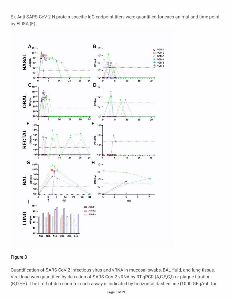
Viral RNA (vRNA) was puri�ed from whole blood, oral, nasal and rectal mucosa, and BAL �uid from allcollection days, as well as from lung tissue harvested at necropsy. As we previously reported [10], we wereunable to detect SARS-CoV-2 vRNA in whole blood by RT-qPCR, nor were we able to recover infectiousvirus in the plasma fraction by plaque assay, con�rming a lack of either cell-associated or freely-circulating virus in the peripheral blood. SARS-CoV-2 vRNA and infectious virus was detected in the nasalmucosa from all animals as early as 2 dpi, with vRNA persisting in a single animal up to 15 dpi (Figure

Page 8/19
3A, B). Likewise, vRNA was detected in oral swabs from all animals beginning 2-3 dpi before falling belowthe limit of detection by 7 dpi, while low quantities of infectious virus (1-2 log10 PFU/mL) were onlyisolated from three animals (AGM-4, AGM-5, and AGM-6) (Figure 3C, D). Remarkably, vRNA wastransiently shed from the lower gastrointestinal tract up to 28 dpi (AGM-4 and AGM-6), althoughinfectious virus could only be recovered from the rectal swab of a single animal (AGM-3) 4-5 dpi (Figure3E, F). vRNA was detected in BAL �uid from 4/6 animals 3 dpi and up to 7 dpi in all three animals heldpast 5 dpi, while infectious virus was recovered from 3/6 animals (Figure 3G, H). Detectable quantities ofvRNA were absent from lungs harvested during necropsy of AGMs euthanized 34 dpi, while 6-9 log10
GEq/g were detected from all three animals euthanized at 5 dpi (Figure 3I).
Gross pathology, histopathology, and immunohistochemistry
Necropsy was performed on all animals following euthanasia, and lungs were collected for grossexamination and histopathological analysis. Consistent with our previous study utilizing a combined i.n.and i.t. inoculation route [10], all AGMs displayed varying degrees of pulmonary consolidation withhyperemia and hemorrhage, characterized by depressed and patchy dark red to light pink regions (Fig. 4,arrows). In all AGMs, the most severe lesions were located in the dorsal aspects of the lower lung lobes. Aboard-certi�ed veterinary pathologist approximated lesion severity for each lung lobe (Supp Table 2). AllAGMs at 5 dpi also had segmentally �accid and gas distention of small intestines. There were no othersigni�cant gross lesions.
Histologically, all three AGMs euthanized at 5 dpi developed mild multifocal neutrophilicbronchointerstitial pneumonia (Figure 5A-E, O). Histologic features include acute in�ammation centeredwithin the airways of terminal bronchioles with occasional �ooding of adjacent alveolar spaces withneutrophils, macrophages, �brin, edema, hemorrhage, mucous and rarely multinucleated giant cells (5A,B). In lesser-affected regions alveolar septate were expanded with mixed in�ammatory cells and alveolarspaces contain increased numbers of alveolar macrophages with scattered red blood cells. Ulcerativetracheobronchitis was also present in all three AGMs and characterized by multifocal epithelial erosionassociated with underlying hemorrhage, �brin accumulation and in�ltrating acute in�ammation.Polymerized �brin, highlighted by IHC, colocalized with acute in�ammation within the bronchial lumen,alveolar spaces, alveolar walls and ulcerated regions of the trachea and bronchus (Figure 5C). Fibrin wasalso present within medium and small caliber vessels but was not associated with an obvious adherentthrombus. Trichrome stain of representative lung sections identi�ed modest collagen deposition withinmultifocal regions of alveolar septae (Figure 5D). IHC for SARS-CoV-2 antigen was positive in all threeAGMs associated with pulmonary lesions. Positive IHC labeling was noted diffusely within the cytoplasmof respiratory epithelium of the bronchus (Figure 5O) and less in type I and type II pneumocytes (Figure5E).
Histologically, all three AGMs euthanized at 34 dpi developed moderate multifocal chronic interstitialpneumonia Figure (5F-J). Histologic features include expansion of alveolar septae with macrophages,lymphocytes, and very rarely neutrophils (Figure 5F, G). Wispy, pale eosinophilic, acellular material also

Page 9/19
multifocally expanded the alveolar walls and stained as immature collagen with trichrome staining(Figure 5I). Polymerized �brin was present within medium and small caliber vessels but was notassociated with an obvious adherent thrombus (Figure 5H). No immunolabeling for SARS-CoV-2 wasnoted with IHC in any of the examined tissue sections from this 34 dpi cohort (Figure 5J).
DiscussionWe previously reported the development of the AGM as a promising animal model of human COVID-19[10]. Other studies have subsequently reported similar �ndings [11,12]. The focus of the current studywas to assess a more natural route of human exposure, speci�cally an exposure mimicking an infectionresulting from mucosal exposure to infectious droplets expelled from close quarter exposure to a sneeze,cough, or even speech in order to begin characterization of lung pathology in the early convalescencephase of COVID-19. The disease resulting from the i.n. MAD challenge was largely re�ective of thatobserved with the combination of the i.t. and i.n. routes except it appeared to be somewhat milder interms of length of any fever, less severe signs of pneumonia as evidenced by reduced alveolar �ooding,and a lower prevalence of SARS-CoV-2 infection. [10]. However, the MAD-infected AGMs still developedvirus-induced pneumonia and viral shedding was detected into the early convalescence period. While itappears that inclusion of direct i.t. instillation of SARS-CoV-2 as an exposure route may result in a moresevere disease in AGMs, it is also possible that animal to animal variability may have contributed to themodest difference between the studies. SARS-CoV-2 infection of humans results in a wide spectrum ofdisease ranging from asymptomatic to severe fatal disease so it is not unexpected that there could bevariability among AGMs as well. While the current study employed female AGMs because of animalavailability at the time the work was initiated gender did not affect the outcome when compared tosimilar studies [10-12].
Coagulation dysfunction is a consistent observation in human COVID-19 and has been associated withdisease severity [18-22]. Here, we performed a limited analysis of blood clotting times (PT and aPTT)and circulating �brinogen levels to begin to characterize the coagulopathy in SARS-CoV-2-infected AGMs.Transient increases in aPTT and in circulating �brinogen levels were observed during the acute phase ofinfection. Increases in PT and/or aPTT have been linked to severe human COVID-19 cases in some butnot all studies [18-22]. However, nearly all severe COVID-19 cases have been associated with high levelsof �brinogen [20-22].
Our �ndings regarding lung injury in the three AGMs that were euthanized at 34 dpi during earlyconvalescence are consistent with the limited human COVID-19 studies that have been reported so far. For example, a recent study of �fty-seven COVID-19 patients in China was completed during the earlyconvalescence phase, approximately 30 days after discharge [23]. The study included 40 non-severecases and 17 severe cases. Thirty-one patients (54.3%) had abnormal CT �ndings while abnormalitieswere detected in the pulmonary function tests in 43 (75.4%) of the patients. In a second human study, 21patients recovering from COVID-19 (without severe respiratory distress during the disease course), hadlung abnormalities visible on chest CT at 10 days after initial onset of symptoms [24]. While other studies

Page 10/19
suggest that some of the abnormalities may be resolved over time [25, 26] more research needs to beconducted in this area.
Regarding histopathology, human data is particularly sparse. One small study performed thoracoscopieswith blebs resection and pleurectomies on performed on the 16th and 23rd days from symptoms onset oftwo patients [27]. Despite well-known pulmonary damages induced during the acute phase of COVID-19,the late-phase gross and histological changes include nonspeci�c chronic reparative lesions, similarly towhat we have described in the AGMs at 34 dpi. Grossly in the human study, there was non-speci�cdiffuse pulmonary congestion, edema and hemorrhagic necrosis. Histologically, the main lesions werefocused on alveolar damage with mildly thickened alveolar interstitial tissues with �brosis andmononuclear cellular in�ltration (lymphocytes, plasma cells and multinucleate giant cells). Intravascularhemorrhagic thrombosis was also noted in these specimens.
In summary, we have expanded on our previous development of the combined i.n. and i.t. inoculationmodel of SARS-CoV-2 in AGMs. Importantly, while AGMs challenged with SARS-CoV-2 via the LMA MADexhibited apparently milder clinical illness and disease, hallmark features from our previous study werestill apparent, notably the development of viral pneumonia during the acute phase. The AGM COVID-19model should be useful in future studies to assess disease and develop interventions that improverecovery.
AbbreviationsaPTT: activated partial thromboplastin time; African green monkey (AGM); BAL: bronchoalveolar lavage;COVID-19: Coronavirus Disease 2019; IHC: immunohistochemistry; i.n.: intranasal; i.t.: intratracheal; MAD:Mucosal Atomization Device; PFU: plaque forming unit; prothrombin times (PT); SARS-CoV-2: severeacute respiratory syndrome coronavirus 2
DeclarationsAcknowledgments
The authors would like to thank the UTMB Animal Resource Center for veterinary support for surgery toimplant temperature data loggers and husbandry support of laboratory animals and Dr. Kevin Melody forassistance with animal studies. The virus used in this publication was kindly provided by the EuropeanVirus Archive goes Global (EVAg) project that has received funding from the European Union’s Horizon2020 research and innovation program under grant agreement No 653316.
Author contributions
RWC and TWG conceived and designed the study. DJD, JBG, and TWG performed the SARS-CoV-2challenge experiments. RWC, DJD, CW, JBG, and TWG performed animal procedures and clinicalobservations. KNA and VB performed the clinical pathology assays. VB performed the SARS-CoV-2

Page 11/19
infectivity assays. KNA optimized and performed the PCR. NSD optimized and performed theimmunohistochemistry. CW performed ELISAs. KAF performed necropsies and analysis of the grosspathology, histopathology, and immunohistochemistry. All authors analyzed the clinical pathology,virology, and immunology data. RWC, ANP, KAF, and TWG, wrote the paper. All authors had access to allof the data and approved the �nal version of the manuscript.
Funding
This study was supported by funds from the Department of Microbiology and Immunology, University ofTexas Medical Branch at Galveston, Galveston, TX to TWG. Operations support of the Galveston NationalLaboratory was supported by NIAID/NIH grant UC7AI094660.
Availability of data and materials
The data supporting the conclusions of this article are included within the article.
Ethics approval and consent to participate
All animal studies were approved by the University of Texas Medical Branch (UTMB) Institutional AnimalCare and Use Committee and adhere to the NIH Guide for the Care and Use of Laboratory Animals.
Consent for publication
Not applicable.
Competing interests
The authors declare no competing interests.
References1. World Health Organization. DRAFT landscape of COVID-19 candidate vaccines – 21 July 2020.
Available from: �le:///C:/Users/p/Downloads/novel-coronavirus-landscape-covid-19cf1952c105464714aaaf8c7cd5c5cc8b.pdf.
2. Milken Institute. COVID-19 treatment and vaccine tracker. July 23, 2020. Available from:https://covid-19tracker.milkeninstitute.org/.
3. Chan JF, Zhang AJ, Yuan S, Poon VK, Chan CC, Lee AC, Chan WM, Fan Z, Tsoi HW, Wen L, et al.Simulation of the clinical and pathological manifestations of Coronavirus Disease 2019 (COVID-19)in golden Syrian hamster model: implications for disease pathogenesis and transmissibility. ClinInfect Dis. 2020 Mar 26:ciaa325.
4. Sia SF, Yan LM, Chin AWH, Fung K, Choy KT, Wong AYL, Kaewpreedee P, Perera RAPM, Poon LLM,Nicholls JM, et al. Pathogenesis and transmission of SARS-CoV-2 in golden hamsters. Nature. 2020May 14.

Page 12/19
5. Richard M, Kok A, de Meulder D, Bestebroer TM, Lamers MM, Okba NMA, Fentener van Vlissingen M,Rockx B, Haagmans BL, Koopmans MPG, et al. SARS-CoV-2 is transmitted via contact and via the airbetween ferrets. Nat Commun. 2020;11:3496.
�. Yu P, Qi F, Xu Y, Li F, Liu P, Liu J, Bao L, Deng W, Gao H, Xiang Z, et al. Age-related rhesus macaquemodels of COVID-19. Animal Model Exp Med. 2020;3:93-97.
7. Munster VJ, Feldmann F, Williamson BN, van Doremalen N, Pérez-Pérez L, Schulz J, Meade-White K,Okumura A, Callison J, Brumbaugh B, et al. Respiratory disease in rhesus macaques inoculated withSARS-CoV-2. Nature. 2020 May 12.
�. Rockx B, Kuiken T, Herfst S, Bestebroer T, Lamers MM, Oude Munnink BB, de Meulder D, vanAmerongen G, van den Brand J, Okba NMA, et al. Comparative pathogenesis of COVID-19, MERS, andSARS in a nonhuman primate model. Science. 2020;368:1012-1015.
9. Lu S, Zhao Y, Yu W, Yang Y, Gao J, Wang J, Kuang D, Yang M, Yang J, Ma C, et al. Comparison ofSARS-CoV-2 infections among 3 species of non-human primates. bioRxiv. 2020 April8:2020.04.08.031807v2.
10. Woolsey C, Borisevich V, Prasad AN, Agans KN, Deer DJ, Dobias NS, Heymann JC, Foster SL, LevineCB, Medina L, et al. Establishment of an African green monkey model for COVID-19. bioRxiv. 2020May 17:2020.05.17.100289.
11. Blair R, Vaccari M, Doyle-Meyers LA, Roy CJ, Russell-Lodrigue K, Fahlberg M, Monjure CJ,Bedding�eld B, Plante KS, Plante JA, et al. ARDS and Cytokine Storm in SARS-CoV-2 InfectedCaribbean Vervets. bioRxiv. 2020 June 18:2020.06.18.157933.
12. Hartman AL, Nambulli S, McMillen CM, White AG, Tilston-Lunel NL, Albe JR, Cottle E, Dunn M, FryeLJ, Gilliland TH, et al. SARS-CoV-2 infection of African green monkeys results in mild respiratorydisease discernible by PET/CT imaging and prolonged shedding of infectious virus from bothrespiratory and gastrointestinal tracts. bioRxiv. 2020 June 20: 2020.06.20.137687.
13. Mire CE, Satter�eld BA, Geisbert JB, Agans KN, Borisevich V, Yan L, Chan YP, Cross RW, Fenton KA,Broder CC, Geisbert TW. Pathogenic Differences between Nipah Virus Bangladesh and MalaysiaStrains in Primates: Implications for Antibody Therapy. Sci Rep. 2016;6:30916.
14. Geisbert JB, Borisevich V, Prasad AN, Agans KN, Foster SL, Deer DJ, Cross RW, Mire CE, Geisbert TW,Fenton KA. An Intranasal Exposure Model of Lethal Nipah Virus Infection in African Green Monkeys.J Infect Dis. 2020;221(Supplement_4):S414-S418.
15. Xie X, Li Y, Sun H, Liu L. Exhaled droplets due to talking and coughing. J R Soc Interface. 2009;6(Suppl 6):S703-14.
1�. Capobianchi MR, Rueca M, Messina F, Giombini E, Carletti F, Colavita F, Castilletti C, Lalle E, Bordi L,Vairo F, et al. Molecular characterization of SARS-CoV-2 from the �rst case of COVID-19 in Italy. ClinMicrobiol Infect. 2020;26:954-956.
17. Centers for Disease Control and Prevention. Research Use Only 2019-Novel Coronavirus (2019-nCoV)Real-time RT-PCR Primers and Probes. 2020. Available from: https://www.cdc.gov/coronavirus/2019-
Page 13/19
ncov/lab/rt-pcr-panel-primer-probes.html.
1�. Chen L, Yu J, He W, Chen L, Yuan G, Dong F, Chen W, Cao Y, Yang J, Cai L, et al. Risk factors for deathin 1859 subjects with COVID-19. Leukemia. 2020 Jun 16:1-11.
19. Bonetti G, Manelli F, Patroni A, Bettinardi A, Borrelli G, Fiordalisi G, Marino A, Menol� A, Saggini S,Volpi R, et al. Laboratory predictors of death from coronavirus disease 2019 (COVID-19) in the areaof Valcamonica, Italy. Clin Chem Lab Med. 2020;58:1100-1105.
20. Di Minno MND, Calcaterra I, Lupoli R, Storino A, Spedicato GA, Maniscalco M, Di Minno A, AmbrosinoP. Hemostatic Changes in Patients with COVID-19: A Meta-Analysis with Meta-Regressions. J ClinMed. 2020;9:E2244.
21. Lin J, Yan H, Chen H, He C, Lin C, He H, Zhang S, Shi S, Lin K. COVID-19 and Coagulation Dysfunctionin Adults: A Systematic Review and Meta-analysis. J Med Virol. 2020 Jul 24.
22. Zhu J, Pang J, Ji P, Zhong Z, Li H, Li B, Zhang J, Lu J. Coagulation dysfunction is associated withseverity of COVID-19: a meta-analysis. J Med Virol. 2020 Jul 24.
23. Huang Y, Tan C, Wu J, Chen M, Wang Z, Luo L, Zhou X, Liu X, Huang X, Yuan S, et al. Impact ofcoronavirus disease 2019 on pulmonary function in early convalescence phase. Respir Res.2020;21:163.
24. Pan F, Ye T, Sun P, Gui S, Liang B, Li L, Zheng D, Wang J, Hesketh R, Yang L, Zheng C. Time Course ofLung Changes at Chest CT during Recovery from Coronavirus Disease 2019 (COVID-19). Radiology2020; 295:715-721.
25. Liu C, Ye L, Xia R, Zheng X, Yuan C, Wang Z, Lin R, Shi D, Gao Y, Yao J, et al. Chest CT and ClinicalFollow-up of Discharged Patients with COVID-19 in Wenzhou City, Zhejiang, China. Ann Am ThoracSoc. 2020 Jul 21.
2�. Wang Y, Dong C, Hu Y, Li C, Ren Q, Zhang X, Shi H, Zhou M. Temporal Changes of CT Findings in 90Patients with COVID-19 Pneumonia: A Longitudinal Study. Radiology 2020; 296:E55-E64.
27. Aiol� A, Bruni B, Biraghi T, Montisci A, Miceli A, Baronio B, Khor D, Cirri S, Donatelli F, Clemente C,Bona D. Late histological �ndings in symptomatic COVID-19 patients: A case report. Medicine(Baltimore). 2020 Jul;99:e21046.
Figures

Page 14/19
Figure 1
Hematological features of SARS-CoV-2 infection in AGMs. Blood gas (A, B), selected leukocytepopulations (C-G), and coagulation assays (H-J) are shown. For parameters where fold change is used,fold change was determined by baseline (0 dpi) subtraction of each time point for each animal.Statistical signi�cance was determined in Graphpad Prism 8.4.3 by mixed-effects analysis with theGeisser-Greenhouse correction without the assumption of sphericity, with multiple comparisons made

Page 15/19
using Dunnett’s post-hoc test and all comparisons made to baseline values (0 dpi). Asterisks denotesigni�cance: * = p ≤ 0.05, ** = p ≤ 0.01, *** = p ≤ 0.001. Two-tailed p-values were computed for allcomparisons.
Figure 2
Serum neutralization and binding antibody titers in SARS-CoV-2 infected AGMs. Total anti-SARS-CoV-2serum neutralization activity was determined for each animal by PRNT50 at the indicated time points (A-
Page 16/19
E). Anti-SARS-CoV-2 N protein speci�c IgG endpoint titers were quanti�ed for each animal and time pointby ELISA (F).
Figure 3
Quanti�cation of SARS-CoV-2 infectious virus and vRNA in mucosal swabs, BAL �uid, and lung tissue.Viral load was quanti�ed by detection of SARS-CoV-2 vRNA by RT-qPCR (A,C,E,G,I) or plaque titration(B,D,F,H). The limit of detection for each assay is indicated by horizontal dashed line (1000 GEq/mL for

Page 17/19
RT-qPCR, 25 PFU/mL for plaque titration). For both assays, data shown is the mean of two technicalreplicates of the same biological sample. Arrow in (G) indicates day of challenge. For panel I, RUL: rightupper lung; RML: right middle lung; RLL: right lower lung; LUL: left upper lung; LML: left middle lung; LLL:left lower lung.
Figure 4

Page 18/19
Gross lung pathology in AGMs infected with SARS-CoV-2. Dorsal view of lungs from AGM-1 (A), AGM-2(B) and AGM-3(C) euthanized at 5 dpi with SARS-CoV-2 exhibiting mild to moderate locally extensivepulmonary consolidation with hyperemia and hemorrhage. Dorsal view of lungs from AGM-4 (D), AGM-5(E) and AGM-6 (F) euthanized at 34 dpi with SARS-CoV-2 exhibiting mild to marked locally extensivepulmonary consolidation with hyperemia and hemorrhage. Dorsal view of control lungs with nosigni�cant lesions from SARS-CoV-2 negative AGM (G).
Figure 5

Page 19/19
Comparative pulmonary histologic lesions in AGMs infected with SARS-CoV-2. Representative tissues ofAGM from 5 dpi (A-E & O) and 34 dpi (F-J). SARS-CoV-2 naïve tissues from an AGM (K-N). H&E staining atlow magni�cation (20x) (A, F, & K) and higher magni�cation (40x) (B, G, L, and F inset) of pulmonaryalveolar septae and alveolar spaces. Moderate neutrophilic bronchiolitis and alveolitis and mildinterstitial pneumonia with congestion (A & B) Moderate lymphohistocytic interstitial pneumonia withcongestion, mild alveolar wall �brosis (F & G) and moderate perivascular lymphocytic cuffs (F inset). Nosigni�cant lesions (K & L). IHC for anti-�brin antigen (red) (C, H & M). Alveolar spaces are partially tocompletely �ooded with �brin (C) Minimal intravascular �brin labeling (H) and no signi�cant �brinimmunolabeling (M). Trichrome special stain for collagen (blue) (D, I & N). Minimal to mild alveolar wallcollagen deposition (D) moderate alveolar wall collagen deposition (I) and minimal collagen staining ofalveolar wall basement membranes (N). IHC labeling for anti-SARS-CoV2 antigen (red) (E, J & O). IHCpositive type I pneumoncytes (black arrows) and type II pneumoncytes (white arrow) localized withalveolar in�ammation (E), No immonolabeling (J) and IHC positive labeling of respiratory epithelium ofthe bronchus (O). Images captured at 20x (M, D, I, N, & J) and 40x (C, H, E, & O).
Supplementary Files
This is a list of supplementary �les associated with this preprint. Click to download.
SUPPLEMENTARYFIGURE1TEMPERATURE.tif
SupplemenaryTable1.docx
SupplementaryTable2Revised.docx