contribution of propriospinal neurons to recovery of hand ... · tetanus neurotoxin. applying the...
TRANSCRIPT
Contribution of propriospinal neurons to recovery ofhand dexterity after corticospinal tract lesionsin monkeysTakamichi Tohyamaa,b,1,2, Masaharu Kinoshitac, Kenta Kobayashid,e, Kaoru Isaa, Dai Watanabef, Kazuto Kobayashig,Meigen Liub, and Tadashi Isaa,e,h,3
aDepartment of Developmental Physiology, National Institute for Physiological Sciences, Okazaki 444-8585, Japan; bDepartment of Rehabilitation Medicine,Graduate School of Medicine, Keio University, Tokyo 160-8582, Japan; cDepartment of Physiology, Hirosaki University Graduate School of Medicine,Hirosaki 036-8562, Japan; dSection of Viral Vector Development, National Institute for Physiological Sciences, Okazaki 444-8585, Japan; eThe GraduateUniversity for Advanced Studies (SOKENDAI), Hayama 240-0193, Japan; fDepartment of Molecular and System Biology, Graduate School of Biostudies,Kyoto University, Kyoto 606-8501, Japan; gDepartment of Molecular Genetics, Institute of Biomedical Sciences, Fukushima Medical University School ofMedicine, Fukushima 960-1295, Japan; and hDepartment of Neuroscience, Graduate School of Medicine and Faculty of Medicine, Kyoto University, Kyoto606-8501, Japan
Edited by Peter L. Strick, University of Pittsburgh, Pittsburgh, PA, and approved December 5, 2016 (received for review July 12, 2016)
The direct cortico-motoneuronal connection is believed to beessential for the control of dexterous hand movements, such asprecision grip in primates. It was reported, however, that evenafter lesion of the corticospinal tract (CST) at the C4–C5 segment,precision grip largely recovered within 1–3 mo, suggesting thatthe recovery depends on transmission through intercalated neu-rons rostral to the lesion, such as the propriospinal neurons (PNs)in the midcervical segments. To obtain direct evidence for thecontribution of PNs to recovery after CST lesion, we applied apathway-selective and reversible blocking method using doubleviral vectors to the PNs in six monkeys after CST lesions at C4–C5. In four monkeys that showed nearly full or partial recovery,transient blockade of PN transmission after recovery caused partialimpairment of precision grip. In the other two monkeys, CST le-sions were made under continuous blockade of PN transmissionthat outlasted the entire period of postoperative observation (3–4.5 mo). In these monkeys, precision grip recovery was notachieved. These results provide evidence for causal contributionof the PNs to recovery of hand dexterity after CST lesions; PNtransmission is necessary for promoting the initial stage recovery;however, their contribution is only partial once the recoveryis achieved.
spinal cord injury | plasticity | neural circuit | nonhuman primates |viral vector
As a potential treatment for brain and spinal cord injury, re-generation of the injured axons has been attempted in animal
models, often using rodents with corticospinal tract (CST) lesions.Regeneration of the injured CST fibers was facilitated by treatmentssuch as peripheral nerve graft (1), application of an antibody againstneurite growth inhibitors IN-1 (2), combined application of theantibody and neurotrophic factor NT-3 (3), and transplantation ofneural stem cells derived from induced pluripotent stem cells (4). Amore recent study showed that regenerated CST axons increasedthe connection to the spinal motoneurons (MNs) after spinal cordinjury in monkeys (5). However, in these studies, the causal con-tribution of such regenerated fibers to the functional recovery wasnot directly demonstrated. Therefore, recent debates address thequestion whether these therapies should target repairing theinjured CST fibers and/or facilitating compensation by indirectcortico-motoneuronal (CM) connections via other descendingmotor pathways (6–9).Traditionally, the neural control of dexterous hand move-
ments in higher primates has primarily been associated withdevelopment of the direct pathway from the motor cortex toMNs, known as the CM pathway (10–12). Lesion of the CST atthe brainstem level in nonhuman primates caused near-permanentloss of dexterous hand movements (13). The authors also argued for
partial compensation of grasping movements by the brainstem-mediated descending pathways such as the rubrospinal tract (14).More recent studies on the neural basis of functional recovery fol-lowing CST lesions revealed that a variety of plastic changes inneural circuits occurred in the supraspinal structures including themotor and premotor cortices (15), and in the connectivity from thenucleus accumbens to motor cortex (16, 17). At the more caudallevel, sprouting of the midline-crossing CST fibers was revealed inthe lower cervical segment of monkeys with hemisected spinal cords(18). In addition, the “indirect” CM pathways via interneurons suchas the propriospinal neurons (PNs), which are located in the mid-cervical segments and project to hand/arm MNs in the lower cer-vical segments, and/or reticulospinal neurons (RSNs) in thepontomedullary reticular formation might also be involved in me-diating cortical commands to MNs of forelimb muscles in animalmodels with CST lesions (19–21). To demonstrate which pathwayscausally contribute to recovery after damage to the CST, selectiveand reversible manipulation of particular pathways is needed.
Significance
There are different views about the targets of regenerativetherapies to induce functional recovery in patients with motorparalysis following brain and spinal cord injury: whether weshould aim at repairing the injured corticospinal tract or atfacilitating compensation by other descending motor path-ways. To help answer this question, we used double viralvectors to reversibly and selectively block the propriospinalneurons (PNs), one of the major intercalated neurons mediat-ing cortical commands to motoneurons, in monkeys with par-tial spinal cord injury. We demonstrated causal roles of the PN-mediated pathway in promoting recovery of hand dexterityafter the lesion. Thus, targeting the PNs might lead to de-veloping effective treatment to facilitate recovery after spinalcord injury.
Author contributions: T.T., M.K., M.L., and T.I. designed research; T.T., M.K., and T.I.performed research; T.T. and K.I. performed histology; Kenta Kobayashi, D.W., and Ka-zuto Kobayashi provided viral vectors; T.T. analyzed data; and T.T. and T.I. wrotethe paper.
The authors declare no conflict of interest.
This article is a PNAS Direct Submission.1Present address: Division of Cerebral Integration, Department of System Neuroscience,National Institute for Physiological Sciences, Okazaki 444-8585, Japan.
2Present address: Department of Rehabilitation Medicine, Keio University School of Med-icine, Tokyo 160-8582, Japan.
3To whom correspondence should be addressed. Email: [email protected].
This article contains supporting information online at www.pnas.org/lookup/suppl/doi:10.1073/pnas.1610787114/-/DCSupplemental.
604–609 | PNAS | January 17, 2017 | vol. 114 | no. 3 www.pnas.org/cgi/doi/10.1073/pnas.1610787114
Historically, much less attention has been paid to the “indirect”CM pathways in the control of dexterous hand movements comparedwith the direct CM pathway, because they were supposed not to beinvolved in the control of fine digit movements. During the last de-cade, however, lesion and electrophysiological studies in nonhumanprimates suggested that the pathway through the PNs might be in-volved in the control of dexterous hand movements in the intact state(21, 22). However, this suggestion was based on the indirect evidenceobtained by comparing the behavioral effects caused by transection ofthe CST at different rostrocaudal levels. Recently, however, a tech-nique was developed that can selectively and reversibly controltransmission via neurons comprising a particular pathway using twodifferent kinds of viral vectors and doxycycline (Dox)-inducibletetanus neurotoxin. Applying the technique to the PNs in intactmonkeys revealed that the PN-mediated pathway could carry thecommand for dexterous hand movements in the intact state (23).However, contribution of the PN-mediated pathway to recoveryafter lesion of the direct CM pathway in the monkey remainsunclear. In the present study, to examine whether the PNs causallycontribute to recovery of dexterous hand movements after damageto the CST, we applied the pathway-selective and reversibleblocking method to the PNs in monkeys after lesioning the CSTat C4–C5.
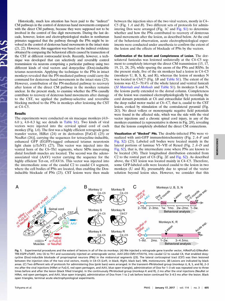
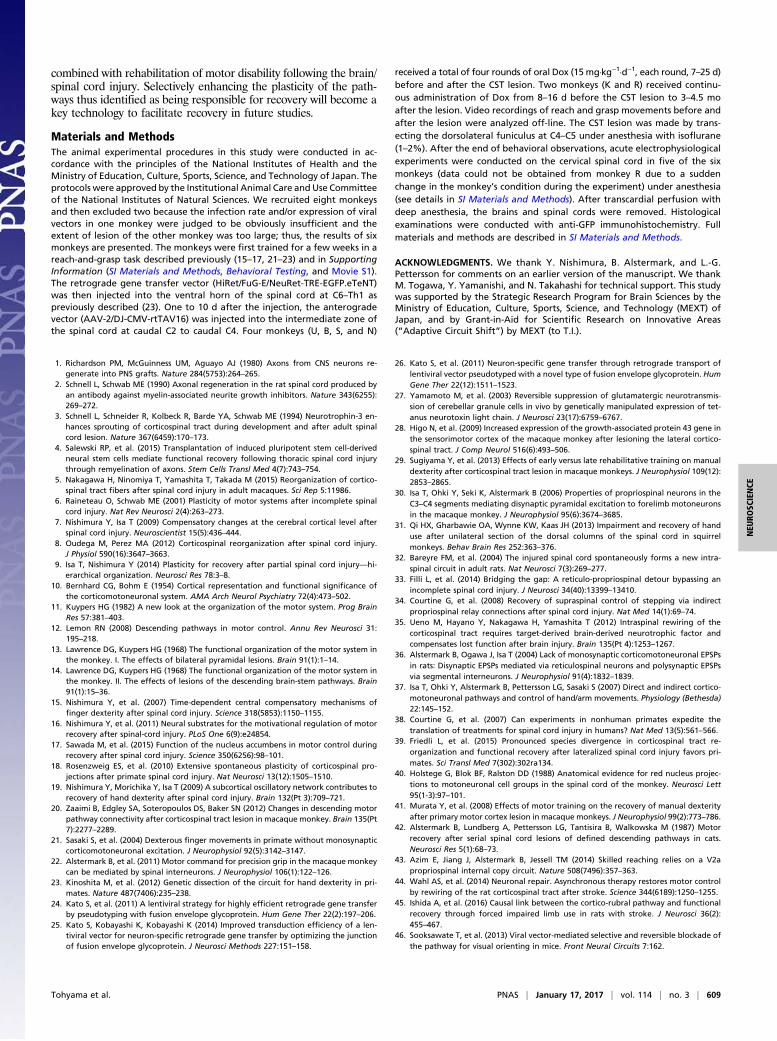
ResultsThe experiments were conducted on six macaque monkeys (4.0–6.0 y, 4.0–8.3 kg; see details in Table S1). Two kinds of viralvectors were injected into the cervical spinal cord of eachmonkey (Fig. 1A). The first was a highly efficient retrograde genetransfer vector, HiRet (24) or its derivatives [FuG-E (25) orNeuRet (26)], carrying the sequences for tetracycline-inducible,enhanced GFP (EGFP)-tagged enhanced tetanus neurotoxinlight chain (eTeNT) (27). This vector was injected into theventral horn of the C6–Th1 segments, where MNs innervatingdistal forelimb muscles are located. The second was the adeno-associated viral (AAV) vector carrying the sequence for thehighly efficient Tet-on, rtTAV16. This vector was injected intothe intermediate zone of the caudal C2 to caudal C4 segment,where the cell bodies of PNs are located, thus enabling the Dox-inducible blockade of PNs (23). CST lesions were then made
between the injection sites of the two viral vectors, mostly in C4–C5 (Fig. 1 A and B). Two different sets of protocols for admin-istering Dox were arranged (Fig. 1C and Fig. S1) to determinewhether and how the PNs contributed to recovery of dexteroushand movements after the lesion, as described below. At the endof the behavioral observation, acute electrophysiological exper-iments were conducted under anesthesia to confirm the extent ofthe lesion and the effects of blockade of PNs by the vectors.
Confirmation of the Extent and Completeness of Lesion. The dor-solateral funiculus was lesioned unilaterally at the C4–C5 seg-ment to completely interrupt the direct CM transmission (15, 17,19, 21, 28, 29), while sparing the axons of the PNs and RSNs. Inthe present study, five of the six monkeys had a lesion in C4–C5(monkeys U, B, S, K, and R), whereas the lesion of monkey Nwas located in C6/C7 (Fig. 1B and Table S1). The extent of thelesions was 42.5−70.4% of the whole lateral and ventral funiculi(SI Materials and Methods and Table S1). In monkeys S and N,the lesions partly extended to the dorsal column. Completenessof the lesion was examined electrophysiologically by recording thecord dorsum potentials at C6 and extracellular field potentials inthe deep radial motor nuclei at C6−C7, that is, caudal to the CSTlesion, evoked by stimulation of the contralateral pyramid (Fig.2G). No direct volleys or monosynaptic negative field potentialswere found in the affected side, which was the side with the viralvector injections and a chronic spinal cord injury, in any of themonkeys examined (a representative is shown in Fig. 2H), revealingthat the lesions completely abolished the direct CM connections.
Visualization of “Blocked” PNs. The double-infected PNs were vi-sualized with anti-GFP immunohistochemistry (Fig. 2 A–F andFig. S2) (23). Labeled cell bodies were located mainly in thelateral portions of laminae VI−VII of Rexed (Fig. 2 A–D andFig. S2), that is, the intermediate zone where PNs are known tobe located (30). Their longitudinal distribution extended fromC2 to the rostral part of C6 (Fig. 2E and Fig. S2). As describedabove, the CST lesion was located mainly in C4–C5. Therefore,some GFP-labeled cells were located caudal to the lesion in twomonkeys (U and R), presumably due to spread of the vectorsolution beyond lesion sites. However, we consider that this
C6/C7
Monkey UA B
C4–C5
C4–C5
C4–C5
C Monkeys U, B, S, N
Monkeys K, R
Acute exp
.
2mo.1–8d. 1–2w. 3–4.5mo.
AAV
NeuRet or
HiRet
Dox
1mo.
Dox
HiRet or
FuG-E
2–3mo.2–10d. 2w–2mo.1w.
Dox2–3 ro
unds
1–3w.
DoxLesio
n
Behavioral analysis
Behavioral analysis
Lesion
Acute exp
.
C4–C5
AAV
AAV
Monkey B
Monkey S Monkey N
Monkey K Monkey R
Dox1–2 ro
unds
C4–C5
CST lesio
n
Spinal cordAAV-2/DJ-CMV-rtT
AV16
HiRet
or FuG-E
or NeuRet
TRE-EGFP.eTeNT
C2–C4
C6–Th1
MN
PNs
Muscles
CST
HiRetor FuG-E
or NeuRet
C4–C5
Fig. 1. Experimental procedures and the extent of lesions in all of the six monkeys. (A) We injected a retrograde gene transfer vector, HiRet/FuG-E/NeuRet-TRE-EGFP.eTeNT, into C6 to Th1 and successively injected an anterograde vector, AAV-2/DJ-CMV-rtTAV16, into caudal C2 to caudal C4, that enabled doxy-cycline (Dox)-inducible blockade of propriospinal neurons (PNs) in the midcervical segments (23). The lateral corticospinal tract (CST) was then lesionedbetween the injection sites of the two viral vectors, mostly in C4–C5 (Left, in black; Right, black bar). MN, motoneurons. (B) Lesions are indicated by blackareas. (C) Two different sets of protocols for administering Dox (pink bars) were arranged. In the transient–PN-blocked group (monkeys U, B, S, and N), 2–3mo after the viral injections (HiRet or FuG-E, red open pentagon, and AAV, blue open triangle), administration of Dox for 1–3 wk was repeated one to threetimes before and after the lesion (black filled triangle). In the continuously PN-blocked group (monkeys K and R), 2 mo after the viral injections (NeuRet orHiRet, red open pentagon, and AAV, blue open triangle), administration of Dox from 1 to 2 wk before lesion continued for 3–4.5 mo after the lesion. Blackopen triangles, terminal acute electrophysiological experiments.
Tohyama et al. PNAS | January 17, 2017 | vol. 114 | no. 3 | 605
NEU
ROSC
IENCE
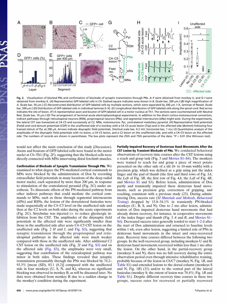
would not affect the main conclusion of this study (Discussion).Axons and boutons of GFP-labeled cells were found in the motornuclei at C6–Th1 (Fig. 2F), suggesting that the blocked cells weredirectly connected with MNs innervating distal forelimb muscles.
Confirmation of Blockade of Synaptic Transmission Through PNs. Weexamined to what degree the PN-mediated cortical commands toMNs were blocked by the administration of Dox by recordingextracellular field potentials in many locations of the deep radialmotor nuclei, each separated by more than 200 μm, in responseto stimulation of the contralateral pyramid (Fig. 2G) under an-esthesia. To dissociate effects of the PN-mediated pathway fromother indirect pathways that could mediate the cortical com-mands to MNs, such as those through segmental interneurons(sINs) and RSNs, the lesions of the dorsolateral funiculus weremade sequentially at the C4–C5 level on the unaffected side andthen at the C2 levels on both sides during the acute experiments(Fig. 2G). Strychnine was injected i.v. to reduce glycinergic in-hibition from the CST. The amplitudes of the disynaptic fieldpotentials in the affected side were significantly smaller thanthose in the unaffected side after acute C4–C5 CST lesion on theunaffected side (Fig. 2 H and I, and Fig. S3), suggesting thatsynaptic transmission through the propriospinal and retic-ulospinal pathways in the affected side were much weakercompared with those in the unaffected side. After additional C2CST lesion on the unaffected side (Fig. 2I and Fig. S3) and onthe affected side (Fig. S3), the amplitudes were very small,suggesting that contribution of the reticulospinal pathway wasminor in both sides. These findings revealed that synaptictransmission presumably through the PNs was blocked by 74.2–93.3% [mean (SD), 83.1 (8.2)] compared with the unaffectedside in four monkeys (U, S, N, and K), whereas no significantblocking was observed in monkey B, as will be discussed later. Nodata were obtained from monkey R due to a sudden change inthe monkey’s condition during the experiment.
Partially Impaired Recovery of Dexterous Hand Movements After theCST Lesion by Transient Blockade of PNs. We conducted behavioralobservations of recovery time courses after the CST lesions usinga reach and grasp task (Fig. 3 and Movies S1–S4). The monkeyswere trained to reach for and grasp a piece of sweet potatopresented on the other side of a slit (8- to 10-mm width) with aprecision grip, which was defined as a grip using just the indexfinger and the pad of thumb (the first and third rows of Fig. 3A,the Left of Fig. 3B, the first row of Fig. 4A, the Left of Fig. 4B,and Movies S1 and S3). Before lesion, administration of Doxpartly and transiently impaired these dexterous hand move-ments, such as precision grip, correctness of gripping, andreaching, consistent with a previous study (23). During admin-istering Dox, success rate (SI Materials and Methods, BehavioralTesting) dropped by 15.8–34.1% in transiently PN-blockedmonkeys (U, B, S, and N). One to 2 mo after lesion, adminis-tration of Dox impaired dexterous hand movements that hadalready shown recovery, for instance, in cooperative movementsof the index finger and thumb (Fig. 3 A and B, and Movies S1–S4). Decreased success rates generally tended to start 1–2 d afterthe start of Dox administration and returned to baseline levelswithin 1 wk, even after lesion, suggesting a limited role of PNs indexterous hand movements in the intact and once-recoveredstate. Recovery time courses differed between the following twogroups. In the well-recovered group, including monkeys U and B,dexterous hand movements recovered within less than 1 mo afterthe lesion. On the other hand, in the partial-recovery group(monkeys S and N), there was no full recovery during the entireobservation period even through intensive rehabilitative training,probably because of the lesion in C6/C7 (monkey N, Fig. 1B, andTable S1) and extended lesions to the dorsal column (monkeys Sand N, Fig. 1B) (31) and/or to the ventral part of the lateralfuniculus (monkey S; the extent of lesion was 70.4%; Fig. 1B andTable S1). Despite different recovery time courses in these twogroups, success rates for recovered or partially recovered
-
+
-
+
-
-
+
+
MNs
RSN
C4–C5lesion
Field rec.
C2lesion
D
GBAC4
100
50
0I-II
I IV V VIVII VIII IX
: GFP+ cellCI-III
IVVVIVIIVIII
IXX
E F
Layer
H
Fiel
dC
DP
Una
ffect
ed s
ide
C4–
C5
acut
e le
sion
Th1
Affe
cted
sid
eC
4–C
5 le
sion
I
Am
plitu
de (m
V)
C4–C
5 (9
)
*
: Affected side
0.6
0.5
0.4
0.3
0.2
0.1
0
Pyr
PNs
sINs
Unaffected side
**
X0
20
40
60GFP+ cells per 400 μm
C2 C3 C4 C5 C6Segment
GFP+ cells (%)
CD
PFi
eld
No (1
4)
C2 (6
)C4
–C5
(4)
Lesio
n (n
)
Fig. 2. Visualization of blocked PNs and confirmation of blockade of synaptic transmission through PNs. A–F were obtained from monkey U, and G–I wereobtained from monkey K. (A) Representative GFP-labeled cells in C4. Dashed square indicates area shown in B. (Scale bar, 200 μm.) (B) High magnification ofA. (Scale bar, 50 μm.) (C) Reconstructed distribution of GFP-labeled cells by multiple sections, which were separated by 200 μm. I–X, laminae of Rexed. (Scalebar, 500 μm.) (D) Distribution of GFP-labeled cells in individual laminae (I–X). (E) Longitudinal distribution of GFP-labeled cells along the spinal cord. Red arrowindicates the site of lesion. (F) A representative axon and bouton of GFP-labeled cell in a motor nucleus at Th1. The sections were counterstained with NeutralRed. (Scale bar, 10 μm.) (G) The arrangement of terminal acute electrophysiological experiments. In addition to the direct cortico-motoneuronal connection,indirect pathways through reticulospinal neurons (RSN), propriospinal neurons (PNs), and segmental interneurons (sINs) might exist. During the experiments,the lateral CST was transected at C4–C5 and successively at C2. MNs, motoneurons. Pyr, contralateral medullary pyramid. (H) Representative field potentials(Field) and cord dorsum potentials (CDP) in the unaffected side of a monkey with a C4–C5 acute lesion (Top) and in the affected side (Bottom) following fourtrained stimuli of Pyr at 200 μA. Arrows indicate disynaptic field potentials. (Vertical scale bar, 0.2 mV; horizontal bar, 1 ms.) (I) Quantitative analysis of theamplitudes of the disynaptic field potentials with no lesion, a C4–C5 lesion, and a C2 lesion on the unaffected side, and with a C4–C5 lesion on the affectedside. The numbers of records are shown in parentheses. The box plots represent the 25th and 75th percentiles of the data. *P < 0.01 (the Wilcoxon test).
606 | www.pnas.org/cgi/doi/10.1073/pnas.1610787114 Tohyama et al.
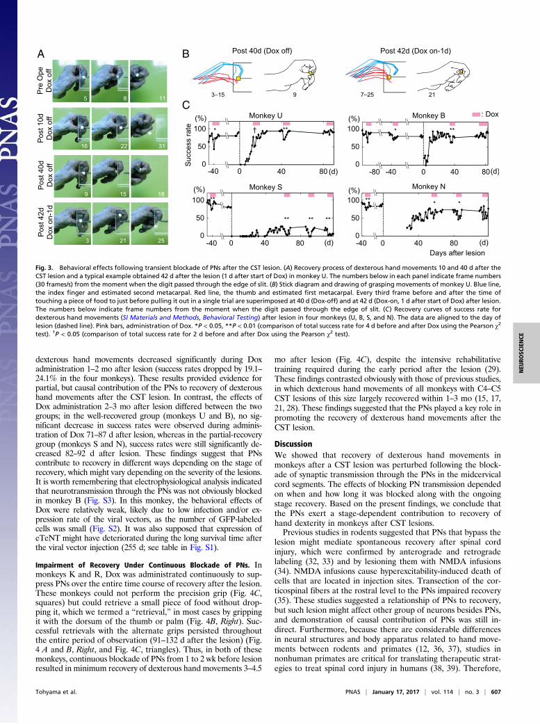
dexterous hand movements decreased significantly during Doxadministration 1–2 mo after lesion (success rates dropped by 19.1–24.1% in the four monkeys). These results provided evidence forpartial, but causal contribution of the PNs to recovery of dexteroushand movements after the CST lesion. In contrast, the effects ofDox administration 2–3 mo after lesion differed between the twogroups; in the well-recovered group (monkeys U and B), no sig-nificant decrease in success rates were observed during adminis-tration of Dox 71–87 d after lesion, whereas in the partial-recoverygroup (monkeys S and N), success rates were still significantly de-creased 82–92 d after lesion. These findings suggest that PNscontribute to recovery in different ways depending on the stage ofrecovery, which might vary depending on the severity of the lesions.It is worth remembering that electrophysiological analysis indicatedthat neurotransmission through the PNs was not obviously blockedin monkey B (Fig. S3). In this monkey, the behavioral effects ofDox were relatively weak, likely due to low infection and/or ex-pression rate of the viral vectors, as the number of GFP-labeledcells was small (Fig. S2). It was also supposed that expression ofeTeNT might have deteriorated during the long survival time afterthe viral vector injection (255 d; see table in Fig. S1).
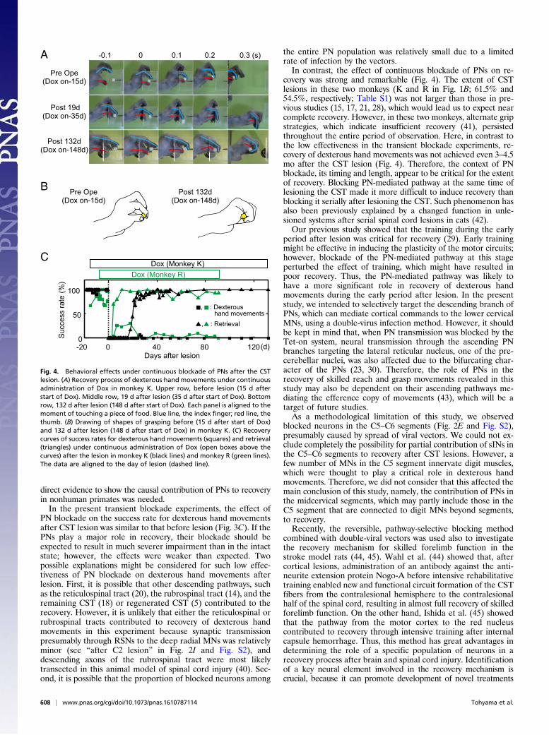
Impairment of Recovery Under Continuous Blockade of PNs. Inmonkeys K and R, Dox was administrated continuously to sup-press PNs over the entire time course of recovery after the lesion.These monkeys could not perform the precision grip (Fig. 4C,squares) but could retrieve a small piece of food without drop-ping it, which we termed a “retrieval,” in most cases by grippingit with the dorsum of the thumb or palm (Fig. 4B, Right). Suc-cessful retrievals with the alternate grips persisted throughoutthe entire period of observation (91–132 d after the lesion) (Fig.4 A and B, Right, and Fig. 4C, triangles). Thus, in both of thesemonkeys, continuous blockade of PNs from 1 to 2 wk before lesionresulted in minimum recovery of dexterous hand movements 3–4.5
mo after lesion (Fig. 4C), despite the intensive rehabilitativetraining required during the early period after the lesion (29).These findings contrasted obviously with those of previous studies,in which dexterous hand movements of all monkeys with C4–C5CST lesions of this size largely recovered within 1–3 mo (15, 17,21, 28). These findings suggested that the PNs played a key role inpromoting the recovery of dexterous hand movements after theCST lesion.
DiscussionWe showed that recovery of dexterous hand movements inmonkeys after a CST lesion was perturbed following the block-ade of synaptic transmission through the PNs in the midcervicalcord segments. The effects of blocking PN transmission dependedon when and how long it was blocked along with the ongoingstage recovery. Based on the present findings, we conclude thatthe PNs exert a stage-dependent contribution to recovery ofhand dexterity in monkeys after CST lesions.Previous studies in rodents suggested that PNs that bypass the
lesion might mediate spontaneous recovery after spinal cordinjury, which were confirmed by anterograde and retrogradelabeling (32, 33) and by lesioning them with NMDA infusions(34). NMDA infusions cause hyperexcitability-induced death ofcells that are located in injection sites. Transection of the cor-ticospinal fibers at the rostral level to the PNs impaired recovery(35). These studies suggested a relationship of PNs to recovery,but such lesion might affect other group of neurons besides PNs,and demonstration of causal contribution of PNs was still in-direct. Furthermore, because there are considerable differencesin neural structures and body apparatus related to hand move-ments between rodents and primates (12, 36, 37), studies innonhuman primates are critical for translating therapeutic strat-egies to treat spinal cord injury in humans (38, 39). Therefore,
A B
C
Fig. 3. Behavioral effects following transient blockade of PNs after the CST lesion. (A) Recovery process of dexterous hand movements 10 and 40 d after theCST lesion and a typical example obtained 42 d after the lesion (1 d after start of Dox) in monkey U. The numbers below in each panel indicate frame numbers(30 frames/s) from the moment when the digit passed through the edge of slit. (B) Stick diagram and drawing of grasping movements of monkey U. Blue line,the index finger and estimated second metacarpal. Red line, the thumb and estimated first metacarpal. Every third frame before and after the time oftouching a piece of food to just before pulling it out in a single trial are superimposed at 40 d (Dox-off) and at 42 d (Dox-on, 1 d after start of Dox) after lesion.The numbers below indicate frame numbers from the moment when the digit passed through the edge of slit. (C) Recovery curves of success rate fordexterous hand movements (SI Materials and Methods, Behavioral Testing) after lesion in four monkeys (U, B, S, and N). The data are aligned to the day oflesion (dashed line). Pink bars, administration of Dox. *P < 0.05, **P < 0.01 (comparison of total success rate for 4 d before and after Dox using the Pearson χ2
test). †P < 0.05 (comparison of total success rate for 2 d before and after Dox using the Pearson χ2 test).
Tohyama et al. PNAS | January 17, 2017 | vol. 114 | no. 3 | 607
NEU
ROSC
IENCE
direct evidence to show the causal contribution of PNs to recoveryin nonhuman primates was needed.In the present transient blockade experiments, the effect of
PN blockade on the success rate for dexterous hand movementsafter CST lesion was similar to that before lesion (Fig. 3C). If thePNs play a major role in recovery, their blockade should beexpected to result in much severer impairment than in the intactstate; however, the effects were weaker than expected. Twopossible explanations might be considered for such low effec-tiveness of PN blockade on dexterous hand movements afterlesion. First, it is possible that other descending pathways, suchas the reticulospinal tract (20), the rubrospinal tract (14), and theremaining CST (18) or regenerated CST (5) contributed to therecovery. However, it is unlikely that either the reticulospinal orrubrospinal tracts contributed to recovery of dexterous handmovements in this experiment because synaptic transmissionpresumably through RSNs to the deep radial MNs was relativelyminor (see “after C2 lesion” in Fig. 2I and Fig. S2), anddescending axons of the rubrospinal tract were most likelytransected in this animal model of spinal cord injury (40). Sec-ond, it is possible that the proportion of blocked neurons among
the entire PN population was relatively small due to a limitedrate of infection by the vectors.In contrast, the effect of continuous blockade of PNs on re-
covery was strong and remarkable (Fig. 4). The extent of CSTlesions in these two monkeys (K and R in Fig. 1B; 61.5% and54.5%, respectively; Table S1) was not larger than those in pre-vious studies (15, 17, 21, 28), which would lead us to expect nearcomplete recovery. However, in these two monkeys, alternate gripstrategies, which indicate insufficient recovery (41), persistedthroughout the entire period of observation. Here, in contrast tothe low effectiveness in the transient blockade experiments, re-covery of dexterous hand movements was not achieved even 3–4.5mo after the CST lesion (Fig. 4). Therefore, the context of PNblockade, its timing and length, appear to be critical for the extentof recovery. Blocking PN-mediated pathway at the same time oflesioning the CST made it more difficult to induce recovery thanblocking it serially after lesioning the CST. Such phenomenon hasalso been previously explained by a changed function in unle-sioned systems after serial spinal cord lesions in cats (42).Our previous study showed that the training during the early
period after lesion was critical for recovery (29). Early trainingmight be effective in inducing the plasticity of the motor circuits;however, blockade of the PN-mediated pathway at this stageperturbed the effect of training, which might have resulted inpoor recovery. Thus, the PN-mediated pathway was likely tohave a more significant role in recovery of dexterous handmovements during the early period after lesion. In the presentstudy, we intended to selectively target the descending branch ofPNs, which can mediate cortical commands to the lower cervicalMNs, using a double-virus infection method. However, it shouldbe kept in mind that, when PN transmission was blocked by theTet-on system, neural transmission through the ascending PNbranches targeting the lateral reticular nucleus, one of the pre-cerebellar nuclei, was also affected due to the bifurcating char-acter of the PNs (23, 30). Therefore, the role of PNs in therecovery of skilled reach and grasp movements revealed in thisstudy may also be dependent on their ascending pathways me-diating the efference copy of movements (43), which will be atarget of future studies.As a methodological limitation of this study, we observed
blocked neurons in the C5–C6 segments (Fig. 2E and Fig. S2),presumably caused by spread of viral vectors. We could not ex-clude completely the possibility for partial contribution of sINs inthe C5–C6 segments to recovery after CST lesions. However, afew number of MNs in the C5 segment innervate digit muscles,which were thought to play a critical role in dexterous handmovements. Therefore, we did not consider that this affected themain conclusion of this study, namely, the contribution of PNs inthe midcervical segments, which may partly include those in theC5 segment that are connected to digit MNs beyond segments,to recovery.Recently, the reversible, pathway-selective blocking method
combined with double-viral vectors was used also to investigatethe recovery mechanism for skilled forelimb function in thestroke model rats (44, 45). Wahl et al. (44) showed that, aftercortical lesions, administration of an antibody against the anti-neurite extension protein Nogo-A before intensive rehabilitativetraining enabled new and functional circuit formation of the CSTfibers from the contralesional hemisphere to the contralesionalhalf of the spinal cord, resulting in almost full recovery of skilledforelimb function. On the other hand, Ishida et al. (45) showedthat the pathway from the motor cortex to the red nucleuscontributed to recovery through intensive training after internalcapsule hemorrhage. Thus, this method has great advantages indetermining the role of a specific population of neurons in arecovery process after brain and spinal cord injury. Identificationof a key neural element involved in the recovery mechanism iscrucial, because it can promote development of novel treatments
-20 0 40 1200
50
100
80
Pre Ope(Dox on-15d)
Post 19d(Dox on-35d)
Post 132d(Dox on-148d)
-0.1 0 0.1 0.2 0.3 (s)S
ucce
ss ra
te (%
)
Days after lesion
A
CDox (Monkey K)
Dox (Monkey R)
(d)
, : Dexterous hand movements
, : Retrieval
B Pre Ope(Dox on-15d)
Post 132d(Dox on-148d)
Fig. 4. Behavioral effects under continuous blockade of PNs after the CSTlesion. (A) Recovery process of dexterous hand movements under continuousadministration of Dox in monkey K. Upper row, before lesion (15 d afterstart of Dox). Middle row, 19 d after lesion (35 d after start of Dox). Bottomrow, 132 d after lesion (148 d after start of Dox). Each panel is aligned to themoment of touching a piece of food. Blue line, the index finger; red line, thethumb. (B) Drawing of shapes of grasping before (15 d after start of Dox)and 132 d after lesion (148 d after start of Dox) in monkey K. (C) Recoverycurves of success rates for dexterous hand movements (squares) and retrieval(triangles) under continuous administration of Dox (open boxes above thecurves) after the lesion in monkey K (black lines) and monkey R (green lines).The data are aligned to the day of lesion (dashed line).
608 | www.pnas.org/cgi/doi/10.1073/pnas.1610787114 Tohyama et al.
combined with rehabilitation of motor disability following the brain/spinal cord injury. Selectively enhancing the plasticity of the path-ways thus identified as being responsible for recovery will become akey technology to facilitate recovery in future studies.
Materials and MethodsThe animal experimental procedures in this study were conducted in ac-cordance with the principles of the National Institutes of Health and theMinistry of Education, Culture, Sports, Science, and Technology of Japan. Theprotocols were approved by the Institutional Animal Care and Use Committeeof the National Institutes of Natural Sciences. We recruited eight monkeysand then excluded two because the infection rate and/or expression of viralvectors in one monkey were judged to be obviously insufficient and theextent of lesion of the other monkey was too large; thus, the results of sixmonkeys are presented. The monkeys were first trained for a few weeks in areach-and-grasp task described previously (15–17, 21–23) and in SupportingInformation (SI Materials and Methods, Behavioral Testing, and Movie S1).The retrograde gene transfer vector (HiRet/FuG-E/NeuRet-TRE-EGFP.eTeNT)was then injected into the ventral horn of the spinal cord at C6–Th1 aspreviously described (23). One to 10 d after the injection, the anterogradevector (AAV-2/DJ-CMV-rtTAV16) was injected into the intermediate zone ofthe spinal cord at caudal C2 to caudal C4. Four monkeys (U, B, S, and N)
received a total of four rounds of oral Dox (15 mg·kg−1·d−1, each round, 7–25 d)before and after the CST lesion. Two monkeys (K and R) received continu-ous administration of Dox from 8–16 d before the CST lesion to 3–4.5 moafter the lesion. Video recordings of reach and grasp movements before andafter the lesion were analyzed off-line. The CST lesion was made by trans-ecting the dorsolateral funiculus at C4–C5 under anesthesia with isoflurane(1–2%). After the end of behavioral observations, acute electrophysiologicalexperiments were conducted on the cervical spinal cord in five of the sixmonkeys (data could not be obtained from monkey R due to a suddenchange in the monkey’s condition during the experiment) under anesthesia(see details in SI Materials and Methods). After transcardial perfusion withdeep anesthesia, the brains and spinal cords were removed. Histologicalexaminations were conducted with anti-GFP immunohistochemistry. Fullmaterials and methods are described in SI Materials and Methods.
ACKNOWLEDGMENTS. We thank Y. Nishimura, B. Alstermark, and L.-G.Pettersson for comments on an earlier version of the manuscript. We thankM. Togawa, Y. Yamanishi, and N. Takahashi for technical support. This studywas supported by the Strategic Research Program for Brain Sciences by theMinistry of Education, Culture, Sports, Science, and Technology (MEXT) ofJapan, and by Grant-in-Aid for Scientific Research on Innovative Areas(“Adaptive Circuit Shift”) by MEXT (to T.I.).
1. Richardson PM, McGuinness UM, Aguayo AJ (1980) Axons from CNS neurons re-generate into PNS grafts. Nature 284(5753):264–265.
2. Schnell L, Schwab ME (1990) Axonal regeneration in the rat spinal cord produced byan antibody against myelin-associated neurite growth inhibitors. Nature 343(6255):269–272.
3. Schnell L, Schneider R, Kolbeck R, Barde YA, Schwab ME (1994) Neurotrophin-3 en-hances sprouting of corticospinal tract during development and after adult spinalcord lesion. Nature 367(6459):170–173.
4. Salewski RP, et al. (2015) Transplantation of induced pluripotent stem cell-derivedneural stem cells mediate functional recovery following thoracic spinal cord injurythrough remyelination of axons. Stem Cells Transl Med 4(7):743–754.
5. Nakagawa H, Ninomiya T, Yamashita T, Takada M (2015) Reorganization of cortico-spinal tract fibers after spinal cord injury in adult macaques. Sci Rep 5:11986.
6. Raineteau O, Schwab ME (2001) Plasticity of motor systems after incomplete spinalcord injury. Nat Rev Neurosci 2(4):263–273.
7. Nishimura Y, Isa T (2009) Compensatory changes at the cerebral cortical level afterspinal cord injury. Neuroscientist 15(5):436–444.
8. Oudega M, Perez MA (2012) Corticospinal reorganization after spinal cord injury.J Physiol 590(16):3647–3663.
9. Isa T, Nishimura Y (2014) Plasticity for recovery after partial spinal cord injury—hi-erarchical organization. Neurosci Res 78:3–8.
10. Bernhard CG, Bohm E (1954) Cortical representation and functional significance ofthe corticomotoneuronal system. AMA Arch Neurol Psychiatry 72(4):473–502.
11. Kuypers HG (1982) A new look at the organization of the motor system. Prog BrainRes 57:381–403.
12. Lemon RN (2008) Descending pathways in motor control. Annu Rev Neurosci 31:195–218.
13. Lawrence DG, Kuypers HG (1968) The functional organization of the motor system inthe monkey. I. The effects of bilateral pyramidal lesions. Brain 91(1):1–14.
14. Lawrence DG, Kuypers HG (1968) The functional organization of the motor system inthe monkey. II. The effects of lesions of the descending brain-stem pathways. Brain91(1):15–36.
15. Nishimura Y, et al. (2007) Time-dependent central compensatory mechanisms offinger dexterity after spinal cord injury. Science 318(5853):1150–1155.
16. Nishimura Y, et al. (2011) Neural substrates for the motivational regulation of motorrecovery after spinal-cord injury. PLoS One 6(9):e24854.
17. Sawada M, et al. (2015) Function of the nucleus accumbens in motor control duringrecovery after spinal cord injury. Science 350(6256):98–101.
18. Rosenzweig ES, et al. (2010) Extensive spontaneous plasticity of corticospinal pro-jections after primate spinal cord injury. Nat Neurosci 13(12):1505–1510.
19. Nishimura Y, Morichika Y, Isa T (2009) A subcortical oscillatory network contributes torecovery of hand dexterity after spinal cord injury. Brain 132(Pt 3):709–721.
20. Zaaimi B, Edgley SA, Soteropoulos DS, Baker SN (2012) Changes in descending motorpathway connectivity after corticospinal tract lesion in macaque monkey. Brain 135(Pt7):2277–2289.
21. Sasaki S, et al. (2004) Dexterous finger movements in primate without monosynapticcorticomotoneuronal excitation. J Neurophysiol 92(5):3142–3147.
22. Alstermark B, et al. (2011) Motor command for precision grip in the macaque monkeycan be mediated by spinal interneurons. J Neurophysiol 106(1):122–126.
23. Kinoshita M, et al. (2012) Genetic dissection of the circuit for hand dexterity in pri-mates. Nature 487(7406):235–238.
24. Kato S, et al. (2011) A lentiviral strategy for highly efficient retrograde gene transferby pseudotyping with fusion envelope glycoprotein. Hum Gene Ther 22(2):197–206.
25. Kato S, Kobayashi K, Kobayashi K (2014) Improved transduction efficiency of a len-tiviral vector for neuron-specific retrograde gene transfer by optimizing the junctionof fusion envelope glycoprotein. J Neurosci Methods 227:151–158.
26. Kato S, et al. (2011) Neuron-specific gene transfer through retrograde transport oflentiviral vector pseudotyped with a novel type of fusion envelope glycoprotein. HumGene Ther 22(12):1511–1523.
27. Yamamoto M, et al. (2003) Reversible suppression of glutamatergic neurotransmis-sion of cerebellar granule cells in vivo by genetically manipulated expression of tet-anus neurotoxin light chain. J Neurosci 23(17):6759–6767.
28. Higo N, et al. (2009) Increased expression of the growth-associated protein 43 gene inthe sensorimotor cortex of the macaque monkey after lesioning the lateral cortico-spinal tract. J Comp Neurol 516(6):493–506.
29. Sugiyama Y, et al. (2013) Effects of early versus late rehabilitative training on manualdexterity after corticospinal tract lesion in macaque monkeys. J Neurophysiol 109(12):2853–2865.
30. Isa T, Ohki Y, Seki K, Alstermark B (2006) Properties of propriospinal neurons in theC3–C4 segments mediating disynaptic pyramidal excitation to forelimb motoneuronsin the macaque monkey. J Neurophysiol 95(6):3674–3685.
31. Qi HX, Gharbawie OA, Wynne KW, Kaas JH (2013) Impairment and recovery of handuse after unilateral section of the dorsal columns of the spinal cord in squirrelmonkeys. Behav Brain Res 252:363–376.
32. Bareyre FM, et al. (2004) The injured spinal cord spontaneously forms a new intra-spinal circuit in adult rats. Nat Neurosci 7(3):269–277.
33. Filli L, et al. (2014) Bridging the gap: A reticulo-propriospinal detour bypassing anincomplete spinal cord injury. J Neurosci 34(40):13399–13410.
34. Courtine G, et al. (2008) Recovery of supraspinal control of stepping via indirectpropriospinal relay connections after spinal cord injury. Nat Med 14(1):69–74.
35. Ueno M, Hayano Y, Nakagawa H, Yamashita T (2012) Intraspinal rewiring of thecorticospinal tract requires target-derived brain-derived neurotrophic factor andcompensates lost function after brain injury. Brain 135(Pt 4):1253–1267.
36. Alstermark B, Ogawa J, Isa T (2004) Lack of monosynaptic corticomotoneuronal EPSPsin rats: Disynaptic EPSPs mediated via reticulospinal neurons and polysynaptic EPSPsvia segmental interneurons. J Neurophysiol 91(4):1832–1839.
37. Isa T, Ohki Y, Alstermark B, Pettersson LG, Sasaki S (2007) Direct and indirect cortico-motoneuronal pathways and control of hand/arm movements. Physiology (Bethesda)22:145–152.
38. Courtine G, et al. (2007) Can experiments in nonhuman primates expedite thetranslation of treatments for spinal cord injury in humans? Nat Med 13(5):561–566.
39. Friedli L, et al. (2015) Pronounced species divergence in corticospinal tract re-organization and functional recovery after lateralized spinal cord injury favors pri-mates. Sci Transl Med 7(302):302ra134.
40. Holstege G, Blok BF, Ralston DD (1988) Anatomical evidence for red nucleus projec-tions to motoneuronal cell groups in the spinal cord of the monkey. Neurosci Lett95(1-3):97–101.
41. Murata Y, et al. (2008) Effects of motor training on the recovery of manual dexterityafter primary motor cortex lesion in macaque monkeys. J Neurophysiol 99(2):773–786.
42. Alstermark B, Lundberg A, Pettersson LG, Tantisira B, Walkowska M (1987) Motorrecovery after serial spinal cord lesions of defined descending pathways in cats.Neurosci Res 5(1):68–73.
43. Azim E, Jiang J, Alstermark B, Jessell TM (2014) Skilled reaching relies on a V2apropriospinal internal copy circuit. Nature 508(7496):357–363.
44. Wahl AS, et al. (2014) Neuronal repair. Asynchronous therapy restores motor controlby rewiring of the rat corticospinal tract after stroke. Science 344(6189):1250–1255.
45. Ishida A, et al. (2016) Causal link between the cortico-rubral pathway and functionalrecovery through forced impaired limb use in rats with stroke. J Neurosci 36(2):455–467.
46. Sooksawate T, et al. (2013) Viral vector-mediated selective and reversible blockade ofthe pathway for visual orienting in mice. Front Neural Circuits 7:162.
Tohyama et al. PNAS | January 17, 2017 | vol. 114 | no. 3 | 609
NEU
ROSC
IENCE