conserved inhibitory mechanism and competent atp binding ... · active site motif and the exposed...
TRANSCRIPT

Conserved Inhibitory Mechanism and Competent ATPBinding Mode for Adenylyltransferases with Fic FoldArnaud Goepfert1,2, Frederic V. Stanger1,2, Christoph Dehio1*, Tilman Schirmer2*
1 Focal Area Infection Biology, Biozentrum, University of Basel, Basel, Switzerland, 2 Focal Area Structural Biology and Biophysics, Biozentrum, University of Basel, Basel,
Switzerland
Abstract
The ubiquitous FIC domain is evolutionarily conserved from bacteria to human and has been shown to catalyze AMPtransfer onto protein side-chain hydroxyl groups. Recently, it was predicted that most catalytically competent Fic proteinsare inhibited by the presence of an inhibitory helix ainh that is provided by a cognate anti-toxin (class I), or is part of the N-or C-terminal part of the Fic protein itself (classes II and III). In vitro, inhibition is relieved by mutation of a conservedglutamate of ainh to glycine. For the class III bacterial Fic protein NmFic from Neisseria meningitidis, the inhibitorymechanism has been elucidated. Here, we extend above study by including bacterial class I and II Fic proteins VbhT fromBartonella schoenbuchensis and SoFic from Shewanella oneidensis, respectively, and the respective E-.G mutants.Comparative enzymatic and crystallographic analyses show that, in all three classes, the ATP substrate binds to the wild-type FIC domains, but with the a-phosphate in disparate and non-competent orientations. In the E-.G mutants, however,the tri-phosphate moiety is found reorganized to the same tightly bound structure through a unique set of hydrogen bondswith Fic signature motif residues. The c-phosphate adopts the location that is taken by the inhibitory glutamate in wild-typeresulting in an a-phosphate orientation that can be attacked in-line by a target side-chain hydroxyl group. The latter isproperly registered to the Fic active center by main-chain b-interactions with the b-hairpin flap. These data indicate that theactive site motif and the exposed edge of the flap are both required to form an adenylylation-competent Fic protein.
Citation: Goepfert A, Stanger FV, Dehio C, Schirmer T (2013) Conserved Inhibitory Mechanism and Competent ATP Binding Mode for Adenylyltransferases withFic Fold. PLoS ONE 8(5): e64901. doi:10.1371/journal.pone.0064901
Editor: Eric Cascales, Centre National de la Recherche Scientifique, Aix-Marseille Universite, France
Received March 13, 2013; Accepted April 19, 2013; Published May 30, 2013
Copyright: � 2013 Goepfert et al. This is an open-access article distributed under the terms of the Creative Commons Attribution License, which permitsunrestricted use, distribution, and reproduction in any medium, provided the original author and source are credited.
Funding: This work was supported by grants 31003A 312979 and 3100 138414 from the Swiss National Science Foundation (to CD and TS, respectively) andgrant 51RT 0_126008 (InfectX) in the frame of the SystemsX.ch Swiss Initiative for Systems Biology (to CD). The funders had no role in study design, data collectionand analysis, decision to publish, or preparation of the manuscript.
Competing Interests: The authors have declared that no competing interests exist.
* E-mail: [email protected] (TS); [email protected] (CD)
Introduction
Adenylyl transferases (ATases) utilize adenosine triphosphate
(ATP) to covalently modify proteins, nucleic acids, or small
molecules with adenosine monophosphate (AMP), a reaction
known as adenylylation or AMPylation. The ubiquitous FIC
domain (pfam 02661) found in proteins of all domains of life and
viruses has only recently been shown to confer ATase activity.
Thus, the bacterial T3SS effector protein VopS from Vibrio
parahaemolyticus and the surface antigen IbpA from Histophilus somni
covalently attach the bulky AMP moiety onto a specific threonine
or tyrosine, respectively, of the switch I region of Rho family
GTPases [1,2]. This abrogates binding of downstream effectors
and results in actin cytoskeleton collapse and concomitant cell
detachment and death. Mutational and bioinformatics analysis
indicated that Fic proteins containing a strictly conserved
HxFx(D/E)GNGRxxR signature motif in the active center
typically display adenylylation activity [1,2,3,4,5], while Fic
proteins with an active center deviating from this consensus are
considered to have adopted different activities. Indeed, the host-
targeted effector protein AnkX of Legionella pneumophila exhibiting
an HxFxDANGRxxV signature motif displays phosphocholina-
tion activity towards the GTPase Rab1 [6].
The FIC domain is structurally characterized by a conserved
central core of four helices (a2 to a5) that is flanked by three
helices (a1, a6 and a7) found in diverse dispositions in different Fic
proteins [3,7]. Helices a4 and a5 are joined by a loop that together
with the N-terminal cap of helix a5 forms the active center
represented by a signature motif with the consensus sequence
HxFx(D/E)GNGRxxR. The catalytic mechanism of adenylylation
was deduced from the crystal structure of the second FIC domain
of IbpA in complex with the adenylylated Cdc42 target [4] and
from biochemical studies [5] and shown to involve nucleophilic
attack of the target side-chain hydroxyl onto the ATP a-
phosphate. The triphosphate binding site at the anionic nest at
the N-terminus of helix a5 was characterized by the crystal
structure of BepA from Bartonella henselae in complex with
pyrophosphate, the side product of the reaction [3]. An ATP
substrate complex structure was obtained recently for the Fic
protein of Neisseria meningitidis [8] corroborating the catalytic
mechanism. The histidine of the signature motif is critical for
deprotonation of the incoming target hydroxyl group [5], whereas
the phenylylanine is part of the hydrophobic core of the domain.
The remaining residues of the motif are involved in ATP/Mg2+
binding and loop stabilization [3,8].
We recently demonstrated that the Fic protein VbhT from
Bartonella schoenbuchensis causes bacterial growth arrest when
overexpressed in Bartonella or E. coli and that this effect can be
repressed by co-expression with the anti-toxin VbhA, a small
protein encoded upstream of VbhT [8]. As shown by structure
PLOS ONE | www.plosone.org 1 May 2013 | Volume 8 | Issue 5 | e64901

analysis, VbhA forms a tight complex with the FIC domain of
VbhT with the conserved glutamate (Einh) from the inhibitory
helix ainh partly obstructing the ATP binding site, which gave a
first clue regarding the inhibitory mechanism mediated by VbhA
binding.
Exhaustive bioinformatic analysis coupled with homology
modeling revealed that the (S/T)xxxE(G/N) signature motif of
ainh is not only found in several other putative anti-toxin sequences
coded immediately upstream of Fic proteins, but is often part of
the FIC domain itself either preceding helix a1 or immediately
following helix a7 [8]. Thus, a classification system was introduced
grouping the Fic proteins for which an anti-toxin with an
inhibitory helix ainh had been found into class I and those with
an equivalent of ainh in the N- or C-terminal part of the Fic
protein into classes II and III, respectively. Indeed, 90% of the Fic
proteins with the canonical FIC signature motif could be classified
accordingly, suggesting that all these enzymes are inhibited in their
enzymatic activity.
The physiological stimulus or condition for relief of ainh-
mediated inhibition is not yet known. For T4SS Fic proteins of
class I (such as VbhT or BepA [9]), however, it appears likely that,
for injection into host cells, the Fic protein has to unfold and will
be translocated without the antitoxin. For class II and III proteins,
detachment, unfolding, or proteolytic cleavage of the ainh helix
may cause relief of inhibition. In fact, a truncation mutant of the
class III Fic protein from N. meningitidis (NmFic) lacking the entire
C-terminal ainh helix showed strong ATase activity and allowed to
study the catalytic and inhibitory mechanism in detail [8]. A more
subtle means to relieve inhibition, which is applicable to Fic
proteins of all three classes, is the replacement of the inhibitory
glutamate by glycine. In vivo, such E-.G mutations showed a
detrimental effect on bacterial growth [8]. For the human HYPE
protein (class II), the corresponding mutant protein catalyzed
in vitro AMP transfer to the small GTPases Rac1 and Cdc42,
whereas only marginal effect was seen with the wild-type proteins
[8].
Here, we assayed in a systematic approach Fic representatives of
the three Fic classes and their E-.G mutants for in vitro
adenylylation showing that the mutation causes inhibition relief
across the Fic classes. Binding of ATP substrate or AMPPNP
substrate analog to the wild-type and the E-.G mutant proteins
was studied by protein crystallography to reveal the inhibitory
mechanism and to get further insight into catalysis. This yielded a
consistent molecular mechanism that most likely applies to most
adenylylation competent Fic proteins irrespective of class.
Materials and Methods
CloningThe full-length vbhA gene and part of the vbhT gene (amino acid
residues 1–248, His6-tagged) were amplified from plasmid
pPE0021 and cloned into the pRSF-Duet1 vector leading to
plasmid pAG0077 (VbhA/VbhT(FIC)). The full-length vbhA gene
and part of the vbhT gene encoding the FIC domain (amino acid
residues 1–198, His6-tagged) were PCR-amplified from plasmid
pPE0021 and cloned into the pRSF-Duet1 vector (pFVS0011). A
two-base pair mutation is then introduced in pFVS0011 to obtain
plasmid pFVS0065 (VbhAE24G/VbhT(FIC)). The fic gene of
Neisseria meningitidis was PCR-amplified with an N-terminal
His6-tag from Neisseria meningitidis from coding region of amino
acid residues 11–191 to generate plasmid expressing NmFic
(pFVS0015). The E186G mutant construct (NmFicE186G,
pFVS0059) was generated by introducing a two-base pair
mutation in pFVS0015. The fic gene of Shewanella oneidensis was
PCR-amplified from plasmid (ASU biodesign institute, Clone ID
SoCD00104192) and cloned with an N-terminal His6-tag into
pRSF-Duet1 (pFVS0040). The SoFicE73G plasmid (pFVS0058)
was generated by introducing a two-base pair point mutation in
pFVS0040.
Protein Expression and PurificationVectors pAG0077 (VbhA/VbhT(FIC)), pFVS0040 (SoFic) and
pFVS0015 (NmFic) were transformed into E.coli BL21 (DE3). E.
coli cultures were grown at 37uC in LB medium supplemented with
50 mg/ml of kanamycin to an OD595 of 0.6 before induction with
0.3 mM IPTG for 16 h at 23uC. Vectors pFVS0065 (VbhAE24G/
VbhT(FIC)), pFVS0059 (NmFicE186G), pFVS0058 (SoFicE73G)
were transformed into BL21-AI cells. Cells were incubated in
750 ml LB medium supplemented with 50 mg/ml kanamycin and
1% glucose at 37uC at 200 rpm until an OD595 value of 1.5 was
reached. Bacterial pellets were resuspended in 1 L of Terrific
Broth media containing 50 mg/ml21 kanamycin. Protein expres-
sion was induced at 23uC with 0.1% arabinose and 0.1 mM IPTG
for 23 h at 200 rpm.
Cells containing overexpressed VbhA/VbhT(FIC) and NmFic
were resuspended in lysis buffer containing 20 mM Tris (pH 7.5),
250 mM NaCl, and 25 mM imidazole and disrupted using French
press. Cell debris were pelleted by ultracentrifugation and the
supernatant was applied to a His-Trap column (GE Healthcare).
The proteins were eluted with a gradient of elution buffer
containing 20 mM Tris (pH 7.5), 250 mM NaCl, and 500 mM
imidazole. The proteins were then concentrated and injected on a
Superdex 75 16/60 gel filtration column (GE Healthcare)
equilibrated with 10 mM Tris (pH 7.6) and 100 mM NaCl. The
pure proteins were concentrated to 3.7 mg/ml for VbhA/
VbhT(FIC) and 30 mg/ml for NmFic.
The same purification protocol as described above was used for
VbhAE24G/VbhT(FIC) and NmFicE186G with an additional
intermediate purification step. After affinity purification, the
proteins were adjusted to 20 mM Tris (pH 8.5), 25 mM NaCl,
applied to a Resource-Q anion exchange column (Amersham
Biosciences), and eluted with a linear gradient of 1 M NaCl. Peak
fractions were concentrated and further purified by gel filtration
chromatography. Purified proteins in 10 mM Tris (pH 7.6),
100 mM NaCl were concentrated to 4.1 mg/ml for VbhAE24G/
VbhT(FIC) and 33 mg/ml for NmFicE186G. Cells containing
overexpressed SoFic and SoFicE73G were resuspended in lysis
buffer containing 50 mM HEPES (pH 8.0), 50 mM NaCl, 1 mM
TCEP, 10% glycerol and 10 mM Imidazole and disrupted using
French press. Cell debris were pelleted by ultracentrifugation and
the supernatant was applied to a His-Trap column (GE
Healthcare). The proteins were eluted with a gradient of elution
buffer containing 50 mM HEPES (pH 8.0), 50 mM NaCl, 1 mM
TCEP, 10% glycerol and 300 mM imidazole. The proteins were
then concentrated and injected on a Superdex 75 16/60 gel
filtration column (GE Healthcare) equilibrated with 20 mM
HEPES (pH 8.0), 200 mM NaCl and 1 mM TCEP. The pure
proteins were concentrated to 21.8 mg/ml for SoFic and 12 mg/
ml for SoFicE73G.
Protein CrystallizationFor crystallization, the hanging-drop vapor diffusion method
was used with 1 ml protein solution mixed with 1 ml reservoir
solution. The VbhA/VbhT(FIC) and VbhAE24G/VbhT(FIC)
complexes were concentrated to 3.7 mg/ml and 4.1 mg/ml,
respectively, and crystallized at 20uC using a reservoir solution
composed of 15% (w/v) PEG 4000, 0.1 M MES pH 6.5. Whereas,
the wild-type crystal was soaked with 5 mM ATP, and 5 mM
Inhibition and ATP Binding Mode of Fic Proteins
PLOS ONE | www.plosone.org 2 May 2013 | Volume 8 | Issue 5 | e64901

MgCl2, the mutant was co-crystallized with 10 mM ATP, and
10 mM MgCl2. For data collection, crystals were transferred to
reservoir solutions supplemented with 20% glycerol and flash
frozen in liquid nitrogen. SoFic and SoFicE73G were concentrated
to 21.8 mg/ml and 12 mg/ml, respectively, and co-crystallized
with either 5 mM ATP or 5 mM AMPPNP and supplemented
with 5 mM MgCl2 in a solution composed of 21% (w/v) PEG
3350 and 0.2 M NaF pH 7.1 at 4uC. For data collection, crystals
of the protein-ligand complex were cryoprotected by transfer to a
reservoir solution supplemented with 15% (v/v) PEG 200 and
flash cooled in liquid nitrogen. For crystallization of NmFicE186G
(33 mg/ml), a reservoir solution composed of 4 M potassium
formate, 0.1 M Bis-Tris propane pH 9.0, 2% (w/v) PEG MME
2000 was used. Crystals were soaked with 5 mM AMPPNP and
5 mM MgCl2 and then cryoprotected with 20% glycerol prior
flash-cooling in liquid nitrogen.
Data Collection, Structure Determination, andRefinement
Diffraction data were collected at the Swiss Light Source at
100 K and processed using XDS [10]. The structures were solved
by molecular replacement using the apo structures of VbhA/
VbhT(FIC) (PDB code 3SHG), SoFic (PDB code 3EQX) or
NmFic (PDB code 2G03) as search models using Phaser [11].
Several rounds of iterative model building and refinement were
performed using Coot [12] and PHENIX [13] or REFMAC5
[14], respectively. 5% of the data were excluded from refinement
and used for cross-validation. The geometry of the final model was
assessed using MolProbity [15] showing .99% of the residues in
the core and allowed regions of the Ramachandran plot. Data
collections and refinement statistics are summarized in Table 1.
The atomic coordinates and structure factors of VbhA/
VbhT(FIC)/ATP, VbhAE24G/VbhT(FIC)/ATP, SoFic/ATP, So-
FicE73G/AMPPNP, and NmFicE186G/AMPPNP have been de-
posited in the Protein Data Bank under accession codes 3ZC7,
3ZCB, 3ZCN, 3ZEC and 3ZLM, respectively. The figures were
generated with Dino (A. Philippsen unpublished, http://www.
dino3d.org).
In vitro Adenylylation AssayAdenylylation activity of VbhA/VbhT(FIC), SoFic and NmFic
constructs was assessed by incubating 125 ng, 1.25 mg and 2.5 mg
of purified protein, respectively, with 10 mCi a-32P-ATP (Hart-
mann Analytic) in a buffer containing 50 mM Tris pH 8.0,
150 mM NaCl, 0.1 mM EGTA, 15 mM MgCl2, and protease
inhibitor cocktail (Roche). Reactions were incubated for 1 h at
30uC, resolved by SDS–PAGE, and subjected to autoradiography.
Results
Constitutive Inhibition is Relieved by Truncation of theInhibitory Glutamate in All Three Fic Classes
For the comparative structure/function study on the inhibitory
mechanism of Fic proteins from the various classes we chose as
representatives the FIC domain of VbhT (residues 1 to 198) from
Bartonella schoenbuchensis in complex with its cognate antitoxin
VbhA (VbhA/VbhT(FIC); class I), Fic protein SO_4266 from
Shewanella oneidensis (SoFic; class II) and Fic protein NMB0255
from Neisseria meningitidis (NmFic; class III).
Auto-adenylylation is a convenient read-out to assess adenylyla-
tion activity of Fic proteins. It does not require the presence of a
physiological protein target that may, in fact, not yet been known
as in the case of SoFic. Autoradiographies of SDS-PAGE gels after
incubation with a-32P-ATP (Fig. 1) show that auto-adenylylation is
virtually absent in the wild-type Fic proteins of all three classes, i.e.
for VbhA/VbhT(FIC), SoFic, and NmFic (see also ref. 8), but is
drastically boosted in the respective E-.G mutants suggesting a
common inhibitory mechanism.
ATP Binds to Wild-type Fic Proteins in Disparate andCatalytically Incompetent Conformations
Fig. 2 shows the high-resolution structures of VbhA/VbhT(FIC)
(class I) and SoFic (class II), both in complex with ATP. Whereas
VbhA/VbhT(FIC) crystallized isomorphously to the unliganded
wild-type crystals ([8], PDB code 3SHG), SoFic yielded crystals of
monoclinic space group, i.e. distinct to the orthorhombic form of
the apo structure ([16], PDB code 3EQX). In the two structures
the nucleotide is clearly visible, albeit with elevated B-factors
(40 A2) in VbhA/VbhT(FIC). Only marginal structural changes
are induced upon substrate binding (rms deviations between the
Ca-positions of apo and complex form of 0.4 A and 0.8 A for
VbhA/VbhT(FIC) and SoFic, respectively).
In both structures the ATP substrate is found at analogous sites
(Fig. 2) with the base filling a pocket formed by a4, a6, and the b-
hairpin flap, the ribose 39-hydroxyl H-bonded to the conserved
glutamate of ainh, and the triphosphate moiety interacting with the
anionic nest formed by the N-terminus of a5. The same binding
mode has been observed for class III NmFic [8]. In all three
structures, also the ribose 29-hydroxyl is forming an H-bond, but
to non-homologous protein side-chains. Similarly, the binding sub-
site for the base is not conserved on the residue level. However, in
each case, hydrophobic residues are contributed by helix a6 and
by the flap. A weak H-bond is formed between the adenine N3
and N133 in VbhT(FIC). A homologous interaction (with N104) is
found in NmFic [8].
Most relevant for catalysis is the orientation of the a-phosphate
that has to be accessible for nucleophilic attack by the target side-
chain hydroxyl group. In VbhA/VbhT(FIC) and SoFic, as in
NmFic [8], the position that is in-line with the scissile Pa-O3abond is not accessible for an attacking group (Fig. 2). Such a group
positioned there would severely clash with atoms of the enzyme.
Thus, in Fic proteins of all three classes, catalytically non-
competent orientation of the a-phosphate appears to be the reason
for the lack of adenylylation activity.
Interestingly, while the a-phosphate is locked in a secured
position in each of the structures, it shows distinct orientations
among the three proteins that can be traced back to differences in
the binding mode of the b- and c-phosphates (Fig. 2C). Though
interacting with the same protein groups (anionic nest; histidine,
asparagine, and first arginine of the signature motif), the detailed
H-bonding patterns are different (e.g. the main chain amide of the
second glycine of the motif interacts with the bridging O3b in
VbhA/VbhT(FIC), and with the non-bridging O1b in SoFic).
It seems that during convergent evolution of ainh-mediated
adenylylation inhibition in the different Fic protein classes no strict
constraints for the ATP binding mode were operational apart from
the requirement for a non-competent orientation for the reacting
phosphate.
Truncation of the Inhibitory Glutamate Allows the ATPSubstrate to Bind in a Catalysis Competent Conformation
Relief of Fic protein inhibition was achieved previously by
expression of VbhT without its cognate antitoxin VbhA or by
replacing in NmFic the SxxxE inhibition motif by AxxxA or –
most drastically - by deleting the entire ainh [8]. The conserved
glutamate of ainh, Einh, was identified to be crucial for the
inhibitory effect, since mere truncation of its side-chain (E-.G
Inhibition and ATP Binding Mode of Fic Proteins
PLOS ONE | www.plosone.org 3 May 2013 | Volume 8 | Issue 5 | e64901

mutation) rendered recombinantly overexpressed Fic proteins of
all three classes toxic to E. coli [8].
In vitro, the mutation has a drastic effect in that auto-
adenylylation is boosted in all three representative Fic proteins
(Fig. 1). This opens the door for studying the action of any active
Fic protein in vivo, even without knowing the physiological stimulus
for inhibition relief.
To reveal the underlying inhibition relief mechanism, crystal
structures of the three mutant proteins in complex with ATP or
AMPPNP were determined to high resolution. Although, in
solution the mutants show auto-adenylylation, no such modifica-
tion is observed in the crystal structures. For NmFicE186G this is
not surprising, since the complex structure has been obtained by
soaking and auto-adenylylation would require partial unfolding of
ainh carrying the modifiable tyrosine (Y183) [8]. The VbhAE24G/
VbhT(FIC) and SoFicE73G complexes were co-crystallized. Since
we do not see adenylylated residues, the extend of modifications
may be either minor, locate to flexible loops or only the
unmodified fraction may have crystallized. Figs. 3A–C show that
in all three cases, the nucleotide is well resolved and, in contrast to
Table 1. Data collection and refinement statistics.
Protein VbhA/VbhT(FIC) VbhAE24G/VbhT(FIC) SoFic SoFicE73G NmFicE186G
Ligand ATP ATP ATP AMPPNP AMPPNP
PDB code 3ZC7 3ZCB 3ZCN 3ZEC 3ZLM
Data collection
Wavelength (A) 1.000 0.979 0.979 0.979 1.000
Detector MAR225 CCD PILATUS 2M MAR225 CCD MAR225 CCD PILATUS 2M
Space group C2 C2 P21 P212121 P6422
Cell dimensions
a, b, c (A) 106.5, 40.6, 73.7 106.5, 40.3, 73.9 37.8, 164.9, 70.2 71.3, 80.6, 141.8 149.1, 149.1, 76.4
a b c (u) 90.0, 121.6, 90.0 90.0, 121.4, 90.0 90.0, 94.4, 90.0 90.0, 90.0, 90.0 90.0, 90.0, 120.0
Resolution (A) 45.422.1 (2.222.1) 45.421.9 (2.121.9) 35.521.7 (1.821.7) 42.722.2 (2.322.2) 49.322.0 (2.122.0)
Rsym or Rmerge (%) 8.5 (33.8) 5.7 (33.4) 4.4 (41.3) 10.5 (52.8) 6.3 (72.7)
CC(1/2) (%) 99.8 (93.6) 99.9 (93.1) 99.9 (87.1) 99.8 (90.3) 100.0 (97.9)
I/s 18.9 (5.7) 14.5 (3.4) 22.9 (3.4) 17.6 (4.5) 31.1 (5.7)
Completeness (%) 99.2 (92.3) 99.2 (97.0) 99.5 (96.7) 100.0 (100.0) 99.9 (100.0)
Multiplicity 5.4 (4.9) 3.6 (3.6) 3.9 (3.4) 7.4 (7.5) 21.4 (22.6)
Refinement
Resolution (A) 15.022.10 30.021.94 15.021.70 15.022.20 30.022.00
No. reflections 15,769 (2,342) 18,923 (1,355) 93,100 (2,837) 42,085 (3,956) 32,490 (2,338)
Rwork/Rfree [%] 16.6/23.0 19.4/23.4 16.7/20.2 16.5/21.1 18.2/19.9
Mol./a.u 1 1 2 2 1
No. atoms
Protein 2172 2011 5961 5984 1458
Ligand/ion 1 ATP 1 ATP, 1 MG 2 ATP 2 ANP, 1 MG 1 ANP, 1 MG
Water 226 134 981 695 135
Average B (A2)
Protein 22.0 25.0 21.9 20.7 45.9
Ligand/ion 39.7 27.5/11.4 21.9 12.9/28.9 40.1/64.8
Water 28.4 32.1 33.7 28.0 49.3
R.m.s deviations
Bond lengths (A) 0.007 0.011 0.008 0.009 0.010
Bond angles (u) 1.0 1.3 1.2 1.2 1.2
Values for the highest resolution shell are shown in brackets.doi:10.1371/journal.pone.0064901.t001
Figure 1. AMP transfer catalyzed by Fic proteins and theirinhibition-relieved variants. Autoradiography of VbhA/VbhT(FIC),SoFic and NmFic (wt, wild type; E/G, E-.G mutant) after incubation withradioactively labeled a-32P-ATP.doi:10.1371/journal.pone.0064901.g001
Inhibition and ATP Binding Mode of Fic Proteins
PLOS ONE | www.plosone.org 4 May 2013 | Volume 8 | Issue 5 | e64901

Figure 2. Crystal structures of wild-type Fic proteins representing classes I to II in complex with ATP substrate. (A) VbhA/VbhT(FIC), (B)SoFic. Structures are shown in cartoon representation (red, FIC core as defined by PFAM; yellow, active site loop and N-terminal end of helix a5; dark-green, inhibitory helix ainh). In (A), the fold of the antitoxin is shown in dark-green and steel-blue. Selected residues are shown in full with theinhibitory glutamate (E24 or E73, respectively) colored in dark. The 2Fo-Fc simulated annealing omit maps covering the ligand are contoured at 1.1 s.In both structures, the orientation of the a-phosphate prevents nucleophilic attack of a putative target side-chain hydroxyl onto the ATP substrate,since the position inline with the scissile Pa-O3a bond (magenta star) is unattainable. C) Stereo view of the superposition of the ATP nucleotides
Inhibition and ATP Binding Mode of Fic Proteins
PLOS ONE | www.plosone.org 5 May 2013 | Volume 8 | Issue 5 | e64901
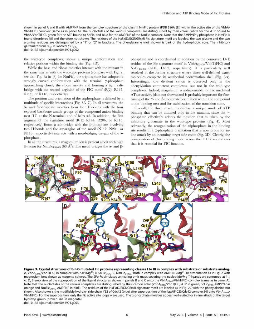
the wild-type complexes, shows a unique conformation and
relative position within the binding site (Fig. 3D).
While the base and ribose moieties interact with the mutant in
the same way as with the wild-type proteins (compare with Fig. 2,
see also Fig. 3a in [8] for NmFic), the triphosphate has adopted a
strongly curved conformation with the terminal c-phosphate
approaching closely the ribose moiety and forming a tight salt-
bridge with the second arginine of the FIC motif (R(2): R147,
R209, or R118, respectively).
The position and orientation of the triphosphate is defined by a
multitude of specific interactions (Fig. 3A–C). In all structures, the
a- and b-phosphate moieties form four H-bonds with the four
exposed backbone amide groups of the compound anion binding
nest [17] at the N-terminal end of helix a5. In addition, the first
arginine of the signature motif (R(1): R144, R206, or R115,
respectively) forms a salt-bridge with the b-phosphate involving
two H-bonds and the asparagine of the motif (N142, N204, or
N113, respectively) interacts with a non-bridging oxygen of the a-
phosphate.
In all the structures, a magnesium ion is present albeit with high
B-factor for NmFicE186G (63 A2). The metal bridges the a- and b-
phosphate and is coordinated in addition by the conserved D/E
residue of the Fic signature motif in VbhAE24G/VbhT(FIC) and
SoFicE73G (E140, D202, respectively). It is particularly well
resolved in the former structure where three well-defined water
molecules complete its octahedral coordination shell (Fig. 3A).
Interestingly, the divalent cation is observed only in the
adenylylation competent complexes, but not in the wild-type
complexes. Indeed, magnesium is indispensable for Fic mediated
ATase activity (data not shown) and is probably important for fine-
tuning of the a- and b-phosphate orientation within the compound
anion binding nest and for stabilization of the transition state.
Overall, the three structures display a unique mode of ATP
binding that can be attained only in the mutants, since the c-
phosphate effectively adopts the position that is taken by the
inhibitory glutamate in the wild-type proteins (Fig. 4). Most
relevantly, the reorganization of the triphosphate in the binding
site results in a a-phosphate orientation that is now prone for in-
line attack by an incoming target side-chain (Fig. 3D). Clearly, the
conservation of this binding mode across the FIC classes shows
that it is essential for FIC function.
shown in panel A and B with AMPPNP from the complex structure of the class III NmFic protein (PDB 3S6A [8]) within the active site of the VbhA/VbhT(FIC) complex (same as in panel A). The nucleotides of the various complexes are distinguished by their colors (white for the ATP bound toVbhA/VbhT(FIC), green for the ATP bound to SoFic, and blue for the AMPPNP of the NmFic complex. Note that the AMPPNP c-phosphate in NmFic isfound disordered [8] and therefore not shown. The residues of the HxFx(D/E)GNGRxxR Fic signature motif are labeled, the two glycine and the twoarginine residues are distinguished by a "1" or "2" in brackets. The phenylalanine (not shown) is part of the hydrophobic core. The inhibitoryglutamate from ainh is labeled as Einh.doi:10.1371/journal.pone.0064901.g002
Figure 3. Crystal structures of E-.G mutated Fic proteins representing classes I to III in complex with substrate or substrate analog.A, VbhAE24G/VbhT(FIC) in complex with ATP/Mg2+; B, SoFicE73G, C, NmFicE186G, both in complex with AMPPNP/Mg2+. Representation as in Fig. 2 withmagnesium ions shown as magenta spheres. The 2Fo-Fc simulated annealing omit maps covering the nucleotide/Mg2+ ligands are contoured at 1.1s. D, Stereo view of the superposition of the ligand structures shown in panels B and C onto the VbhAE24G/VbhT(FIC) complex (same as in panel A).Note that the nucleotides of the various complexes are distinguished by their carbon color (VbhAE24G/VbhT(FIC) ATP in green, SoFicE73G AMPPNP inorange and NmFicE186G AMPPNP in pink). The residues of the HxFx(D/E)GNGRxxR signature motif are labeled as in Fig. 2C with the phenylalanine notshown. Also shown is the modifiable hydroxyl side-chain Y32 of Cdc42 (blue) after superposition of the IbpA(FIC2)/Cdc42 complex [4] onto VbhAE24G/VbhT(FIC). For the superposition, only the Fic active site loops were used. The a-phosphate moieties appear well-suited for in-line attack of the targethydroxyl group (broken line in magenta).doi:10.1371/journal.pone.0064901.g003
Inhibition and ATP Binding Mode of Fic Proteins
PLOS ONE | www.plosone.org 6 May 2013 | Volume 8 | Issue 5 | e64901

Target Registration to the FIC Active SiteThe conservation of the FIC active site and the ATP substrate
binding mode prompts for a precise alignment of the incoming
side-chain hydroxyl with the scissile Pa-O3a bond. The beta-
hairpin flap partly covering the active site appears to represent a
"target dock" that ensures this precise positioning of the target
backbone stretch immediately following the modifiable hydroxyl
side-chain and thus registers the side-chain to the active site as has
been proposed before (2). This was deduced mainly from the only
known Fic protein/target complex structure IbpA(FIC)/Cdc42 [4]
where the AMPylated Y32 of Cdc42 is part of a segment (switch 1
loop) in extended conformation and complements inter-molecu-
larly the b-hairpin of the flap (Fig. 5A).
This notion is further corroborated by the structure of the wild-
type VbhA/VbhT(FIC) complex presented here that revealed
additional density close to the flap above the active site (Fig. 5B).
This was interpreted as a four residue peptide in extended
conformation that is associated antiparallely to the edge of the
two-stranded b-hairpin of the flap via three main chain-main
chain H-bonds. Location and side-chain densities are consistent
with the peptide representing residues 203 to 206 of a symmetry
mate (note that the ordered part of the VbhT(FIC) construct ends
with residue F197). Very similarly, peptide density is present at an
equivalent location in the A-chain of SoFicE73G and could be
attributed to the N-terminus (residues 0 to 3) of a symmetry related
B-chain as also reported for the isomorphous crystal structure of
wild-type SoFic (Fig. 5C) [16].
Figure 4. Comparison of triphosphate nucleotide structures as bound to wild-type and E-.G mutated Fic proteins from class I to III.Stereo views of the ligand structures after superposition of the FIC domains (not shown). Also shown is the inhibitory glutamate of the wild-typestructures. A, ATP as bound to VbhA/VbhT wild-type (white) and the E24G mutant (dark green). B, ATP and AMPPNP as bound to SoFic wild-type(green) and the E73G mutant (orange), respectively. C, AMPPNP as bound to NmFic (blue) and the E186G mutant (pink). Note that the AMPPNP c-phosphate in NmFic is found disordered [8] and therefore not shown.doi:10.1371/journal.pone.0064901.g004
Inhibition and ATP Binding Mode of Fic Proteins
PLOS ONE | www.plosone.org 7 May 2013 | Volume 8 | Issue 5 | e64901
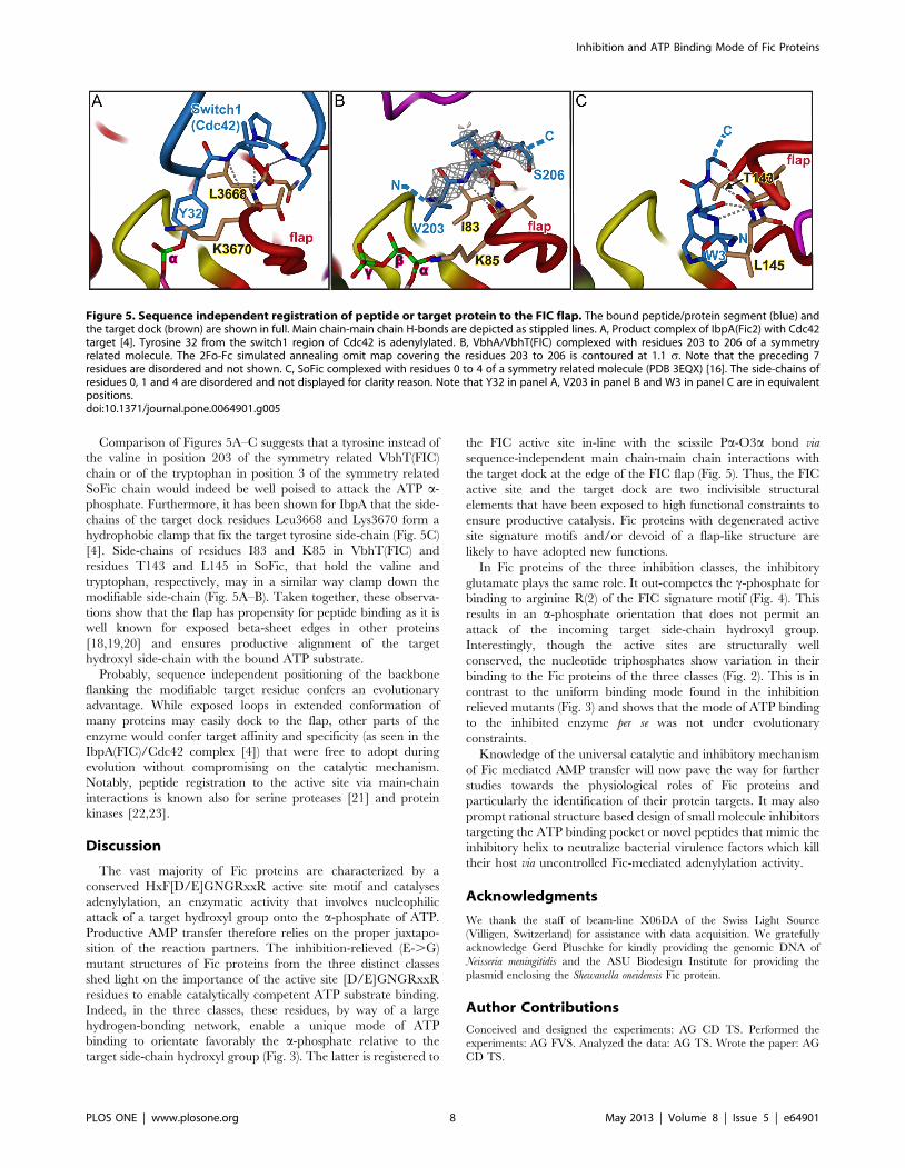
Comparison of Figures 5A–C suggests that a tyrosine instead of
the valine in position 203 of the symmetry related VbhT(FIC)
chain or of the tryptophan in position 3 of the symmetry related
SoFic chain would indeed be well poised to attack the ATP a-
phosphate. Furthermore, it has been shown for IbpA that the side-
chains of the target dock residues Leu3668 and Lys3670 form a
hydrophobic clamp that fix the target tyrosine side-chain (Fig. 5C)
[4]. Side-chains of residues I83 and K85 in VbhT(FIC) and
residues T143 and L145 in SoFic, that hold the valine and
tryptophan, respectively, may in a similar way clamp down the
modifiable side-chain (Fig. 5A–B). Taken together, these observa-
tions show that the flap has propensity for peptide binding as it is
well known for exposed beta-sheet edges in other proteins
[18,19,20] and ensures productive alignment of the target
hydroxyl side-chain with the bound ATP substrate.
Probably, sequence independent positioning of the backbone
flanking the modifiable target residue confers an evolutionary
advantage. While exposed loops in extended conformation of
many proteins may easily dock to the flap, other parts of the
enzyme would confer target affinity and specificity (as seen in the
IbpA(FIC)/Cdc42 complex [4]) that were free to adopt during
evolution without compromising on the catalytic mechanism.
Notably, peptide registration to the active site via main-chain
interactions is known also for serine proteases [21] and protein
kinases [22,23].
Discussion
The vast majority of Fic proteins are characterized by a
conserved HxF[D/E]GNGRxxR active site motif and catalyses
adenylylation, an enzymatic activity that involves nucleophilic
attack of a target hydroxyl group onto the a-phosphate of ATP.
Productive AMP transfer therefore relies on the proper juxtapo-
sition of the reaction partners. The inhibition-relieved (E-.G)
mutant structures of Fic proteins from the three distinct classes
shed light on the importance of the active site [D/E]GNGRxxR
residues to enable catalytically competent ATP substrate binding.
Indeed, in the three classes, these residues, by way of a large
hydrogen-bonding network, enable a unique mode of ATP
binding to orientate favorably the a-phosphate relative to the
target side-chain hydroxyl group (Fig. 3). The latter is registered to
the FIC active site in-line with the scissile Pa-O3a bond via
sequence-independent main chain-main chain interactions with
the target dock at the edge of the FIC flap (Fig. 5). Thus, the FIC
active site and the target dock are two indivisible structural
elements that have been exposed to high functional constraints to
ensure productive catalysis. Fic proteins with degenerated active
site signature motifs and/or devoid of a flap-like structure are
likely to have adopted new functions.
In Fic proteins of the three inhibition classes, the inhibitory
glutamate plays the same role. It out-competes the c-phosphate for
binding to arginine R(2) of the FIC signature motif (Fig. 4). This
results in an a-phosphate orientation that does not permit an
attack of the incoming target side-chain hydroxyl group.
Interestingly, though the active sites are structurally well
conserved, the nucleotide triphosphates show variation in their
binding to the Fic proteins of the three classes (Fig. 2). This is in
contrast to the uniform binding mode found in the inhibition
relieved mutants (Fig. 3) and shows that the mode of ATP binding
to the inhibited enzyme per se was not under evolutionary
constraints.
Knowledge of the universal catalytic and inhibitory mechanism
of Fic mediated AMP transfer will now pave the way for further
studies towards the physiological roles of Fic proteins and
particularly the identification of their protein targets. It may also
prompt rational structure based design of small molecule inhibitors
targeting the ATP binding pocket or novel peptides that mimic the
inhibitory helix to neutralize bacterial virulence factors which kill
their host via uncontrolled Fic-mediated adenylylation activity.
Acknowledgments
We thank the staff of beam-line X06DA of the Swiss Light Source
(Villigen, Switzerland) for assistance with data acquisition. We gratefully
acknowledge Gerd Pluschke for kindly providing the genomic DNA of
Neisseria meningitidis and the ASU Biodesign Institute for providing the
plasmid enclosing the Shewanella oneidensis Fic protein.
Author Contributions
Conceived and designed the experiments: AG CD TS. Performed the
experiments: AG FVS. Analyzed the data: AG TS. Wrote the paper: AG
CD TS.
Figure 5. Sequence independent registration of peptide or target protein to the FIC flap. The bound peptide/protein segment (blue) andthe target dock (brown) are shown in full. Main chain-main chain H-bonds are depicted as stippled lines. A, Product complex of IbpA(Fic2) with Cdc42target [4]. Tyrosine 32 from the switch1 region of Cdc42 is adenylylated. B, VbhA/VbhT(FIC) complexed with residues 203 to 206 of a symmetryrelated molecule. The 2Fo-Fc simulated annealing omit map covering the residues 203 to 206 is contoured at 1.1 s. Note that the preceding 7residues are disordered and not shown. C, SoFic complexed with residues 0 to 4 of a symmetry related molecule (PDB 3EQX) [16]. The side-chains ofresidues 0, 1 and 4 are disordered and not displayed for clarity reason. Note that Y32 in panel A, V203 in panel B and W3 in panel C are in equivalentpositions.doi:10.1371/journal.pone.0064901.g005
Inhibition and ATP Binding Mode of Fic Proteins
PLOS ONE | www.plosone.org 8 May 2013 | Volume 8 | Issue 5 | e64901

References
1. Worby CA, Mattoo S, Kruger RP, Corbeil LB, Koller A, et al. (2009) The fic
domain: regulation of cell signaling by adenylylation. Molecular cell 34: 93–103.2. Yarbrough ML, Li Y, Kinch LN, Grishin NV, Ball HL, et al. (2009) AMPylation
of Rho GTPases by Vibrio VopS disrupts effector binding and downstreamsignaling. Science 323: 269–272.
3. Palanivelu DV, Goepfert A, Meury M, Guye P, Dehio C, et al. (2011) Fic
domain-catalyzed adenylylation: insight provided by the structural analysis ofthe type IV secretion system effector BepA. Protein science : a publication of the
Protein Society 20: 492–499.4. Xiao J, Worby CA, Mattoo S, Sankaran B, Dixon JE (2010) Structural basis of
Fic-mediated adenylylation. Nature structural & molecular biology 17: 1004–
1010.5. Luong P, Kinch LN, Brautigam CA, Grishin NV, Tomchick DR, et al. (2010)
Kinetic and structural insights into the mechanism of AMPylation by VopS Ficdomain. The Journal of biological chemistry 285: 20155–20163.
6. Mukherjee S, Liu X, Arasaki K, McDonough J, Galan JE, et al. (2011)Modulation of Rab GTPase function by a protein phosphocholine transferase.
Nature 477: 103–106.
7. Kinch LN, Yarbrough ML, Orth K, Grishin NV (2009) Fido, a novelAMPylation domain common to fic, doc, and AvrB. PloS one 4: e5818.
8. Engel P, Goepfert A, Stanger FV, Harms A, Schmidt A, et al. (2012)Adenylylation control by intra- or intermolecular active-site obstruction in Fic
proteins. Nature 482: 107–110.
9. Schulein R, Guye P, Rhomberg TA, Schmid MC, Schroder G, et al. (2005) Abipartite signal mediates the transfer of type IV secretion substrates of Bartonella
henselae into human cells. Proceedings of the National Academy of Sciences ofthe United States of America 102: 856–861.
10. Kabsch W (2010) Xds. Acta crystallographica Section D, Biological crystallog-raphy 66: 125–132.
11. McCoy AJ, Grosse-Kunstleve RW, Adams PD, Winn MD, Storoni LC, et al.
(2007) Phaser crystallographic software. Journal of applied crystallography 40:658–674.
12. Emsley P, Lohkamp B, Scott WG, Cowtan K (2010) Features and developmentof Coot. Acta crystallographica Section D, Biological crystallography 66: 486–
501.
13. Adams PD, Afonine PV, Bunkoczi G, Chen VB, Davis IW, et al. (2010)PHENIX: a comprehensive Python-based system for macromolecular structure
solution. Acta crystallographica Section D, Biological crystallography 66: 213–
221.
14. Murshudov GN, Vagin AA, Dodson EJ (1997) Refinement of macromolecular
structures by the maximum-likelihood method. Acta crystallographica Section
D, Biological crystallography 53: 240–255.
15. Chen VB, Arendall WB 3rd, Headd JJ, Keedy DA, Immormino RM, et al.
(2010) MolProbity: all-atom structure validation for macromolecular crystallog-
raphy. Acta crystallographica Section D, Biological crystallography 66: 12–21.
16. Das D, Krishna SS, McMullan D, Miller MD, Xu Q, et al. (2009) Crystal
structure of the Fic (Filamentation induced by cAMP) family protein SO4266
(gi|24375750) from Shewanella oneidensis MR-1 at 1.6 A resolution. Proteins
75: 264–271.
17. Watson JD, Milner-White EJ (2002) A novel main-chain anion-binding site in
proteins: the nest. A particular combination of phi,psi values in successive
residues gives rise to anion-binding sites that occur commonly and are found
often at functionally important regions. Journal of molecular biology 315: 171–
182.
18. Hill CP, Yee J, Selsted ME, Eisenberg D (1991) Crystal structure of defensin
HNP-3, an amphiphilic dimer: mechanisms of membrane permeabilization.
Science 251: 1481–1485.
19. Nassar N, Horn G, Herrmann C, Scherer A, McCormick F, et al. (1995) The 2.2
A crystal structure of the Ras-binding domain of the serine/threonine kinase c-
Raf1 in complex with Rap1A and a GTP analogue. Nature 375: 554–560.
20. Doyle DA, Lee A, Lewis J, Kim E, Sheng M, et al. (1996) Crystal structures of a
complexed and peptide-free membrane protein-binding domain: molecular basis
of peptide recognition by PDZ. Cell 85: 1067–1076.
21. Wilmouth RC, Clifton IJ, Robinson CV, Roach PL, Aplin RT, et al. (1997)
Structure of a specific acyl-enzyme complex formed between beta-casomorphin-
7 and porcine pancreatic elastase. Nature structural biology 4: 456–462.
22. Hubbard SR (1997) Crystal structure of the activated insulin receptor tyrosine
kinase in complex with peptide substrate and ATP analog. The EMBO journal
16: 5572–5581.
23. Yang J, Cron P, Good VM, Thompson V, Hemmings BA, et al. (2002) Crystal
structure of an activated Akt/protein kinase B ternary complex with GSK3-
peptide and AMP-PNP. Nature structural biology 9: 940–944.
Inhibition and ATP Binding Mode of Fic Proteins
PLOS ONE | www.plosone.org 9 May 2013 | Volume 8 | Issue 5 | e64901