consequences of the photodynamic treatment of resting and … · 2012-04-04 · consequences of the...
TRANSCRIPT
Ž .Immunopharmacology 41 1999 31–44
Consequences of the photodynamic treatment of resting andactivated peripheral T lymphocytes
David W.C. Hunt a,b,), Huijun Jiang a, David J. Granville a,b, Agnes H. Chan a,Simon Leong a, Julia G. Levy a,c
a QLT Photo Therapeutics, 520 West 6th AÕenue, VancouÕer, B.C., Canada V5Z 4H5b CardioÕascular Research Laboratory, Department of Pathology and Laboratory Medicine, St. Paul’s Hospital, UniÕersity of British
Columbia, 1081 Burrard Street, VancouÕer, B.C., Canada V6Z 1Y6c Department of Microbiology and Immunology, Faculty of Science, UniÕersity of British Columbia, 300-6174 UniÕersity BouleÕard,
VancouÕer, B.C., Canada V6T 1W5
Accepted 24 September 1998
Abstract
Ž .The impact of the immunomodulatory photosensitizer benzoporphyrin derivative monoacid ring A BPD-MA, verteporfinand visible light on the survival and surface receptor pattern of resting and activated murine T cells was evaluated. T cellstreated for 48 h with immobilized anti-CD3 monoclonal antibody upregulated expression of the interleukin-2 receptor
Ž . Ž . Ž .a-chain CD25 , transferrin receptor CD71 , the apoptosis-regulating Fas receptor CD95 , contained a greater level of theanti-apoptotic protein Bcl-2 and accumulated significantly more BPD-MA than their unactivated counterparts. Activated Tcells displayed a modestly greater susceptibility to the photodynamic induction of DNA fragmentation than resting T cells.Resting T cells treated with sub-lethal levels of BPD-MA and light did not exhibit changes in surface levels of CD3, CD4,
Ž .CD8, CD28, CD45 or T cell receptor TCR b-chain structures. However, levels of major histocompatibility complexŽ . Ž .MHC class I antigens were decreased while the density of Thy-1.2 CD90 increased on these cells. Photodynamicallytreated T cells failed to express optimal CD25 levels when exposed to the mitogenic anti-CD3 antibody. Activated T cellstreated with sub-lethal levels of BPD-MA and light exhibited lower CD25 levels, a temporary block in cell cycle transition,
Ž .but unaltered expression of MHC Class I, CD3, CD4, CD8, CD45, CD54, CD71, CD122 IL-2R b-chain or TCR b-chainantigens 24 h afterward. Resting and activated T lymphocytes differ in susceptibility to PDT-mediated apoptosis but bothtypes are sensitive to anti-proliferative effects the treatment exerts at sub-lethal photosensitizer levels. The marked sensitivityof activated T cells to photodynamic inactivation likely contributes to the immunomodulatory action of BPD-MA. q 1999Elsevier Science B.V. All rights reserved.
Keywords: Apoptosis; Interleukin-2 receptor; Lymphocyte activation; Photodynamic therapy; Photosensitizers; T lymphocytes
Abbreviations: BPD-MA, benzoporphyrin derivative monoacid ring A; FACS, fluorescence activated cell sorter; ICAM-1, intercellularadhesion molecule-1; FITC, fluorescein isothiocyanate; IL-2R, interleukin-2 receptor; LED, light emitting diode; MHC, major histocompati-
Ž .bility complex; MTT, 3- 4,5-dimethylthiazol-2-yl -2,5-diphenyl tetrazolium bromide; PDT, photodynamic therapy; PE, phycoerythrin; PI,propidium iodide; PWM-SCCM, pokeweed mitogen-stimulated spleen cell-conditioned medium; TCR, T cell receptor
) Corresponding author. Tel.: q1-604-872-7881; Fax: q1-604-875-0001; E-mail: [email protected]
0162-3109r99r$ - see front matter q 1999 Elsevier Science B.V. All rights reserved.Ž .PII: S0162-3109 98 00051-4
( )D.W.C. Hunt et al.r Immunopharmacology 41 1999 31–4432
1. Introduction
Ž .Photodynamic therapy PDT in its conventionalform is a cancer treatment that utilizes light-sensitizecompounds, commonly porphyrin derivatives, to
Žachieve clinical effects Gomer et al., 1989; Hender-.son and Dougherty, 1992 . In comparison to cancer
indications, immunologic applications of this proce-dure have received comparatively little attention. Theporphyrin photosensitizers hematoporphyrin deriva-
Ž . Ž . wtive HpD Elmets and Bowen, 1986 , PhotofrinŽ .Musser and Fiel, 1991 and benzoporphyrin deriva-
Ž .tive monoacid ring A BPD-MA, verteporfinŽ .Simkin et al., 1997 in combination with visiblelight irradiation impaired the immunologically-mediated murine contact hypersensitivity response.BPD-MA has a light absorption spectrum whichpermits photodynamic activation at 690 nm, a wave-length of light which effectively penetrates living
Ž .tissue Levy, 1995 . BPD-MA combined with wholebody red light exposure inhibited the development ofexperimental murine T cell-mediated autoimmune
Žconditions including adjuvant arthritis Chowdhary.et al., 1994; Ratkay et al., 1994 and an adoptively-
transferred form of autoimmune encephalomyelitisŽ .Leong et al., 1996 without altering parameters of
Žcentral immune responsiveness Chowdhary et al.,.1994; Leong et al., 1996 . It is apparent that PDT
may be utilized to influence immune reactivity.No overriding mechanism to account for the im-
munomodulatory action of PDT has been unveiled,although evidence for the selective depletion of acti-
Žvated T lymphocytes has been forwarded Obochi et.al., 1995 . Mitogen-activated murine splenocytes
were more sensitive to photodynamic killing withŽ .HpD than quiescent spleen cells Canti et al., 1981 .
Activated murine spleen cells accumulated greateramounts of BPD-MA and were more susceptible tophotodynamic killing with this compound than their
Ž .resting counterparts Obochi et al., 1995 . As thisearlier investigation employed unseparated spleencells as the test population, it was important todetermine how BPD-MA might affect the survival ofpurified T lymphocytes in order to minimize possibleinfluences of other cell types. Since T lymphocytesplay a pivotal role in many aspects of immunity, it iscritical to define the impact of PDT upon this celltype.
In the present work, it was found that T cellsactivated by antibody cross-linking of the T cell
Ž .receptor TCR complex in vitro were more suscepti-ble to the induction of apoptosis with BPD-MA andlight than their resting counterparts. Furthermore,treatment at sub-lethal levels of BPD-MA and lightinfluenced the expression of specific surface recep-tors and growth characteristics of resting and acti-vated T cells. The photodynamic impairment of acti-vated T lymphocytes may contribute to the im-munomodulatory action of PDT.
2. Materials and methods
2.1. Animals, T cell purification and actiÕation
ŽMale DBAr2 mice Charles River Canada, St..Constance Quebec of 8–12 weeks of age served as
cell donors for all experiments. Animals were keptunder 12 h light:12 h dark and supplied with stan-
Ž .dard rodent laboratory food Ralston Purina andacidified water ad libitum. Animal maintenance wasin accordance with the Canadian Council of AnimalCare guidelines.
RPMI 1640 medium containing 5% heat-in-Ž . Žactivated fetal calf serum FCS , penicillin 100
. Ž .Urml , streptomycin 100 mgrml , all from GibcoŽ .BRL Burlington, Ontario with 50 mM 2-mercapto-
ethanol was used for all cell work. T lymphocyteswere isolated by passing erythrocyte-depleted spleencell suspensions over Type 200 L nylon scrubbed
Ž .wool DuPont Canada, Mississauga, Ontario or TŽcell immuno-affinity R&D Systems, Minneapolis,
.MN columns. Preparations routinely consisted of)85% T cells and -10% B cells as indicated bytheir labeling with the monoclonal antibodies 145-
Ž .2C11 anti-CD3, Leo et al., 1987 and RA3-6B2Ž .anti-CD45R-B220, Coffman, 1982 , respectively.CD4q and CD8q T lymphocytes were presentwithin these preparations in a ratio of approximately2.5:1 as determined by flow cytometric analysis.Fewer than 2% of the isolated cells labeled with the
Ž d. Žanti-MHC Class II I-A antibody 39-10-8 Phar-.Mingen, San Diego, CA .
T cell activation was achieved with immobilizedŽ .anti-CD3 antibody Jenkins et al., 1990 . Mono-
( )D.W.C. Hunt et al.r Immunopharmacology 41 1999 31–44 33
Ž . Žclonal antibody 145-3C11 Leo et al., 1987 Boeh-.ringer Mannheim, Dorval, Quebec was coated onto
Žsterile flat-bottomed polystyrene petri dishes 10. Ž .mlrplate or 96-well microtiter plates 0.1 mlrwell
at 10 mgrml in sterile phosphate-buffered salineŽ .PBS . Microtiter plates were employed for cytotoxi-city assays while petri dishes were used to prepareactivated T cells for surface antigen studies. After 3h at 378C, the coating solution was removed andcontainers were washed 3 times with PBS. T cellsŽ 5 . Ž5=10 cellsrml were cultured in 0.2 ml 96-well
. Ž .plates or 10 ml petri dishes volumes at 378C under5% CO . In select experiments, cultures were sup-2
plemented with recombinant human interleukin-2Ž .rIL-2, Amgen Biologicals, Thousand Oaks, CA .Cells were harvested by gentle swirling and removedby pipet.
2.2. Photodynamic treatment and assessment of cellÕiability
ŽLiposomally-formulated BPD-MA QLT Pho-.toTherapeutics, Vancouver, B.C., Canada was re-
constituted at 2.1 mgrml with sterile, distilled water.Further dilution was with culture medium. All cellwork with the photosensitizer was carried out underlight-attenuated conditions. To test the impact ofPDT upon T cell activation, freshly purified T lym-phocytes were seeded into 96-well microtiter platesat 1=105 cells per well in 0.2 ml. Cells wereincubated with BPD-MA at 378C in the dark for 1 h,exposed to 690 nm light delivered from light emit-
Ž . Ž .ting diodes LED Hunt et al., 1995 and added toanti-CD3 antibody-coated plates. T cells activatedfor 48 h prior to PDT were treated with BPD-MAand light as described above for resting T cellsexcept that these cells remained in the presence ofthe anti-CD3 antibody during all manipulations. Pro-liferative responses and cytotoxicity were assessed
Ž w xby the MTT 3- 4-,5-dimethylthiazol-2-yl -2,4-di-.phenyl tetrazolium bromide, Sigma colorimetric as-
Ž .say Mosmann, 1983; Hunt et al., 1995 . Replicatesof four were performed at each BPD-MA concentra-tion. Color development was terminated after 4 h at378C in the presence of MTT and read with an
Žautomated microtiter plate reader Dynatech, Hamil-.ton, VA at a wavelength of 590 nm. Absorbance
values for wells containing medium alone were sub-
tracted from the result obtained for wells containingcells. Results are given as a percentage of the ab-sorbance obtained for cells treated with light alone.The BPD-MA concentration required to produce a
Ž50% reduction in cell viability lethal dose 50%,. ŽLD or a 50% inhibition inhibitory concentration50
.50%, IC of the response to the anti-CD3 antibody50
was interpolated from regression lines plotting BPD-MA concentration vs. % cell response.
Cell viability was also assessed by propidiumŽ .iodide PI dye exclusion studies. PI is excluded
from living cells. PI was added at 20 mgrml to;1=105 cells and cell fluorescence was read im-mediately using the Elite software package with an
ŽEpics XL flow cytometer Coulter Electronics,.Hialeah, FL . The strong fluorescent signal of PI
clearly distinguishes dead from live cells.To evaluate the effect of PDT upon surface recep-
tor expression of resting T cells, these cells weremaintained for 24 h after treatment in the presence of10% pokeweed mitogen spleen cell conditioned
Žmedium PWM-SCCM, StemCell, Vancouver,.Canada as a source of cell maintenance factors.
Cells were harvested and labeled with monoclonalantibodies as described below.
( )2.3. Fluorescence-actiÕated cell sorter FACS anal-ysis
To assess their purity, activation state or influenceof PDT upon antigen expression, ;5=105 cells in0.2 ml of PBS containing 5% FCS and 0.05% NaN3
were incubated with fluorescein isothiocyanateŽ .FITC -conjugated monoclonal antibodies specific for
Ž d. ŽMHC class I H-2D , CD3 ´-chain Leo et al.,. Ž .1987 , CD4, CD8, IL-2 receptor IL-2R a-chain
Ž . Ž . ŽCD25 Ortega et al., 1984 , CD28 Gross et al.,. Ž . Ž1992 , leukocyte common antigen CD45 Ledbetter
.and Herzenberg, 1979 , intercellular adhesionŽ . Žmolecule-1 ICAM-1, CD54 Scheynius et al.,
. Ž . Ž1993 , Thy-1.2 CD90 Ledbetter and Herzenberg,
. Ž . Ž .1979 , Fas CD95 Ogasawara et al., 1993 , IL-2RŽ . Ž .b-chain CD122 Tanaka et al., 1991 or the TCRŽ . Ž .b-chain Kubo et al., 1989 . A phycoerythrin PE -
conjugated antibody against the transferrin receptorŽ . Ž .CD71 Kemp et al., 1987 was employed. Thedistribution of the structures recognized by thesemonoclonal antibodies is described in Table 1. PE-
( )D.W.C. Hunt et al.r Immunopharmacology 41 1999 31–4434
Table 1Distribution and function of the T lymphocyte surface antigens evaluated in this study
Antigen Cellular distribution Function
CD3 ´-chain pan-T cell TCR signal transductionCD4 helper T cells TCR co-receptor, MHC Class II recognitionCD8 cytotoxic T cells TCR co-receptor, MHC Class I recognition
Ž .CD25 IL-2R a-chain activated lymphocytes IL-2 bindingCD28 most T cells CD80rCD86 counter-receptor, signal transduction
Ž .CD45 leukocyte common antigen pan-leukocyte signal transductionŽ .CD54 ICAM-1 activated leukocytes cell adhesionŽ .CD71 transferrin receptor proliferating cells iron uptakeŽ .CD90 Thy-1.2 pan-T cell signal transduction?Ž .CD95 Fas activated T cells apoptotic signalingŽ .CD122 IL-2R b-chain most T cells IL-2 binding, signal transduction
TCR b-chain most T cells peptide antigen recognitionMHC Class I all nucleated cells peptide antigen presentation
and FITC-conjugated antigen-specific and isotypecontrol monoclonal antibodies were from PharMin-gen. After 30 min on ice, cells were washed twicewith buffer and fixed in 1% p-formaldehyde in PBS.
Ž .Fluorescence signals 5000 cells analyzed were ana-lyzed with the flow cytometer. Dead cells and debriswere gated out. Mean channel fluorescence intensityŽ . Ž .MCFI values in arbitrary units were obtained. Tonormalize data from different experiments, cell sur-face antigen levels were expressed as a percentage ofthe MCFI determined for cells treated with lightalone. A PDT-induced change in the MCFI value of
Ž .G20% with a standard deviation S.D. less thanthis difference was considered significantly differentfrom the control result. Exposure of light-protected,resting and anti-CD3 activated T cells to BPD-MAup to 200 ngrml did not alter the labeling efficiency
Žof the antibodies utilized in this study data not.shown .
2.4. Cell cycle analysis
T cell cycle status was evaluated using PI to stainŽ . Žnuclear DNA Fleming et al., 1993 . Cells 5=
5 .10 rtube were washed twice with PBS and treatedwith 1 ml of PBS containing 1% Triton X-100, PIŽ . Ž10 mgrml and DNAse-free, RNAse A 100
.mgrml , all from Sigma, for 30 min at 48C in theŽ .dark. Fluorescence 5000 nuclei analyzed was mea-
sured by flow cytometry on a logarithmic scale at620 nm. The resultant histogram was divided into
Ž .nuclei with 2N DNA G0rG1 stages , G2N DNAŽ .SrG2rM stages and total DNA.
2.5. Preparation of cell extracts and detection ofBcl-2
To prepare whole cell lysates, T lymphocyteswere first washed twice with ice-cold PBS. Cell
Žpellets were treated with 1 ml of lysis buffer 1%Nonidet P-40, 10% glycerol, 137 mM NaCl, 20 mMTris pH 8.0 supplemented with 1 mM phenylmethyl-
Ž .sulfonyl fluoride, aprotinin 0.15 Urml and 1 mM.sodium orthovanadate for 20 min on ice and cen-
trifuged for 10 min at 15,800=g at 48C. Cell extractprotein concentrations were determined with the
Ž . ŽBCAe assay Pierce, Rockford, IL and separated 6.mg proteinrlane by sodium dodecyl sulfate poly-
acrylamide gel electrophoresis in 12% gels underŽ .reducing conditions Laemmli, 1970 . Proteins were
transferred to nitrocellulose membrane at 100 V for60 min. Membranes were blocked for 30 min atroom temperature in PBS with 5% skim milk powder
Ž .and 0.05% Tween 20 PBS-T and treated for 45 minat room temperature with a rabbit IgG anti-Bcl-2
Žantibody Santa Cruz Biotechnology, Santa Cruz,.CA at 1 mgrml. Membranes were washed with
PBS-T and then probed with anti-rabbit IgG-Ž . Žhorseradish peroxidase Santa Cruz 1:5000 in PBS-
.T with 1% skim milk powder for 30 min at roomtemperature. Membranes were washed extensively

( )D.W.C. Hunt et al.r Immunopharmacology 41 1999 31–44 35
with PBS-T and proteins were revealed with theenhanced chemiluminescence detection systemŽ .Amersham, Arlington Heights, IL . Blots were
Žviewed with an HP ScanJet 4c Hewlett Packard,.Palo Alto, CA and band densities were measured
Žusing 1D Image Analysis Software Eastman Kodak,.Rochester, NY .
2.6. BPD-MA uptake
To assess the association of BPD-MA with restingand anti-CD3 activated T cells, 5=105 cells were
Žincubated with the photosensitizer 0, 10 or 100.ngrml in 0.5 ml of medium containing 5% FCS at
378C in the dark for 1 h. Cells were washed once andanalyzed immediately with the flow cytometer usinga laser excitation wavelength of 488 nm and an
Ž .emission wavelength of 690 nm Hunt et al., 1995 .MCFI values were obtained for each sample.
2.7. Detection of DNA fragmentation
DNA degradation was measured by PI stainingŽ .and flow cytometry Nicoletti et al., 1991 . Three
hours following light irradiation, 5=105 cells werewashed twice with ice-cold PBS and fixed in 80%ethanol at 48C for 1 h. Cells were washed with PBS
Ž .and stained with PI 50 mgrml in PBS withŽ .DNAse-free RNAse 5 Urml . Fluorescence was
measured by single parameter flow cytometry. TheŽ .percentage of apoptotic hypodiploid -2N DNA
cells was calculated from the resultant histogram.PDT-induced DNA fragmentation was calculatedfrom the equation:
PDT-treated cells % -2N DNA -light-treated cells % -2N DNAŽ . Ž .DNA fragmentations =100.
100-light-treated cells % -2N DNA .Ž .
3. Results
3.1. T cell characterization and induction of DNAfragmentation by PDT
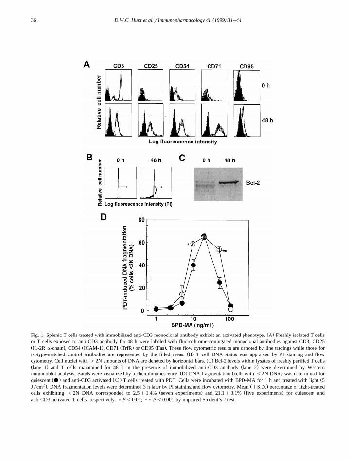
Freshly purified CD3q splenic T cells expressedŽ .low CD54, CD71 and CD95 levels Fig. 1A . CD25
was undetectable for these cells. T cells culturedwith immobilized anti-CD3 monoclonal antibody for48 h exhibited positive surface expression of CD25,CD54, CD71 and CD95. CD3 levels were lower onanti-CD3 activated T cells than resting T cells. Timecourse studies demonstrated that T cell CD25 levelsincreased from 0 to 72 h and were declining by 96 h
Žafter exposure to the anti-CD3 antibody data not.shown . Evaluation of cell cycle status indicated that
Ž .the vast majority ;98% of freshly isolated T cellsŽ .contained 2N amounts of DNA Fig. 1B . After 48 h
in the presence of the anti-CD3 antibody, a sizeableŽ .proportion ;25% of cell nuclei contained )2N
levels of DNA. Bcl-2 levels were over 5-fold higherwithin extracts prepared from activated T cells thanresting T cells as determined by Western im-munoblot analysis and gel band image densitometryŽ .Fig. 1C . T cells maintained in medium for 48 h didnot metabolize the MTT dye, while T cells culturedwith anti-CD3 antibody for 48 h exhibited strong
Ž .MTT dye reduction activity data not shown . Forresting and anti-CD3 activated T cells, PDT-media-ted DNA fragmentation was detected at photosensi-tizer concentrations from 5 to 50 ngrml at 3 h
Ž .post-irradiation Fig. 1D . At BPD-MA concentra-tions of 10 or 50 ngrml, a significantly greaterproportion of activated T cells exhibited photody-namically-induced DNA fragmentation than restingT cells. Fewer quiescent T cells treated with BPD-MA at 50 ngrml exhibited DNA fragmentation than
Ž .at certain lower concentrations 10 and 20 ngrml ofthe photosensitizer. Similar levels of DNA fragmen-tation were produced by 3 h for light-irradiatedanti-CD3 activated T cells treated with BPD-MA at10, 20 or 50 ngrml. PDT-induced DNA fragmenta-tion was not detectable at this sampling time forresting or activated T cells when BPD-MA concen-trations of -5 or at 100 ngrml were employed.
3.2. BPD-MA uptake by resting and actiÕated T cells
BPD-MA uptake by 48 h anti-CD3 activated Tcells was significantly greater than that of freshlyisolated T cells as determined by flow cytometryŽ .Fig. 2A . At 690 nm, the fluorescence strength ofanti-CD3 activated T cells was approximately 2.5
( )D.W.C. Hunt et al.r Immunopharmacology 41 1999 31–4436
Ž .Fig. 1. Splenic T cells treated with immobilized anti-CD3 monoclonal antibody exhibit an activated phenotype. A Freshly isolated T cellsor T cells exposed to anti-CD3 antibody for 48 h were labeled with fluorochrome-conjugated monoclonal antibodies against CD3, CD25Ž . Ž . Ž . Ž .IL-2R a-chain , CD54 ICAM-1 , CD71 TrfR or CD95 Fas . These flow cytometric results are denoted by line tracings while those for
Ž .isotype-matched control antibodies are represented by the filled areas. B T cell DNA status was appraised by PI staining and flowŽ .cytometry. Cell nuclei with )2N amounts of DNA are denoted by horizontal bars. C Bcl-2 levels within lysates of freshly purified T cells
Ž . Ž .lane 1 and T cells maintained for 48 h in the presence of immobilized anti-CD3 antibody lane 2 were determined by WesternŽ . Ž .immunoblot analysis. Bands were visualized by a chemiluminescence. D DNA fragmentation cells with -2N DNA was determined for
Ž . Ž . Žquiescent v and anti-CD3 activated ` T cells treated with PDT. Cells were incubated with BPD-MA for 1 h and treated with light 52 . Ž .Jrcm . DNA fragmentation levels were determined 3 h later by PI staining and flow cytometry. Mean "S.D. percentage of light-treated
Ž . Ž .cells exhibiting -2N DNA corresponded to 2.5"1.4% seven experiments and 21.1"3.1% five experiments for quiescent andanti-CD3 activated T cells, respectively. ) P-0.01; )) P-0.001 by unpaired Student’s t-test.

( )D.W.C. Hunt et al.r Immunopharmacology 41 1999 31–44 37
Ž .Fig. 2. A T cells activated for 48 h with anti-CD3 antibody accumulate greater amounts of BPD-MA than freshly isolated T cells. Cellswere incubated with BPD-MA for 1 h at 378C, washed with medium and cell-associated fluorescence determined immediately by flowcytometry at an absorbance wavelength of 690 nm. The fluorescence of cells not exposed to BPD-MA corresponds to cell autofluorescence.
Ž .MCFI values were determined from three to four independent experiments. B Profiles of cell-associated BPD-MA fluorescence for restingŽ . Ž . Ž .and 48 h anti-CD3 activated T lymphocytes incubated with BPD-MA at 0 light lines , 10 hatched lines or 100 bold lines ngrml are
presented. ) P-0.005; )) P-0.001 by paired Student’s t-test.
times greater than of resting cells whether these cellswere incubated with BPD-MA at 10 or 100 ngrml.The autofluorescence of 48 h anti-CD3 activated T
Ž .cells MCFIs0.57"0.14 was modestly greaterŽthan that of freshly isolated T cells MCFIs0.33"
.0.10 . Representative flow cytometric profiles forBPD-MA-associated fluorescence of resting and acti-vated T cells are presented in Fig. 2B.
3.3. Influence of PDT upon the surface receptorpattern of resting T cells
Surface TCR b-chain, CD3, CD4, CD8, CD28and CD45 levels for unactivated T cells treated with
BPD-MA and light and then maintained at 378C for24 h were no different than those treated with light
Ž .alone Fig. 3A . CD90 levels exhibited a dose-re-lated increase of up to 60% at this sampling time.MHC Class I levels were approximately 20% lowerafter treatment at the highest concentration of BPD-MA. There was no significant difference in the levelof interaction of isotype control antibodies withlight-treated or PDT-treated cells. In the absence oflight, BPD-MA did not affect the surface receptor
Žexpression levels of inactivated T cells data not.shown . Flow cytometric profiles of the labeling
patterns obtained for CD45, CD90 and MHC Class Iare shown in Fig. 3B.

( )D.W.C. Hunt et al.r Immunopharmacology 41 1999 31–4438
Fig. 3. Treatment of resting T cells with PDT increases surfaceŽ .CD90 Thy-1.2 expression and lowers levels of MHC Class I
Ž .antigens. A Freshly purified T cells were incubated with BPD-Ž . Ž . Ž .MA at 1 cross-hatched , 2.5 I or 5 B ngrml for 1 h at 378C,
Ž 2 .exposed to 690 nm light 5 Jrcm and maintained for 24 h inmedium containing 10% PWM-SCCM. Cells were labeled withmonoclonal antibodies against different surface antigens and thenanalyzed by flow cytometry. Results, given as a percentage of theMCFI value obtained for cells treated with light alone, wereobtained in three to four independent experiments. CD8 expres-sion was not evaluated for cells treated at the lowest BPD-MA
Ž .concentration. B Flow cytometric profiles for MHC Class I,Ž .CD45 and CD90 line tracings and the isotype control antibodies
Ž .filled areas for T cells analyzed 24 h following PDT are shown.Results for CD45 exhibit an example of antigen expression unal-tered after PDT. MCFI values obtained with the control andantigen-specific antibodies are given within each panel.
3.4. T cells treated with BPD-MA and light exhibit areduced proliferatiÕe potential and depressed CD25leÕels
For light-irradiated resting T cells treated over alimited concentration range of BPD-MA, DNA frag-
mentation levels produced by 3 h post-irradiationwere inversely related to the capacity of these cellsto proliferate in response to immobilized anti-CD3antibody as determined by MTT dye reduction 48 h
Ž .later Fig. 4A . Maximum levels of DNA fragmenta-tion were produced with BPD-MA at 20 ngrml. At
Ž .higher concentrations 50 and 100 ngrml of BPD-MA, little or no PDT-induced DNA fragmentationwas detectable although these cells did not prolifer-ate in the presence of the anti-CD3 antibody. For Tcells treated with PDT prior to exposure to anti-CD3antibody, the IC for BPD-MA corresponded to50
Ž .3.5"0.3 ngrml three experiments , as determinedby the MTT method. Cell viability was preserved at
Žhigher concentrations of BPD-MA IC s6.2"0.350.ngrml, three experiments as assessed by PI dye
exclusion studies. In comparison to T cells treatedwith light alone prior to their exposure to anti-CD3antibody, CD25 levels of PDT-treated cells werelower in a drug dose-related manner, resulting inapproximately 75% lower CD25 levels at the highest
Ž . Ž .concentration 5 ngrml of BPD-MA Fig. 4B .Representative flow cytometric profiles for these
Ž .studies are shown Fig. 4C . For light-irradiated Tcells treated with BPD-MA at 1 and 2.5 ngrml, butnot 5 ngrml, supplementation with rIL-2 was associ-ated with CD25 expression levels equivalent to thoseof light-treated control cells after 48 h in the pres-ence of anti-CD3 antibody.
3.5. Influence of PDT on surface antigen expressionand cell cycle status of anti-CD3 actiÕated T cells
For 48 h anti-CD3 activated T cells, the LD for50ŽBPD-MA corresponded to 3.3"0.9 ngrml five
.independent experiments as determined by MTTassays performed 24 h after light irradiation. For
Ž .these experiments, assay absorbance 590 nm valuesfor light-treated anti-CD3 activated T cells averaged
Ž .0.656"0.119 above background levels ;0.100 .TCR b-chain, CD3, CD4, CD8, CD45, CD71, IL-2R
Ž .b-chain CD122 and MHC Class I expression levelsof anti-CD3 activated T cells treated with BPD-MAand light 24 h previously were no different than
Ž .those of light-irradiated T cells Fig. 5A . CD95expression was modestly higher on PDT-treated cellswhile CD25 levels were lower in a drug dose-related

( )D.W.C. Hunt et al.r Immunopharmacology 41 1999 31–44 39
Ž .Fig. 4. Resting T cells treated with BPD-MA and light exhibit reduced proliferation and depressed CD25 levels. A Freshly-purified T cellsŽ 2 .were treated with BPD-MA and light 5 Jrcm and seeded into microtiter plate wells pre-coated with anti-CD3 antibody. DNA
Ž . Ž .fragmentation ^ was assessed 3 h post-irradiation seven experiments . Cell responses were assessed 48 h post-irradiation by MTT dyeŽ . Ž . Ž .reduction v and PI dye exclusion ` assays performed in parallel. For cells pretreated with light alone 100% response , PI studies
Ž .indicated that 62.5"2.6% were viable while absorbance 590 nm values for these T cells averaged 0.781"0.203 above backgroundŽ . Ž . Ž .;0.100 by MTT assay three experiments . B CD25 expression by T cells pretreated with PDT and cultured with anti-CD3 antibody for48 h was evaluated by flow cytometric analysis. Results are given as the percentage of the result obtained with cells treated with light aloneŽ . Ž .MCFIs26.9"8.0 units, four experiments . C CD25 expression for T cells treated with light alone or BPD-MA and light 48 h before
Ž .followed by exposure to immobilized anti-CD3 antibody without or with rIL-2 200 Urml was assessed. Cells were stained with anti-CD25Ž . Ž .line tracings or the isotype control antibody filled areas . MCFI values obtained with these reagents are given in each panel. Results fromone of four experiments are shown.
manner 24 h after PDT, resulting in approximately40% lower CD25 levels at the highest concentrationof BPD-MA. CD25 expression levels of anti-CD3activated T cells approximated control MCFI values
Ž .by 48 h after the photodynamic treatment Fig. 5B .There was no difference in the degree of interaction
of the control antibodies with light or PDT-treatedanti-CD3 activated T cells. In the absence of light,BPD-MA did not affect the surface receptor expres-
Žsion levels of anti-CD3 activated T cells data not.shown . The extremely bright expression of CD90 by
these cells prevented reliable assessment of the influ-

( )D.W.C. Hunt et al.r Immunopharmacology 41 1999 31–4440
Fig. 5. The photodynamic treatment of anti-CD3 activated T lymphocytes does not alter CD3, CD4, CD8, CD28, CD45, CD54, CD71,CD122 or MHC Class I expression but results in depressed CD25 levels, a modest elevation in CD95 expression and a transient block in cell
Ž . Ž . Ž . Ž . Ž 2 .cycle progression. A Activated T cells treated with BPD-MA at 1 cross-hatched , 2.5 I or 5 B ngrml and light 5 Jrcm werelabeled 24 h later with monoclonal antibodies against mouse T cell surface structures and analyzed by flow cytometry. Relative antigen
Ž . Žexpression levels means with S.D. are given as a percentage of the MCFI value of cells treated with light alone three to ten independent. Žexperiments . For anti-CD3 activated T cells treated with light, MCFI values for CD25 expression levels averaged 69.3"26.6 units 10. Ž .experiments . B Flow cytometric results for 48 h anti-CD3 activated T cells treated with BPD-MA and light 24 or 48 h before and labeled
Ž . Ž . Ž .with the isotype control antibody shaded areas or anti-CD25 line tracings and their respective MCFI values are presented. C Cell cycleanalyses were performed on T cells treated with BPD-MA and light 24 or 48 h previously using PI staining and flow cytometric analysis.
Ž .The percentage of nuclei residing in the G0rG1 phases of the cell cycle 2N DNA is given within each panel and this region is denoted byhorizontal bars.
ence of PDT upon levels of this marker. In compari-son to light-irradiated anti-CD3 activated T cells, agreater percentage of T cells treated with BPD-MAand light were in the G0rG1 phases of the cell cycle
Ž .upon evaluation 24 h post-PDT Fig. 5C . At 48 h
post-PDT, the DNA profile of photodynamically-treated cells was indistinguishable from that of cellstreated with light alone, with all groups having com-parable percentages of cells with 2N amounts ofDNA.
( )D.W.C. Hunt et al.r Immunopharmacology 41 1999 31–44 41
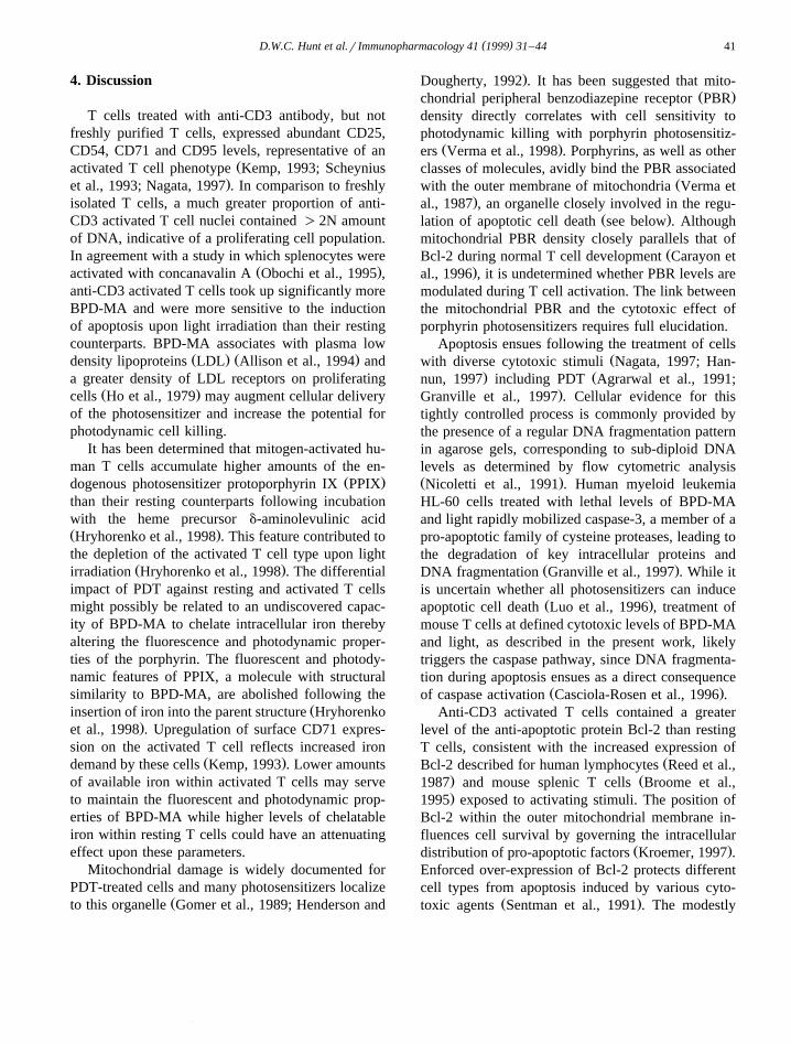
4. Discussion
T cells treated with anti-CD3 antibody, but notfreshly purified T cells, expressed abundant CD25,CD54, CD71 and CD95 levels, representative of an
Žactivated T cell phenotype Kemp, 1993; Scheynius.et al., 1993; Nagata, 1997 . In comparison to freshly
isolated T cells, a much greater proportion of anti-CD3 activated T cell nuclei contained )2N amountof DNA, indicative of a proliferating cell population.In agreement with a study in which splenocytes were
Ž .activated with concanavalin A Obochi et al., 1995 ,anti-CD3 activated T cells took up significantly moreBPD-MA and were more sensitive to the inductionof apoptosis upon light irradiation than their restingcounterparts. BPD-MA associates with plasma low
Ž . Ž .density lipoproteins LDL Allison et al., 1994 anda greater density of LDL receptors on proliferating
Ž .cells Ho et al., 1979 may augment cellular deliveryof the photosensitizer and increase the potential forphotodynamic cell killing.
It has been determined that mitogen-activated hu-man T cells accumulate higher amounts of the en-
Ž .dogenous photosensitizer protoporphyrin IX PPIXthan their resting counterparts following incubationwith the heme precursor d-aminolevulinic acidŽ .Hryhorenko et al., 1998 . This feature contributed tothe depletion of the activated T cell type upon light
Ž .irradiation Hryhorenko et al., 1998 . The differentialimpact of PDT against resting and activated T cellsmight possibly be related to an undiscovered capac-ity of BPD-MA to chelate intracellular iron therebyaltering the fluorescence and photodynamic proper-ties of the porphyrin. The fluorescent and photody-namic features of PPIX, a molecule with structuralsimilarity to BPD-MA, are abolished following the
Žinsertion of iron into the parent structure Hryhorenko.et al., 1998 . Upregulation of surface CD71 expres-
sion on the activated T cell reflects increased ironŽ .demand by these cells Kemp, 1993 . Lower amounts
of available iron within activated T cells may serveto maintain the fluorescent and photodynamic prop-erties of BPD-MA while higher levels of chelatableiron within resting T cells could have an attenuatingeffect upon these parameters.
Mitochondrial damage is widely documented forPDT-treated cells and many photosensitizers localize
Žto this organelle Gomer et al., 1989; Henderson and
.Dougherty, 1992 . It has been suggested that mito-Ž .chondrial peripheral benzodiazepine receptor PBR
density directly correlates with cell sensitivity tophotodynamic killing with porphyrin photosensitiz-
Ž .ers Verma et al., 1998 . Porphyrins, as well as otherclasses of molecules, avidly bind the PBR associated
Žwith the outer membrane of mitochondria Verma et.al., 1987 , an organelle closely involved in the regu-
Ž .lation of apoptotic cell death see below . Althoughmitochondrial PBR density closely parallels that of
ŽBcl-2 during normal T cell development Carayon et.al., 1996 , it is undetermined whether PBR levels are
modulated during T cell activation. The link betweenthe mitochondrial PBR and the cytotoxic effect ofporphyrin photosensitizers requires full elucidation.
Apoptosis ensues following the treatment of cellsŽwith diverse cytotoxic stimuli Nagata, 1997; Han-
. Žnun, 1997 including PDT Agrarwal et al., 1991;.Granville et al., 1997 . Cellular evidence for this
tightly controlled process is commonly provided bythe presence of a regular DNA fragmentation patternin agarose gels, corresponding to sub-diploid DNAlevels as determined by flow cytometric analysisŽ .Nicoletti et al., 1991 . Human myeloid leukemiaHL-60 cells treated with lethal levels of BPD-MAand light rapidly mobilized caspase-3, a member of apro-apoptotic family of cysteine proteases, leading tothe degradation of key intracellular proteins and
Ž .DNA fragmentation Granville et al., 1997 . While itis uncertain whether all photosensitizers can induce
Ž .apoptotic cell death Luo et al., 1996 , treatment ofmouse T cells at defined cytotoxic levels of BPD-MAand light, as described in the present work, likelytriggers the caspase pathway, since DNA fragmenta-tion during apoptosis ensues as a direct consequence
Ž .of caspase activation Casciola-Rosen et al., 1996 .Anti-CD3 activated T cells contained a greater
level of the anti-apoptotic protein Bcl-2 than restingT cells, consistent with the increased expression of
ŽBcl-2 described for human lymphocytes Reed et al.,. Ž1987 and mouse splenic T cells Broome et al.,.1995 exposed to activating stimuli. The position of
Bcl-2 within the outer mitochondrial membrane in-fluences cell survival by governing the intracellular
Ž .distribution of pro-apoptotic factors Kroemer, 1997 .Enforced over-expression of Bcl-2 protects differentcell types from apoptosis induced by various cyto-
Ž .toxic agents Sentman et al., 1991 . The modestly
( )D.W.C. Hunt et al.r Immunopharmacology 41 1999 31–4442
greater sensitivity to DNA fragmentation of activatedT cells, despite elevated Bcl-2 expression, suggeststhat additional regulatory factors influence the sur-vival of photodynamically-treated T cells. Relativelevels of other Bcl-2 family members of apoptosis-
Ž .regulating factors Reed, 1997 may be important inthis regard. At relatively high cytotoxic concentra-tions of BPD-MA, fewer T cells exhibited DNAfragmentation after photoirradiation than at lower yetstill cytotoxic concentrations of the photosensitizer.Outer membrane integrity may be rapidly compro-mised after the light treatment of T cells exposed tohigher amounts of BPD-MA and the caspase path-way may not be invoked and necrotic death ensues.In comparison to quiescent T cells, a greater propor-tion of anti-CD3 activated T cells exhibited DNAfragmentation at a higher concentration of BPD-MAafter light treatment. It is uncertain whether this doseeffect is related to differences in membrane stabilityor the intracellular distribution of BPD-MA betweenthese T cell forms.
Resting T cell levels of CD3, CD4, CD8, CD28,CD45 and TCR b-chain were unchanged 24 h afterPDT at sub-lethal levels of BPD-MA and light.MHC Class I density was modestly diminished whileCD90 expression levels were increased after thisphotodynamic treatment. All of the above markers,with the exception of CD90, are transmembraneproteins. The carboxy-terminus of CD90 is anchoredwithin the cell membrane by a glycosylphos-phatidylinositol moiety and molecules of this typeexhibit greater lateral mobility within the membrane
Ž .than transmembrane proteins Robinson, 1991 .ŽMembrane perturbations produced by PDT Girotti,
.1990 could conceivably affect the distribution anddensity of CD90 on the cell surface.
Resting T cells treated with sub-lethal levels ofBPD-MA and light exhibited deficient proliferativeresponses and low CD25 expression following expo-sure to the mitogenic anti-CD3 antibody. For thesecells, high levels of DNA fragmentation detected at 3h post-treatment corresponded to a complete loss ofviability and an absence of proliferation as deter-mined 48 h later. Importantly, at lower levels ofBPD-MA, photodynamically-treated T cells re-mained viable after a treatment which lead to little orno proliferation. Oxidative stress provided by hydro-gen peroxide or tumor-associated macrophages re-
duced CD3 z-chain, but not CD3 ´-chain, levels ofmouse splenic T cells and responses of these cells to
Ž .a specific antigen Otsuji et al., 1996 . AlthoughCD3 z-chain levels were not evaluated, the sub-opti-mal responses produced by photodynamically-treatedT cells was not associated with altered CD3 ´-chainlevels, a change which might have affected the ca-pacity of these cells to respond to the anti-CD3antibody. Addition of rIL-2 to cultures of T cellspreviously treated with PDT was partially effectiveat restoring CD25 levels indicating that PDT-treatedT cells may have a reduced capacity to elaborateIL-2. Pre-treatment of human blood MNC withnanomolar levels of PPIX and ultraviolet A lightsuppressed mitogen and rIL-2-stimulated prolifera-tion by preventing up-regulation of the IL-2R com-
Ž .plex Barrett et al., 1994 . PPIX and BPD-MA havesimilar molecular configurations and may producecomparable cellular effects as a result of the oxida-tive stress the photoactivation of these compoundsplaces upon immune cells.
The lower CD25 expression levels of anti-CD3activated T cells treated with BPD-MA and light wasa notable effect since no alteration in surface densityof MHC Class I, CD3, CD4, CD8, CD54, CD71,CD122 or TCR b-chain was observed. This dose-re-lated effect upon these cells was transient and CD25expression levels as well as the cell cycle statusreturned to control values by 48 h post-PDT. Bindingof IL-2 to its multimeric abg high affinity receptorprofoundly influences T lymphocyte biology by pro-moting the induction of nuclear proto-oncogenes,
Žcell proliferation and differentiation Minami et al.,.1993 . A depression in T cell CD25 expression
produced by PDT would presumably lower IL-2Rsignaling activity, slow cell cycle transit and limit Tcell differentiation. Treatment of whole blood frompatients infected with the human immunodeficiencyvirus with BPD-MA and light reduced the proportionof MNC which expressed CD25 suggesting a selec-tive depletion of activated lymphocytes from these
Ž .samples North et al., 1993 . These findings couldalso be partially explained by a dampening of CD25expression produced by the photodynamic treatment.CD95 expression levels of activated T cells weremodestly increased 24 h following photodynamictreatment. Upregulated CD95 expression may lowerthe cellular threshold to apoptosis upon interaction
( )D.W.C. Hunt et al.r Immunopharmacology 41 1999 31–44 43
with cells expressing its counter receptor, Fas-ligandŽ .Nagata, 1997 . Relationships between PDT andCD95 signaling in the induction of T lymphocyte
Žapoptosis are under study within the laboratory Jiang.et al., submitted manuscript .
PDT with BPD-MA may produce beneficial out-comes in autoimmune conditions through an accen-tuated accumulation in activated autoreactive T lym-phocytes and the induction of cell death at lownanomolar drug levels upon photoirradiation. Fur-thermore, at sub-lethal photosensitizer and light lev-els, the proliferation and differentiation of this T cellform may be restricted. The present work indicatesthat activated T cells may be selectively targeted forphotodynamic inactivation even in the setting ofupregulated expression of the anti-apoptotic factorBcl-2. This attribute may contribute to the effective-ness of PDT for the treatment of human autoimmunedisease.
References
Agrarwal, M.L., Clay, M.E., Harvey, E.J., Evans, H.H., Antunez,A.R., Oleinick, N.L., 1991. Photodynamic therapy inducesrapid cell death by apoptosis in L5178Y mouse lymphomacells. Cancer Res. 51, 5993–5996.
Allison, B.A., Pritchard, P.H., Levy, J.G., 1994. Evidence forlow-density lipoprotein-mediated uptake of benzoporphyrinderivative. Br. J. Cancer 69, 833–839.
Barrett, K.E., Yen, A., Bigby, T.D., Montisano, D., Gigli, I.,1994. Inhibition of human peripheral blood lymphocyte func-tion by protoporphyrin and longwave ultraviolet light. J. Im-munol. 153, 3286–3294.
Broome, H.E., Dargan, C.M., Bessant, E.F., Krajewski, S., Reed,J.C., 1995. Apoptosis and Bcl-2 expression in cultured murinesplenic T cells. Immunology 84, 375–382.
Canti, G., Marelli, O., Ricci, L., Nicolin, A., 1981. Haematopor-phyrin-treated murine lymphocytes: in vitro inhibition of DNAsynthesis and light-mediated inactivation of cells responsiblefor GVHR. Photochem. Photobiol. 34, 589–594.
Carayon, P., Portier, M., Dussossoy, D., Bord, A., Petitpretre, G.,Canat, X., Le Fur, G., Casellas, P., 1996. Involvement ofperipheral benzodiazepine receptors in the protection ofhematopoietic cells against oxygen radical damage. Blood 87,3170–3178.
Casciola-Rosen, L., Nicholson, D.W., Chong, T., Rowan, K.R.,Thornberry, N.A., Miller, D.K., Rosen, A., 1996.ApopainrCPP32 cleaves proteins that are essential for cellularrepair: a fundamental principle of apoptotic death. J. Exp.Med. 183, 1957–1964.
Chowdhary, R.K., Ratkay, L.G., Neyndorff, H.C., Richter, A.,Obochi, M., Waterfield, J., Levy, J.G., 1994. The use of
transcutaneous photodynamic therapy in the prevention ofadjuvant-enhanced arthritis in MRLrlpr mice. Clin. Immunol.Immunopathol. 72, 255–263.
Coffman, B., 1982. Surface antigen expression and immuno-globulin rearrangement during mouse pre-B cell development.Immunol. Rev. 69, 5–23.
Elmets, C.A., Bowen, K.D., 1986. Immunological suppression inmice treated with hematoporphyrin derivative photoradiation.Cancer Res. 46, 1608–1611.
Fleming, W.H., Alpren, E.J., Uchida, N., Ikuta, K., Spangrude,G.J., Weismann, I.L., 1993. Functional heterogeneity is asso-ciated with the cell cycle status of murine hematopoietic stemcells. J. Cell Biol. 122, 897–902.
Girotti, A.W., 1990. Photodynamic lipid peroxidation in biologi-cal systems. Photochem. Photobiol. 51, 497–509.
Gomer, C.J., Rucker, N., Ferrario, A., Wong, S., 1989. Propertiesand applications of photodynamic therapy. Radiat. Res. 120,1–18.
Granville, D.J., Levy, J.G., Hunt, D.W.C., 1997. Photodynamictherapy induces caspase-3 activation in HL-60 cells. CellDeath and Different 4, 623–628.
Gross, J.A., Callas, E., Allison, J.P., 1992. Identification anddistribution of the costimulatory receptor CD28 in the mouse.J. Immunol. 149, 380–388.
Hannun, Y.A., 1997. Apoptosis and the dilemma of cancerchemotherapy. Blood 89, 1845–1853.
Henderson, B.W., Dougherty, T.J., 1992. How does photodynamictherapy work?. Photochem. Photobiol. 55, 145–157.
Ho, Y.K., Smith, G.R., Brown, M.S., Goldstein, J.L., 1979. LowŽ .density lipoprotein LDL receptor activity in human acute
myelogenous leukaemia cells. Blood 56, 1099–1114.Hryhorenko, E., Rittenhouse-Diakun, Harvey, N.S., Morgan, J.,
Stewart, C.C., Oseroff, A.R., 1998. Characterization of en-dogenous protoporphyrin IX induced by d-amino-levulinicacid in resting and activated peripheral blood lymphocytes byfour-color flow cytometry. Photochem. Photobiol. 67, 565–572.
Hunt, D.W.C., Jiang, H.J., Levy, J.G., Chan, A.H., 1995. Sensitiv-ity of activated murine peritoneal macrophages to photody-namic killing with benzoporphyrin derivative. Photochem.Photobiol. 61, 417–421.
Jenkins, M.K., Chen, C., Jung, G., Mueller, D.L., Shwartz, R.H.,1990. Inhibition of antigen-specific proliferation of type 1murine T cell clones after stimulation with immobilized anti-CD3 monoclonal antibody. J. Immunol. 144, 16–22.
Kemp, J.D., 1993. The role of iron binding proteins in lymphocytephysiology and pathology. J. Clin. Immunol. 13, 81–92.
Kemp, J.D., Thorson, J.A., McAlmont, T.H., Horowitz, M., Cow-dery, J.S., Ballas, Z.K., 1987. Role of the transferrin receptorin lymphocyte growth: a rat IgG monoclonal antibody againstthe murine transferrin receptor produces highly selective inhi-bition of T and B cell activation protocols. J. Immunol. 138,2422–2426.
Kroemer, G., 1997. The proto-oncogene Bcl-2 and its role inregulating apoptosis. Nature Med. 3, 614–620.
Kubo, R.T., Born, W., Kappler, J.W., Marrack, P., Pigeon, M.,1989. Characterization of a monoclonal antibody which de-
( )D.W.C. Hunt et al.r Immunopharmacology 41 1999 31–4444
tects all murine ab T cell receptors. J. Immunol. 142, 2736–2742.
Laemmli, U.K., 1970. Cleavage of structural proteins during theassembly of the head of bacteriophage T4. Nature 227, 680–685.
Ledbetter, J.A., Herzenberg, L.A., 1979. Xenogenic monoclonalantibodies to mouse lymphoid differentiation antigens. Im-munol. Rev. 47, 63–90.
Leo, O., Foo, M., Sachs, D.H., Samelson, L.E., Bluestone, J.,1987. Identification of a monoclonal antibody specific for amurine T3 polypeptide. Proc. Natl. Acad. Sci. U.S.A. 84,1374–1378.
Leong, S., Chan, A.H., Levy, J.G., Hunt, D.W.C., 1996. Transcu-taneous photodynamic therapy alters the development of anadoptively transferred form of murine experimental autoim-mune encephalomyelitis. Photochem. Photobiol. 64, 751–757.
Levy, J.G., 1995. Photodynamic therapy. Trends Biotech. 13,14–18.
Luo, Y., Chang, C.K., Kessel, D., 1996. Rapid initiation ofapoptosis by photodynamic therapy. Photochem. Photobiol.63, 528–534.
Minami, Y., Kono, T., Miyazaki, T., Taniguchi, T., 1993. TheIL-2 receptor complex: Its structure, function, and target genes.Annu. Rev. Immunol. 11, 245–267.
Mosmann, T., 1983. Rapid colorimetric assay for cellular growthand survival: application to proliferation and cytotoxicity as-says. J. Immunol. Methods 65, 55–63.
Musser, D.A., Fiel, R.J., 1991. Cutaneous photosensitising andimmunosuppressive effects of a series of tumour localizingporphyrins. Photochem. Photobiol. 53, 119–123.
Nagata, S., 1997. Apoptosis by death factor. Cell 88, 355–365.Nicoletti, I., Migliorati, G., Pagliacci, M.C., Grignani, F., Ric-
cardi, C., 1991. A rapid and simple method for measuringthymocyte apoptosis by propidium iodide and flow cytometry.J. Immunol. Methods 139, 271–316.
North, J., Neyndorff, H., Levy, J.G., 1993. Photosensitizers asvirucidal agents. J. Photochem. Photobiol. 17, 99–108.
Obochi, M.O.K., Canaan, A.J., Jain, A.K., Richter, A.M., Levy,J.G., 1995. Targeting activated lymphocytes with photody-namic therapy: susceptibility of mitogen-stimulated splenic
Ž .lymphocytes to benzoporphyrin derivative BPD photosensiti-zation. Photochem. Photobiol. 62, 169–175.
Ogasawara, J., Watanabe-Fukunaga, R., Adachi, M., Matsuzama,A., Kusugai, T., Kitamura, Y., Itoh, N., Suda, T., Nagata, S.,1993. Lethal effects of the anti-Fas antibody in mice. Nature364, 806–809.
Ortega, G.O., Robb, R.J., Shevach, E.M., Malek, T.R., 1984. Themurine IL 2 receptor: I. Monoclonal antibodies that definedistinct functional eptitopes on activated T cells and react withactivated B cells. J. Immunol. 133, 1970–1975.
Otsuji, M., Yoshimitsu, K., Aoe, T., Okamoto, Y., Saito, T., 1996.Oxidative stress by tumor-derived macrophages suppresses theexpression of CD3z chain of T-cell receptor complex andantigen-specific T-cell responses. Proc. Natl. Acad. Sci. U.S.A.93, 13119–13124.
Ratkay, L.G., Chowdhary, R.K., Neyndorff, H.C., Tonzetich, J.,Waterfield, J.D., Levy, J.G., 1994. Photodynamic therapy: acomparison with other immunomodulatory treatments of adju-vant-enhanced arthritis in MRLrlpr mice. Clin. Immunol.Immunopathol. 95, 373–377.
Reed, J.C., 1997. Double identity for proteins of the Bcl-2 family.Nature 387, 773–776.
Reed, J.C., Tsujimoto, Y., Alpers, J.D., Croce, C.M., Nowell,P.C., 1987. Regulation of bcl-2 proto-oncogene expressionduring normal human lymphocyte proliferation. Science 236,1295–1299.
Robinson, P.J., 1991. Phosphatidylinositol membrane anchors andT-cell activation. Immunol. Today 12, 35–41.
Scheynius, A., Camp, R.L., Pure, E., 1993. Reduced contactsensitivity reactions in mice treated with monoclonal antibod-ies to leukocyte function-associated molecule-1 and intercellu-lar adhesion molecule-1. J. Immunol. 150, 655–663.
Sentman, C.L., Shutter, J.R., Hochenberry, D., Kanagawa, O.,Korsmeyer, S.J., 1991. Bcl-2 inhibits multiple forms of apop-tosis but not negative selection in thymocytes. Cell 67, 879–888.
Simkin, G.O., King, D.E., Levy, J.G., Chan, A.H., Hunt, D.W.C.,1997. Inhibition of contact hypersensitivity with differentanalogs of benzoporphyrin derivative. Immunopharmacology37, 221–230.
Tanaka, T., Tsudo, M., Karasuyama, H., Kitamura, F., Kono, T.,Hatakeyama, M., Taniguchi, T., Miyasaka, M., 1991. A novelmonoclonal antibody against murine IL-2 receptor b chain.Characterization of receptor expression in normal lymphoidcells and EL-4 cells. J. Immunol. 147, 2222–2229.
Verma, A., Nye, J.S., Snyder, S.H., 1987. Porphyrins are endoge-Ž .nous ligands for the mitochondrial peripheral-type benzodi-
azepine receptor. Proc. Natl. Acad. Sci. U.S.A. 84, 2256–2260.Verma, A., Facchina, S.L., Hirsch, D.J., Song, S.-Y., Dillahey,
L.F., Williams, J.R., Snyder, S.H., 1998. Photodynamic tumortherapy: mitochondrial benzodiazepine receptors as a therapeu-tic target. Mol. Med. 4, 40–45.