compartment syndrome: pathophysiology, clinical presentations, treatment, and prevention in human...
TRANSCRIPT

State of the Art Review Journal of Veterinary Emergency and Critical Care 22(3) 2012, pp 291–302doi: 10.1111/j.1476-4431.2012.00750.x
Compartment syndrome: pathophysiology,clinical presentations, treatment, andprevention in human and veterinary medicineLindsey K. Nielsen, DVM and Megan Whelan, DVM, DACVECC
Abstract
Objective – To review the human and veterinary literature pertaining to all forms of compartment syndrome(CS).Data Sources – Data sources included scientific reviews and original research publications from the human andveterinary literature.Human Data Synthesis – While CS affecting the extremities has been recognized in people for decades, otherforms of CS in the abdominal and thoracic cavities are recently gaining more attention. The role of CS incritically ill people is a rapidly growing area of interest. More research on prevention and treatment of CS isbeing conducted in people because some studies have found mortality rates as high as 80% for those sufferingfrom these conditions.Veterinary Data Synthesis – While a significant amount of experimental studies of CS have been performedon small animals, there is a marked lack of primary veterinary studies. The majority of the veterinary literatureincludes case reports and series, and many of these studies were published over a decade ago. However, theincreased recognition of CS in people has sparked an interest in veterinary critical care medicine and this hasbeen demonstrated by the recent increased evaluation of compartment pressures in veterinary patients.Conclusions – CS is a complex clinical condition where increased pressure within a compartment can causesignificant adverse effects within the compartment as well as throughout the body. Systemic inflammatoryresponses and local ischemia-reperfusion elements can contribute to the detrimental effects seen in CS. Thiscascade of events results in increased mortality rates and contributes to the development of CS elsewhere.A better understanding of CS will help veterinarians improve patient care and outcome. Future studies onincidence, prevention, and treatment of CSs in the critical care patient are needed in veterinary medicine.
(J Vet Emerg Crit Care 2012; 22(3): 291–302) doi: 10.1111/j.1476-4431.2012.00750.x
Keywords: complication, intraorgan hypertension, monitoring, perfusion
Abbreviations
ACS abdominal compartment syndromeAV arteriovenousCS compartment syndromeECS extremity compartment syndromeGDV gastric dilatation volvulusIAH intra-abdominal hypertensionIAP intra-abdominal pressureNG nasogastric
From the Angell Animal Medical Center, Boston, MA 02130.
The authors declare no conflict of interest.
Address correspondence and reprint requests toDr. Megan Whelan, Angell Animal Medical Center, 350 South HuntingtonAve, Boston, MA 02130, USA.Email: [email protected] December 14, 2010; Accepted March 25, 2012.
NSAID nonsteroidal anti-inflammatory drugPD peritoneal dialysisRAAS renin angiotensin aldosterone systemROS reactive oxygen speciesSECS secondary extremity compartment syndromeSIRS systemic inflammatory response syndromeTCS thoracic compartment syndromeWSACS World Society on Abdominal Compartment
Syndrome
Introduction
Compartment syndrome (CS) is defined as the dysfunc-tion of organs or tissues within a compartment thatdevelops secondary to increased pressures within thatcompartment.1 The increased pressure within the com-partment limits the blood supply resulting in reduced or
C© Veterinary Emergency and Critical Care Society 2012 291

L.K. Nielsen & M. Whelan
absent perfusion to the tissues. Eventually this processcan cause physiologic dysfunction in tissues, leading toirreversible damage, and in some cases death.2 CS canaffect many organ systems and is being recognized withincreasing frequency in people as a compounding fac-tor in many critically ill states. This is especially true forabdominal compartment syndrome (ACS). An increasedrecognition of ACS has led to early monitoring of at-riskpatients, and earlier interventions for those affected.3
However, CS have not been extensively investigated inveterinary patients. There has been some research deter-mining normal pressure levels of various compartmentsin veterinary patients.4, 5 In addition, there have beensome case reports and series, but CS as a whole has goneunrecognized and under-reported in critically ill veteri-nary patients.6–11 The goal of this article is to review thepathophysiology underlying CS, discuss the most com-mon clinical manifestations, and to review treatment andprevention strategies in the management of CS in peopleand veterinary patients.
Pathophysiology
There are 2 theorized pathways that may lead to theeventual cellular hypoxia resulting in CS. The first the-ory used to explain CS is known as the "arteriovenouspressure gradient theory." The second theory is knownas the "ischemia-reperfusion syndrome theory." Eventhough these 2 theories can be considered complimen-tary, the "arteriovenous pressure gradient theory" is themore widely accepted theory in explaining CS becauseof early experiments and studies corroborating the de-scribed mechanism of this theory.1, 12, 13 This theory pro-poses that CS develops because of increased pressurewithin the tissue resulting in an increase in pressure ofthe veins in the compartment, and that this causes a de-crease in the arteriovenous (AV) pressure gradient withinthat compartment.14 This decrease in pressure gradientresults in decreased oxygen delivery, which then resultsin ischemia of the affected organs or tissues within thecompartment.
The second theory of CS states that as the pressurewithin the individual compartment increases the intersti-tial fluid pressure will rise above the capillary pressure,and when this happens there is an inability to perfuse theorgans or tissues within that compartment. Following aperiod when perfusion is impaired, the ensuing reper-fusion causes a massive production of reactive oxygenspecies (ROS) in addition to the decreased oxygen deliv-ery. A vicious cycle of hypoxia, anaerobic metabolism,further vasoconstriction, and continued cellular damageensues.15 The underlying principle of this theory of CSis that the ischemia and subsequent reperfusion is the
cause of the edema and cellular damage characteristic ofthis condition.
An awareness of both theories can help clinicians ap-preciate the pathogenesis of CS. Elements of both pres-sure gradients as well as ischemia and reperfusion con-tribute to the development of CS and its systemic effects.
Primary and Secondary Forms CS
Central to CS is the dysfunction of organs or tissueswithin a compartment that develops secondary to in-creased pressures within that compartment.1 CSs canalso be further subdivided into primary or secondaryCS. Primary CS occurs as a result of an injury or dis-ease process within the affected compartment. In the hu-man literature this is sometimes referred to as a CS thatstems from the initial insult, hemorrhage, or edema.16
Examples of primary CS include hemorrhage, signifi-cant edema due to a primary injury, obstruction of bloodsupply (such as thrombosis), or soft tissue or orthopedicinjury to that compartment.17–19
Secondary CS in contrast is caused by an injury or dis-ease outside the compartment that is affected. Instead anextra-compartment contributing factor or factors resultsin a CS away from the site of injury or disease. In the hu-man literature this is often known as a CS that occurs sec-ondary to iatrogenic factors.16 An example is secondaryextremity compartment syndrome (SECS) where the fas-cial planes of muscle bellies form a compartment in thelimb. SECS can develop following fluid resuscitation ofpatients suffering from systemic inflammatory responsesyndrome and shock. Moreover, SECS was found to bean indicator of increased mortality in 1 human study.20
The most common cause of secondary CS is overag-gressive fluid resuscitation,18,a but other causes includeconstrictive bandaging or improper surgical placementor positioning,19 prolonged preoperative times, or inap-propriately chosen surgical approach.21 An experimentalstudy investigating fluid rates for intraosseous deliveryof fluids in dogs found that high rates of fluid admin-istration resulted in CS of that limb, demonstrating aniatrogenic cause of SECS in dogs.22 Careful administra-tion of fluid therapy could potentially lower the rates ofsecondary CS seen in people and veterinary patients.
Extremity or skeletal muscle CSThis occurs with marked increases in the content or re-duction in the volume of a muscle compartment result-ing in increased pressure within this compartment. Thisincreased pressure results in ischemia of the affectedmuscle bodies and can lead to loss of muscle or limbfunction and eventual muscle death. Skeletal muscle CSis the most widely recognized CS entity in veterinary
292 C© Veterinary Emergency and Critical Care Society 2012, doi: 10.1111/j.1476-4431.2012.00750.x

Compartment syndrome in human and veterinary medicine
medicine. It is also known as extremity compartmentsyndrome (ECS).
ECS can be a result of 3 factors: (1) decreased compart-mental volume within the extremity compartment, (2) in-creased tissue or fluid volume within the compartment,or (3) externally applied pressure on the extremity.23 Thepathophysiology behind the above-mentioned changesresulting in ECS has been explained using parts of theischemia-reperfusion model.24 The neutrophil invasionof the area along with the associated cytokine produc-tion caused during the ischemic and reperfusion phasesappear to have a significant impact on the severity of thesyndrome.25
In people these causes of ECS are further broken downinto orthopedic (ie, fracture) related, vascular related,iatrogenic, or soft tissue related.19 The most commoncauses in people include fractures (with tibial fracturesbeing the most common to result in ECS), blunt soft tis-sue injury to a limb, arterial injuries, venous thrombosis,burns, and prolonged limb compression.17, 19 SecondaryECS can also occur. An experimental study in dogs thatinduced hemorrhagic shock and then resuscitated the an-imals with high volumes of crystalloids resulted in a sec-ondary increase in extremity compartment pressures.26
Although snake bite wounds were thought to lead toan extremity CS that might require a fasciotomy in thepast,27 fasciotomy in these cases has fallen out of fa-vor and is not recommended in cases where anti-veninis available. Fasciotomy is reserved for rare and extremecases in which the CS has not responded to medical man-agement with anti-venin in people, and at this time theveterinary literature is following the recommendationsset forth in the human literature.27, 28
A normal intracompartmental pressure in humanmuscles is 10–12 mm Hg (13.6–16.3 cm H2O).19 Normalvalues in dogs have not been specifically published butare assumed to be similar to those of people since dogsare frequently used as experimental models for humanECS.22, 24–26 The normal values in dogs typically cited inthese studies is approximately 5.7 ± 5.1 mm Hg (7.8 ±6.9 cm H2O).24 Normal intracompartmental pressure forcat and horse muscles have not been documented at thistime.
In people, the most important clinical sign is pain ofthe limb, but other important clinical signs include pal-pable tenseness of the limb, parasthesia or paresis of thelimb, or pulselessness in the limb.17 While veterinary pa-tients might mask pain more readily than people, painwas also the most common complaint in veterinary pa-tients later found to be experiencing ECS secondary to abandage injury.29
A definitive diagnosis of ECS is made with measure-ment of the intracompartmental pressure. This can bedone using several devices. The first is by inserting a
needle into the compartment and then using differentforms of manometry to measure the pressure.19 Morerecently a noninvasive measurement of intracompart-mental pressure using a near-infrared spectroscopy de-vice has been developed in people.30 While diagnosisrequires confirmation of elevated compartmental pres-sure, clinical signs indicative of ECS warrant interven-tion without confirmation of an increased pressure inhuman medicine. In veterinary patients this may also bea more acceptable approach due to the difficulty mea-surement poses. However, with a decreased ability toassess clinical signs, obtaining an actual pressure of thecompartment may be advantageous.
ECS is still considered a relatively rare process in vet-erinary medicine. However, of all the CSs it is probablythe most extensively discussed in the veterinary liter-ature. In dogs, CS secondary to specific muscle bodyinjuries can contribute to worsening of muscular con-tractures from the injury, and the infraspinatus musclemay be more susceptible than others based on a reviewof muscle contractures in small animals.31 Athletic, largebreed dogs that sustain injuries to their infraspinatusor supraspinatus muscles are most at risk for sufferingacute CS, and early intervention is critical to help pre-vent the development of fibrous replacement of muscletissue.32 Just as in people, fractures or trauma-relatedinjuries to the extremities has been documented as acommon cause of ECS in dogs.6–8 There is speculationthat "limber tail," a term used to describe muscle dam-age to the tail in large breed dogs, may be a variantof an ECS,32 and further investigation into this theoryis warranted. There have also been 2 recent case reportswhere intramuscular neoplasia has resulted in the devel-opment of ECS in dogs.9, 10 Both dogs had intramuscularhemangiosarcoma, which has a tendency to bleed intoits confined compartment and might have contributedto the development of CS.
In large animal medicine, CS has been most recog-nized in the form of a postanesthetic myopathy. Horsesthat have undergone anesthesia can develop a signifi-cant lameness postanesthesia that could be secondary toCS.33 The AV gradient theory of CS can develop alongwith hypotension and poor positioning during anesthe-sia, and this can result in ECS in horses. The gluteal andtricep muscles appear to be the most susceptible.33 Clin-ical signs in addition to the lameness postoperatively in-clude pain, sweating, trembling, and presence of a firm,swollen muscle belly in the affected limb that may or maynot have neurologic deficits.33 Additionally, many horsesseem unable to extend the digit and carpus of the affectedlimb.33–35 Two experimental studies36, 37 confirmed, re-spectively, circulatory compromise and increased intra-compartmental pressure in anesthetized horses, and thatprotective and padded surfaces decreased the elevation
C© Veterinary Emergency and Critical Care Society 2012, doi: 10.1111/j.1476-4431.2012.00750.x 293

L.K. Nielsen & M. Whelan
of compartmental pressure seen in anesthetized horses.One case series34 confirmed the presence of increasedintracompartmental pressures in a horse with this sus-pected condition. Another case report of 2 horses38 withthis suspected condition found histopathologic lesionsconsistent with ischemia-reperfusion injury in the af-fected muscle bellies. In another case report with 2horses suspected to be suffering from postanesthesiaECS,35 there was improvement and recovery followingfasciotomy of the affected muscle bellies.
The definitive treatment for ECS is surgical decom-pression, typically via fasciotomy.15, 39 Fasciotomy is rec-ommended when pressures in the compartment riseabove 30 mm Hg (40.8 cm H2O), or within 30 mm Hg(40.8 cm H2O) of the patient’s diastolic pressure if clin-ical signs are also consistent with ECS.17, 40 Up to 80%of those affected may require delayed primary closureof the incision site in addition to the initial decompres-sive procedure.39 Vacuum-assisted closure may speedrecovery.17 Additionally, while hyperbaric oxygen ther-apy is not a readily accessible treatment modality in themajority of veterinary facilities, an experimental studyin which ECS was induced in dogs found that the useof hyperbaric oxygen therapy significantly improvedoutcome.41
Pain management is of the utmost importance in treat-ment in human medicine17 and, aside from decompres-sion, should also be considered a cornerstone of treat-ment in veterinary patients. It can also be theorized thatthe ischemia-reperfusion aspect of the ECS would ben-efit from the administration of anti-inflammatories andantioxidants.42 A recent study found that administrationof the nonsteroidal anti-inflammatory drug (NSAID) in-domethacin to rats following experimentally inducedECS resulted in decreased muscle necrosis as well asincreased perfusion to the compartment for those giventhe study drug.43 A study of experimentally inducedECS in dogs found that treating with a cyclooxygenaseinhibitor, lysine-acetyl-salicylate, resulted in decreasedthromboxane levels as well as overall lower compart-mental pressures.44 These studies and our knowledge ofECS in people suggest that in addition to decompression,the use of pain management and anti-inflammatoriesmay benefit veterinary patients with ECS. Althoughthere has been no research into the use of antioxidantsas being beneficial in ECS, it would seem in theory thattheir utilization might be helpful in veterinary and hu-man patients.
Prevention of ECS may be achieved in those patientsthat have sustained orthopedic trauma by operating onthem as soon as they are stable for anesthesia. However,it can be more difficult to have a surgeon available insmaller hospitals or after hours. Although closed frac-tures are generally considered a nonemergent surgical
procedure, there is a decreased risk of ECS when surgeryis not delayed45 and this may be an argument for pursu-ing surgery as soon as possible in patients with fracturesfollowing initial stabilization. If left untreated, ECS mayresult in loss of the limb, in which case amputation wouldbe needed. In severe cases that are untreated or in whichtreatment is delayed, complications such as systemic in-flammatory response syndrome or acute kidney injuryhave resulted in death in people.19
In summary, although detection of pain in veterinarypatients may be more complicated than in people, in clin-ical presentations where ECS might be suspected mea-surement of extremity compartment pressure should beconsidered. A clinical picture consistent with the syn-drome can serve as sufficient reason for intervention.Confirmation of ECS can be achieved by measuring in-tracompartmental pressures, and if these measurementsare consistent with elevated intracompartmental pres-sure, surgical intervention is warranted. Certain situa-tions may allow clinicians to take measures to prevent thedevelopment of ECS, such as using protective paddingfor large animal species about to undergo anesthesia. Fu-ture treatments with antioxidants or NSAIDs may provebeneficial in patients suffering acute ECS. While some in-dividual small animals may suffer fewer consequencesfrom a limb amputation, limb amputation may not bean option for other large breed dogs and large animals.Therefore clinicians should be proactive in the preven-tion and treatmentof ECS.
Abdominal compartment syndromeACS is a syndrome in which increased intra-abdominalpressure (IAP) results in progressive intra-abdominal or-gan dysfunction as well as detrimental effects on thecardiovascular, respiratory, and CNS systems.18 Intra-abdominal hypertension (IAH) is defined as a sustainedor repeated pathological elevation in IAP ≥ 12 mm Hg(16.3 cm H2O). In people the consequences of IAH in-clude a secondary increase in intrathoracic pressure,which results in decreased left ventricular complianceand decreased ventricular filling, as well as a tertiary in-crease in intracranial pressure due to obstruction of cere-bral venous blood outflow.46 Additionally, people withACS are more likely to develop kidney failure and hada much higher mortality, with 1 study having only 20%of those with ACS surviving.47 Primary ACS has alsobeen found to result in decreased to absent mesentericlymph flow, which was then associated with gut walledema and could thus further worsen IAH.48 ACS hasserious consequences and knowledge of the syndromeis very important for human and veterinary emergencyclinicians and surgeons. The diagnosis of IAH and ACSis becoming much more widely reported in the human
294 C© Veterinary Emergency and Critical Care Society 2012, doi: 10.1111/j.1476-4431.2012.00750.x

Compartment syndrome in human and veterinary medicine
Table 1: A summary of systemic effects caused by intra-abdominal hypertension and abdominal compartment syndrome
Organ system effected Pathophysiology
Cardiovascular Compression of the vena cava resulting in decreased left ventricular filling and decreased cardiac output;Decreased left ventricular compliance because of a secondary rise in intrathoracic pressures;Direct myocardial ischemia
CNS Increased intracranial pressure secondary to a combination of decreased cardiac output and obstruction of cerebralvenous outflow causing a decreased cerebral perfusion pressure
Renal Increased pressure in the abdomen resulting in compression of the urinary collecting ducts and renal vessels, leading tooliguria
Lymphatics Decreased to absent mesenteric lymph flow secondary to compression from the increased abdominal pressurePulmonary Decreased diaphragmatic excursion secondary to compression from the abdomen’s distension, resulting in decreased
ventilation and hypoxia;Intra-abdominal pressure is also transmitted into the thorax through the diaphragm resulting in increased intrathoracic
pressuresGastrointestinal Impaired wound healing;
Gut wall edema secondary to increased inflammatory mediators, decreased lymphatic flow, and capillary leakage;Potential translocation of bacteria through compromised gastrointestinal tract
Hepatic Decreased hepatic blood flow resulting in hypoxia and hepatic dysfunction;Possible acute hepatic failure
literature over the last several years, and with a betterunderstanding of the syndrome and its consequencesmay help veterinary medicine follow suit.
Normal IAP in people is approximately 5–7 mm Hg(6.8–9.5 cm H2O).3 Two studies have determined normalvalues for IAP in small animals. In dogs they have beenreported to be 1.5–5.1 mm Hg (2–7 cm H2O),4 while incats they have been reported to be 3.8–6.5 mm Hg (5.2–8.8 cm H2O).5 In standing horses IAP is not expected toexceed 5.1 mm Hg (7 cm H2O), and in recumbent horsesIAP should not exceed 7.4 mm Hg (10 cm H2O).49
Previously, a diagnosis of ACS in people was madewith a combination of: (1) IAH with a pressure >25 mmHg (>34 cm H2O), (2) one of the following: oliguria, in-creased pulmonary pressure, hypoxia, decreased cardiacoutput, hypotension, or acidosis, and (3) improvementwith abdominal decompression.50 Others suggested a di-agnosis when there was IAH in combination with a rigidand tense abdomen, an increased peak inspiratory pres-sure, renal dysfunction, and hemodynamic instabilityrequiring catecholamines for management as the criteriafor diagnosing ACS.51 However, the recent World So-ciety on Abdominal Compartment Syndrome (WSACS)has given a more specific definition stating that ACS isdefined as a sustained IAP >20 mm Hg (>27.2 cm H2O),with or without an abdominal perfusion pressure of <60mm Hg that is associated with new organ dysfunctionor failure.3
The systems known to be affected in ACS includecardiovascular, pulmonary, CNS, renal, heptic, gastroin-testinal, and lymphatic. The cardiovascular changesoccur because of decreased venous return due tocompression on the vena cava, which results in decreasedventricular filling and a decreased cardiac output.52 Di-
aphragmatic excursion is decreased, resulting in de-creased functional residual capacity and impaired pul-monary function. The increased intrathoracic pressurethen results in obstruction of cerebral venous outflow.52
These changes in combination with the decreased car-diac output results in a decreased cerebral perfusionpressure.46 Direct compression of the renal veins and uri-nary collecting system results in oliguria and the kidneyischemia results in stimulation of the renin angiotensinaldosterone system (RAAS), all resulting in a decreasedurinary output.52 Gastrointestinal changes include im-paired wound healing and gut wall edema.2, 52 Addi-tionally, rodent models of ACS have shown an increasedincidence of bacterial translocation in rats.53 Lastly, thehepatic system can be expected to suffer hypoxia andedema just like other organs in the abdominal cavity.2
In a canine model of ACS, progressive increases in IAPresulted in worsening impairment of hepatic blood flowwith subsequent hypoxia and dysfunction.54 A summaryof all the different systemic effects of IAH and ACS canbe found in Table 1.
The confirmation of a diagnosis of ACS, requires thepresence of IAH. IAP is most frequently measured viaplacement of a urinary catheter. When the pressure ismeasured with a urinary catheter in people the valuesobtained are generally similar, although possibly slightlylower than the actual IAP (by 1 mm Hg [1.4 cm H2O]).55
Measurement of IAP in people has been standardizedwith guidelines from the WSACS which recommendsthat patient’s IAP be measured with a urinary catheterand measurements should be taken at end-expirationin the supine position and zeroed to the level of mid-axillary.56 Specific factors that have been found to alterIAP measurement in people include positioning, body
C© Veterinary Emergency and Critical Care Society 2012, doi: 10.1111/j.1476-4431.2012.00750.x 295
L.K. Nielsen & M. Whelan
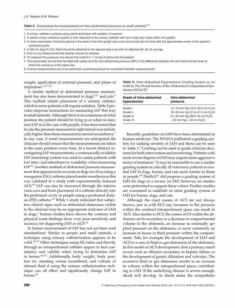
Table 2: Instructions for measurement of intra-abdominal pressures in small animals5, 61
1. A urinary catheter is placed using sterile technique with sedation if required.2. A sterile urinary collection system is then attached to the urinary catheter with two 3-way stop cocks within the system.3. A water manometer should be placed at the level of the first upright stop cock and should also be level with the approximate center of the patient’s
symphysis pubis.4. A 250 mL bag of 0.9% NaCl should be attached to the second stop cock with an attached 20–35 mL syringe.5. Prior to any measurement the bladder should be emptied.6. To measure the pressure one should first instill 0.5–1 mL/kg of saline into the bladder.7. The manometer should then be filled with saline and the intra-abdominal pressure (IAP) is the difference between the zero level and the level at
which the meniscus of the saline lies.8. If serial measurements are to be performed, positioning should be consistent between measurements.
weight, application of external pressure, and phase ofrespiration.3, 57–60
A similar method of abdominal pressure measure-ment has also been demonstrated in dogs4, 61 and cats.5
This method entails placement of a urinary catheter,which in some patients will require sedation. Table 2 pro-vides stepwise instructions for measuring IAP this wayin small animals. Although there is no consensus on whatposition the patient should be lying in or when to mea-sure IAP as is the case with people, it has been noted thatin cats the pressure measured in right lateral was statisti-cally higher than those measured in sternal recumbency.5
In any case, if serial measurements are anticipated theclinician should ensure that the measurements are takenin the same position every time. In a recent abstract in-vestigating IAP measurements, a commercially availableIAP measuring system was used in canine patients withlow intra- and interobserver variability when measuringIAP.62 Another method of abdominal pressure measure-ment that appears to be accurate in dogs involves using anasogastric (NG) catheter placed under anesthesia as thiswas validated in a study with experimentally inducedACS.63 IAP can also be measured through the inferiorvena cava and from placement of a catheter directly intothe peritoneal cavity in people using a peritoneal dialy-sis (PD) catheter.18 While 1 study indicated that subjec-tive clinical signs such as abdominal distension visibleto the clinician may be an appropriate indicator of IAHin dogs,4 human studies have shown the contrary andphysical exam findings show very poor sensitivity andaccuracy for diagnosing IAH or ACS.64
In horses measurement of IAP has not yet been wellstandardized. Similar to people and small animals, atechnique using urinary catheterization appears to bevalid.50, 65 Other techniques using NG tubes and directlythrough an intraperitoneal catheter appear to lack con-sistency and validity when trying to determine IAPin horses.66, 67 Additionally, body weight, body posi-tion (ie, standing versus recumbent), and volume ofinfused fluid if using the urinary catheterization tech-nique can all effect and significantly change IAP inhorses.66
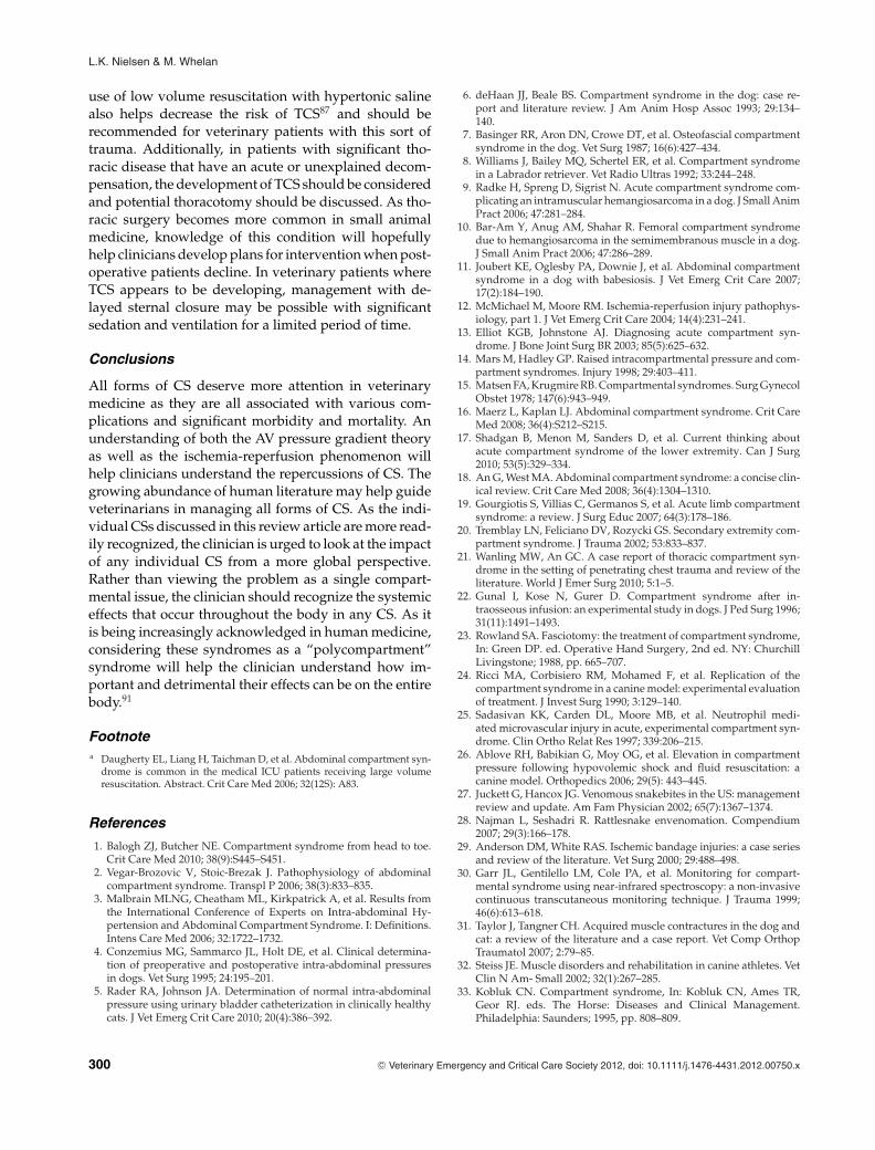
Table 3: Intra-abdominal Hypertension Grading System as setforth by The World Society of the Abdominal Compartment Syn-drome (WSACS)3
Grade of intra-abdominal Intra-abdominalhypertension pressure
Grade I 12–15 mm Hg (16.3–20.4 cm H2O)Grade II 16–20 mm Hg (21.8–27.2 cm H2O)Grade III 21–25 mm Hg (28.6–34 cm H2O)Grade IV >25 mm Hg (>34 cm H2O)
Recently guidelines on IAH have been determined inhuman medicine. The WSACS published a grading sys-tem for ranking severity of IAH and these can be seenin Table 3.3 Grading can be used to guide clinician deci-sions for both intervention and monitoring. Patients withmore severe degrees of IAH may require more aggressiveforms of treatment.3 It may be reasonable to use a similargrading system in critically ill veterinary patients as nor-mal IAP in dogs, horses, and cats seem similar to thosein people.68 Drellich61 did propose a grading system ofIAH for dogs in a review on IAP; however, no studieswere performed to support these values. Further studiesare warranted to establish an ideal grading system ofIAH for horses, dogs, and cats.
Although the exact causes of ACS are not alwaysknown, just as with ECS any increases in the pressurewithin the confined intraperitoneal space can result inACS. Also similar to ECS, the causes of CS within the ab-domen can be secondary to a decrease in compartmentalvolume in the abdomen, an increase in externally ap-plied pressure on the abdomen, or most commonly anincrease in tissue or fluid pressure within the compart-ment. Take for example the development of IAH andACS in a case of fluid or gas distension of the abdomen.In this model of ACS development, first a primary insultoccurs such as effusion secondary to hepatic failure orthe development of gastric dilatation and volvulus. Theexcessive fluid or gas distension results in an increasein volume within the intraperitoneal space, contribut-ing to IAH. If the underlying disease is severe enough,shock will develop. In shock states the sympathetic
296 C© Veterinary Emergency and Critical Care Society 2012, doi: 10.1111/j.1476-4431.2012.00750.x

Compartment syndrome in human and veterinary medicine
nervous system is stimulated and blood is preferentiallyshifted toward the brain and heart due to vasoconstric-tion elsewhere. This results in a relative state of cellularhypoxia in the gut, triggering the release of cytokines,the production of ROS, and a decrease in the overall lev-els and production of ATP. Proinflammatory cytokines,ROS, and decreased ATP resulting in dysfunction of cel-lular pumps such as the Na-K-ATPase pump all result incellular edema, which results in further swelling of tis-sues and increases IAPs.52 If decompression is performedvia drainage of ascites or trocharization or decompres-sion of the stomach, this may result in reperfusion injuryto the abdominal organs, activation of proinflammatorycytokines further increasing IAP, and resulting in IAH.Eventually this IAH may become severe enough to causeorgan dysfunction, satisfying criteria for ACS.
Just as with other CSs, causes or risk factors can beprimary, occurring within the abdomen or pelvic cav-ity, or secondary when an insult outside of the peri-toneal cavity results in development of ACS.67, 69 Themost common risk factors cited in human literature forIAH that may or may not lead to ACS include severeshock, sepsis, or pancreatitis.2 Those at risk include:those receiving high volume resuscitative fluids,18,a anypatient with acute respiratory distress syndrome (ARDS)or undergoing mechanical ventilation,47 those who havesuffered severe abdominal or pelvic injury that may ormay not have undergone surgical interventions,18, 51, 52
and any patient that has already experienced emer-gency laparotomy.70 Other conditions in which ACShas been documented include gastrointestinal perfora-tions, bile peritonitis, abdominal masses, and pregnantindividuals.70, 71 The WSACS presented another way tothink about these conditions in groups rather than as alist of diseases associated with ACS.3 These groupingsare summarized in Table 4.
Table 4: Conditions at risk for the development of IAH and sub-sequent ACS3
Condition Example
Diminished abdominal wallcompliance
Counterpressure applied by a bellywrap
Positioning and immobility in thecritically ill
Increased intra-luminalcontents
IleusGI obstructionGI paresis
Increased abdominalcontents
Free gas, blood, ascites in theabdomen
Abdominal massAny condition resulting in
capillary leakage inassociation with fluidtherapy
SepsisSystemic inflammatory response
syndrome (SIRS)ARDSPancreatitis
The veterinary literature on ACS is very limited con-sisting of a few review papers as well as some case re-ports. The normal values for IAP in cats were recentlyevaluated, and better standards for measurement in catswere established in this landmark study.5 Normal IAPvalues in dogs have also been documented, and in thisstudy it was acknowledged that dogs’ IAP is mildly el-evated postlaparatomy.4 It was also determined that el-evated abdominal pressures were found commonly incanine patients with a disease that resulted in a visiblydistended abdomen.4 One case report described a dogwith babesiosis that developed IAH and subsequent res-piratory distress and renal dysfunction consistent withACS.11 An experimental study in cats in which chronicobstructive pancreatitis was induced found that thesecats also commonly suffered from IAH.72 Although ban-daging of the abdomen is sometimes used to provideabdominal counterpressure to help slow bleeding, it hasbeen suggested that this may be contraindicated as itmay increase the risk for development of ACS.73 How-ever, the presence of IAH has yet to be verified as acomponent in animals with a hemoabdomen.
In large animal medicine the presence of ACS has yetto be definitely diagnosed. However, a case report did ac-knowledge the presence of IAH in 2 critically ill horses.74
Common clinical situations where large animal clini-cians suspect ACS may play a role in equine medicineinclude colitis, hydrops, colonic displacement, uroperi-toneum, pregnancy, and lymphatic obstruction.49, 74 Ad-ditionally, the use of abdominal insufflation with CO2
in horses undergoing laproscopy appeared to have sig-nificant negative cardiopulmonary affects in dorsally re-cumbent horses, suggesting a component of ACS.75 Fur-ther studies into this found that these same negativeeffects were not seen when the insufflation occurred instanding horses.76, 77
In summary, while there have been limited reportsof IAH and ACS, the veterinary literature has acknowl-edged that IAH and ACS have many negative systemiceffects. A better understanding of the condition as wellas implementing IAP monitoring will most likely lead tobetter management in critically ill veterinary patients.65
As with ECS, the treatment for ACS is decompression,and this is definitively accomplished via a decompres-sive laparatomy and potentially management with anopen abdomen postoperatively in certain human cases.78
Following decompression patients with ACS have anincrease in cardiac index, tidal volume, oxygen deliv-ery, and urine output, while heart rate, central venouspressure (CVPs), positive inspiratory pressure (PIP), andlactate all decrease to more acceptable levels.51, 70 Ad-ditionally, a recent prospective study looking at inter-vention with an open abdomen to decrease the devel-opment of ACS in people found that these interventions
C© Veterinary Emergency and Critical Care Society 2012, doi: 10.1111/j.1476-4431.2012.00750.x 297

L.K. Nielsen & M. Whelan
significantly improved survival rates from 50% to 72%.79
There were also trends toward decreased ICU stay anddays on the mechanical ventilator for patients managedwith an open abdomen, but these findings were not sta-tistically significant.79 Although management of an openabdomen is more easily achieved in people, it shouldbe noted that ACS is the only situation in which post-operative open abdomen management is the definitiverecommendation.78 Open peritoneal drainage followingsurgery has been successfully used in veterinary patientswith abdominal sepsis80, 81 but has fallen out of favor inthis patient population with the development of closedsuction drains, except for cases with overt gross con-tamination that cannot be lavaged or debrided clean.82
However, in cases at risk for or with confirmed IAH andACS it may be reasonable to consider using open peri-toneal drainage postlaparotomy.
As ACS becomes more widely recognized, the focuswill hopefully shift to prevention. The consequencesof surgery may worsen the patient’s condition sincethe surgical insult can further exacerbate the inflam-matory response, and this also advocates for earliermonitoring and prevention. Aggressive monitoring inthose at risk may help identify IAH before ACS andorgan dysfunction develops. Depending on the under-lying cause of the IAH, some nonsurgical interventionsthat might help reduce IAP prior to ACS include gas-tric decompression, rectal decompression with enemas,changing body position, diuretics to remove excess fluidand edema from tissues, paracentesis to remove excessfluid from the abdominal cavity, hemodialysis, and neu-romuscular blockade.18 The WSACS also set up spe-cific medical management recommendations for thosewith known IAH in an effort to lower the IAP andprevent the development of ACS that would requiresurgical intervention.56 Their recommendations foundthat sedation, analgesia, or neuromuscular blockade canall improve abdominal wall compliance and thereforelower IAP.18 To help with emptying luminal contentswithin the gastrointestinal tract, the use of prokinet-ics should be instituted and emptying of the stomachvia an NG tube or instituting enemas to empty thecolon should be considered.18 Overzealous fluid ther-apy should be avoided and the use of hypertonic flu-ids that will lower the overall fluid volume adminis-tered is recommended.56 Another study found that theuse of lower volume colloid fluid therapy as opposed tohigh volume crystalloid therapy was beneficial in keep-ing IAP within normal limits while those receiving thecrystalloids did go on to develop IAH.83 If oliguria oranuria has already developed, then the use of diureticsor dialysis should be considered.56 Attempts at drain-ing specific areas found to have gas or fluid taking upabdominal space can be made, but if the medical man-
agement strategies are not working the clinician shouldnot hesitate to pursue surgical decompression.3, 56 Ad-ditionally, when surgery is deemed necessary for treat-ment of ACS or when a patient must undergo abdom-inal surgery but there is a risk of postoperative devel-opment of ACS, use of epidural analgesia in people hasbeen found to significantly decrease IAP in postoperativeabdominal surgery patients.84 These preventative andtreatment measures have not been investigated in veteri-nary medicine as of yet. However, all of these recommen-dations from human medicine might be helpful for theveterinary clinician when ACS is suspected, or when apatient is deemed at risk for development of ACS. If ACSis confirmed or if medical measures to decrease IAH fail,surgical decompression may be necessary in veterinarypatients.
In people with IAH that went on to develop ACS,survival has been documented to be as low as 20%.47
Obviously the detrimental consequences of ACS includ-ing cardiovascular compromise, renal compromise, de-creased pulmonary function, and potential CNS dam-age all make it a very dangerous condition that shouldbe monitored for, prevented if possible, and rapidlytreated with surgical decompression when it does de-velop. Based on the large amount of human literatureon the subject and the smaller amount of veterinary lit-erature, more aggressive monitoring of our veterinarypatients for IAH and ACS, instituting preventative mea-sures when possible, and treating once a diagnosis ofACS is confirmed should be recommended. MonitoringIAP via placement with a urinary catheter might be moredifficult in some small animal patients, mainly femalecats and dogs where urinary catheter placement mightrequire anesthesia. Also, consistent measurement proto-cols for measuring IAP in horses have not been estab-lished, leaving another hurdle for large animal cliniciansto overcome. Hopefully this will not stop the veterinaryclinician from attempting monitoring for IAH in those atrisk. Additional monitoring with measurements of pul-monary function and cardiac output in conjunction withIAP measurements help physicians confirm a diagnosisof ACS, but these measurements are also not always asclosely or easily monitored in veterinary patients. Thismakes the diagnosis of both IAH and ACS more difficultin both small and large animals.
Certain preventative measures as recommended fromthe human literature might be easier to implement thanothers with veterinary patients. More frequent adminis-tration of epidurals in those undergoing surgery deemedto be at risk for postoperative IAH may increase the pre-vention of ACS in veterinary patients. NG tube place-ment for emptying of luminal contents and enemas toempty colonic contents are easily achievable, but otherpreventative measures like dialysis or neuromuscular
298 C© Veterinary Emergency and Critical Care Society 2012, doi: 10.1111/j.1476-4431.2012.00750.x

Compartment syndrome in human and veterinary medicine
blockades might prove to be difficult for most veterinarypractices.
When ACS is strongly suspected or confirmed in vet-erinary patients, surgical decompression should be rec-ommended. Following surgical decompression peopleare typically managed with an open abdomen, and thedecision to manage our veterinary patients in this man-ner should be made on a case-by-case basis. People man-aged with an open abdomen postoperatively have goodsuccess rates78 and this might be achievable in veteri-nary patients as well.80, 81 Lastly, the element of financialcapability is often a large component to decision makingin veterinary patients, and because of this the recom-mendation of surgical decompression for treatment ofACS might not be as economically feasible in veterinarymedicine as it is with people.
Thoracic compartment syndromeThoracic compartment syndrome (TCS) is the dysfunc-tion of organs and tissues within the thoracic cavity sec-ondary to an increased intrathoracic pressure. In peopleit has also been referred to as a “tight mediastinum.”1
TCS is the rarest form of CS and is just now gainingmore recognition in human medicine. The most com-mon situation in which TCS is encountered is followingopen heart or intrathoracic surgery. Human cardiologistshave acknowledged this phenomenon for the last coupleof decades and have found that after intrathoracic surg-eries a significant amount of edema and inflammationsecondary to tissue manipulation at surgery results ina significant increase in intrathoracic pressure followingsternal closure.85
Clinical signs are vague and include increasing airwaypressure, decreasing cardiac output, worsening acidosis,and hemodynamic instability.21, 86 TCS is the 1 CS wherediagnosis does not require measurement of pressures butthe syndrome is suspected based on worsening clinicalsigns and then confirmed via response to therapy (ie,decompressive thoracotomy).
Risk factors for TCS include overaggressive resuscita-tive fluid therapy during a prolonged preoperative pe-riod, significant chest trauma resulting in tissue edema,significant intrathoracic bleeding, and patients under-going open heart surgery.21, 87 The most documentedand recognized cause of TCS is myocardial and pul-monary edema secondary to manipulation in open heartsurgery.85 The development of edema from manipula-tion and secondary to concurrent fluid therapy can causeTCS to develop as soon as the chest is closed. Other docu-mented cases include significant thoracic trauma, whichmight result in significant intrathoracic hemorrhage,21, 87
or complications from routine intrathoracic surgery.86
It has been hypothesized that components of ischemia-reperfusion syndrome may contribute to the postopera-
tive development of TCS,21 but in any case where edemaor hemorrhage is involved, direct compression on thelungs or heart can contribute to impaired functionality.
At this time no documented cases of TCS have beenreported in the veterinary literature. It is not knownwhether the condition does not occur or is just not recog-nized in veterinary patients. In theory, situations whereit may be encountered in veterinary medicine includepostoperative intrathoracic surgery or in patients withextensive thoracic trauma.
As with all other CSs, the treatment of TCS en-tails decompression via opening the chest surgically,and delayed sternal closure following decompression isadvocated.1 The sternum exerts a significant amount ofpressure on the thoracic cavity and the heart specifically,and opening the sternum is similar to relieving pressureexerted on the heart in pericardial disease.88 Although in-fection and subsequent sepsis seem like significant com-plications associated with delayed sternal closure, in onestudy of 150 children undergoing delayed sternal closurenone died from sepsis and only 15 developed minor skininfections at the site of surgery.85
In theory, chest tubes would eliminate the possibil-ity of TCS developing, but they were present in sev-eral documented human cases where the syndromedeveloped.86, 87 Therefore, chest tubes are not preven-tative of TCS.86, 87 As with other CSs, aggressive painmanagement might help prevent development of TCS.There is still some controversy on the ideal approachto thoracotomy (sternal versus lateral) and whether ornot 1 approach is more painful than the other. A hu-man study of 815 thoracotomies found that those un-dergoing median sternotomy had a shorter postopera-tive hospital stay as opposed to those undergoing lateralthoracotomies.89 Additionally, a preliminary experimen-tal study in dogs found that the intercostal approach maybe more painful because of secondary nerve entrapmentdepending on the closure technique used.90 This sug-gests that using the median sternotomy approach maybe advantageous to the patient, and with regards to TCSit might enable the surgeon to quickly reopen the areacausing the increase in intrathoracic pressure to relievethe consequences of TCS.
In summary, TCS is one of the CSs that relies exclu-sively on the clinician for identification, so an aware-ness of the syndrome is very important. Although situ-ations where TCS may arise are very rare in veterinarymedicine, an awareness of the condition may help clini-cians make appropriate surgical recommendations in sit-uations where patients are decompensating. One of themore commonly encountered situations where aware-ness of TCS would be very beneficial in small animalpatients is after sustaining thoracic trauma. In cases ofsignificant hemothorax and pulmonary contusions the
C© Veterinary Emergency and Critical Care Society 2012, doi: 10.1111/j.1476-4431.2012.00750.x 299
L.K. Nielsen & M. Whelan
use of low volume resuscitation with hypertonic salinealso helps decrease the risk of TCS87 and should berecommended for veterinary patients with this sort oftrauma. Additionally, in patients with significant tho-racic disease that have an acute or unexplained decom-pensation, the development of TCS should be consideredand potential thoracotomy should be discussed. As tho-racic surgery becomes more common in small animalmedicine, knowledge of this condition will hopefullyhelp clinicians develop plans for intervention when post-operative patients decline. In veterinary patients whereTCS appears to be developing, management with de-layed sternal closure may be possible with significantsedation and ventilation for a limited period of time.
Conclusions
All forms of CS deserve more attention in veterinarymedicine as they are all associated with various com-plications and significant morbidity and mortality. Anunderstanding of both the AV pressure gradient theoryas well as the ischemia-reperfusion phenomenon willhelp clinicians understand the repercussions of CS. Thegrowing abundance of human literature may help guideveterinarians in managing all forms of CS. As the indi-vidual CSs discussed in this review article are more read-ily recognized, the clinician is urged to look at the impactof any individual CS from a more global perspective.Rather than viewing the problem as a single compart-mental issue, the clinician should recognize the systemiceffects that occur throughout the body in any CS. As itis being increasingly acknowledged in human medicine,considering these syndromes as a “polycompartment”syndrome will help the clinician understand how im-portant and detrimental their effects can be on the entirebody.91
Footnotea Daugherty EL, Liang H, Taichman D, et al. Abdominal compartment syn-
drome is common in the medical ICU patients receiving large volumeresuscitation. Abstract. Crit Care Med 2006; 32(12S): A83.
References
1. Balogh ZJ, Butcher NE. Compartment syndrome from head to toe.Crit Care Med 2010; 38(9):S445–S451.
2. Vegar-Brozovic V, Stoic-Brezak J. Pathophysiology of abdominalcompartment syndrome. Transpl P 2006; 38(3):833–835.
3. Malbrain MLNG, Cheatham ML, Kirkpatrick A, et al. Results fromthe International Conference of Experts on Intra-abdominal Hy-pertension and Abdominal Compartment Syndrome. I: Definitions.Intens Care Med 2006; 32:1722–1732.
4. Conzemius MG, Sammarco JL, Holt DE, et al. Clinical determina-tion of preoperative and postoperative intra-abdominal pressuresin dogs. Vet Surg 1995; 24:195–201.
5. Rader RA, Johnson JA. Determination of normal intra-abdominalpressure using urinary bladder catheterization in clinically healthycats. J Vet Emerg Crit Care 2010; 20(4):386–392.
6. deHaan JJ, Beale BS. Compartment syndrome in the dog: case re-port and literature review. J Am Anim Hosp Assoc 1993; 29:134–140.
7. Basinger RR, Aron DN, Crowe DT, et al. Osteofascial compartmentsyndrome in the dog. Vet Surg 1987; 16(6):427–434.
8. Williams J, Bailey MQ, Schertel ER, et al. Compartment syndromein a Labrador retriever. Vet Radio Ultras 1992; 33:244–248.
9. Radke H, Spreng D, Sigrist N. Acute compartment syndrome com-plicating an intramuscular hemangiosarcoma in a dog. J Small AnimPract 2006; 47:281–284.
10. Bar-Am Y, Anug AM, Shahar R. Femoral compartment syndromedue to hemangiosarcoma in the semimembranous muscle in a dog.J Small Anim Pract 2006; 47:286–289.
11. Joubert KE, Oglesby PA, Downie J, et al. Abdominal compartmentsyndrome in a dog with babesiosis. J Vet Emerg Crit Care 2007;17(2):184–190.
12. McMichael M, Moore RM. Ischemia-reperfusion injury pathophys-iology, part 1. J Vet Emerg Crit Care 2004; 14(4):231–241.
13. Elliot KGB, Johnstone AJ. Diagnosing acute compartment syn-drome. J Bone Joint Surg BR 2003; 85(5):625–632.
14. Mars M, Hadley GP. Raised intracompartmental pressure and com-partment syndromes. Injury 1998; 29:403–411.
15. Matsen FA, Krugmire RB. Compartmental syndromes. Surg GynecolObstet 1978; 147(6):943–949.
16. Maerz L, Kaplan LJ. Abdominal compartment syndrome. Crit CareMed 2008; 36(4):S212–S215.
17. Shadgan B, Menon M, Sanders D, et al. Current thinking aboutacute compartment syndrome of the lower extremity. Can J Surg2010; 53(5):329–334.
18. An G, West MA. Abdominal compartment syndrome: a concise clin-ical review. Crit Care Med 2008; 36(4):1304–1310.
19. Gourgiotis S, Villias C, Germanos S, et al. Acute limb compartmentsyndrome: a review. J Surg Educ 2007; 64(3):178–186.
20. Tremblay LN, Feliciano DV, Rozycki GS. Secondary extremity com-partment syndrome. J Trauma 2002; 53:833–837.
21. Wanling MW, An GC. A case report of thoracic compartment syn-drome in the setting of penetrating chest trauma and review of theliterature. World J Emer Surg 2010; 5:1–5.
22. Gunal I, Kose N, Gurer D. Compartment syndrome after in-traosseous infusion: an experimental study in dogs. J Ped Surg 1996;31(11):1491–1493.
23. Rowland SA. Fasciotomy: the treatment of compartment syndrome,In: Green DP. ed. Operative Hand Surgery, 2nd ed. NY: ChurchillLivingstone; 1988, pp. 665–707.
24. Ricci MA, Corbisiero RM, Mohamed F, et al. Replication of thecompartment syndrome in a canine model: experimental evaluationof treatment. J Invest Surg 1990; 3:129–140.
25. Sadasivan KK, Carden DL, Moore MB, et al. Neutrophil medi-ated microvascular injury in acute, experimental compartment syn-drome. Clin Ortho Relat Res 1997; 339:206–215.
26. Ablove RH, Babikian G, Moy OG, et al. Elevation in compartmentpressure following hypovolemic shock and fluid resuscitation: acanine model. Orthopedics 2006; 29(5): 443–445.
27. Juckett G, Hancox JG. Venomous snakebites in the US: managementreview and update. Am Fam Physician 2002; 65(7):1367–1374.
28. Najman L, Seshadri R. Rattlesnake envenomation. Compendium2007; 29(3):166–178.
29. Anderson DM, White RAS. Ischemic bandage injuries: a case seriesand review of the literature. Vet Surg 2000; 29:488–498.
30. Garr JL, Gentilello LM, Cole PA, et al. Monitoring for compart-mental syndrome using near-infrared spectroscopy: a non-invasivecontinuous transcutaneous monitoring technique. J Trauma 1999;46(6):613–618.
31. Taylor J, Tangner CH. Acquired muscle contractures in the dog andcat: a review of the literature and a case report. Vet Comp OrthopTraumatol 2007; 2:79–85.
32. Steiss JE. Muscle disorders and rehabilitation in canine athletes. VetClin N Am- Small 2002; 32(1):267–285.
33. Kobluk CN. Compartment syndrome, In: Kobluk CN, Ames TR,Geor RJ. eds. The Horse: Diseases and Clinical Management.Philadelphia: Saunders; 1995, pp. 808–809.
300 C© Veterinary Emergency and Critical Care Society 2012, doi: 10.1111/j.1476-4431.2012.00750.x
Compartment syndrome in human and veterinary medicine
34. Norman WM, Williams R, Dodman NH et al. Postanesthetic com-partmental syndrome in a horse. J Am Vet Med Assoc 1989;195(4):502–504.
35. Sullins KE, Heath RB, Turner AS et al. Possible antebrachial flexorcompartment syndrome as a cause of lameness in 2 horses. EquineVet J 1987; 19(2):147–150.
36. Norman WM, Dodman NH, Court MH. Interstitial pH and pres-sure in the dependent biceps femoris muscle of laterally recumbentanesthetized horses. Vet Surg 1988; 17(4):234–239.
37. Lindsay WA, Pascoe PJ, McDonell WN et al. Effect of protectivepadding on forelimb intracompartmental muscle pressures in anes-thetized horses. Am J Vet Res 1985; 46(3):688–691.
38. Dodman NH, Williams R, Court MH et al. Postanesthetic hindlimb adductor myopathy in five horses. J Am Vet Med Assoc 1988;193(1):83–86.
39. Bae DS, Kadiyala RR, Waters PM. Acute compartment syndromein children: contemporary diagnosis, treatment and outcome. J PedOrthop 2001; 21:680–688.
40. McQueen MM, Gaston P, Court-Brown CM. Acute compartmentsyndrome: who is at risk? J Bone Joint Surg 2000; 82B:200–203.
41. Strauss MB, Hargens AR, Gershuni DH, et al. Reduction of skeletalmuscle necrosis using hyperbaric oxygen. J Bone Joint Surg 1983;65A:656–662.
42. McMicheal M. Ischemia-reperfusion injury assessment and treat-ment, part 2. J Vet Emerg Crit Care 2004; 14(4):242–252.
43. Manjoo A, Sanders D, Lawendy A, et al. Indomethacin reduces celldamage. J Orthop Trauma 2010; 24(9):526–529.
44. Dabby D, Greif F, Yaniv M, et al. Thromboxane A2 in the post-ischemic acute compartmental syndrome. Arch Surg 1998; 133:953–956.
45. McQueen MM, Christie J, Court-Brown CM. Acute compartmentsyndrome in the tibial diaphyseal fractures. J Bone Joint Surg 1996;78-B(1):95–98.
46. Vegar-Brozovic V, Brezak J, Brozovic I. Intraabdominal hyper-tension: pulmonary and cerebral complications. Transpl P 2008;40(4):1190–1192.
47. Vidal MG, Weisser JR, Gonzalez F, et al. Incidence and clinical effectsof intra-abdominal hypertension in critically ill patients. Crit CareMed 2008; 36(6):1823–1831.
48. Moore-Olufemi SD, Xue H, Allen SJ, et al. Effects of primary andsecondary intra-abdominal hypertension on mesenteric lymph flow:implications for the abdominal compartment syndrome. Shock 2005;23:571–575.
49. Southwood LL, Wilkins PA. Measurement of intra-abdominal pres-sure in horses. In: 23rd Annual Forum of the American College ofVeterinary Internal Medicine Conference 2005 Proceedings; 2005:Baltimore, USA. pp. 192–194.
50. Mayberry JC. Prevention of abdominal compartment syndrome.Lancet 1999; 354(20):1749–1750.
51. Ertel W, Oberholzer A, Platz A, et al. Incidence and clinical patternof the abdominal compartment syndrome after “damage control”laparotomy in 311 patients with severe abdominal and/or pelvictrauma. Crit Care Med 2000; 28(6):1747–1753.
52. Walker J, Criddle LM. Pathophysiology and management of ab-dominal compartment syndrome. Am J Crit Care 2003; 12:367–371.
53. Diebel LN, Dulchavsky SA, Brown WJ. Splanchnic ischemia and bac-terial translocation in abdominal compartment syndrome. J Trauma1997; 43(5):852–855.
54. Diebel LN, Wilson RF, Dulchavsky SA, et al. Effect of increasedintra-abdominal pressure on hepatic arterial, portal venous, andhepatic microcirculatory blood flow. J Trauma 1992; 33(2):279–282.
55. Lee SL, Anderson JT, Kraut EJ, et al. A simplified approach tothe diagnosis of elevated intra-abdominal pressure. J Trauma 2002;52(6):1169–1172.
56. Malbrain MLNG, Cheatham ML, Kirkpatrick A, et al. Results fromthe International Conference of Experts on Intra-abdominal Hyper-tension and Abdominal Compartment Syndrome. II: Recommenda-tions. Intens Care Med 2007; 33:951–962.
57. Cheatham ML, De Waele JJ, De Laet I, et al. The impact of bodyposition on intra-abdominal pressure measurement: a multicenteranalysis. Crit Care Med 2009; 37(7):2187–2190.
58. Hering R, Hermann W, Vorwerk R, et al. The effects of prone po-sitioning on intra-abdominal pressure and cardiovascular and re-nal function in patients with acute lung injury. Anesth Analg 2001;92:1226–1231.
59. Torquato JA, Lucato JJJ, Antunes T, et al. Interaction between intra-abdominal pressure and positive end expiratory pressure. Clinics2009; 64(2):105–112.
60. Carlotti APCP, Carvalho WB. Abdominal compartment syndrome:a review. Pediatr Crit Care Med 2009; 10(1):115–120.
61. Drellich S. Intraabdominal pressure, In: Silverstein DC, Hopper K.eds. Small Animal Critical Care Medicine. St. Louis: Saunders: Else-vier Inc.; 2009, pp. 872–874.
62. Fetner M, Prittie J. Evaluation of transvesicular intra-abdominalpressure measurement in hospitalized dogs. J Vet Emerg Crit Care2012; 22(2):230–238.
63. Engum SA, Kogon B, Jensen E, et al. Gastric tonometry and directintra-abdominal pressure monitoring in abdominal compartmentsyndrome. J Ped Surg 2002; 37(2):214–218.
64. Kirkpatrick AW, Brennemen FD, McLean RF, et al. Is clinical exam-ination an accurate indicator of raised intra-abdominal pressure incritically injured patients? Can J Surg 2000; 43(3):207–211.
65. Wilkins PA. Abdominal compartment syndrome in equinemedicine. In: 23rd Annual Forum of the American College of Vet-erinary Internal Medicine Conference 2005 Proceedings; 2005: Bal-timore, USA. pp. 190–191.
66. Munsterman AS, Hanson RR. Comparison of direct and indirectmethods for intra-abdominal pressure measurement in normalhorses. J Vet Emerg Crit Care 2009; 19(6):545–553.
67. Munsterman AS, Hanson RR. Evaluation of gastric pressures as anindirect method for measurement of intra-abdominal pressure inhorses. J Vet Emerg Crit Care 2011; 21(1):29–35.
68. Serrano S. Abdominal compartment syndrome: what’s new. In: Pro-ceedings of the 16th IVECCS; 2010: San Antonio, USA. pp. 453–455.
69. Kopelman T, Harris C, Miller R et al. Abdominal compartment syn-drome in patients with isolated extraperitoneal injuries. J Trauma2000; 49:744–749.
70. Meldrum DR, Moore FA, Moore EE, et al. Prospective characteri-zation and selective management of the abdominal compartmentsyndrome. Am J Surg 1997; 174:667–673.
71. Williams M, Simms HH. Abdominal compartment syndrome: casereports and implications in critically ill patients. Am Surg 1997;63:555–558.
72. Karanjia ND, Widdison AL, Leung F, et al. Compartment syndromein experimental chronic obstructive pancreatitis: effect of decom-pressing the main pancreatic duct. Brit J Surg 1994; 81:259–264.
73. Herold LV, Devey JJ, Kirby R, et al. Clinical evaluation and man-agement of hemoperitoneum in dogs. J Vet Emerg Crit Care 2008;18(1):40–53.
74. Brosnahan MM, Holbrook TC, Gilliam LL, et al. Intra-abdominal hy-pertension in two adult horses. J Vet Emerg Crit Care 2009; 19(2):174–180.
75. Donaldson LL, Trostle SS, White NA. Cardiopulmonary changesassociated with abdominal insufflation of CO2 in mechanically ven-tilated, dorsally recumbent, halothane anaesthetized horses. EquineVet J 1998; 30(2):144–151.
76. Cruz AM, Kerr CL, Boure LP et al. Cardiovascular effects of insuf-flation of the abdomen with CO2 in standing horses sedated withdetomidine. Am J Vet Res 2004; 65(3):357–361.
77. Latimer FG, Eades SC, Pettifer G, et al. Cardiopulmonary, blood, andperitoneal fluid alterations associated with abdominal insufflationof CO2 in standing horses. Equine Vet J 2003; 35(3):283–290.
78. Diaz JJ, Cullinane DC, Dutton WD, et al. The management of theopen abdomen in trauma and emergency general surgery: part 1-damage control. J Trauma 2010; 68(6):1425–1438.
79. Cheatam ML, Safcsak K. Is the evolving management of intra-abdominal hypertension and abdominal compartment syndromeimproving survival? Crit Care Med 2010; 38(2):402–407.
C© Veterinary Emergency and Critical Care Society 2012, doi: 10.1111/j.1476-4431.2012.00750.x 301

L.K. Nielsen & M. Whelan
80. Greenfield CL, Walshaw R. Open peritoneal drainage for treatmentof contaminated peritoneal cavity and septic peritonitis in dogs andcats: 24 cases (1980-1986). J Am Vet Med Assoc 1987; 191(1):100–105.
81. Staatz AJ, Monnet E, Seim HB. Open peritoneal drainage versusprimary closure for the treatment of septic peritonitis in dogs andcats: 42 cases (1993-1999). Vet Surg 2002; 31(2):174–180.
82. Beal MW. Peritoneal drainage techniques. In: Silverstein DC, Hop-per K. eds. Small Animal Critical Care Medicine. St. Louis: Saunders,Elsevier Inc.; 2009, pp. 638–641.
83. O’Mara MS, Slater H, Goldfarb IW, et al. A prospective, random-ized evaluation of intra-abdominal pressure with crystalloid andcolloid resuscitation in burn patients. J Trauma 2005; 58(5):1011–1018.
84. Hakobyan RV, Mkhoyan GG. Epidural analgesia decreases intra-abdominal pressure in postoperative patients with primaryintra-abdominal hypertension. Acta Clinica Belgica 2008; 63:86–92.
85. Iyer RS, Jacobs JP, de Leval MR, et al. Outcomes after delayed sternal
closure in pediatric heart operations: a 10 year experience. AnnThorac Surg 1997; 63:489–491.
86. Rizzo AG, Sample GA. Thoracic compartment syndrome sec-ondary to a thoracic procedure. Chest 2003; 124(3):1164–1168.
87. Kaplan LJ, Trooskin SZ, Santora TA. Thoracic compartment syn-drome. J Trauma 1996; 40(2):291–293.
88. Fumary AP, Magovern JA, Simpson KA, et al. Prolonged open ster-notomy and delayed sternal closure after cardiac operations. AnnThorac Surg 1992; 54:233–239.
89. Asaph JW, Handy JR, Grunkemeier GL. Median sternotomy versusthoracotomy to resect primary lung cancer: analysis of 815 cases.Ann Thorac Surg 2000; 70:373–379.
90. Rooney MA, Mehl M, Monnet E. Intercostal thoracotomy closure:transcostal sutures as a less painful alternative to circumcostal su-ture placement. Vet Surg 2004; 33(3):209–213.
91. Malbrain MLNG, Wilmer A. The polycompartment syndrome: to-wards an understanding of the interactions between the differentcompartments! Intens Care Med 2007; 33:1869–1872.
302 C© Veterinary Emergency and Critical Care Society 2012, doi: 10.1111/j.1476-4431.2012.00750.x