pathophysiology and pathogenesis of...
TRANSCRIPT
1
Pathophysiology and Pathogenesis of Osteomyelitis
Mayank Roy1, Jeremy S. Somerson1, Kevin G. Kerr2 and Jonathan L. Conroy2 1University of Texas Health Science Centre, San Antonio, Texas
2Harrogate District Hospital, North Yorkshire 1USA
2UK
1. Introduction
The term osteomyelitis encompasses a broad group of infectious diseases characterized by infection of the bone and/or bone marrow. The pathogenesis of these diseases can follow acute, subacute or chronic courses and involves a range of contributory host and pathogen factors. A commonly used aetiological classification distinguishes between three types of osteomyelitis: acute or chronic haematogenous disease seeded by organisms in the bloodstream, local spread from a contiguous source of infection and secondary osteomyelitis related to vascular insufficiency.
1.1 Acute haematogenous osteomyelitis
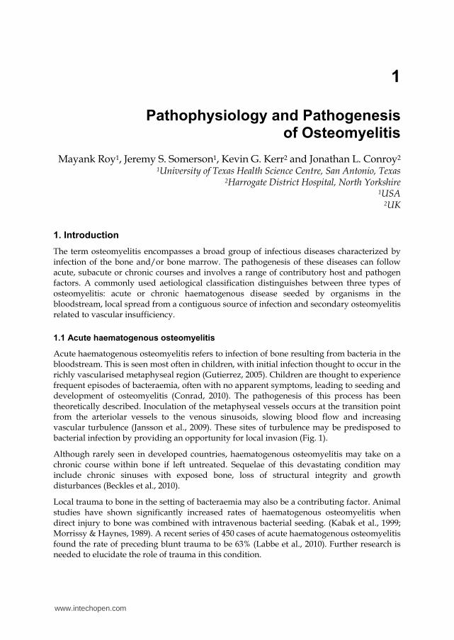
Acute haematogenous osteomyelitis refers to infection of bone resulting from bacteria in the bloodstream. This is seen most often in children, with initial infection thought to occur in the richly vascularised metaphyseal region (Gutierrez, 2005). Children are thought to experience frequent episodes of bacteraemia, often with no apparent symptoms, leading to seeding and development of osteomyelitis (Conrad, 2010). The pathogenesis of this process has been theoretically described. Inoculation of the metaphyseal vessels occurs at the transition point from the arteriolar vessels to the venous sinusoids, slowing blood flow and increasing vascular turbulence (Jansson et al., 2009). These sites of turbulence may be predisposed to bacterial infection by providing an opportunity for local invasion (Fig. 1).
Although rarely seen in developed countries, haematogenous osteomyelitis may take on a chronic course within bone if left untreated. Sequelae of this devastating condition may include chronic sinuses with exposed bone, loss of structural integrity and growth disturbances (Beckles et al., 2010).
Local trauma to bone in the setting of bacteraemia may also be a contributing factor. Animal studies have shown significantly increased rates of haematogenous osteomyelitis when direct injury to bone was combined with intravenous bacterial seeding. (Kabak et al., 1999; Morrissy & Haynes, 1989). A recent series of 450 cases of acute haematogenous osteomyelitis found the rate of preceding blunt trauma to be 63% (Labbe et al., 2010). Further research is needed to elucidate the role of trauma in this condition.
www.intechopen.com
Osteomyelitis
4
Fig. 1. Schematic drawing showing the vascular supply to the physis. The callout represents a detailed view of the physis. The red arrow indicates an area of transition. These transitional zones show increased turbulence and allow for local invasion. (Image used with permission from Dr. Kaye Wilkins)
1.2 Vertebral osteomyelitis
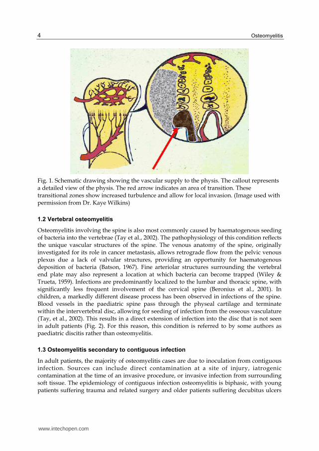
Osteomyelitis involving the spine is also most commonly caused by haematogenous seeding of bacteria into the vertebrae (Tay et al., 2002). The pathophysiology of this condition reflects the unique vascular structures of the spine. The venous anatomy of the spine, originally investigated for its role in cancer metastasis, allows retrograde flow from the pelvic venous plexus due a lack of valvular structures, providing an opportunity for haematogenous deposition of bacteria (Batson, 1967). Fine arteriolar structures surrounding the vertebral end plate may also represent a location at which bacteria can become trapped (Wiley & Trueta, 1959). Infections are predominantly localized to the lumbar and thoracic spine, with significantly less frequent involvement of the cervical spine (Beronius et al., 2001). In children, a markedly different disease process has been observed in infections of the spine. Blood vessels in the paediatric spine pass through the physeal cartilage and terminate within the intervertebral disc, allowing for seeding of infection from the osseous vasculature (Tay, et al., 2002). This results in a direct extension of infection into the disc that is not seen in adult patients (Fig. 2). For this reason, this condition is referred to by some authors as paediatric discitis rather than osteomyelitis.
1.3 Osteomyelitis secondary to contiguous infection
In adult patients, the majority of osteomyelitis cases are due to inoculation from contiguous infection. Sources can include direct contamination at a site of injury, iatrogenic contamination at the time of an invasive procedure, or invasive infection from surrounding soft tissue. The epidemiology of contiguous infection osteomyelitis is biphasic, with young patients suffering trauma and related surgery and older patients suffering decubitus ulcers
www.intechopen.com
Pathophysiology and Pathogenesis of Osteomyelitis
5
Fig. 2. MRI scan showing disc space infection. The lack of normal disc signal at the circled segment (black arrow) represents infection. This type of spinal infection is seen more commonly in children. (Image used with permission from Dr. Kaye Wilkins).
(Mader et al., 1999). Chronic infection often results, with clinical courses complicated by loss of bone structural integrity and soft tissue envelope disturbance.
The progression of disease in localized osteomyelitis is characterized by a cycle of microbial invasion, vascular disruption, necrosis and sequestration. The host inflammatory response, discussed in detail below, results in obstruction of small vessels due to coagulopathy and oedema. As a result of this, cortical bone undergoes necrosis and is detached from surrounding live bone, creating an area known as a sequestrum. This provides a fertile environment for further bacterial invasion and progression continues. Simultaneously, induction of bone begins at the intact periosteum, forming a layer of viable osseous tissue around the site of infection known as involucrum. This mechanism is thought to result from an inflammatory reaction of the periosteum.
Osteomyelitis of the diabetic foot represents a common form of localized infection. Aetiological factors have been thought to include peripheral neuropathy with associated superficial ulceration and peripheral vascular disease. However, a large recent study of risk factors for osteomyelitis in 1666 diabetic patients found no association of osteomyelitis with either peripheral neuropathy or vascular disease (Lavery et al., 2009). History and physical examination findings associated with increased relative risk for osteomyelitis in this study
www.intechopen.com
Osteomyelitis
6
included a previous history of foot ulceration prior to enrolment, the presence of multiple foot wounds or wounds that penetrated deep to bone or joint. This supports prior literature suggesting that clinical ability to probe bone directly in a diabetic ulcer is diagnostic of underlying osteomyelitis (Grayson et al., 1995).
2. Host factors
The pathogenesis of osteomyelitis is a complex process involving interactions between a host and an infectious agent. The host’s inflammatory response to a pathogen can further the physical spread of disease by clearing space in bone. Predisposing genetic differences in immune function are increasingly seen as an aetiological factor in some cases of osteomyelitis. Acquired factors such as diseases causing immune or vascular compromise and implantation of foreign materials are frequently involved in the disease process as well.
2.1 Inflammatory response to infection
The unique demarcated environment of osteomyelitis results in a high-grade local inflammatory host response with systemic effects ranging from minimal to severe. The initial host response to infection of bone is characterized by a local increase in proinflammatory cytokines. Involvement of human monocyte cells in this process has been well-described. When presented with Staphylococcus aureus cells or bacterial cell wall components such as peptidoglycan (PepG) or lipopolysaccharide (LPS), monocytes secrete large amounts of interleukin 1-beta (IL-1beta), IL-6, IL-8, tumour necrosis factor alpha (TNF-alpha) and macrophage inflammatory protein 1-alpha (MIP-1alpha) (Fullilove et al., 2000; Klosterhalfen et al., 1996; Wang et al., 2000). This has been confirmed in an in vivo animal model demonstrating up-regulation of cytokines following intravenous infusion of PepG and LPS (Ruud et al., 2007).
Matrix metalloproteases, a zinc-dependent group of endopeptidases, have been proposed as a key element of bone loss in osteomyelitis. These enzymes are secreted by mesenchymal stromal cells and osteoblasts and work to degrade the extracellular matrix (ECM) in various ways. MMPs have also been shown to activate osteoclast function, leading directly to cell-mediated bone resorption (Ortega et al., 2003). Future therapeutic interventions may target these inflammatory pathways to influence progression of disease.
2.2 Genetics
The role of genetics in the pathogenesis of osteomyelitis is a field of growing research interest. This has partly been driven by new technologies that quickly and affordably perform DNA sequencing of targeted areas. Multiple genetic differences have been identified between patients with osteomyelitis and control subjects, indicating possible hereditary susceptibilities. A recent study identified polymorphisms resulting in upregulation of MMPs with significantly higher frequency in patients with osteomyelitis than in healthy controls (Angel Hugo Montes et al., 2010). The mutation may cause an increase in osteoblast MMP1 production, which has been linked to osteodestructive activity in metastasis (Lu et al., 2009) and inflammatory arthropathy (Neidhart et al., 2009). The IL-1α (-889 TT) genotype has also been found to be more common in patients with osteomyelitis. (VÃctor Asensi et al., 2003; Tsezou et al., 2008). Mutations in the G(-248)A
www.intechopen.com
Pathophysiology and Pathogenesis of Osteomyelitis
7
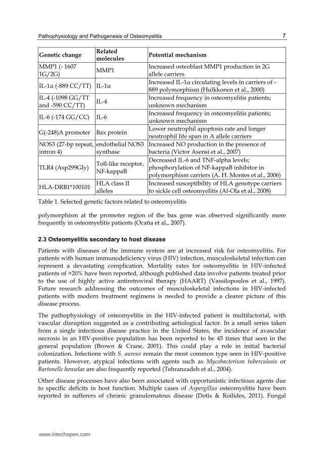
Genetic change Related molecules
Potential mechanism
MMP1 (- 1607 1G/2G)
MMP1 Increased osteoblast MMP1 production in 2G allele carriers
IL-1α (-889 CC/TT) IL-1α Increased IL-1α circulating levels in carriers of -889 polymorphism (Hulkkonen et al., 2000)
IL-4 (-1098 GG/TT and -590 CC/TT)
IL-4 Increased frequency in osteomyelitis patients; unknown mechanism
IL-6 (-174 GG/CC) IL-6 Increased frequency in osteomyelitis patients; unknown mechanism
G(-248)A promoter Bax protein Lower neutrophil apoptosis rate and longer neutrophil life span in A allele carriers
NOS3 (27-bp repeat, intron 4)
endothelial NOS3 synthase
Increased NO production in the presence of bacteria (Victor Asensi et al., 2007)
TLR4 (Asp299Gly) Toll-like receptor, NF-kappaB
Decreased IL-6 and TNF-alpha levels; phosphorylation of NF-kappaB inhibitor in polymorphism carriers (A. H. Montes et al., 2006)
HLA-DRB1*100101 HLA class II alleles
Increased susceptibility of HLA genotype carriers to sickle cell osteomyelitis (Al-Ola et al., 2008)
Table 1. Selected genetic factors related to osteomyelitis
polymorphism at the promoter region of the bax gene was observed significantly more frequently in osteomyelitis patients (Ocaña et al., 2007).
2.3 Osteomyelitis secondary to host disease
Patients with diseases of the immune system are at increased risk for osteomyelitis. For patients with human immunodeficiency virus (HIV) infection, musculoskeletal infection can represent a devastating complication. Mortality rates for osteomyelitis in HIV-infected patients of >20% have been reported, although published data involve patients treated prior to the use of highly active antiretroviral therapy (HAART) (Vassilopoulos et al., 1997). Future research addressing the outcomes of musculoskeletal infections in HIV-infected patients with modern treatment regimens is needed to provide a clearer picture of this disease process.
The pathophysiology of osteomyelitis in the HIV-infected patient is multifactorial, with vascular disruption suggested as a contributing aetiological factor. In a small series taken from a single infectious disease practice in the United States, the incidence of avascular necrosis in an HIV-positive population has been reported to be 45 times that seen in the general population (Brown & Crane, 2001). This could play a role in initial bacterial colonization. Infections with S. aureus remain the most common type seen in HIV-positive patients. However, atypical infections with agents such as Mycobacterium tuberculosis or Bartonelle henselae are also frequently reported (Tehranzadeh et al., 2004).
Other disease processes have also been associated with opportunistic infectious agents due to specific deficits in host function. Multiple cases of Aspergillus osteomyelitis have been reported in sufferers of chronic granulomatous disease (Dotis & Roilides, 2011). Fungal
www.intechopen.com
Osteomyelitis
8
invasion of bone is facilitated in these patients due to defective phagocyte function. In patients suffering from sickle cell disease, microvascular changes lead to predisposition for bone infection. While authors disagree as to whether Salmonella or Staphlyococcus osteomyelitis represents the most common form of bone infection seen in the sickle cell population, published literature uniformly supports a higher rate of Salmonella osteomyelitis than in the general population (Hernigou et al., 2010; Smith, 1996). The pathogenesis of Salmonella osteomyelitis in sickle cell patients may be related to gastrointestinal mini-infarction and resultant bacteraemic episodes. Bone infarction due to impaired microcirculation and impaired opsonisation has also been suggested to play a role (Wilson & Thomas, 1979). Clinical understanding of predisposition and altered pathophysiology of osteomyelitis in patients with these underlying illnesses is required for prompt diagnosis and appropriate treatment.
2.4 Implanted materials and osteomyelitis
Surgically implanted devices in and around bone represent a risk factor for the development of osteomyelitis. Due to the high global rate of total hip and knee replacement, endoprostheses represent an increasingly common source of infection, although infections of other implants such as orthopaedic internal fixation devices are also commonly seen. Stainless steel, titanium, and titanium alloys are the most commonly used materials for osteosynthesis implants, although biodegradable polymers such as poly(L-lactide) are regularly used in non-load bearing fractures, eg, some areas of maxillofacial surgery. The differences between stainless steel and titanium are well documented (Arens et al., 1996; Melcher et al., 1994), with stainless steel implants being associated with significantly greater infection rates than titanium implants. A possible reason for this is the fact that soft tissue adheres firmly to titanium-implant surfaces (Gristina, 1987; Perren, 1991), whilst a known reaction to steel implants is the formation of a fibrous capsule, enclosing a liquid filled void (Gristina, 1987). Bacteria can spread and multiply freely in this unvascularized space, which is also less accessible to the host defence mechanisms. Electro-polishing titanium and titanium alloys has been shown to be more cytocompatible to fibroblasts in static culture conditions than standard surfaces (Meredith et al., 2005). Coatings based on human protein such as albumin or human serum have been shown to reduce S. aureus and S. epidermidis adhesion to the surface (Kinnari et al., 2005). Poly(l-lysine)-grafted-poly(ethylene glycol) (PLL- g-PEG) coatings have been extensively studied for use in biomedical applications, and are highly effective in reducing the adsorption of blood serum, blood plasma and single proteins, such as fibrinogen and albumin (Tosatti et al., 2003). It is also known that fibroblast and osteoblast cell adhesion and spreading on metal oxide surfaces coated with PLL-g-PEG is strongly reduced in comparison to uncoated oxide surfaces (VandeVondele et al., 2003).There has also been interest in coating osteosynthesis implants (stainless steel, titanium, or titanium alloy) with a thin layer of antibiotic-loaded biocompatible, biodegradable polymer, such as polylactic-co-glycolic acid (PLGA) (Price et al., 1996), and poly(D,L-lactide) (PDLLA) (Gollwitzer et al., 2003). The ideology behind this is that the antibiotic is slowly eluted locally at high concentration from the polymer coating as it degrades. Various antibiotics have been studied, including gentamicin (Gollwitzer, et al., 2003), ciprofloxacin (Makinen et al., 2005) and vancomycin (Adams et al., 2009). However, the main concern with all of these antibiotics is the development of bacterial resistance. To prevent this, the amount of antibiotic eluted from the implant must remain above the
www.intechopen.com
Pathophysiology and Pathogenesis of Osteomyelitis
9
minimal inhibitory concentration (MIC) value of the selected antibiotic for the time the implant is in the body. A novel idea to prevent bacterial colonization on external fixation devices and wires was described by Forster et al (Forster et al., 2004), who fitted gentamicin-coated polyurethane sleeves over the pins and wires of the external fixation device. The sleeves substantially reduced the incidence of pin tract infections caused by S. epidermidis, and elution tests revealed that the concentration of gentamicin in the pin tract remained above the 4 μg/ml MIC breakpoint for gentamicin for up to 26 weeks. To date no surface modification or coating fully prevents bacterial adhesion, however, many of the methods discussed have decreased the numbers of adherent bacteria significantly. An important factor to help the fight against infection is the development of biocompatible surfaces or coatings that allow fibroblast and osteoblast cells to adhere and proliferate, leading to soft- and hard-tissue integration and vascularization, while preventing bacterial adhesion. This tissue-covered implant surface then confronts bacteria with an integrated viable tissue layer with a functional host defence mechanism, and may therefore be the best solution we have so far in conquering bacterial adhesion (Harris & Richards, 2006).
The majority of these infections can be traced to intraoperative contamination rather than haematogenous spread (Gillespie, 1990). Because of this, absolute sterility of the operating theatre and implants must be ensured during implantation. The pathogenesis of implant-related infections of bone is related to interactions between the device and local granulocytes, which impairs host clearance of microbes (Zimmerli & Sendi, 2011). Treatment of these infections is complicated by the propensity of infectious agents to form biofilms on implanted surfaces, as discussed in detail below.
3. Pathogen factors
The initial event in the localization of infection appears to be adhesion of the bacteria to the extracellular matrix (ECM). Various factors govern this adhesion process. Once a bacteria reaches the biomaterial surface by haematogenous route they acquire a conditioning film of ECM proteins. Osteoblast play an active role in the internalization of the bacteria. Subsequently a multi-layered biofilm is made by the bacteria, which protects it from phagoctytosis and antibiotics.
3.1 Extracellular matrix attachment and adhesins
The ECM is a biologically active layer composed of a complex mixture of macromolecules, such as fibronectin, fibrinogen, albumin, vitronectin, and collagen. Host cell adhesion, migration, proliferation, and differentiation are all influenced by the composition and structural organization of the surrounding ECM. Interaction between host cells and the ECM is known to be mediated by specific receptors such as integrins, which are composed of and ß units and link many ECM proteins to the eukaryotic cellular cytoskeleton (Ruoslahti, 1991). The ECM not only serves as a substrate for host cells, but also for colonizing bacteria. If an infection is to develop, pathogenic bacteria must cling to the tissue in order to overcome removal by physical forces. As well as using non-specific hydrophobic and electrostatic forces to interact with their hosts, bacteria have surface proteins with specific affinity for components of the ECM and for plasma proteins. These proteins are often called ECM-binding proteins (ECMBPs) or MSCRAMMs (microbial surface components recognizing adhesive matrix molecules). The S. aureus proteins responsible for
www.intechopen.com
Osteomyelitis
10
binding to fibronectin (fibronectin binding protein; fnbp), collagen (collagen binding protein; cna) and fibrinogen (clumping factor; cifA and cifB) are the best-studied ECMBPs (Flock, 1999). Peacock et al. showed that seven putative virulence genes in S. aureus, including the adhesin genes fnbA and cna, the toxin genes sej, eta and hlg, and icaA, which are involved in biofilm production, were found to be associated with invasive isolates (Peacock et al., 2002). Some studies have shown that immunization with cna can protect against septic death (Nilsson et al., 1998; Smeltzer & Gillaspy, 2000). Smeltzer concluded in his study that the inclusion of immunogens derived from conserved adhesins (e.g., fnbpA and clfA) would be required to achieve maximum effectiveness. However, failure to include cna would result in an immune response that would not necessarily limit the ability of a cna-positive strain to colonize musculoskeletal tissues (Smeltzer & Gillaspy, 2000). Besides collagen binding, S. aureus cells isolated from patients with osteomyelitis bind to bone sialoprotein suggesting that sialoprotein binding may also serve to localize the infection to bone tissue (Ryden et al., 1989).
Capsular polysaccharides expressed on the bacterial cell surface are a major virulence factor known to promote evasion of or interference with the host immune system. Binding of S. aureus to bone collagen is clearly associated with the protein ‘adhesin’ and is inhibited by the presence of a capsule on the bacterium. The latter has been demonstrated by experiments utilizing S. aureus strains Cowan and Wood. Strain Cowan (originally isolated from a patient with septic arthritis) lacks a capsule and demonstrates extensive binding to purified type I collagen. Strain Wood is encapsulated and demonstrated very poor binding ability to purified type I collagen in the same study (Buxton et al., 1990).
3.2 Attachment to biomaterial surfaces
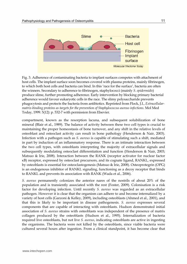
S. aureus is a common cause of metal-biomaterial, bone-joint, and soft-tissue infections (Petty et al., 1985), while S. epidermidis is more common with polymer-associated implant infections (von Eiff et al., 2002). It has been shown that both fibrinogen (Brokke et al., 1991) and fibronectin (Fischer et al., 1996) deposited in vivo onto the implant surface mediate bacterial adherence. Bacteria compete with host cells for attachment to the implant surface, a phenomenon that has been referred to as ‘the race for the surface’ (Gristina, 1987) (Fig. 3). Once a biomaterial has been implanted, they acquire a conditioning film of ECM proteins (Baier et al., 1984).
3.3 Role of osteoblasts
The skeleton is a dynamic organ system, in a state of perpetual turnover which is continually remodelled by the actions of two cell types (Henderson & Nair, 2003). Osteoblasts are responsible for the deposition of bone matrix; they are found on bone surfaces and are derived from mesenchymal osteoprogenitor cells. These cells secrete osteoid, a mixture of bone matrix proteins primarily made up of type I collagen (over 90%), proteoglycans such as decorin and biglycan, glycoproteins such as fibronectin, osteonectin and tenascin-C, osteopontin, osteocalcin and bone sialoprotein, oriented along stress lines (Mackie, 2003).The opposing action of bone matrix removal is performed by osteoclasts, multinucleate cells that are derived from the macrophage-monocyte lineage. These cells express large quantities of a vacuolar-type H(+)-ATPase on their cell surface, along with chloride channel 7 (ClC 7) enabling localized hydrochloric acid secretion into a closed
www.intechopen.com
Pathophysiology and Pathogenesis of Osteomyelitis
11
Fig. 3. Adherence of contaminating bacteria to implant surfaces competes with attachment of host cells. The implant surface soon becomes covered with plasma proteins, mainly fibrinogen, to which both host cells and bacteria can bind. In this ‘race for the surface’, bacteria are often the winners. Secondary to adherence to fibrinogen, staphylococci (mainly S. epidermidis) produce slime, further promoting adherence. Early intervention by blocking primary bacterial adherence would favour eukaryotic cells in the race. The slimy polysaccharide prevents phagocytosis and protects the bacteria from antibiotics. Reprinted from Flock, J.I., Extracellular-matrix-binding proteins as targets for the prevention of Staphylococcus aureus infections. Mol Med Today, 1999. 5(12): p. 532-7 with permission from Elsevier.
compartment, known as the resorption lacuna, and subsequent solubilization of bone mineral (Blair et al., 1989). The balance of activity between these two cell types is crucial to maintaining the proper homeostasis of bone turnover, and any shift in the relative levels of osteoblast and osteoclast activity can result in bone pathology (Henderson & Nair, 2003). Infection with a pathogen such as S. aureus is capable of stimulating such a shift, mediated in part by induction of an inflammatory response. There is an intimate interaction between the two cell types, with osteoblasts interpreting the majority of extracellular signals and subsequently modulating osteoclast differentiation and function (Henderson & Nair, 2003; Matsuo & Irie, 2008). Interaction between the RANK (receptor activator for nuclear factor κB) receptor, expressed by osteoclast precursors, and its cognate ligand, RANKL, expressed by osteoblasts is essential for osteoclastogenesis (Matsuo & Irie, 2008). Osteoprotegrin (OPG) is an endogenous inhibitor of RANKL signaling, functioning as a decoy receptor that binds to RANKL and prevents its association with RANK (Wada et al., 2006).
S. aureus permanently colonizes the anterior nares of the nostrils of about 20% of the population and is transiently associated with the rest (Foster, 2009). Colonisation is a risk factor for developing infection. Until recently S. aureus was regarded as an extracellular pathogen. However it is clear that the organism can adhere to and become internalized by a variety of host cells (Garzoni & Kelley, 2009), including osteoblasts (Ahmed et al., 2001), and that this is likely to be important in disease pathogenesis. S. aureus expresses several components that are capable of interacting with osteoblasts. Hudson demonstrated initial association of S. aureus strains with osteoblasts was independent of the presence of matrix collagen produced by the osteoblasts (Hudson et al., 1995). Internalization of bacteria required live osteoblasts, but not live S. aureus, indicating osteoblasts are active in ingesting the organisms. The bacteria were not killed by the osteoblasts, since viable bacteria were cultured several hours after ingestion. From a clinical standpoint, it has become clear that
www.intechopen.com
Osteomyelitis
12
patients can have recurrent attacks of osteomyelitis after completion of therapy even when causative organisms cannot be isolated (Craigen et al., 1992). The observation that S. aureus can be internalized by osteoblasts may be relevant to this clinical problem.
Uptake is promoted by fibronectin binding proteins that capture fibronectin and use it as a bridge between bacteria and the a5b1 integrin (Sinha et al., 1999). Integrin clustering results in signaling that leads to bacterial uptake into phagocytic vesicles. The mechanism of invasion differs between S. aureus and S. epidermidis and the latter does not gain entry via the fibronectin-integrin α5β1 mechanism (Khalil et al., 2007). The level of expression of the alternative sigma factor, σB, affects fnbA expression and the fibronectin binding ability of S. aureus strains correlates with the level of internalization of bacteria by osteoblasts suggesting that σB-mediated up-regulation of FnBP expression may facilitate invasion (Nair et al., 2003). Once internalized bacteria can escape the phagosome and cause necrosis (Wright & Nair, 2010). Slow growing variants (called small colony variants) often emerge allowing the bacteria and the infection to persist (von Eiff, Bettin et al., 1997). These bacteria are mutant forms of Staphylococcus that may have an adaptive advantage enabling persistent bone colonisation. Small colony variants can be associated with both refractory and relapsing infections that are poorly responsive to standard treatment regimens. Their decreased metabolic activity and decreased a-toxin production may enable them to survive intracellularly and to exhibit decreased susceptibility to antibiotics (von Eiff, Heilmann et al., 1997). Because of their slow growth, atypical colonial morphology, and other altered phenotypes, these organisms may be missed or incorrectly identified by clinical laboratories (Proctor et al., 1995).
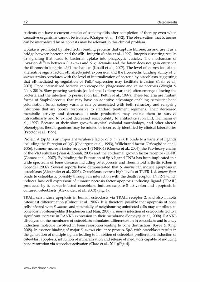
Protein A (SpA) is an important virulence factor of S. aureus. It binds to a variety of ligands including the Fc region of IgG (Cedergren et al., 1993), Willebrand factor (O'Seaghdha et al., 2006), tumour necrosis factor receptor-1 (TNFR-1) (Gomez et al., 2006), the Fab-heavy chains of the Vh3 subclass (Viau & Zouali, 2005) and the epidermal growth factor receptor (EGFR) (Gomez et al., 2007). By binding the Fc portion of SpA ligand TNFa has been implicated in a wide spectrum of bone diseases including osteoporosis and rheumatoid arthritis (Chen & Goeddel, 2002). Several reports have demonstrated that S. aureus can induce apoptosis in osteoblasts (Alexander et al., 2003). Osteoblasts express high levels of TNFR-1. S. aureus SpA binds to osteoblasts, possibly through an interaction with the death receptor TNFR-1 which induces host cell expression of tumour necrosis factor apoptosis inducing ligand (TRAIL) produced by S. aureus-infected osteoblasts induces caspase-8 activation and apoptosis in cultured osteoblasts (Alexander, et al., 2003) (Fig. 4).
TRAIL can induce apoptosis in human osteoclasts via TRAIL receptor 2, and also inhibits osteoclast differentiation (Colucci et al., 2007). It is therefore possible that apoptosis of bone cells infected with S. aureus, and potentially of neighbouring uninfected cells may contribute to bone loss in osteomyelitis (Henderson and Nair, 2003). S. aureus infection of osteoblasts led to a significant increase in RANKL expression in their membrane (Somayaji et al., 2008). RANKL displayed on the membrane of osteoblasts stimulates differentiation in osteoclasts and is a key induction molecule involved in bone resorption leading to bone destruction (Boyce & Xing, 2008). In essence binding of major S. aureus virulence protein, SpA with osteoblasts results in the generation of multiple signals leading to inhibition of osteoblast proliferation, induction of osteoblast apoptosis, inhibition of mineralization and release of mediators capable of inducing bone resorption via osteoclast activation (Claro et al., 2011)(Fig. 4).
www.intechopen.com
Pathophysiology and Pathogenesis of Osteomyelitis
13
Fig. 4. Proposed mechanism of Staphylococcus aureus – osteoblast interaction. Claro, T., et al., Staphylococcus aureus protein A binds to osteoblasts and triggers signals that weaken bone in osteomyelitis. PLoS One, 2011. 6(4): p. e18748
3.4 Biofilm formation
A biofilm is defined as a microbially derived sessile community, typified by cells that are attached to a substratum, interface, or to each other, are embedded in a matrix of extracellular polymeric substance, and exhibit an altered phenotype with regard to growth, gene expression, and protein production (Donlan & Costerton, 2002). Biofilm depth can vary, from a single cell layer to a thick community of cells surrounded by a thick polymeric milieu. Structural analyses have shown that these thick biofilms possess a complex architecture in which microcolonies can exist in distinct pillar or mushroom-shaped structures (Costerton et al., 1995), through which an intricate channel network runs. These channels provide access to environmental nutrients even in the deepest areas of the biofilm.
By adopting this sessile mode of life, biofilm-embedded microorganisms benefit from a number of advantages over their planktonic counterparts:
1. The capability of the extracellular matrix to seize and concentrate a number of environmental nutrients, such as carbon, nitrogen, and phosphate (Beveridge et al., 1997).
2. The facilitation of resistance to a number of removal tactics, such as elimination by antimicrobial agents, shear stress, host phagocytic clearance, and host oxygen radical and protease defences. This innate resistance to antimicrobial factors is mediated through very low metabolic levels and radically down-regulated rates of cell division of the deeply entrenched micro-organisms.
3. The potential for dispersion via detachment. Microcolonies may detach under the direction of mechanical fluid shear or through a genetically programmed response that mediates the detachment process (Boyd & Chakrabarty, 1994). Under the direction of fluid flow, this microcolony travels to other regions of the host system to attach and
www.intechopen.com
Osteomyelitis
14
promote biofilm formation in previously uninfected areas. In addition, detachment and seeding of virgin surfaces may be accomplished by the migration of single, motile cells from the cores of attached microcolonies (Sauer et al., 2002). Therefore, this advantage allows an enduring bacterial source population that is resilient against antimicrobial agents and the host immune response, while simultaneously enabling continuous shedding to encourage bacterial spread.
Formation of biofilm is a two-stage process in which bacteria first attach to a substrate (e.g., bone) and then attach to each other as the biofilm grows and matures. The two-stage process is consistent with the scenario described for S. epidermidis, which is a common cause of infections involving in-dwelling medical devices. In this case, the initial attachment appears to be dependent on the production of one or more protein adhesins, whereas the subsequent aggregation of bacteria into a biofilm is dependent on the production of exopolysaccharide adhesins (Heilmann et al., 1996). It is known that once a biofilm has formed, the bacteria within the biofilm are protected from phagocytosis and antibiotics (Hoyle & Costerton, 1991), and a mouse bacteraemia model found that the biofilm enhanced S. aureus virulence factors, such as the α-toxin (Caiazza & O'Toole, 2003; Thakker et al., 1998). A final detachment (or dispersal) phase involves the detachment of single cells or cell clusters by various mechanisms and is believed to be crucial for the dissemination of the bacteria, in the case of pathogens to new infection sites in the human body.
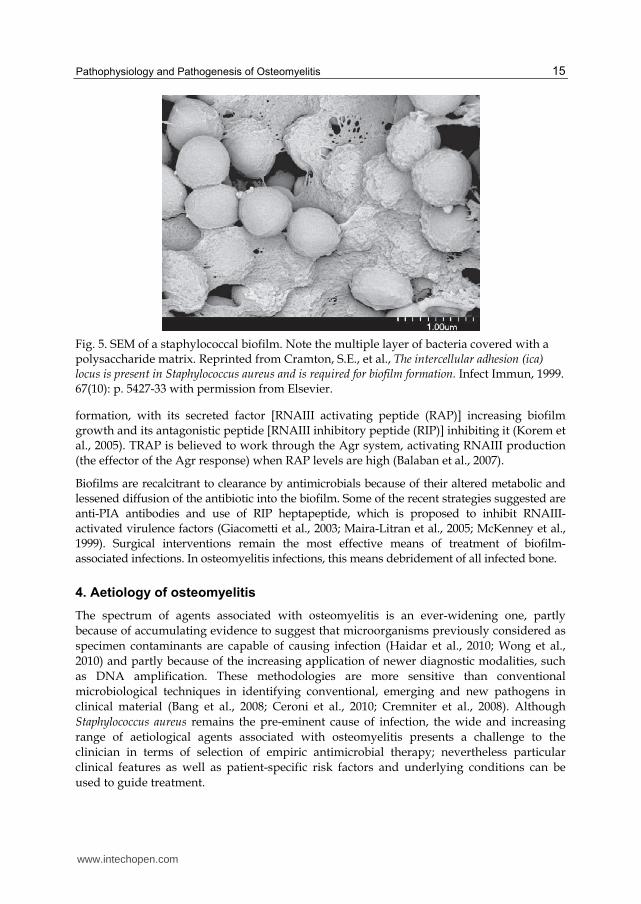
Staphylococcus spp. can produce a multilayered biofilm embedded within a glycocalyx, or slime layer. The glycocalyx develops on devitalized tissue and bone, or on medically implanted devices, to produce an infection (Akiyama et al., 1993). Early studies described the solid component of the glycocalyx as primarily composed of teichoic acids (80%) and staphylococcal and host proteins (Hussain et al., 1993). In recent years, the polysaccharide intercellular adhesin (PIA) has been found in many S. aureus strains (Cramton et al., 1999), and is required for biofilm formation and bacterium-bacterium adhesion (Fig. 6). PIA is produced in vitro from UDP-N-acetylglucosamine via products of the intercellular adhesion (ica) locus (Cramton, et al., 1999). The genes and products of the ica locus [icaR (regulatory) and icaADBC (biosynthetic) genes] have been demonstrated to be necessary for biofilm formation and virulence, and are up-regulated in response to anaerobic growth, such as the conditions seen in the biofilm environment (Cramton et al., 2001). Another important component of the staphylococcal biofilm is extracellular DNA (eDNA). The discovery that this substance is an important component of biofilms was recently made in P. aeruginosa (Whitchurch et al., 2002). Rice et al. very recently showed that eDNA is important for biofilm formation and adherence in S. aureus, and that this DNA release seems to be, at least in part, mediated through the cidA murein hydrolase (Rice et al., 2007). This gene has been shown to be a holin homologue involved in cell lysis, and it is thought that this gene allows S. aureus biofilm cells to lyse and release DNA into the extracellular milieu.
Many factors seem to play a role in regulation of biofilm. The agr quorum sensing (QS) system, a central regulator of virulence, has been shown to down-regulate genes of cell wall-associated adherence factors (Chan et al., 2004). This would lead to lesser adherence and thus, indirectly, decreased initial biofilm formation. As well, the agr system has been shown to up-regulate the expression of detergent-like peptides that seem to increase biofilm detachment (Kong et al., 2006), and mutation of the system leads to increased biofilm growth. Another regulatory system, Target of RAP (TRAP), has been implicated in biofilm
www.intechopen.com
Pathophysiology and Pathogenesis of Osteomyelitis
15
Fig. 5. SEM of a staphylococcal biofilm. Note the multiple layer of bacteria covered with a polysaccharide matrix. Reprinted from Cramton, S.E., et al., The intercellular adhesion (ica) locus is present in Staphylococcus aureus and is required for biofilm formation. Infect Immun, 1999. 67(10): p. 5427-33 with permission from Elsevier.
formation, with its secreted factor [RNAIII activating peptide (RAP)] increasing biofilm growth and its antagonistic peptide [RNAIII inhibitory peptide (RIP)] inhibiting it (Korem et al., 2005). TRAP is believed to work through the Agr system, activating RNAIII production (the effector of the Agr response) when RAP levels are high (Balaban et al., 2007).
Biofilms are recalcitrant to clearance by antimicrobials because of their altered metabolic and lessened diffusion of the antibiotic into the biofilm. Some of the recent strategies suggested are anti-PIA antibodies and use of RIP heptapeptide, which is proposed to inhibit RNAIII-activated virulence factors (Giacometti et al., 2003; Maira-Litran et al., 2005; McKenney et al., 1999). Surgical interventions remain the most effective means of treatment of biofilm-associated infections. In osteomyelitis infections, this means debridement of all infected bone.
4. Aetiology of osteomyelitis
The spectrum of agents associated with osteomyelitis is an ever-widening one, partly because of accumulating evidence to suggest that microorganisms previously considered as specimen contaminants are capable of causing infection (Haidar et al., 2010; Wong et al., 2010) and partly because of the increasing application of newer diagnostic modalities, such as DNA amplification. These methodologies are more sensitive than conventional microbiological techniques in identifying conventional, emerging and new pathogens in clinical material (Bang et al., 2008; Ceroni et al., 2010; Cremniter et al., 2008). Although Staphylococcus aureus remains the pre-eminent cause of infection, the wide and increasing range of aetiological agents associated with osteomyelitis presents a challenge to the clinician in terms of selection of empiric antimicrobial therapy; nevertheless particular clinical features as well as patient-specific risk factors and underlying conditions can be used to guide treatment.
www.intechopen.com
Osteomyelitis
16
As noted above, osteomyelitis can be broadly classified according to source of infection: spread from a contiguous site or following haematogenous seeding. The latter is more likely to be associated with monomicrobial infection while the former is often polymicrobial in origin including obligately anaerobic bacteria Osteomyelitis in individuals with vascular insufficiency including patients with diabetes mellitus is also frequently polymicrobial (Powlson & Coll, 2010).
There are also aetiological associations with patient age. In neonates, for example, the bacteria most frequently associated with acute haematogenous osteomyelitis are those which cause neonatal sepsis, notably Lancefield group B streptococci (Streptococcus agalactiae) and Escherichia coli as well as S. aureus (Dessi et al., 2008). In older children, S. aureus infection predominates and in some countries, such as the US, community-acquired methicillin-resistant strains (CA-MRSA) are increasingly recognized (Vander Have et al., 2009). Kingella kingae has also emerged in recent years as an important cause of osteomyelitis in children (Dubnov-Raz et al., 2008). In contrast, Haemophilus influenzae infections, once common in patients aged under five years, have markedly declined as a result of vaccination against Pittman type b strains of this bacterium (Howard et al., 1999). In adults, as with younger patients, S. aureus is the most frequent agent of infection.
Risk factor/feature Microorganism
Geographic location Mycobacterium tuberculosis Brucella species (Colmenero et al., 2008)
Dimorphic fungi e.g. Coccidiodes immitis (Holley et al., 2002)
Intravenous drug use
Staphylococcus aureus Pseudomonas aeruginosa (Miskew et al., 1983) Candida albicans (Lafont et al., 1994) Eikenella corrodens ( “needle lickers’ osteomyelitis”) (Swisher et al., 1994)
Post-human or animal bite
Staphylococcus aureus Pasteurella multocida (Jarvis et al., 1981)
Eikenella corrodens (Schmidt & Heckman, 1983) Obligate anaerobes (Brook, 2008)
Vertebral osteomyelitis
Staphylococcus aureus Coagulase-negative staphylococci Propionibacterium acnes (post-spinal surgery) (Kowalski, Berbari, Huddleston, Steckelberg, Mandrekar et al., 2007; Kowalski, Berbari, Huddleston, Steckelberg, & Osmon, 2007) Escherichia coli (McHenry et al., 2002)
Pseudomonas aeruginosa (Patzakis et al., 1991)
Prosthetic devices Staphylococcus aureus Coagulase-negative staphylococci Propionibacterium acnes (Lew & Waldvogel, 2004)
Puncture wounds of the foot Pseudomonas aeruginosa (“sneaker osteomyelitis”) (Dixon & Sydnor, 1993)
Table 2. Aetiological association
www.intechopen.com
Pathophysiology and Pathogenesis of Osteomyelitis
17
Osteomyelitis in patients with immunocompromise, both congenital and acquired, can be caused by an extremely wide range of conventional and opportunistic pathogens including fungi (See section 2.3). Examples of other aetiological associations are shown in Table 2.
5. References
Adams, C. S., Antoci, V., Jr., Harrison, G., Patal, P., Freeman, T. A., Shapiro, I. M., et al. (2009). Controlled release of vancomycin from thin sol-gel films on implant surfaces successfully controls osteomyelitis. Journal of Orthopaedic Research: Official Publication of the Orthopaedic Research Society, Vol. 27, No. 6, pp. (701-709)
Ahmed, S., Meghji, S., Williams, R. J., Henderson, B., Brock, J. H., & Nair, S. P. (2001). Staphylococcus aureus fibronectin binding proteins are essential for internalization by osteoblasts but do not account for differences in intracellular levels of bacteria. Infect Immun, Vol. 69, No. 5, pp. (2872-2877), 0019-9567
Akiyama, H., Torigoe, R., & Arata, J. (1993). Interaction of Staphylococcus aureus cells and silk threads in vitro and in mouse skin. J Dermatol Sci, Vol. 6, No. 3, pp. (247-257), 0923-1811
Al-Ola, K., Mahdi, N., Al-Subaie, A. M., Ali, M. E., Al-Absi, I. K., & Almawi, W. Y. (2008). Evidence for HLA class II susceptible and protective haplotypes for osteomyelitis in pediatric patients with sickle cell anemia. Tissue Antigens, Vol. 71, No. 5, pp. (453-457),
Alexander, E. H., Rivera, F. A., Marriott, I., Anguita, J., Bost, K. L., & Hudson, M. C. (2003). Staphylococcus aureus - induced tumor necrosis factor - related apoptosis - inducing ligand expression mediates apoptosis and caspase-8 activation in infected osteoblasts. BMC Microbiol, Vol. 3, No., pp. (5), 1471-2180
Arens, S., Schlegel, U., Printzen, G., Ziegler, W. J., Perren, S. M., & Hansis, M. (1996). Influence of materials for fixation implants on local infection. An experimental study of steel versus titanium DCP in rabbits. J Bone Joint Surg Br, Vol. 78, No. 4, pp. (647-651), 0301-620X
Asensi, V., Alvarez, V., Valle, E., Meana, A., Fierer, J., Coto, E., et al. (2003). IL-1 alpha (-889) promoter polymorphism is a risk factor for osteomyelitis. American Journal of Medical Genetics. Part A, Vol. 119A, No. 2, pp. (132-136)
Asensi, V., Montes, A. H., Valle, E., Ocaña, M. G., Astudillo, A., Alvarez, V., et al. (2007). The NOS3 (27-bp repeat, intron 4) polymorphism is associated with susceptibility to osteomyelitis. Nitric Oxide: Biology and Chemistry / Official Journal of the Nitric Oxide Society, Vol. 16, No. 1, pp. (44-53)
Baier, R. E., Meyer, A. E., Natiella, J. R., Natiella, R. R., & Carter, J. M. (1984). Surface properties determine bioadhesive outcomes: methods and results. J Biomed Mater Res, Vol. 18, No. 4, pp. (327-355), 0021-9304
Balaban, N., Cirioni, O., Giacometti, A., Ghiselli, R., Braunstein, J. B., Silvestri, C., et al. (2007). Treatment of Staphylococcus aureus biofilm infection by the quorum-sensing inhibitor RIP. Antimicrob Agents Chemother, Vol. 51, No. 6, pp. (2226-2229), 0066-4804
Bang, D., Herlin, T., Stegger, M., Andersen, A. B., Torkko, P., Tortoli, E., et al. (2008). Mycobacterium arosiense sp. nov., a slowly growing, scotochromogenic species causing osteomyelitis in an immunocompromised child. Int J Syst Evol Microbiol, Vol. 58, No. Pt 10, pp. (2398-2402), 1466-5026
www.intechopen.com
Osteomyelitis
18
Batson, O. V. (1967). The vertebral system of veins as a means for cancer dissemination. Progress in Clinical Cancer, Vol. 3, No., pp. (1-18)
Beckles, V. L. L., Jones, H. W., & Harrison, W. J. (2010). Chronic haematogenous osteomyelitis in children: a retrospective review of 167 patients in Malawi. The Journal of Bone and Joint Surgery. British Volume, Vol. 92, No. 8, pp. (1138-1143)
Beronius, M., Bergman, B., & Andersson, R. (2001). Vertebral Osteomyelitis in Goteborg, Sweden: A Retrospective Study of Patients During 1990-95. Scandinavian Journal of Infectious Diseases, Vol. 33, No. 7, pp. (527-532)
Beveridge, T. J., Makin, S. A., Kadurugamuwa, J. L., & Li, Z. (1997). Interactions between biofilms and the environment. FEMS Microbiol Rev, Vol. 20, No. 3-4, pp. (291-303), 0168-6445
Blair, H. C., Teitelbaum, S. L., Ghiselli, R., & Gluck, S. (1989). Osteoclastic bone resorption by a polarized vacuolar proton pump. Science, Vol. 245, No. 4920, pp. (855-857), 0036-8075
Boyce, B. F., & Xing, L. (2008). Functions of RANKL/RANK/OPG in bone modeling and remodeling. Arch Biochem Biophys, Vol. 473, No. 2, pp. (139-146), 1096-0384
Boyd, A., & Chakrabarty, A. M. (1994). Role of alginate lyase in cell detachment of Pseudomonas aeruginosa. Appl Environ Microbiol, Vol. 60, No. 7, pp. (2355-2359), 0099-2240
Brokke, P., Dankert, J., Carballo, J., & Feijen, J. (1991). Adherence of coagulase-negative staphylococci onto polyethylene catheters in vitro and in vivo: a study on the influence of various plasma proteins. J Biomater Appl, Vol. 5, No. 3, pp. (204-226), 0885-3282
Brook, I. (2008). Microbiology and management of joint and bone infections due to anaerobic bacteria. J Orthop Sci, Vol. 13, No. 2, pp. (160-169), 0949-2658
Brown, P., & Crane, L. (2001). Avascular necrosis of bone in patients with human immunodeficiency virus infection: report of 6 cases and review of the literature. Clinical Infectious Diseases: An Official Publication of the Infectious Diseases Society of America, Vol. 32, No. 8, pp. (1221-1226)
Buxton, T. B., Rissing, J. P., Horner, J. A., Plowman, K. M., Scott, D. F., Sprinkle, T. J., et al. (1990). Binding of a Staphylococcus aureus bone pathogen to type I collagen. Microb Pathog, Vol. 8, No. 6, pp. (441-448), 0882-4010
Caiazza, N. C., & O'Toole, G. A. (2003). Alpha-toxin is required for biofilm formation by Staphylococcus aureus. J Bacteriol, Vol. 185, No. 10, pp. (3214-3217), 0021-9193
Cedergren, L., Andersson, R., Jansson, B., Uhlen, M., & Nilsson, B. (1993). Mutational analysis of the interaction between staphylococcal protein A and human IgG1. Protein Eng, Vol. 6, No. 4, pp. (441-448), 0269-2139
Ceroni, D., Cherkaoui, A., Ferey, S., Kaelin, A., & Schrenzel, J. (2010). Kingella kingae osteoarticular infections in young children: clinical features and contribution of a new specific real-time PCR assay to the diagnosis. J Pediatr Orthop, Vol. 30, No. 3, pp. (301-304), 1539-2570
Chan, C., Burrows, L. L., & Deber, C. M. (2004). Helix induction in antimicrobial peptides by alginate in biofilms. J Biol Chem, Vol. 279, No. 37, pp. (38749-38754), 0021-9258
Chen, G., & Goeddel, D. V. (2002). TNF-R1 signaling: a beautiful pathway. Science, Vol. 296, No. 5573, pp. (1634-1635), 1095-9203
www.intechopen.com
Pathophysiology and Pathogenesis of Osteomyelitis
19
Claro, T., Widaa, A., O'Seaghdha, M., Miajlovic, H., Foster, T. J., O'Brien, F. J., et al. (2011). Staphylococcus aureus protein A binds to osteoblasts and triggers signals that weaken bone in osteomyelitis. PLoS One, Vol. 6, No. 4, pp. (e18748), 1932-6203
Colmenero, J. D., Ruiz-Mesa, J. D., Plata, A., Bermudez, P., Martin-Rico, P., Queipo-Ortuno, M. I., et al. (2008). Clinical findings, therapeutic approach, and outcome of brucellar vertebral osteomyelitis. Clin Infect Dis, Vol. 46, No. 3, pp. (426-433), 1537-6591
Colucci, S., Brunetti, G., Cantatore, F. P., Oranger, A., Mori, G., Pignataro, P., et al. (2007). The death receptor DR5 is involved in TRAIL-mediated human osteoclast apoptosis. Apoptosis, Vol. 12, No. 9, pp. (1623-1632), 1360-8185
Conrad, D. A. (2010). Acute Hematogenous Osteomyelitis. Pediatrics in Review, Vol. 31, No. 11, pp. (464-471),
Costerton, J. W., Lewandowski, Z., Caldwell, D. E., Korber, D. R., & Lappin-Scott, H. M. (1995). Microbial biofilms. Annu Rev Microbiol, Vol. 49, No., pp. (711-745), 0066-4227
Craigen, M. A., Watters, J., & Hackett, J. S. (1992). The changing epidemiology of osteomyelitis in children. J Bone Joint Surg Br, Vol. 74, No. 4, pp. (541-545), 0301-620X
Cramton, S. E., Gerke, C., Schnell, N. F., Nichols, W. W., & Gotz, F. (1999). The intercellular adhesion (ica) locus is present in Staphylococcus aureus and is required for biofilm formation. Infect Immun, Vol. 67, No. 10, pp. (5427-5433), 0019-9567
Cramton, S. E., Ulrich, M., Gotz, F., & Doring, G. (2001). Anaerobic conditions induce expression of polysaccharide intercellular adhesin in Staphylococcus aureus and Staphylococcus epidermidis. Infect Immun, Vol. 69, No. 6, pp. (4079-4085), 0019-9567
Cremniter, J., Bauer, T., Lortat-Jacob, A., Vodovar, D., Le Parc, J. M., Emile, J. F., et al. (2008). Prosthetic hip infection caused by Tropheryma whipplei. J Clin Microbiol, Vol. 46, No. 4, pp. (1556-1557), 1098-660X
Dessi, A., Crisafulli, M., Accossu, S., Setzu, V., & Fanos, V. (2008). Osteo-articular infections in newborns: diagnosis and treatment. J Chemother, Vol. 20, No. 5, pp. (542-550), 1973-9478
Dixon, R. S., & Sydnor, C. H. t. (1993). Puncture wound pseudomonal osteomyelitis of the foot. J Foot Ankle Surg, Vol. 32, No. 4, pp. (434-442), 1067-2516
Donlan, R. M., & Costerton, J. W. (2002). Biofilms: survival mechanisms of clinically relevant microorganisms. Clin Microbiol Rev, Vol. 15, No. 2, pp. (167-193), 0893-8512
Dotis, J., & Roilides, E. (2011). Osteomyelitis due to Aspergillus species in chronic granulomatous disease: an update of the literature. Mycoses, [Epub ahead of print]
Dubnov-Raz, G., Scheuerman, O., Chodick, G., Finkelstein, Y., Samra, Z., & Garty, B. Z. (2008). Invasive Kingella kingae infections in children: clinical and laboratory characteristics. Pediatrics, Vol. 122, No. 6, pp. (1305-1309), 1098-4275
Fischer, B., Vaudaux, P., Magnin, M., el Mestikawy, Y., Proctor, R. A., Lew, D. P., et al. (1996). Novel animal model for studying the molecular mechanisms of bacterial adhesion to bone-implanted metallic devices: role of fibronectin in Staphylococcus aureus adhesion. J Orthop Res, Vol. 14, No. 6, pp. (914-920), 0736-0266
Flock, J. I. (1999). Extracellular-matrix-binding proteins as targets for the prevention of Staphylococcus aureus infections. Mol Med Today, Vol. 5, No. 12, pp. (532-537), 1357-4310
www.intechopen.com

Osteomyelitis
20
Forster, H., Marotta, J. S., Heseltine, K., Milner, R., & Jani, S. (2004). Bactericidal activity of antimicrobial coated polyurethane sleeves for external fixation pins. J Orthop Res, Vol. 22, No. 3, pp. (671-677), 0736-0266
Foster, T. J. (2009). Colonization and infection of the human host by staphylococci: adhesion, survival and immune evasion. Vet Dermatol, Vol. 20, No. 5-6, pp. (456-470), 1365-3164
Fullilove, S., Jellis, J., Hughes, S. P., Remick, D. G., & Friedland, J. S. (2000). Local and systemic concentrations of tumour necrosis factor-alpha, interleukin-6 and interleukin-8 in bacterial osteomyelitis. Transactions of the Royal Society of Tropical Medicine and Hygiene, Vol. 94, No. 2, pp. (221-224),
Garzoni, C., & Kelley, W. L. (2009). Staphylococcus aureus: new evidence for intracellular persistence. Trends Microbiol, Vol. 17, No. 2, pp. (59-65), 0966-842X
Giacometti, A., Cirioni, O., Gov, Y., Ghiselli, R., Del Prete, M. S., Mocchegiani, F., et al. (2003). RNA III inhibiting peptide inhibits in vivo biofilm formation by drug-resistant Staphylococcus aureus. Antimicrob Agents Chemother, Vol. 47, No. 6, pp. (1979-1983), 0066-4804
Gillespie, W. J. (1990). Infection in total joint replacement. Infectious Disease Clinics of North America, Vol. 4, No. 3, pp. (465-484),
Gollwitzer, H., Ibrahim, K., Meyer, H., Mittelmeier, W., Busch, R., & Stemberger, A. (2003). Antibacterial poly(D,L-lactic acid) coating of medical implants using a biodegradable drug delivery technology. J Antimicrob Chemother, Vol. 51, No. 3, pp. (585-591), 0305-7453
Gomez, M. I., O'Seaghdha, M., Magargee, M., Foster, T. J., & Prince, A. S. (2006). Staphylococcus aureus protein A activates TNFR1 signaling through conserved IgG binding domains. J Biol Chem, Vol. 281, No. 29, pp. (20190-20196), 0021-9258
Gomez, M. I., Seaghdha, M. O., & Prince, A. S. (2007). Staphylococcus aureus protein A activates TACE through EGFR-dependent signaling. EMBO J, Vol. 26, No. 3, pp. (701-709), 0261-4189
Grayson, M. L., Gibbons, G. W., Balogh, K., Levin, E., & Karchmer, A. W. (1995). Probing to bone in infected pedal ulcers. A clinical sign of underlying osteomyelitis in diabetic patients. JAMA: The Journal of the American Medical Association, Vol. 273, No. 9, pp. (721-723),
Gristina, A. G. (1987). Biomaterial-centered infection: microbial adhesion versus tissue integration. Science, Vol. 237, No. 4822, pp. (1588-1595), 0036-8075
Gutierrez, K. (2005). Bone and Joint Infections in Children. Pediatric Clinics of North America, Vol. 52, No. 3, pp. (779-794),
Haidar, R., Najjar, M., Der Boghossian, A., & Tabbarah, Z. (2010). Propionibacterium acnes causing delayed postoperative spine infection: review. Scand J Infect Dis, Vol. 42, No. 6-7, pp. (405-411), 1651-1980
Harris, L. G., & Richards, R. G. (2006). Staphylococci and implant surfaces: a review. Injury, Vol. 37 Suppl 2, No., pp. (S3-14), 0020-1383
Heilmann, C., Schweitzer, O., Gerke, C., Vanittanakom, N., Mack, D., & Gotz, F. (1996). Molecular basis of intercellular adhesion in the biofilm-forming Staphylococcus epidermidis. Mol Microbiol, Vol. 20, No. 5, pp. (1083-1091), 0950-382X
Henderson, B., & Nair, S. P. (2003). Hard labour: bacterial infection of the skeleton. Trends Microbiol, Vol. 11, No. 12, pp. (570-577), 0966-842X
www.intechopen.com
Pathophysiology and Pathogenesis of Osteomyelitis
21
Hernigou, P., Daltro, G., Flouzat-Lachaniette, C.-H., Roussignol, X., & Poignard, A. (2010). Septic arthritis in adults with sickle cell disease often is associated with osteomyelitis or osteonecrosis. Clinical Orthopaedics and Related Research, Vol. 468, No. 6, pp. (1676-1681)
Holley, K., Muldoon, M., & Tasker, S. (2002). Coccidioides immitis osteomyelitis: a case series review. Orthopedics, Vol. 25, No. 8, pp. (827-831, 831-822), 0147-7447
Howard, A. W., Viskontas, D., & Sabbagh, C. (1999). Reduction in osteomyelitis and septic arthritis related to Haemophilus influenzae type B vaccination. J Pediatr Orthop, Vol. 19, No. 6, pp. (705-709), 0271-6798
Hoyle, B. D., & Costerton, J. W. (1991). Bacterial resistance to antibiotics: the role of biofilms. Prog Drug Res, Vol. 37, No., pp. (91-105), 0071-786X
Hudson, M. C., Ramp, W. K., Nicholson, N. C., Williams, A. S., & Nousiainen, M. T. (1995). Internalization of Staphylococcus aureus by cultured osteoblasts. Microb Pathog, Vol. 19, No. 6, pp. (409-419), 0882-4010
Hulkkonen, J., Laippala, P., & Hurme, M. (2000). A rare allele combination of the interleukin-1 gene complex is associated with high interleukin-1 beta plasma levels in healthy individuals. European Cytokine Network, Vol. 11, No. 2, pp. (251-255),
Hussain, M., Wilcox, M. H., & White, P. J. (1993). The slime of coagulase-negative staphylococci: biochemistry and relation to adherence. FEMS Microbiol Rev, Vol. 10, No. 3-4, pp. (191-207), 0168-6445
Jansson, A., Jansson, V., & von Liebe, A. (2009). [Pediatric osteomyelitis]. Der Orthopäde, Vol. 38, No. 3, pp. (283-294)
Jarvis, W. R., Banko, S., Snyder, E., & Baltimore, R. S. (1981). Pasteurella multocida. Osteomyelitis following dog bites. Am J Dis Child, Vol. 135, No. 7, pp. (625-627), 0002-922X
Kabak, S., Tuncel, M., Halici, M., Tutuş, A., Baktir, A., & Yildirim, C. (1999). Role of trauma on acute haematogenic osteomyelitis aetiology. European Journal of Emergency Medicine: Official Journal of the European Society for Emergency Medicine, Vol. 6, No. 3, pp. (219-222),
Khalil, H., Williams, R. J., Stenbeck, G., Henderson, B., Meghji, S., & Nair, S. P. (2007). Invasion of bone cells by Staphylococcus epidermidis. Microbes Infect, Vol. 9, No. 4, pp. (460-465), 1286-4579
Kinnari, T. J., Peltonen, L. I., Kuusela, P., Kivilahti, J., Kononen, M., & Jero, J. (2005). Bacterial adherence to titanium surface coated with human serum albumin. Otol Neurotol, Vol. 26, No. 3, pp. (380-384), 1531-7129
Klosterhalfen, B., Peters, K. M., Tons, C., Hauptmann, S., Klein, C. L., & Kirkpatrick, C. J. (1996). Local and systemic inflammatory mediator release in patients with acute and chronic posttraumatic osteomyelitis. The Journal of Trauma, Vol. 40, No. 3, pp. (372-378),
Kong, K. F., Vuong, C., & Otto, M. (2006). Staphylococcus quorum sensing in biofilm formation and infection. Int J Med Microbiol, Vol. 296, No. 2-3, pp. (133-139), 1438-4221
Korem, M., Gov, Y., Kiran, M. D., & Balaban, N. (2005). Transcriptional profiling of target of RNAIII-activating protein, a master regulator of staphylococcal virulence. Infect Immun, Vol. 73, No. 10, pp. (6220-6228), 0019-9567
www.intechopen.com
Osteomyelitis
22
Kowalski, T. J., Berbari, E. F., Huddleston, P. M., Steckelberg, J. M., Mandrekar, J. N., & Osmon, D. R. (2007). The management and outcome of spinal implant infections: contemporary retrospective cohort study. Clin Infect Dis, Vol. 44, No. 7, pp. (913-920), 1537-6591
Kowalski, T. J., Berbari, E. F., Huddleston, P. M., Steckelberg, J. M., & Osmon, D. R. (2007). Propionibacterium acnes vertebral osteomyelitis: seek and ye shall find? Clin Orthop Relat Res, Vol. 461, No., pp. (25-30), 0009-921X
Labbe, J. L., Peres, O., Leclair, O., Goulon, R., Scemama, P., Jourdel, F., et al. (2010). Acute osteomyelitis in children: The pathogenesis revisited? Orthopaedics & Traumatology: Surgery & Research, Vol. 96, No. 3, pp. (268-275)
Lafont, A., Olive, A., Gelman, M., Roca-Burniols, J., Cots, R., & Carbonell, J. (1994). Candida albicans spondylodiscitis and vertebral osteomyelitis in patients with intravenous heroin drug addiction. Report of 3 new cases. J Rheumatol, Vol. 21, No. 5, pp. (953-956), 0315-162X
Lavery, L. A., Peters, E. J. G., Armstrong, D. G., Wendel, C. S., Murdoch, D. P., & Lipsky, B. A. (2009). Risk factors for developing osteomyelitis in patients with diabetic foot wounds. Diabetes Research and Clinical Practice, Vol. 83, No. 3, pp. (347-352)
Lew, D. P., & Waldvogel, F. A. (2004). Osteomyelitis. The Lancet, Vol. 364, No. 9431, pp. (369-379),
Lu, X., Wang, Q., Hu, G., Van Poznak, C., Fleisher, M., Reiss, M., et al. (2009). ADAMTS1 and MMP1 proteolytically engage EGF-like ligands in an osteolytic signaling cascade for bone metastasis. Vol. 23, No. 16, pp. (1882-1894)
Mackie, E. J. (2003). Osteoblasts: novel roles in orchestration of skeletal architecture. Int J Biochem Cell Biol, Vol. 35, No. 9, pp. (1301-1305), 1357-2725
Mader, J. T., Shirtliff, M., & Calhoun, J. H. (1999). The host and the skeletal infection: classification and pathogenesis of acute bacterial bone and joint sepsis. Best Practice & Research Clinical Rheumatology, Vol. 13, No. 1, pp. (1-20),
Maira-Litran, T., Kropec, A., Goldmann, D. A., & Pier, G. B. (2005). Comparative opsonic and protective activities of Staphylococcus aureus conjugate vaccines containing native or deacetylated Staphylococcal Poly-N-acetyl-beta-(1-6)-glucosamine. Infect Immun, Vol. 73, No. 10, pp. (6752-6762), 0019-9567
Makinen, T. J., Veiranto, M., Knuuti, J., Jalava, J., Tormala, P., & Aro, H. T. (2005). Efficacy of bioabsorbable antibiotic containing bone screw in the prevention of biomaterial-related infection due to Staphylococcus aureus. Bone, Vol. 36, No. 2, pp. (292-299), 8756-3282
Matsuo, K., & Irie, N. (2008). Osteoclast-osteoblast communication. Arch Biochem Biophys, Vol. 473, No. 2, pp. (201-209), 1096-0384
McHenry, M. C., Easley, K. A., & Locker, G. A. (2002). Vertebral osteomyelitis: long-term outcome for 253 patients from 7 Cleveland-area hospitals. Clin Infect Dis, Vol. 34, No. 10, pp. (1342-1350), 1537-6591
McKenney, D., Pouliot, K. L., Wang, Y., Murthy, V., Ulrich, M., Doring, G., et al. (1999). Broadly protective vaccine for Staphylococcus aureus based on an in vivo-expressed antigen. Science, Vol. 284, No. 5419, pp. (1523-1527), 0036-8075
Melcher, G. A., Claudi, B., Schlegel, U., Perren, S. M., Printzen, G., & Munzinger, J. (1994). Influence of type of medullary nail on the development of local infection. An
www.intechopen.com
Pathophysiology and Pathogenesis of Osteomyelitis
23
experimental study of solid and slotted nails in rabbits. J Bone Joint Surg Br, Vol. 76, No. 6, pp. (955-959), 0301-620X
Meredith, D. O., Eschbach, L., Wood, M. A., Riehle, M. O., Curtis, A. S., & Richards, R. G. (2005). Human fibroblast reactions to standard and electropolished titanium and Ti-6Al-7Nb, and electropolished stainless steel. J Biomed Mater Res A, Vol. 75, No. 3, pp. (541-555), 1549-3296
Miskew, D. B., Lorenz, M. A., Pearson, R. L., & Pankovich, A. M. (1983). Pseudomonas aeruginosa bone and joint infection in drug abusers. J Bone Joint Surg Am, Vol. 65, No. 6, pp. (829-832), 0021-9355
Montes, A. H., Asensi, V., Alvarez, V., Valle, E., Ocaña, M. G., Meana, A., et al. (2006). The Toll-like receptor 4 (Asp299Gly) polymorphism is a risk factor for Gram-negative and haematogenous osteomyelitis. Clinical and Experimental Immunology, Vol. 143, No. 3, pp. (404-413)
Montes, A. H., Valle-Garay, E., Alvarez, V., Pevida, M., García Pérez, E., Paz, J., et al. (2010). A functional polymorphism in MMP1 could influence osteomyelitis development. Journal of Bone and Mineral Research: The Official Journal of the American Society for Bone and Mineral Research, Vol. 25, No. 4, pp. (912-919)
Morrissy, R. T., & Haynes, D. W. (1989). Acute hematogenous osteomyelitis: a model with trauma as an etiology. Journal of Pediatric Orthopedics, Vol. 9, No. 4, pp. (447-456)
Nair, S. P., Bischoff, M., Senn, M. M., & Berger-Bachi, B. (2003). The sigma B regulon influences internalization of Staphylococcus aureus by osteoblasts. Infect Immun, Vol. 71, No. 7, pp. (4167-4170), 0019-9567
Neidhart, M., Baraliakos, X., Seemayer, C., Zelder, C., Gay, R. E., Michel, B. A., et al. (2009). Expression of cathepsin K and matrix metalloproteinase 1 indicate persistent osteodestructive activity in long-standing ankylosing spondylitis. Annals of the Rheumatic Diseases, Vol. 68, No. 8, pp. (1334-1339),
Nilsson, I. M., Patti, J. M., Bremell, T., Hook, M., & Tarkowski, A. (1998). Vaccination with a recombinant fragment of collagen adhesin provides protection against Staphylococcus aureus-mediated septic death. J Clin Invest, Vol. 101, No. 12, pp. (2640-2649), 0021-9738
O'Seaghdha, M., van Schooten, C. J., Kerrigan, S. W., Emsley, J., Silverman, G. J., Cox, D., et al. (2006). Staphylococcus aureus protein A binding to von Willebrand factor A1 domain is mediated by conserved IgG binding regions. FEBS J, Vol. 273, No. 21, pp. (4831-4841), 1742-464X
Ocaña, M. G., Valle-Garay, E., Montes, A. H., Meana, A., Cartón, J. A., Fierer, J., et al. (2007). Bax gene G(-248)A promoter polymorphism is associated with increased lifespan of the neutrophils of patients with osteomyelitis. Genetics in Medicine: Official Journal of the American College of Medical Genetics, Vol. 9, No. 4, pp. (249-255)
Ortega, N., Behonick, D., Stickens, D., & Werb, Z. (2003). How Proteases Regulate Bone Morphogenesis. Annals of the New York Academy of Sciences, Vol. 995, No. 1, pp. (109-116)
Patzakis, M. J., Rao, S., Wilkins, J., Moore, T. M., & Harvey, P. J. (1991). Analysis of 61 cases of vertebral osteomyelitis. Clin Orthop Relat Res, Vol., No. 264, pp. (178-183), 0009-921X
www.intechopen.com
Osteomyelitis
24
Peacock, S. J., Moore, C. E., Justice, A., Kantzanou, M., Story, L., Mackie, K., et al. (2002). Virulent combinations of adhesin and toxin genes in natural populations of Staphylococcus aureus. Infect Immun, Vol. 70, No. 9, pp. (4987-4996), 0019-9567
Perren, S. M. (1991). The concept of biological plating using the limited contact-dynamic compression plate (LC-DCP). Scientific background, design and application. Injury, Vol. 22 Suppl 1, No., pp. (1-41), 0020-1383
Petty, W., Spanier, S., Shuster, J. J., & Silverthorne, C. (1985). The influence of skeletal implants on incidence of infection. Experiments in a canine model. J Bone Joint Surg Am, Vol. 67, No. 8, pp. (1236-1244), 0021-9355
Powlson, A. S., & Coll, A. P. (2010). The treatment of diabetic foot infections. J Antimicrob Chemother, Vol. 65 Suppl 3, No., pp. (iii3-9), 1460-2091
Price, J. S., Tencer, A. F., Arm, D. M., & Bohach, G. A. (1996). Controlled release of antibiotics from coated orthopedic implants. J Biomed Mater Res, Vol. 30, No. 3, pp. (281-286), 0021-9304
Proctor, R. A., van Langevelde, P., Kristjansson, M., Maslow, J. N., & Arbeit, R. D. (1995). Persistent and relapsing infections associated with small-colony variants of Staphylococcus aureus. Clin Infect Dis, Vol. 20, No. 1, pp. (95-102), 1058-4838
Rice, K. C., Mann, E. E., Endres, J. L., Weiss, E. C., Cassat, J. E., Smeltzer, M. S., et al. (2007). The cidA murein hydrolase regulator contributes to DNA release and biofilm development in Staphylococcus aureus. Proc Natl Acad Sci U S A, Vol. 104, No. 19, pp. (8113-8118), 0027-8424
Ruoslahti, E. (1991). Integrins. J Clin Invest, Vol. 87, No. 1, pp. (1-5), 0021-9738 Ruud, T. E., Gundersen, Y., Wang, J. E., Foster, S. J., Thiemermann, C., & Aasen, A. O.
(2007). Activation of cytokine synthesis by systemic infusions of lipopolysaccharide and peptidoglycan in a porcine model in vivo and in vitro. Surgical Infections, Vol. 8, No. 5, pp. (495-503)
Ryden, C., Yacoub, A. I., Maxe, I., Heinegard, D., Oldberg, A., Franzen, A., et al. (1989). Specific binding of bone sialoprotein to Staphylococcus aureus isolated from patients with osteomyelitis. Eur J Biochem, Vol. 184, No. 2, pp. (331-336), 0014-2956
Sauer, K., Camper, A. K., Ehrlich, G. D., Costerton, J. W., & Davies, D. G. (2002). Pseudomonas aeruginosa displays multiple phenotypes during development as a biofilm. J Bacteriol, Vol. 184, No. 4, pp. (1140-1154), 0021-9193
Schmidt, D. R., & Heckman, J. D. (1983). Eikenella corrodens in human bite infections of the hand. J Trauma, Vol. 23, No. 6, pp. (478-482), 0022-5282
Sinha, B., Francois, P. P., Nusse, O., Foti, M., Hartford, O. M., Vaudaux, P., et al. (1999). Fibronectin-binding protein acts as Staphylococcus aureus invasin via fibronectin bridging to integrin alpha5beta1. Cell Microbiol, Vol. 1, No. 2, pp. (101-117), 1462-5814
Smeltzer, M. S., & Gillaspy, A. F. (2000). Molecular pathogenesis of staphylcoccal osteomyelitis. Poult Sci, Vol. 79, No. 7, pp. (1042-1049), 0032-5791
Smith, J. A. (1996). Bone disorders in sickle cell disease. Hematology/Oncology Clinics of North America, Vol. 10, No. 6, pp. (1345-1356),
Somayaji, S. N., Ritchie, S., Sahraei, M., Marriott, I., & Hudson, M. C. (2008). Staphylococcus aureus induces expression of receptor activator of NF-kappaB ligand and prostaglandin E2 in infected murine osteoblasts. Infect Immun, Vol. 76, No. 11, pp. (5120-5126), 1098-5522
www.intechopen.com
Pathophysiology and Pathogenesis of Osteomyelitis
25
Swisher, L. A., Roberts, J. R., & Glynn, M. J. (1994). Needle licker's osteomyelitis. Am J Emerg Med, Vol. 12, No. 3, pp. (343-346), 0735-6757
Tay, B. K. B., Deckey, J., & Hu, S. S. (2002). Spinal Infections. J Am Acad Orthop Surg, Vol. 10, No. 3, pp. (188-197),
Tehranzadeh, J., Ter-Oganesyan, R. R., & Steinbach, L. S. (2004). Musculoskeletal disorders associated with HIV infection and AIDS. Part I: Infectious musculoskeletal conditions. Skeletal Radiology, Vol. 33, No. 5, pp. (249-259),
Thakker, M., Park, J. S., Carey, V., & Lee, J. C. (1998). Staphylococcus aureus serotype 5 capsular polysaccharide is antiphagocytic and enhances bacterial virulence in a murine bacteremia model. Infect Immun, Vol. 66, No. 11, pp. (5183-5189), 0019-9567
Tosatti, S., De Paul, S. M., Askendal, A., VandeVondele, S., Hubbell, J. A., Tengvall, P., et al. (2003). Peptide functionalized poly(L-lysine)-g-poly(ethylene glycol) on titanium: resistance to protein adsorption in full heparinized human blood plasma. Biomaterials, Vol. 24, No. 27, pp. (4949-4958), 0142-9612
Tsezou, A., Poultsides, L., Kostopoulou, F., Zintzaras, E., Satra, M., Kitsiou-Tzeli, S., et al. (2008). Influence of interleukin 1alpha (IL-1alpha), IL-4, and IL-6 polymorphisms on genetic susceptibility to chronic osteomyelitis. Clinical and Vaccine Immunology: CVI, Vol. 15, No. 12, pp. (1888-1890),
Vander Have, K. L., Karmazyn, B., Verma, M., Caird, M. S., Hensinger, R. N., Farley, F. A., et al. (2009). Community-associated methicillin-resistant Staphylococcus aureus in acute musculoskeletal infection in children: a game changer. J Pediatr Orthop, Vol. 29, No. 8, pp. (927-931), 1539-2570
VandeVondele, S., Voros, J., & Hubbell, J. A. (2003). RGD-grafted poly-L-lysine-graft-(polyethylene glycol) copolymers block non-specific protein adsorption while promoting cell adhesion. Biotechnol Bioeng, Vol. 82, No. 7, pp. (784-790), 0006-3592
Vassilopoulos, D., Chalasani, P., Jurado, R. L., Workowski, K., & Agudelo, C. A. (1997). Musculoskeletal infections in patients with human immunodeficiency virus infection. Medicine, Vol. 76, No. 4, pp. (284-294)
Viau, M., & Zouali, M. (2005). Effect of the B cell superantigen protein A from S. aureus on the early lupus disease of (NZBxNZW) F1 mice. Mol Immunol, Vol. 42, No. 7, pp. (849-855), 0161-5890
von Eiff, C., Bettin, D., Proctor, R. A., Rolauffs, B., Lindner, N., Winkelmann, W., et al. (1997). Recovery of small colony variants of Staphylococcus aureus following gentamicin bead placement for osteomyelitis. Clin Infect Dis, Vol. 25, No. 5, pp. (1250-1251), 1058-4838
von Eiff, C., Heilmann, C., Proctor, R. A., Woltz, C., Peters, G., & Gotz, F. (1997). A site-directed Staphylococcus aureus hemB mutant is a small-colony variant which persists intracellularly. J Bacteriol, Vol. 179, No. 15, pp. (4706-4712), 0021-9193
von Eiff, C., Peters, G., & Heilmann, C. (2002). Pathogenesis of infections due to coagulase-negative staphylococci. Lancet Infect Dis, Vol. 2, No. 11, pp. (677-685), 1473-3099
Wada, T., Nakashima, T., Hiroshi, N., & Penninger, J. M. (2006). RANKL-RANK signaling in osteoclastogenesis and bone disease. Trends Mol Med, Vol. 12, No. 1, pp. (17-25), 1471-4914
Wang, Z.-M., Liu, C., & Dziarski, R. (2000). Chemokines Are the Main Proinflammatory Mediators in Human Monocytes Activated by Staphylococcus aureus,
www.intechopen.com
Osteomyelitis
26
Peptidoglycan, and Endotoxin. Journal of Biological Chemistry, Vol. 275, No. 27, pp. (20260 -20267)
Whitchurch, C. B., Tolker-Nielsen, T., Ragas, P. C., & Mattick, J. S. (2002). Extracellular DNA required for bacterial biofilm formation. Science, Vol. 295, No. 5559, pp. (1487), 1095-9203
Wiley, A. M., & Trueta, J. (1959). The vascular anatomy of the spine and its relationship to pyogenic vertebral osteomyelitis. The Journal of Bone and Joint Surgery. British Volume, Vol. 41-B, No., pp. (796-809)
Wilson, W. A., & Thomas, E. J. (1979). Activation of the alternative pathway of human complement by haemoglobin. Clinical and Experimental Immunology, Vol. 36, No. 1, pp. (140-144)
Wong, J. S., Seaward, L. M., Ho, C. P., Anderson, T. P., Lau, E. O., Amodeo, M. R., et al. (2010). Corynebacterium accolens-associated pelvic osteomyelitis. J Clin Microbiol, Vol. 48, No. 2, pp. (654-655), 1098-660X
Wright, J. A., & Nair, S. P. (2010). Interaction of staphylococci with bone. Int J Med Microbiol, Vol. 300, No. 2-3, pp. (193-204), 1618-0607
Zimmerli, W., & Sendi, P. (2011). Pathogenesis of implant-associated infection: the role of the host. Seminars in Immunopathology, Vol. 33, No. 3, pp. (295-306)
www.intechopen.com
OsteomyelitisEdited by Prof. Mauricio S. Baptista
ISBN 978-953-51-0399-8Hard cover, 180 pagesPublisher InTechPublished online 23, March, 2012Published in print edition March, 2012
InTech EuropeUniversity Campus STeP Ri Slavka Krautzeka 83/A 51000 Rijeka, Croatia Phone: +385 (51) 770 447 Fax: +385 (51) 686 166www.intechopen.com
InTech ChinaUnit 405, Office Block, Hotel Equatorial Shanghai No.65, Yan An Road (West), Shanghai, 200040, China
Phone: +86-21-62489820 Fax: +86-21-62489821
If you want to learn more about osteomyelitis you should not miss this book. The editors are professionals andscientists working in health sciences and the chapters have been prepared by experts in the field, coveringsubjects related with the fundamentals of osteomyelitis and new diagnosis and treatment tools. You will havethe opportunity to review concepts as well as to learn state-of-the-art alternatives for diagnosis and treatments.
How to referenceIn order to correctly reference this scholarly work, feel free to copy and paste the following:
Mayank Roy, Jeremy S. Somerson, Kevin G. Kerr and Jonathan L. Conroy (2012). Pathophysiology andPathogenesis of Osteomyelitis, Osteomyelitis, Prof. Mauricio S. Baptista (Ed.), ISBN: 978-953-51-0399-8,InTech, Available from: http://www.intechopen.com/books/osteomyelitis/pathophysiology-and-pathogenesis