comparison of two classifications in a the underlying ... filealcohol consumption, drug abuse,...
TRANSCRIPT
Received 06/17/2019 Review began 06/25/2019 Review ended 07/01/2019 Published 07/17/2019
© Copyright 2019Patel et al. This is an open accessarticle distributed under the terms ofthe Creative Commons AttributionLicense CC-BY 3.0., which permitsunrestricted use, distribution, andreproduction in any medium, providedthe original author and source arecredited.
The Underlying Stroke Etiology: AComparison of Two Classifications in aRural SetupAvani R. Patel , Amar R. Patel , Soaham Desai
1. Internal Medicine, Northern California Kaiser Permanente, Fremont, USA 2. Neurology,Pramukhswami Medical College, Karamsad, IND
Corresponding author: Amar R. Patel, [email protected] Disclosures can be found in Additional Information at the end of the article
AbstractIntroductionThis study compares the Trial of Org 10172 in Acute Stroke Treatment (TOAST) and theatherothrombosis, small vessel disease, cardiac pathology, other causes, and dissection(ASCOD) classification performed in a rural hospital setup. Stroke is the second leading causeof death after ischemic heart disease with over 9.5 million new cases of ischemic stroke in2016. Stroke is a complex disease with numerous contributing factors. India needs astandardized stroke classification system, as without one it becomes difficult to collect data onstroke patients, perform follow-ups, and provide appropriate secondary prevention. Astandardized stroke classification system would also help in building a nationwide database inorder to note epidemiological trends of ischemic stroke. This would also create greaterawareness regarding stroke in rural parts of India where healthcare is difficult to access.
Aims and objectivesOur aim was to review all admitted stroke patients’ data and classify their etiology andmechanism based on the TOAST and ASCOD classification systems. The ASCOD classificationhas yet to be utilized in the Indian population. The two classifications are then compared inorder to gain a better insight into which classification is a better fit for the Indian population.Both are based on the etiology of ischemic stroke but the ASCOD classification differs becauseit gives suitable secondary prevention measures based on the diseases linked to stroke. ASCODalso gives a proper indication of the patient’s present causative factor (similar to TOAST) andother factors that can possibly lead to further recurrences. This is different from TOAST,which denotes only a single cause for stroke and eliminates the possibility of other involvedcontributing factors.
Materials and methodsAll patients involved in the study were admitted to a rural Indian hospital from January 2014 toJuly 2016. All the relevant clinical details of each patient were then retrieved from thehospital’s electronic medical record system for the study. We then classified all the patientsbased on the TOAST and ASCOD classification criteria.
ResultsUsing the ASCOD classification, we found that 179 (86%) patients out of 209 had eitheratherothrombosis or small vessel disease. The ASCOD classification also showed substantialevidence that the determined stroke mechanism/etiology is interconnected to multiple causal
1 1 2
Open Access OriginalArticle DOI: 10.7759/cureus.5157
How to cite this articlePatel A R, Patel A R, Desai S (July 17, 2019) The Underlying Stroke Etiology: A Comparison of TwoClassifications in a Rural Setup. Cureus 11(7): e5157. DOI 10.7759/cureus.5157

factors in over 50% of patients. In contrast, the TOAST classification had identified a largernumber of ischemic stroke patients as having an etiology of other and undetermined causes ascompared to the ASCOD classification.
ConclusionThe ASCOD classification is better to use in patients and helps decide the secondary preventionappropriately.
Categories: Internal Medicine, NeurologyKeywords: ischemic stroke, rural healthcare, toast classification, ascod classification,atherothrombosis, cardiac pathology, other causes, dissection, small vessel disease, hypertension
IntroductionThe World Health Organization defines stroke as rapidly developing clinical signs of focal (andsometimes global) disturbance of cerebral function lasting more than 24 hours or leading todeath with no apparent cause other than that of vascular origin [1]. Strokes can be divided intohemorrhagic stroke and ischemic stroke. In our study, we aim to compare two differentetiological classifications of ischemic stroke. Ischemic stroke has been defined as an episode ofneurological dysfunction that is caused by a localized cerebral, spinal, or retinal infarction [2].It is associated with risk factors such as hypertension, diabetes mellitus, smoking, dyslipidemia,alcohol consumption, drug abuse, previous stroke, previous transient ischemic attack, migrainehistory, atrial fibrillation, coronary artery disease, and family history of stroke among first andsecond-degree relatives [3].
Stroke has been determined to be a major leading cause of disability and the second leadingcause of death [4]. In 2016, the Global Burden of Diseases, Injuries, and Risk FactorsStudy (GBD) established that there were 5.5 million (95% uncertainty interval [UI]: 5.3-5.7)deaths and loss of 116.4 million (95% UI: 111.4-121.4) disability-adjusted life-years (DALYs) dueto stroke [5]. GBD also reported that the year 2016 had over 9.5 million new cases of ischemicstroke, of which almost 60% occurred in patients under 70 years of age [6]. Also, there were over2.7 million deaths attributable to ischemic stroke in the year 2016 [6].
Countries like India are seeing an increasing number of stroke cases and believe that largeracademic studies can bring more awareness on a national level. This awareness relates to thecriteria by which ischemic stroke is classified. India has a vast population, and many differencesexist between different regions of the country in relation to stroke. Different studies usedifferent ways of classification regarding stroke etiology and mechanism. A unified system ofassessment and classification of patients of ischemic stroke based on etiology will need to beutilized to compare and contrast stroke cases across the country. Two etiologic classificationsthat are commonly used are the Trial of Org 10172 in Acute Stroke Treatment (TOAST) and theatherothrombosis, small vessel disease, cardiac pathology, other causes, and dissection(ASCOD) classification [7-8]. The TOAST classification subcategories are large arteryatherosclerosis (LAA), small vessel disease (SVO), cardiac embolism (CE), other causes, andundetermined causes [7]. The ASCOD classification subcategories are atherothrombosis (A),small vessel disease (S), cardiac pathology (C), other causes (O), and dissection (D) [8]. TheTOAST classification pinpoints a single determinant as the cause of stroke. In contrast, theASCOD classification lists all possible phenotypes that could potentially cause stroke andgrades each subcategory according to the level of evidence available [8]. There is a lack of dataregarding risk factors, etiology and the mechanism of stroke in India. Worldwide, studies havedemonstrated that not only is stroke a major cause of death in the adult population, but thereis still a lack of data regarding its etiology. Our aim for this study was to review ischemic stroke
2019 Patel et al. Cureus 11(7): e5157. DOI 10.7759/cureus.5157 2 of 19

patients and classify their stroke etiology based on the two classification systems mentionedearlier. We utilized two systems in this study in order to gain insight into which system is abetter fit for describing stroke etiology across the Indian population.
Materials And MethodsThe study population consisted of ischemic stroke patients admitted to a rural Indian hospitalfrom January 2014 to July 2016. The name of the rural hospital is Shree Krishna Hospital and islocated in the western state of Gujarat, India. For the study, 209 patients were selected. Thepatient’s data were collected from the hospital’s electronic medical record system. Relevantdata included each patient’s demographic information, baseline risk factors, presentingcomplaints, stroke severity, diagnostic evaluations, and secondary prevention treatments.Diagnostic evaluations included brain imaging, computed tomography (CT), magneticresonance angiography (MRA), electrocardiogram (ECG), and echocardiography (see Figure 1).Secondary prevention consisted of anticoagulant treatment, antiplatelet treatment, statintreatment, and thrombolysis. Each patient’s personal information was removed from the datain order to preserve patient confidentiality. Patients were classified according to the TOAST andASCOD classification systems [7-8].
2019 Patel et al. Cureus 11(7): e5157. DOI 10.7759/cureus.5157 3 of 19
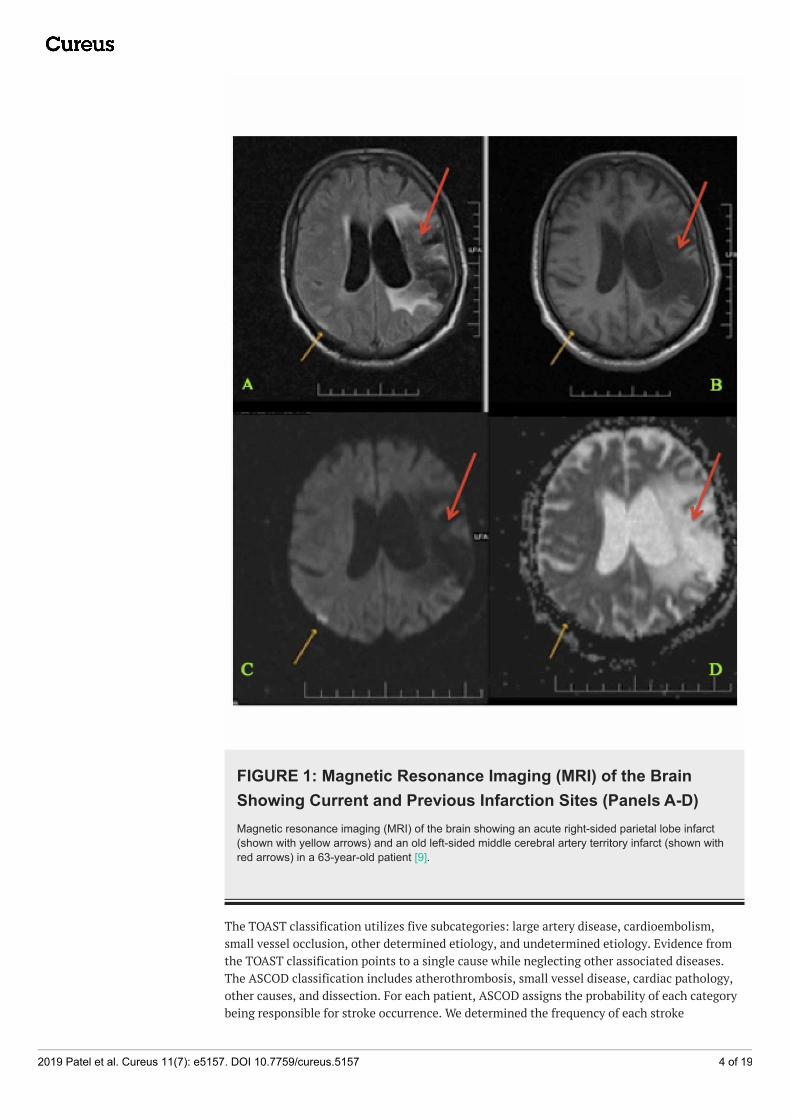
FIGURE 1: Magnetic Resonance Imaging (MRI) of the BrainShowing Current and Previous Infarction Sites (Panels A-D)Magnetic resonance imaging (MRI) of the brain showing an acute right-sided parietal lobe infarct(shown with yellow arrows) and an old left-sided middle cerebral artery territory infarct (shown withred arrows) in a 63-year-old patient [9].
The TOAST classification utilizes five subcategories: large artery disease, cardioembolism,small vessel occlusion, other determined etiology, and undetermined etiology. Evidence fromthe TOAST classification points to a single cause while neglecting other associated diseases.The ASCOD classification includes atherothrombosis, small vessel disease, cardiac pathology,other causes, and dissection. For each patient, ASCOD assigns the probability of each categorybeing responsible for stroke occurrence. We determined the frequency of each stroke
2019 Patel et al. Cureus 11(7): e5157. DOI 10.7759/cureus.5157 4 of 19
etiology/mechanism according to both classification systems and the most predominantmechanism in each classification system.
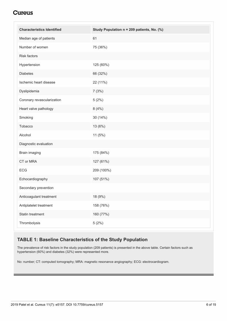
ResultsWe evaluated a total of 209 patients (mean age: 61 years), of which 64% were men and 36% werewomen. The baseline characteristics of the study population were identified and evaluated (seeTable 1). The prevalence of risk factors in our study was as follows: hypertension (60%),diabetes (32%), ischemic heart disease (11%), heart valve pathology (4%), and dyslipidemia(3%). The determined prevalence of personal habits documented was found to be smoking(14%), tobacco (6%), and alcohol (5%). Despite the recognized association between smoking asa risk factor and stroke incidence, there is a high likelihood that patients underreported theirsmoking habit as it is considered socially unacceptable in that area of the world [10]. Using theASCOD classification, we found that A was present in 42% of patients (A1 = 21%, A2 = 8%, A3 =13%), S was present in 44% of patients (S1 = 9%, S2 = 1%, S3 = 34%), C was present in 17% ofpatients (C1 = 6%, C2 = 4%, C3 =7 %), O was present in 5% of patients (O1 = 5%), and D waspresent in <1% of patients (see Figure 2). The TOAST classification showed LAA (33%), SVO(29%), CE (13%), other causes (6%), and undetermined (18%) (see Figure 3). In the ASCODclassification, there was an overlap of disease between grades 1 and 2 (3%) and when extendedto grade 3 the overlap was 26%.
2019 Patel et al. Cureus 11(7): e5157. DOI 10.7759/cureus.5157 5 of 19
Characteristics Identified Study Population n = 209 patients, No. (%)
Median age of patients 61
Number of women 75 (36%)
Risk factors
Hypertension 125 (60%)
Diabetes 66 (32%)
Ischemic heart disease 22 (11%)
Dyslipidemia 7 (3%)
Coronary revascularization 5 (2%)
Heart valve pathology 8 (4%)
Smoking 30 (14%)
Tobacco 13 (6%)
Alcohol 11 (5%)
Diagnostic evaluation
Brain imaging 175 (84%)
CT or MRA 127 (61%)
ECG 209 (100%)
Echocardiography 107 (51%)
Secondary prevention
Anticoagulant treatment 18 (9%)
Antiplatelet treatment 158 (76%)
Statin treatment 160 (77%)
Thrombolysis 5 (2%)
TABLE 1: Baseline Characteristics of the Study PopulationThe prevalence of risk factors in the study population (209 patients) is presented in the above table. Certain factors such ashypertension (60%) and diabetes (32%) were represented more.
No: number; CT: computed tomography; MRA: magnetic resonance angiography; ECG: electrocardiogram.
2019 Patel et al. Cureus 11(7): e5157. DOI 10.7759/cureus.5157 6 of 19
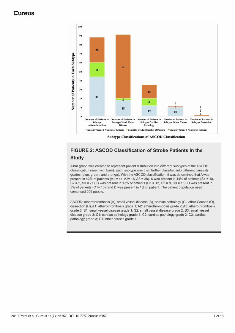
FIGURE 2: ASCOD Classification of Stroke Patients in theStudyA bar graph was created to represent patient distribution into different subtypes of the ASCODclassification (seen with bars). Each subtype was then further classified into different causalitygrades (blue, green, and orange). With the ASCOD classification, it was determined that A waspresent in 42% of patients (A1 = 44, A2= 16, A3 = 28), S was present in 44% of patients (S1 = 18,S2 = 2, S3 = 71), C was present in 17% of patients (C1 = 12, C2 = 8, C3 = 15), O was present in5% of patients (O1= 10), and D was present in 1% of patient. The patient population usedcomprised 209 people.
ASCOD: atherothrombosis (A), small vessel disease (S), cardiac pathology (C), other Causes (O),dissection (D); A1: atherothrombosis grade 1; A2: atherothrombosis grade 2; A3: atherothrombosisgrade 3; S1: small vessel disease grade 1; S2: small vessel disease grade 2; S3: small vesseldisease grade 3; C1: cardiac pathology grade 1; C2: cardiac pathology grade 2; C3: cardiacpathology grade 3; O1: other causes grade 1.
2019 Patel et al. Cureus 11(7): e5157. DOI 10.7759/cureus.5157 7 of 19

FIGURE 3: TOAST Classification of Stroke Patients in theStudyA bar graph was created to represent patient distribution according to the TOAST classification. Thex-axis represents the subtypes of TOAST classification and the y-axis represents the number ofpatients in each subtype. The study population comprised 209 patients. The TOAST classificationshowed LAA (70), SVO (61), CE (27), other causes (13), and undetermined (30).
TOAST: Trial of Org 10172 in Acute Stroke Treatment; LAA: large artery atherosclerosis; SVO:small vessel occlusion; CE: cardioembolism.
Diagnostic evaluation was necessary in both classifications to further categorize each patient.In our study, we found the prevalence of conclusive brain imaging (84%), CT angiography (CTA)or magnetic resonance angiogram (MRA) (61%), ECG (100%), and echocardiography (51%). Theprevalence of incomplete evaluations consisting of brain imaging (16%), CTA or MRA (39%), orechocardiography (49%) was indicated by discharges against medical advice. These patients hadfinancial restrictions, they refused to give consent for the procedure, or their power of attorneyrequested a transfer to another hospital.
DiscussionThe TOAST classification was used to classify subtypes of ischemic stroke. TOAST helpedneurologists to further determine the treatment, the prognosis, and the recurrence of stroke inthese patients [7]. Similarly, the ASCOD classification is a phenotypic classification thatbroadly lists all the possible causes that could lead to stroke. Based on the degree of evidence,each disease can be certain, uncertain, unlikely, negative, or incompletely studied as a link tostroke [8]. Both classifications require an extensive workup, and incomplete investigations canlead to deficiencies in proper classification.
2019 Patel et al. Cureus 11(7): e5157. DOI 10.7759/cureus.5157 8 of 19
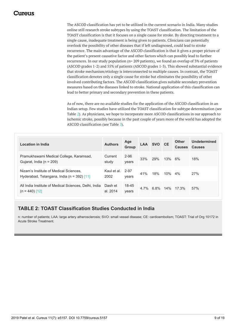
The ASCOD classification has yet to be utilized in the current scenario in India. Many studiesonline still research stroke subtypes by using the TOAST classification. The limitation of theTOAST classification is that it focuses on a single cause for stroke. By directing treatment to asingle cause, inadequate treatment is being given to patients. Clinicians can potentiallyoverlook the possibility of other diseases that if left undiagnosed, could lead to strokerecurrence. The main advantage of the ASCOD classification is that it gives a proper picture ofthe patient’s present causative factor and other factors which can possibly lead to furtherrecurrences. In our study population (n= 209 patients), we found an overlap of 3% of patients(ASCOD grades 1-2) and 55% of patients (ASCOD grades 1-3). This showed substantial evidencethat stroke mechanism/etiology is interconnected to multiple causes. In contrast, the TOASTclassification denotes only a single cause for stroke but eliminates the possibility of otherinvolved contributing factors. The ASCOD classification gives suitable secondary preventionmeasures based on the diseases linked to stroke. National application of this classification canlead to better primary and secondary prevention in these patients.
As of now, there are no available studies for the application of the ASCOD classification in anIndian setup. Few studies have utilized the TOAST classification for subtype determination (seeTable 2). As physicians, we hope to incorporate more ASCOD classifications in our approach toischemic stroke, possibly because in the past couple of years more of the world has adopted theASCOD classification (see Table 3).
Location in India AuthorsAgeGroup
LAA SVO CEOtherCauses
UndeterminedCauses
Pramukhswami Medical College, Karamsad,Gujarat, India (n = 209)
Currentstudy
2-96years
33% 29% 13% 6% 18%
Nizam’s Institute of Medical Sciences,Hyderabad, Telangana, India (n = 392) [11]
Kaul et al.2002
2-97years
41% 18% 10% 4% 27%
All India Institute of Medical Sciences, Delhi, India(n = 440) [12]
Dash etal. 2014
18-45years
4.7% 6.8% 14% 17.3% 57%
TABLE 2: TOAST Classification Studies Conducted in Indian: number of patients; LAA: large artery atherosclerosis; SVO: small vessel disease; CE: cardioembolism; TOAST: Trial of Org 10172 inAcute Stroke Treatment.
2019 Patel et al. Cureus 11(7): e5157. DOI 10.7759/cureus.5157 9 of 19

AuthorNameandYear oftheStudy
StudyPopulation
Observation
Gökçalet al.2017[13]
151 patients
Using the TOAST classification, patient stroke etiology was classified into undetermined(41.1%), CE (19.2%), LAA (13.2%), SVO (11.3%), and other causes (15.2%). Compared to theTOAST classification, ASCO classification assigned fewer patients to undetermined etiologysubtype (26.5%, p<0.001) and SVO category (21.9%, p<0.001). ASCO also assigned morepatients to the LAA group (16.6%).
Arsavaet al.2017[14]
1,816patients
The classification systems were different in their ability to assign stroke etiologies to knownsubtypes; the size of the undetermined category was 53% per the TOAST classification and42% per the ASCO classification (p < 0.001 for all binary comparisons).
Markakiet al.2013[15]
101patients, 84withischemicstroke and17 with aTIA
There was a moderately high agreement between the TOAST and ASCO classifications in allsubtypes. Along with the classification, the one- and four-year mortality rates were observedduring a mean observation period of 28 months, during which 26 patients died. The one- andfour-year mortality rates, respectively, were 0% and 4% in LAA, 23% and 36% in CE, 0% inSVO, 63% and 100% in unknown etiology, and 12% and 29% in the cryptogenic subtype. Forthe ASCO classification, the one-year and four-year mortality rates, respectively, were 0% and6% in LAA, 25% and 36% in CE, 0% in SVO, 0% and 14% in LAA + CE, 16% and 36% in SVO +CE, and 56% and 100% in the undetermined etiology despite complete workup.
Shanget al.2012[16]
425 patientswith firsttimeischemicstroke
There was a moderately high agreement between the TOAST and ASCO classification in allsubtypes except the “undetermined” etiology subtype (16.2% vs. 15.5 %, p = 0.795).
Wolf etal. 2012[17]
103 patients
There was a high agreement between the ASCO and TOAST classifications. With ASCO,grades 1-3 were identified in 60.19% A, 75.73% S, 49.51% C, and 3.88% O. Around 68.93% ofthe patients were classified in more than one category, and only 3.88% remained completelyundetermined. With the TOAST classification, the distribution was 9.71% in A, 23.30% in S,34.95% in C, 1.94% in O, and 30.10% in the undetermined subtype.
TABLE 3: TOAST and ASCOD/ASCO Stroke Etiology Classification Studies ConductedInternationallyTOAST: Trial of Org 10172 in Acute Stroke Treatment; CE: cardioembolism; LAA: large artery atherosclerosis; SVO: small vesselocclusion; ASCOD: atherothrombosis (A), small vessel disease (S), cardiac pathology (C), other causes (O), and dissection (D).
The Hyderabad study showed a similar median age of patients at 54 years compared to a medianage of 61 years in our study [11]. The predominant subtype of ischemic stroke was LAA (41-33%). Undetermined etiology was the second most common in that study [11]. This was mainlyattributed to the lack of the new algorithm proposed by the Stop-Stroke Study TOAST in 2005[18]. The new modifications to the TOAST classification expanded the definitions of SVO and
2019 Patel et al. Cureus 11(7): e5157. DOI 10.7759/cureus.5157 10 of 19

LAA that then decreased the undetermined subtype to 4% [18]. Our study still had a largeproportion of undetermined cases (18%), which were due to incomplete evaluation (14%) andnegative evaluation (4%). The reasons for incomplete evaluation included financial restraints ofthe patients, negative consent by the relatives, request for transfer, or death of the patient. InIndia, financial restraints proved to be the greatest barrier to proper evaluation of a strokepatient. For additional information on the TOAST and ASCOD classifications, see the appendixfor Tables 4-11.
ConclusionsStroke is a complex disease with numerous contributing factors. Without a standardizedprotocol, it becomes difficult to collect data on patients, follow up, and provide treatment. TheASCOD classification is a better fit for patients of the Indian population and helps in decidingsecondary prevention appropriately. However, we need to continue evaluating its applicabilityby motivating more physicians to generate larger prospective studies utilizing the ASCODclassification. Only with further studies can physicians come closer to a more standardizedapproach to ischemic stroke classification.
Appendices
Trial of Org 10172 in Acute Stroke Treatment (TOAST) Classification [7]
Classification of Subtypes of Ischemic Stroke
Large ArteryAtherosclerosis
SmallVesselOcclusion
Cardioembolism
Stroke ofOtherDeterminedEtiology
Stroke ofUndeterminedEtiology
Stroke ofUndeterminedEtiology with aNegativeEvaluation
Stroke ofUndeterminedEtiology with anIncompleteEvaluation
TABLE 4: TOAST Classification [7]A table was created representing the different subtypes of the TOAST Classification [7].
TOAST: Trial of Org 10172 in Acute Stroke Treatment.
2019 Patel et al. Cureus 11(7): e5157. DOI 10.7759/cureus.5157 11 of 19

ASCOD Classification [8]
Classification of Subtypes of Ischemic Stroke
Atherothrombosis (A) Small Vessel Disease (S) Cardiac Pathology (C) Other Causes (O) Dissection (D)
TABLE 5: ASCOD Classification [8]A table was created representing the different causality grades, as an underlying etiology of ischemic stroke as per a subcategory ofthe ASCOD classification [8].
ASCOD: atherothrombosis, small vessel disease, cardiac pathology, other causes, and dissection classification.
The CausalityGrades [8]
ASCOD Atherothrombosis (A) Phenotypes According to Classification [8]
A1: potentiallycausal. A strokethat isatherothromboticis defined as oneof the following:
An ipsilateralatheroscleroticstenosis of 50-99%in an intracranial orextracranial arterysupplying theischemic field.
An ipsilateralatherosclerotic stenosis<50% in an intracranialor extracranial arterywith an endoluminalthrombus supplying theischemic field.
A mobilethrombus inthe aortic arch.
An ipsilateral arterial occlusion inan intracranial or extracranialartery with evidence of underlyingatherosclerotic plaque supplyingthe ischemic field.
A2: the causal linkis uncertain.Defined aspotentially one ofthe following:
An ipsilateralatheroscleroticstenosis of 30-50%in an intracranial orextracranial arterysupplying theischemic field.
An aortic plaque ≥ 4 mm without a mobile lesion.
A3: the causal linkis unlikely, but thedisease ispresent. One ormore of thefollowing may beseen:
A plaque (stenosis<30%) in anintracranial orextracranial artery,which is ipsilateralto the infarct area.
An aortic plaque<4 mm without amobile thrombus.
A stenosisof anydegreethat is notsupplyingthe infarctarea.
A present historyof myocardialinfarction,coronaryrevascularization,or peripheralarterial disease.
An ipsilateral or bilateralatherosclerotic stenosisof 50–99% withbihemispheric MR-DWIlesion present.
A0:atherosclerosis isnot detected. Inorder to rule outatherosclerosis,the followingshould be lookedfor:
A negative findingfor an extracranialarterial stenosis onUS-duplex, CTA,MRA, XRA, orautopsy.
A negative findingfor an intracranialarterial stenosison US-TCD, CTA,MRA, XRA, orautopsy.
A negative finding for an aortic arch atheroma: TEE orCTA with specific assessment of the aortic arch.
2019 Patel et al. Cureus 11(7): e5157. DOI 10.7759/cureus.5157 12 of 19
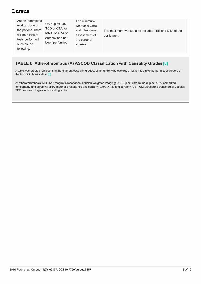
A9: an incompleteworkup done onthe patient. Therewill be a lack oftests performedsuch as thefollowing:
US-duplex, US-TCD or CTA, orMRA, or XRA orautopsy has notbeen performed.
The minimumworkup is extra-and intracranialassessment ofthe cerebralarteries.
The maximum workup also includes TEE and CTA of theaortic arch.
TABLE 6: Atherothrombus (A) ASCOD Classification with Causality Grades [8]A table was created representing the different causality grades, as an underlying etiology of ischemic stroke as per a subcategory ofthe ASCOD classification [8].
A: atherothrombosis; MR-DWI: magnetic resonance diffusion-weighted imaging; US-Duplex: ultrasound duplex; CTA: computedtomography angiography; MRA: magnetic resonance angiography; XRA: X-ray angiography; US-TCD: ultrasound transcranial Doppler;TEE: transesophageal echocardiography.
2019 Patel et al. Cureus 11(7): e5157. DOI 10.7759/cureus.5157 13 of 19

The Causality Grades [8]ASCOD Small Vessel Disease (S) Phenotypes According toClassification [8]
S1: potentially causal. A stroke that will havea combination of a lacunar artery infarctionon an MRI-DWI in an area corresponding tothe symptoms and at least one out of thethree following criteria:
One or several smalldeep older infarct(s) oflacunar type in otherterritories.
The patient has severe white matter lesions,small vessel ischemia, microbleeds, or severedilatation of perivascular spaces.
S2: the causal link is uncertain. It is definedas potentially one of the following:
Only one recentlacunar infarction andno other abnormality isseen on MRI or CT.
The patient has a clinical syndrome suggestiveof a deep branch artery stroke, without anischemic lesion in the appropriate area.
S3: the causal link is unlikely, but thedisease is present.
There are severe whitematter lesionsrepresentative of smallvessel ischemia visibleon an MRI or CT scan.
There aremicrobleedsseen on aT2-weightedMRI.
There isa severedilatation of theperivascularspace visible onT2-weightedMRI.
There are oneor several old,small, anddeep infarctsof the lacunartype.
S0: small vessel disease is not detected. Inorder to rule out small vessel disease, lookfor the following:
Patient has anegativeMRI (T2, FLAIR, GRE,DWI).
There is no appropriate clinical syndromesuggestive of a deep branch artery stroke.
S9: an incomplete workup done on thepatient. There will be a lack of testsperformed such as:
MRI CT scan
TABLE 7: Small Vessel Disease (S) ASCOD Classification with Causality Grades [8]A table was created representing the different causality grades, as an underlying etiology of ischemic stroke as per a subcategory ofthe ASCOD classification [8].
ASCOD: atherothrombosis, small vessel disease, cardiac pathology, other causes, and dissection classification; MRI-DWI: magneticresonance diffusion-weighted imaging; MRI: magnetic resonance imaging; CT: computed tomography; FLAIR: fluid-attenuatedinversion recovery; GRE: gradient echo imaging; DWI: diffusion-weighted imaging.
The CausalityGrades [8]
ASCOD Cardiac Pathology (C) Disease Phenotypes According to Classification [8]
C1: potentiallycausal. A stroke thatis cardiogenic isdefined as anischemic lesion andhas signs of systemicembolism withdetection of at least
Mitral stenosis, mechanical valve, myocardial infarction within four weeks, preceding the cerebralinfarction, mural thrombus in the left cavities, aneurysm of the left ventricle, a history or presenceof documented atrial fibrillation (either paroxysmal, persistent or permanent) or atrial flutter with orwithout left atrial thrombus or spontaneous echo, atrial disease (tachycardia-bradycardiasyndrome), dilated or hypertrophic cardiomyopathies, left ventricle ejection fraction <35%,endocarditis, intracardiac mass, PFO and thrombus in situ, PFO and concomitant pulmonaryembolism or proximal DVT preceding the index cerebral infarction, aforementioned cardiac
2019 Patel et al. Cureus 11(7): e5157. DOI 10.7759/cureus.5157 14 of 19

one of the followingcauses:
pathologies (C1) with single or without obvious cerebral ischemic lesion
C2: the causal link isuncertain.Regardless of thecardiogenic strokepattern, there maybe:
PFO +atrialseptalaneurysm.
PFO andpulmonaryembolism orproximal DVTconcomitant butnot precedingthe indexcerebralinfarction.
Anintracardiacspontaneousechocontrast.
Apicalakinesia ofthe leftventricleanddecreasedejectionfraction (but>35%) inthe patient.
A history ofmyocardialinfarction orpalpitation, andmultiple braininfarction,repeated eitherbilaterally or in twodifferent arterialterritories.
No directcardiac sourceidentified butmultiple braininfarctionspresent and/orevidence ofsystemicemboli.
C3: the causal link isunlikely, but thedisease is present. Itis defined aspotentially one of thefollowing:
PFO, ASA, strands, mitral annulus calcification, calcification aortic valve, non-apical akinesia ofthe left ventricle, transient atrial fibrillation less than 1 minute in duration, atrial hyperexcitability.
C0: cardiacpathology is neitherdetected norsuspected. Workupneeded to rule outcardiac pathology:
The minimum workup needed isa negative ECG and an examinationby a cardiologist.
The maximum workup needed is a negativeECG/telemetry/24-hour Holter ECG/long-term ECGrecording, a negative TEE, a negative TTE for PFO andassessment of left ventricle, a negative cardiac CT/MRI,and a negative abdominal CT/MRI.
C9: an incompleteworkup done on thepatient. Minimumworkup needed torule out cardiogenicshock:
ECG An examination by a trained cardiologist in the absence of cardiac imaging.
TABLE 8: Cardiac Pathology (C) of the ASCOD Classification with Causality Grades[8]A table was created representing the different causality grades, as an underlying etiology of ischemic stroke as per a subcategory ofthe ASCOD classification [8].
ASCOD: atherothrombosis, small vessel disease, cardiac pathology, other causes, and dissection classification; PFO: patent foramenovale; DVT: deep venous thrombosis; ASA: atrial septal aneurysm; ECG: electrocardiogram; TEE: transesophageal echocardiography;TTE: transthoracic echocardiography; CT: computed tomography; MRI: magnetic resonance imaging.
2019 Patel et al. Cureus 11(7): e5157. DOI 10.7759/cureus.5157 15 of 19
The CausalityGrades [8]
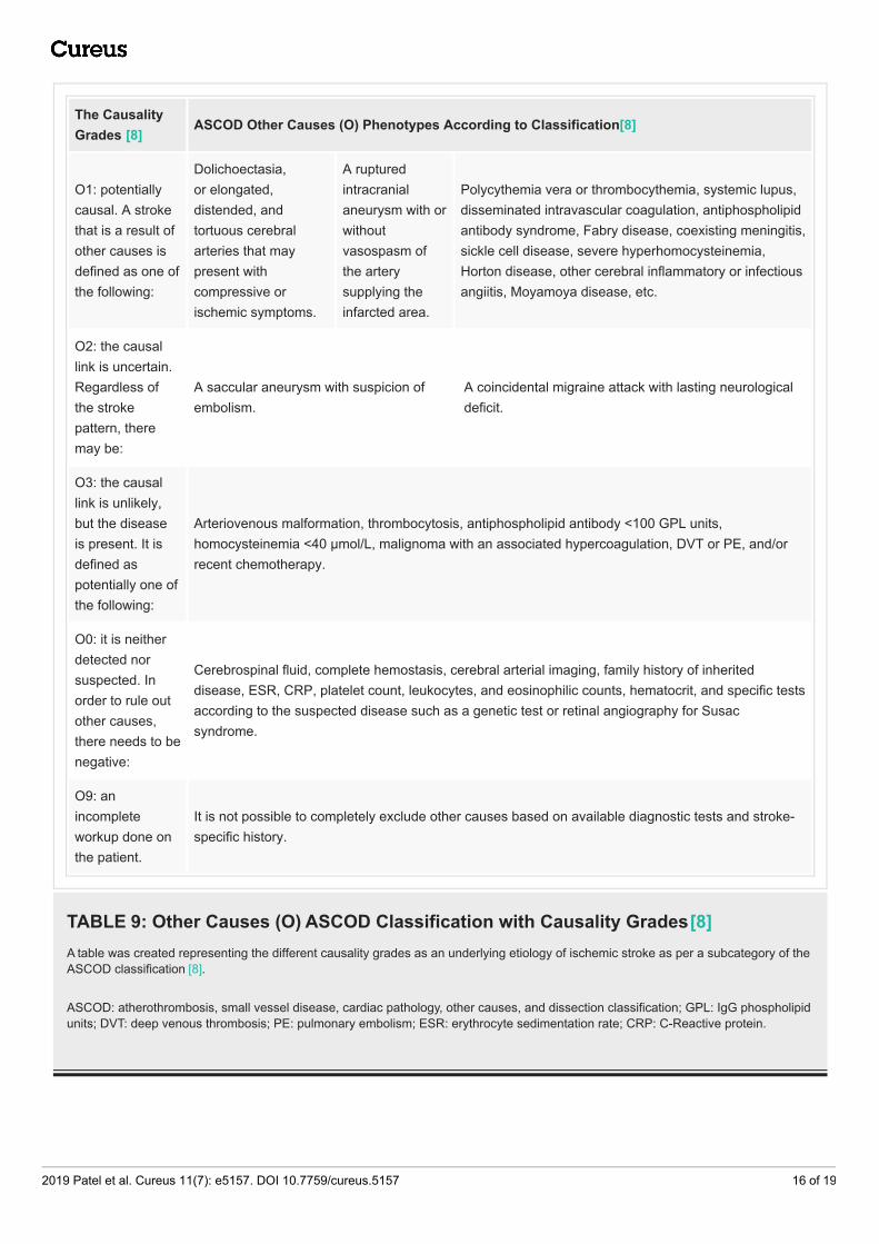
ASCOD Other Causes (O) Phenotypes According to Classification [8]
O1: potentiallycausal. A strokethat is a result ofother causes isdefined as one ofthe following:
Dolichoectasia,or elongated,distended, andtortuous cerebralarteries that maypresent withcompressive orischemic symptoms.
A rupturedintracranialaneurysm with orwithoutvasospasm ofthe arterysupplying theinfarcted area.
Polycythemia vera or thrombocythemia, systemic lupus,disseminated intravascular coagulation, antiphospholipidantibody syndrome, Fabry disease, coexisting meningitis,sickle cell disease, severe hyperhomocysteinemia,Horton disease, other cerebral inflammatory or infectiousangiitis, Moyamoya disease, etc.
O2: the causallink is uncertain.Regardless ofthe strokepattern, theremay be:
A saccular aneurysm with suspicion ofembolism.
A coincidental migraine attack with lasting neurologicaldeficit.
O3: the causallink is unlikely,but the diseaseis present. It isdefined aspotentially one ofthe following:
Arteriovenous malformation, thrombocytosis, antiphospholipid antibody <100 GPL units,homocysteinemia <40 μmol/L, malignoma with an associated hypercoagulation, DVT or PE, and/orrecent chemotherapy.
O0: it is neitherdetected norsuspected. Inorder to rule outother causes,there needs to benegative:
Cerebrospinal fluid, complete hemostasis, cerebral arterial imaging, family history of inheriteddisease, ESR, CRP, platelet count, leukocytes, and eosinophilic counts, hematocrit, and specific testsaccording to the suspected disease such as a genetic test or retinal angiography for Susacsyndrome.
O9: anincompleteworkup done onthe patient.
It is not possible to completely exclude other causes based on available diagnostic tests and stroke-specific history.
TABLE 9: Other Causes (O) ASCOD Classification with Causality Grades [8]A table was created representing the different causality grades as an underlying etiology of ischemic stroke as per a subcategory of theASCOD classification [8].
ASCOD: atherothrombosis, small vessel disease, cardiac pathology, other causes, and dissection classification; GPL: IgG phospholipidunits; DVT: deep venous thrombosis; PE: pulmonary embolism; ESR: erythrocyte sedimentation rate; CRP: C-Reactive protein.
2019 Patel et al. Cureus 11(7): e5157. DOI 10.7759/cureus.5157 16 of 19

The CausalityGrades [8]
ASCOD Dissection (D) Disease Phenotypes According to Classification [8]
D1: potentiallycausal. A strokecaused by dissectioncan be a result ofone of the following:
An arterial dissection demonstrated by ahypersignal on FAT-saturated MRI,autopsy, TOF-MRA, CT scans, increasedarterial diameter.
A demonstration of an arterial dissection by anindirect demonstration or by less sensitive or lessspecific diagnostic test (XRA, echocardiography,CTA, MRA, US) like an arterial stenosis seen withoutdemonstration of the arterial wall hematoma
D2: the causal link isuncertain. It isdefined as potentiallyone of the following:
An arterial dissection diagnosed based ona suggestive clinical history like a pasthistory of dissection or Horner’ssyndrome.
If there is imaging evidence of fibromusculardysplasia of a cerebral artery of an involved cerebralfield present.
D3: the causal link isunlikely, but thedisease is present.Defined aspotentially one of thefollowing;
There is kinking or dolichoectasia(elongated, distended, and tortuouscerebral arteries that may present withcompressive or ischemic symptoms)without complicated aneurysm or plicature.
There are arteries not implicated in the currentischemia that have evidence of fibromusculardysplasia.
D0: dissection isneither detected norsuspected. In orderto rule it out, thefollowing shouldbe done:
A negativefat-saturatedMRI.
AnormalXRA.
There is a lack ofclinical suspicion ofdissection.
There are negative extra- andintracranial cerebral arteryevaluations.
A negativecardiacevaluation.
D9: an incompleteworkup done on thepatient. Minimumworkup needed torule out dissection is:
In patients younger than 60 years and with no evidence of A1, A2, S1, C1, or O1, should undergoMRI or XRA within 15 days of symptom onset.
TABLE 10: Dissection (D) ASCOD Classification with Causality Grades [8]A table was created representing the different causality grades as an underlying etiology of ischemic stroke as per a subcategory of theASCOD classification [8].
ASCOD: atherothrombosis, small vessel disease, cardiac pathology, other causes, and dissection classification; MRI: magneticresonance imaging; TOF-MRA: time-of-flight magnetic resonance angiography; CT: computed tomography; XRA: X-ray angiography;CTA: computed tomography angiography; MRA: magnetic resonance angiography; US: ultrasound.
2019 Patel et al. Cureus 11(7): e5157. DOI 10.7759/cureus.5157 17 of 19
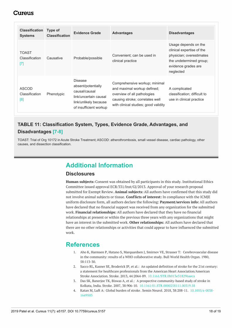
ClassificationSystems
Type ofClassification
Evidence Grade Advantages Disadvantages
TOASTClassification[7]
Causative Probable/possibleConvenient; can be used inclinical practice
Usage depends on theclinical expertise of thephysician; overestimatesthe undetermined group;evidence grades areneglected
ASCODClassification[8]
Phenotypic
Diseaseabsent/potentiallycausal/causallink/uncertain causallink/unlikely becauseof insufficient workup
Comprehensive workup; minimaland maximal workup defined;overview of all pathologiescausing stroke; correlates wellwith clinical studies; good validity
A complicatedclassification; difficult touse in clinical practice
TABLE 11: Classification System, Types, Evidence Grade, Advantages, andDisadvantages [7-8]TOAST: Trial of Org 10172 in Acute Stroke Treatment; ASCOD: atherothrombosis, small vessel disease, cardiac pathology, othercauses, and dissection classification.
Additional InformationDisclosuresHuman subjects: Consent was obtained by all participants in this study. Institutional EthicsCommittee issued approval ECR/331/Inst/GJ/2013. Approval of your research proposalsubmitted for Exempt Review. Animal subjects: All authors have confirmed that this study didnot involve animal subjects or tissue. Conflicts of interest: In compliance with the ICMJEuniform disclosure form, all authors declare the following: Payment/services info: All authorshave declared that no financial support was received from any organization for the submittedwork. Financial relationships: All authors have declared that they have no financialrelationships at present or within the previous three years with any organizations that mighthave an interest in the submitted work. Other relationships: All authors have declared thatthere are no other relationships or activities that could appear to have influenced the submittedwork.
References1. Aho K, Harmsen P, Hatano S, Marquardsen J, Smirnov VE, Strasser T: Cerebrovascular disease
in the community: results of a WHO collaborative study. Bull World Health Organ. 1980,58:113-30.
2. Sacco RL, Kasner SE, Broderick JP, et al.: An updated definition of stroke for the 21st century:a statement for healthcare professionals from the American Heart Association/AmericanStroke Association. Stroke. 2013, 44:2064-89. 10.1161/STR.0b013e318296aeca
3. Das SK, Banerjee TK, Biswas A, et al.: A prospective community-based study of stroke inKolkata, India. Stroke. 2007, 38:906-10. 10.1161/01.STR.0000258111.00319.58
4. Katan M, Luft A: Global burden of stroke . Semin Neurol. 2018, 38:208-11. 10.1055/s-0038-1649503
2019 Patel et al. Cureus 11(7): e5157. DOI 10.7759/cureus.5157 18 of 19

5. GBD 2016 Stroke Collaborators: Global, regional, and national burden of stroke, 1990-2016: asystematic analysis for the Global Burden of Disease Study 2016. Lancet Neurol. 2019, 18:439-58. 10.1016/S1474-4422(19)30034-1
6. World Stroke Organization global stroke fact sheet. (2019). Accessed: May 26, 2019:https://www.world-stroke.org/images/WSO_Global_Stroke_Fact_Sheet_final.pdf.
7. Adams HP Jr, Bendixen BH, Kappelle LJ, Biller J, Love BB, Gordon DL, Marsh EE 3rd:Classification of subtype of acute ischemic stroke. Definitions for use in a multicenter clinicaltrial. TOAST. Trial of Org 10172 in Acute Stroke Treatment. Stroke. 1993, 24:35-41.10.1161/01.STR.24.1.35
8. Amarenco P, Bogousslavsky J, Caplan LR, Donnan GA, Wolf ME, Hennerici MG: The ASCODphenotyping of ischemic stroke (updated ASCO phenotyping). Cerebrovasc Dis. 2013, 36:1-5.10.1159/000352050
9. Patel A R, Patel A R, Desai S: Acute hemiballismus as the presenting feature of parietal lobeinfarction. Cureus. 2019, 11:4675. 10.7759/cureus.4675
10. Boehme AK, Esenwa C, Elkind MS: Stroke risk factors, genetics, and prevention . Circ Res.2017, 120:472-95. 10.1161/CIRCRESAHA.116.308398
11. Kaul S, Sunitha P, Suvarna A, Meena AK, Uma M, Reddy JM: Subtypes of ischemic stroke in ametropolitan city of south India (one year data from hospital based stroke registry). NeurolIndia. 2002, 50:8-14.
12. Dash D, Bhashin A, Pandit AK, Tripathi M, Bhatia R, Prasad K, Padma MV: Risk factors andetiologies of ischemic strokes in young patients: a tertiary hospital study in north India. JStroke. 2014, 16:173-77. 10.5853/jos.2014.16.3.173
13. Gökçal E, Niftaliyev E, Asil T: Etiological classification of ischemic stroke in young patients: acomparative study of TOAST, CCS, and ASCO. Acta Neurol Belg. 2017, 117:643-48.10.1007/s13760-017-0813-8
14. Arsava EM, Helenius J, Avery R, et al.: Assessment of the predictive validity of etiologic strokeclassification. JAMA Neurol. 2017, 74:419-26. 10.1001/jamaneurol.2016.5815
15. Markaki I, Franzén I, Talani C, Loizou L, Kostulas N: Long-term survival of ischemiccerebrovascular disease in the acute inflammatory stroke study, a hospital-based cohortdescribed by TOAST and ASCO. Cerebrovasc Dis. 2013, 35:213-9. 10.1159/000346094
16. Shang Wy, Liu Jy: Stroke subtype classification: a comparative study of ASCO and modifiedTOAST. J Neurol Sci. 2012, 314:66-70. 10.1016/j.jns.2011.10.029
17. Wolf ME, Sauer T, Alonso A, Hennerici MG: Comparison of the new ASCO classification withthe TOAST classification in a population with acute ischemic stroke. J Neurol. 2012, 259:1284-9. 10.1007/s00415-011-6325-1
18. Ay H, Furie KL, Singhal A, Smith WS, Sorensen AG, Koroshetz WJ: An evidence-basedcausative classification system for acute ischemic stroke. Ann Neurol. 2005, 58:688-97.10.1002/ana.20617
2019 Patel et al. Cureus 11(7): e5157. DOI 10.7759/cureus.5157 19 of 19