comparing prokaryotic and eukaryotic cellsfire.biol.wwu.edu/cmoyer/zztemp_fire/biol345_w10/last...
TRANSCRIPT
1
Comparing Prokaryotic and Eukaryotic Cells
Basic unit of living organisms is the cell; the smallest unit capable of life.
“Features” found in all cells:! Ribosomes ! ATP Energy! Cell Membrane ! External Stimuli! Genetic Material ! Regulate Flow! Cytoplasm ! Reproduce
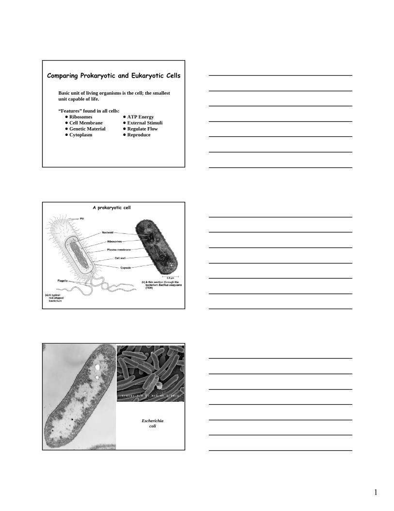
A prokaryotic cell
Escherichiacoli
2
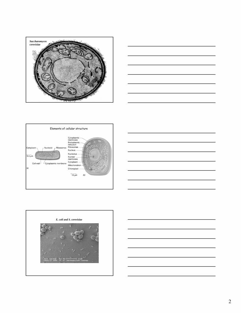
Saccharomycescerevisiae
Elements of cellular structure
E. coli and S. cerevisiae
3
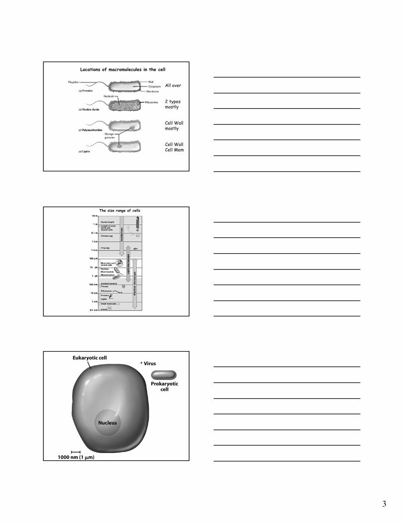
Locations of macromolecules in the cell
All over
2 typesmostly
Cell Wallmostly
Cell WallCell Mem
The size range of cells

4
Size relationship among prokaryotes
A Million times bigger than E. coli!
5
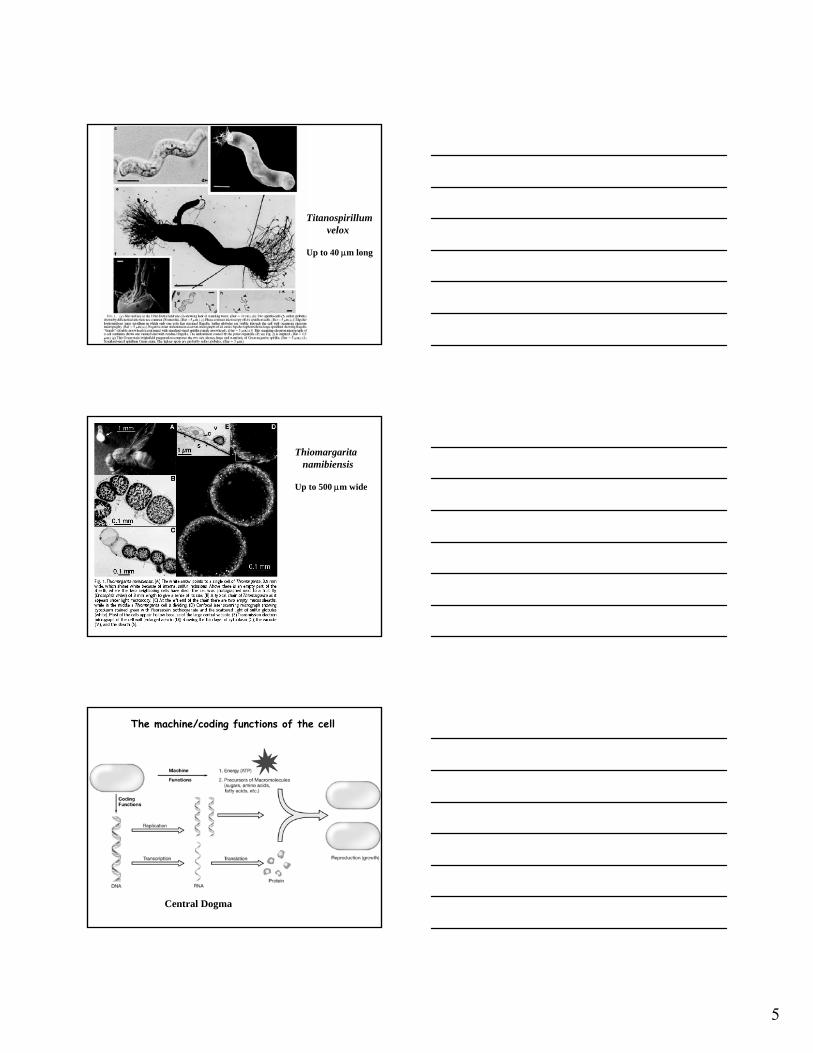
Titanospirillumvelox
Up to 40 μm long
Thiomargaritanamibiensis
Up to 500 μm wide
The machine/coding functions of the cell
Central Dogma
6
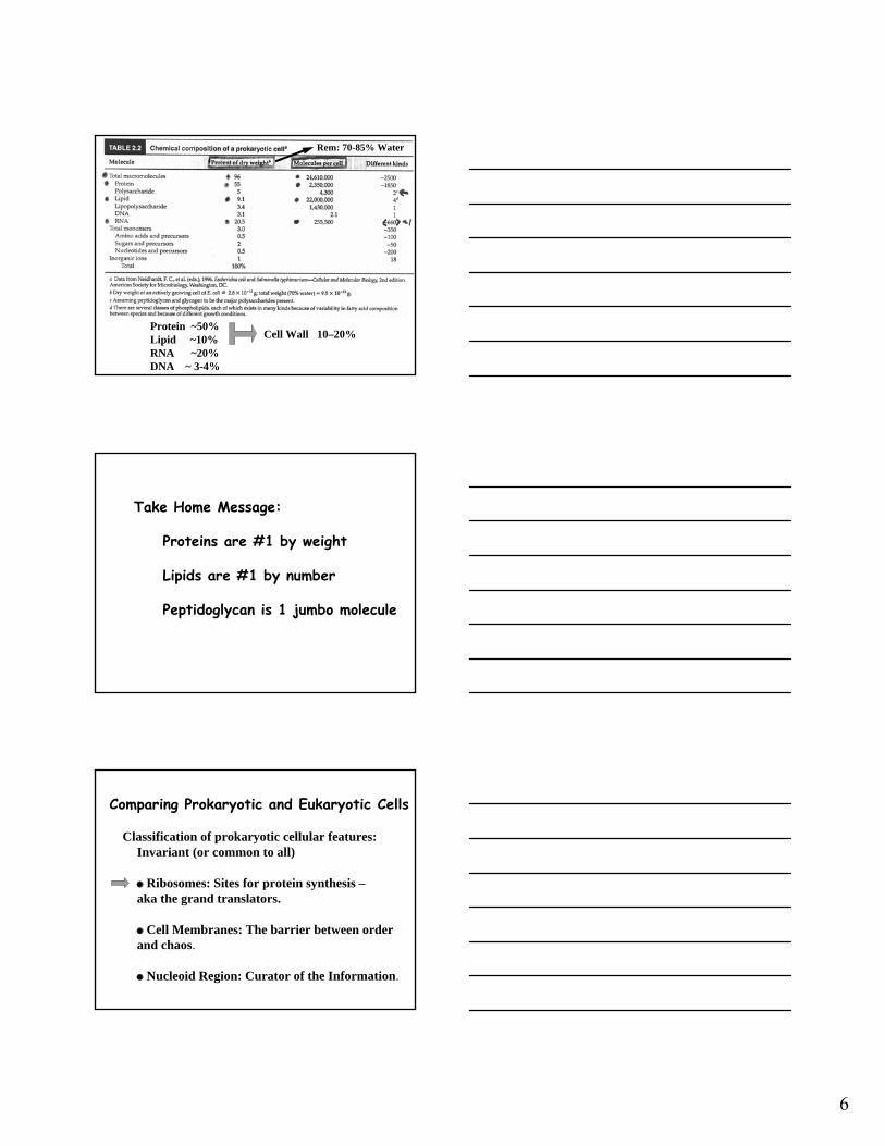
Protein ~50%Lipid ~10%RNA ~20%DNA ~ 3-4%
Cell Wall 10–20%
Rem: 70-85% Water
Take Home Message:
Proteins are #1 by weight
Lipids are #1 by number
Peptidoglycan is 1 jumbo molecule
Comparing Prokaryotic and Eukaryotic Cells
Classification of prokaryotic cellular features:Invariant (or common to all)
Ribosomes: Sites for protein synthesis –aka the grand translators.
Cell Membranes: The barrier between order and chaos.
Nucleoid Region: Curator of the Information.
7
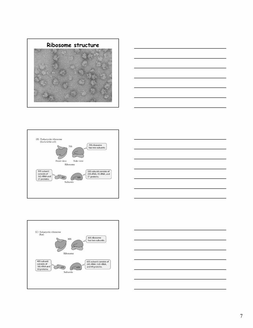
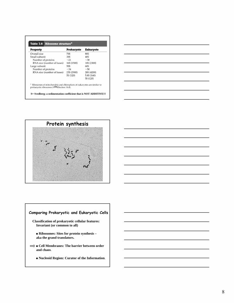
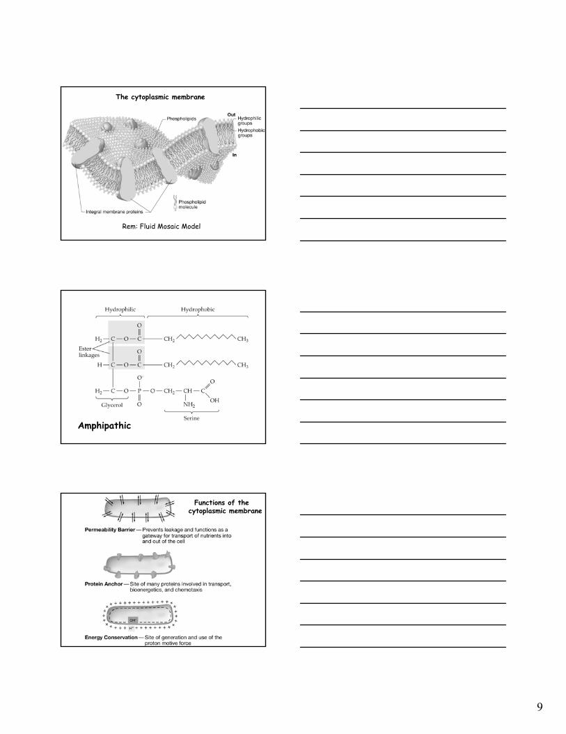
Ribosome structure
8
S= Svedberg; a sedimentation coefficient that is NOT ADDITIVE!!!
Protein synthesis
Comparing Prokaryotic and Eukaryotic Cells
Classification of prokaryotic cellular features:Invariant (or common to all)
Ribosomes: Sites for protein synthesis –aka the grand translators.
Cell Membranes: The barrier between order and chaos.
Nucleoid Region: Curator of the Information.
9
The cytoplasmic membrane
Rem: Fluid Mosaic Model
Amphipathic
Functions of the cytoplasmic membrane

10
Sterol Cholesterol Hopanoid(e.g., Diploptene)
All rigid planar molecules
O2 -Few Bacteria Many Bacteria
11
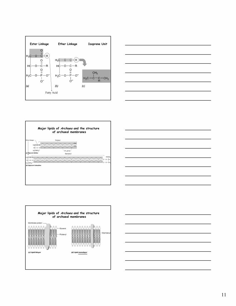
Ester Linkage Ether Linkage Isoprene Unit
Fatty Acid
Major lipids of Archaea and the structureof archaeal membranes
Major lipids of Archaea and the structureof archaeal membranes

12
Archaeal cell membrane structure
Comparing Prokaryotic and Eukaryotic Cells
Classification of prokaryotic cellular features:Invariant (or common to all)
Ribosomes: Sites for protein synthesis –aka the grand translators.
Cell Membranes: The barrier between order and chaos.
Nucleoid Region: Curator of the Information.
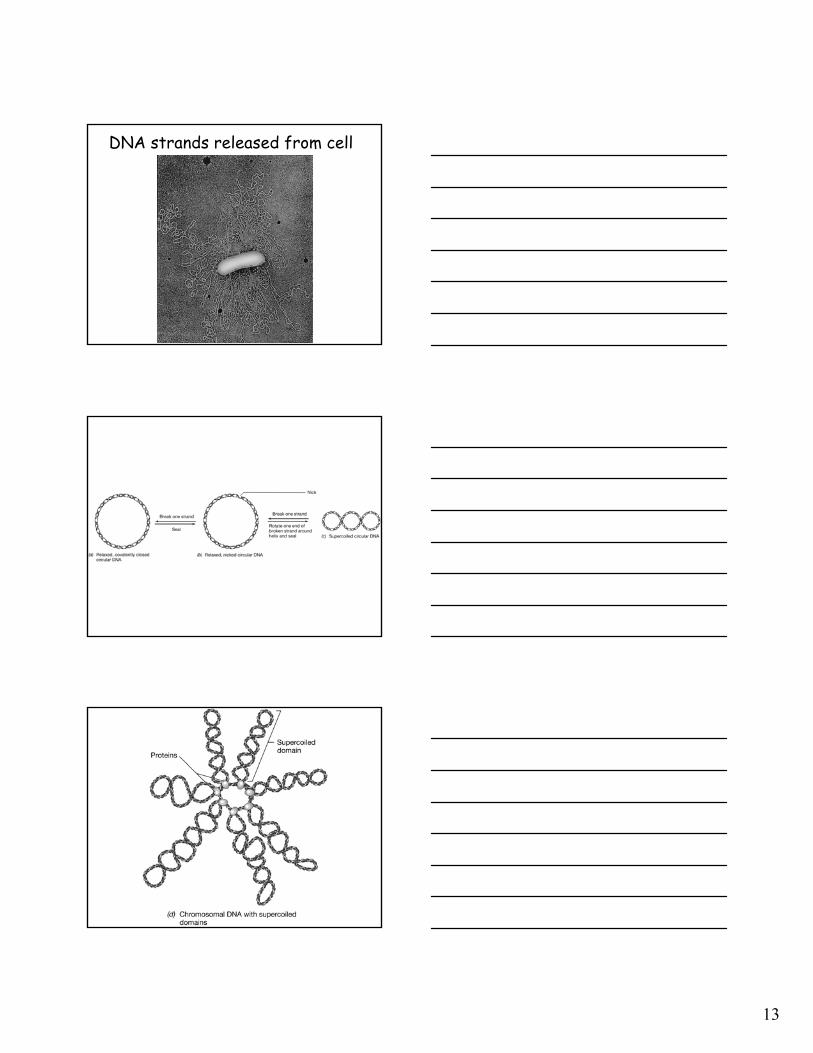
Appearance of DNA by EM
13
DNA strands released from cell
14
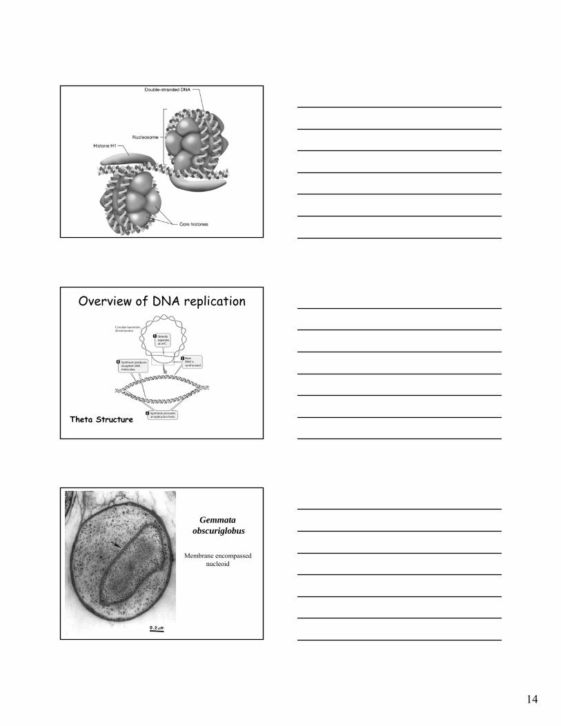
Overview of DNA replication
Theta Structure
Gemmataobscuriglobus
Membrane encompassednucleoid
15
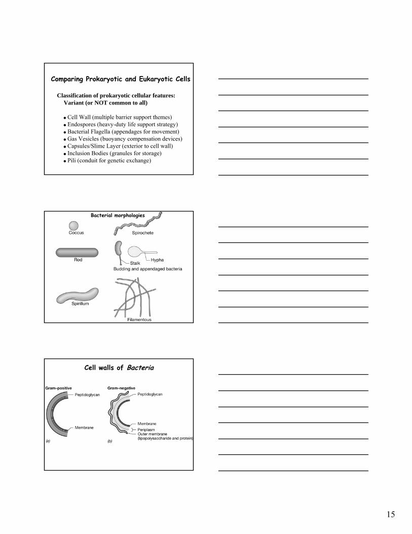
Comparing Prokaryotic and Eukaryotic Cells
Classification of prokaryotic cellular features:Variant (or NOT common to all)
Cell Wall (multiple barrier support themes)Endospores (heavy-duty life support strategy)Bacterial Flagella (appendages for movement)Gas Vesicles (buoyancy compensation devices)Capsules/Slime Layer (exterior to cell wall)Inclusion Bodies (granules for storage)Pili (conduit for genetic exchange)
Bacterial morphologies
Cell walls of Bacteria
16
Cell wall structure
E. coli structure ofpeptidoglycan aka murein
NAG NAM
DAP
Peptidoglycan of a gram-positive bacterium
Bond broken by penicillin

17
DAP or Diaminopimelic acid Lysine
Crossing linking AAs
Overall structure of peptidoglycan
Cell walls of gram-positive and gram-negative bacteria

18
Teichoic acids and the overall structure of the gram-positive cell wall
Summary diagram of the gram-positive cell wall
Cell envelopes of Bacteria

19
Cell envelopes of Bacteria
Structure of the lipopolysaccharideof gram-negative Bacteria
The gram-negative cell wall
20
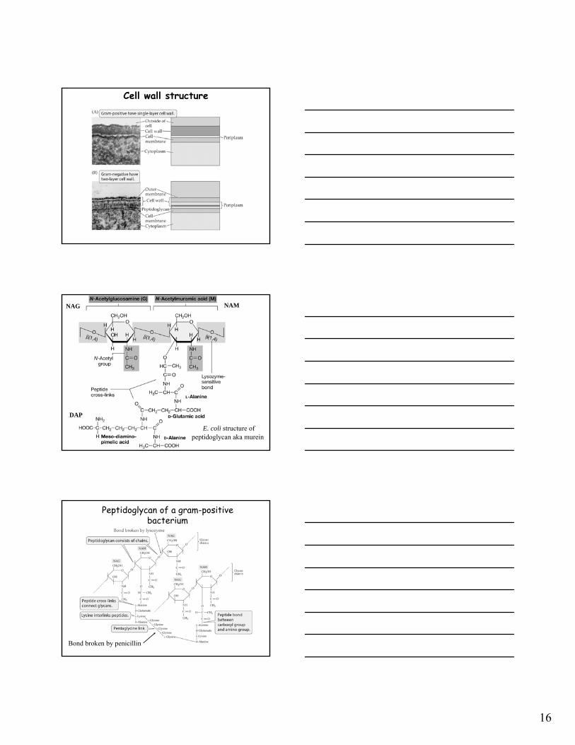
Pseudopeptidoglycanof Archaea
N-Acetyltalosaminuronic acidaka NAT
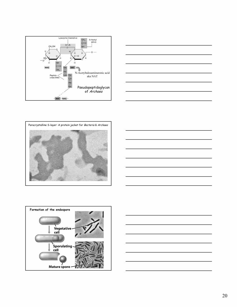
Paracrystalline S-layer: A protein jacket for Bacteria & Archaea
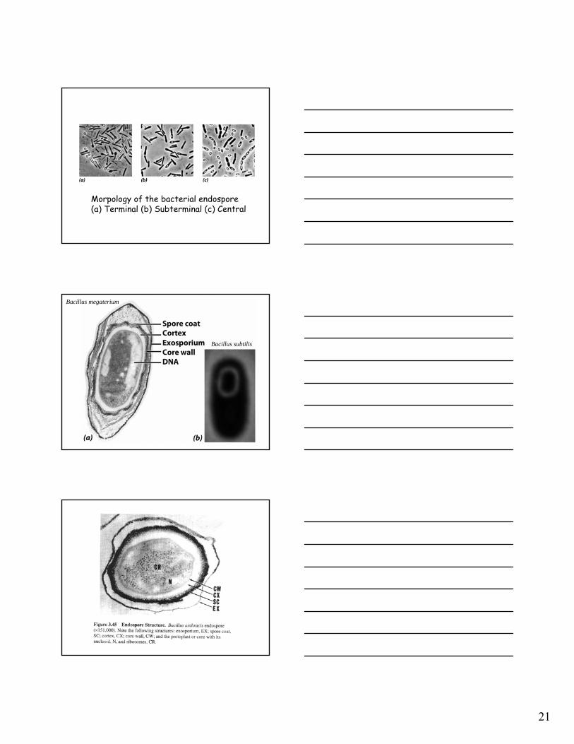
Formation of the endospore
21
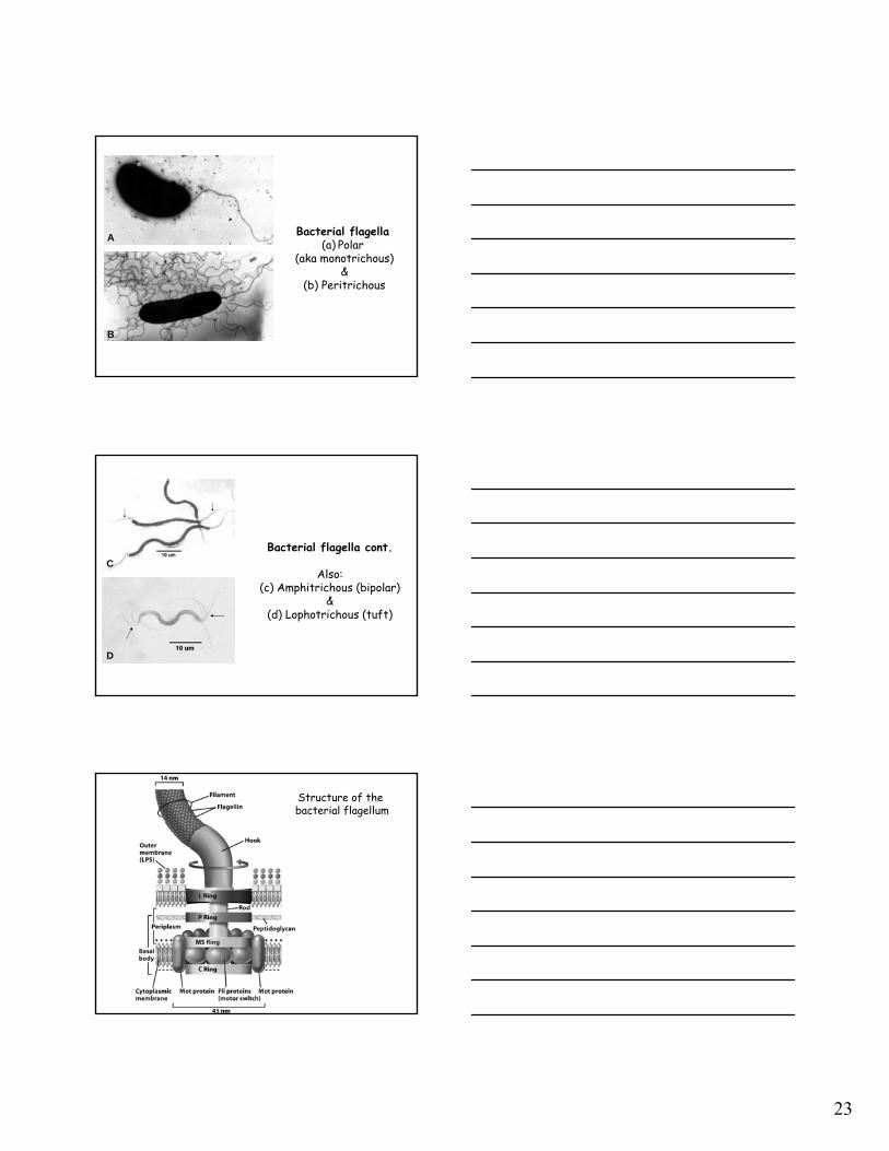
Morpology of the bacterial endospore(a) Terminal (b) Subterminal (c) Central
Bacillus megaterium
Bacillus subtilis

22
(a) Structure of Dipicolinic Acid & (b) crosslinked with Ca++
•
•
• The endospore is a highly resistant differentiated bacterial cell produced by certain gram-positive Bacteria.
• Endospore formation leads to a highly dehydrated structure that contains essential macromolecules and a variety of substances such as calcium dipicolinate and small acid-soluble proteins, absent from vegetative cells.
• Endospores can remain dormant indefinitely but germinate quickly when the appropriate trigger is applied.
Characteristics of Endospore: Take Home Message
23
Bacterial flagella(a) Polar
(aka monotrichous)&
(b) Peritrichous
A
B
Bacterial flagella cont.
Also:(c) Amphitrichous (bipolar)
&(d) Lophotrichous (tuft)
C
D
Structure of the bacterial flagellum
24
Proton Transport-Coupled Rotation of the Flagellum. (A) Mot protein may form a structure having two half-channels. (B) One model for the mechanism of coupling rotation to a proton gradient requires protons to be taken up into the outer half-channel and transferred to the MS ring. The MS ring rotates in a CCW direction, and the protons are released into the inner half-channel. The flagellumis linked to the MS ring and so the flagellum rotates as well.
Flagellar Motility: Relationship of flagellar rotation to bacterial movement.

25
Flagellar Motility: Relationship of flagellar rotation to bacterial movement.
(both)
Chemotaxis Signaling Pathway. Receptors in the plasma membrane initiate a signaling pathway leading to the phosphorylation of the CheY protein. Phosphorylated CheY binds to the flagellar motor and favors CW rotation. When an attractant binds to the receptor, this pathway is blocked, and CCW flagellar rotation and, hence, smooth swimming results. When a repellant binds, the pathway is stimulated, leading to an increased concentration of phosphoylated CheY and, hence, more frequent CW rotation and tumbling.
26
• Motility in most microorganisms is due to flagella.
• In prokaryotes the flagellum is a complex structure made of several proteins, most of which are anchored in the cell wall and cytoplasmic membrane.
• The flagellum filament, which is made of a single kind of protein, rotates at the expense of the proton motive force, which drives the flagellar motor.
Flagellar Motility: Take Home Message
Gliding Motility: Mechanism??
Gas Vesicles(a) Anabaena flos-aquae
(b) Microcystis sp.
A
B

27
The Hammer, Cork, and Bottle Experiment(Before) (After)
Model of how the two proteins that make up the gas vesicle, GvpA and GvpC, interact to form a
watertight but gas-permeable structure.
α-helix
β-sheet
Bacterial Capsules: (a) Acinetobacter sp. (b) Rhizobium trifolii
negative stainA B
28
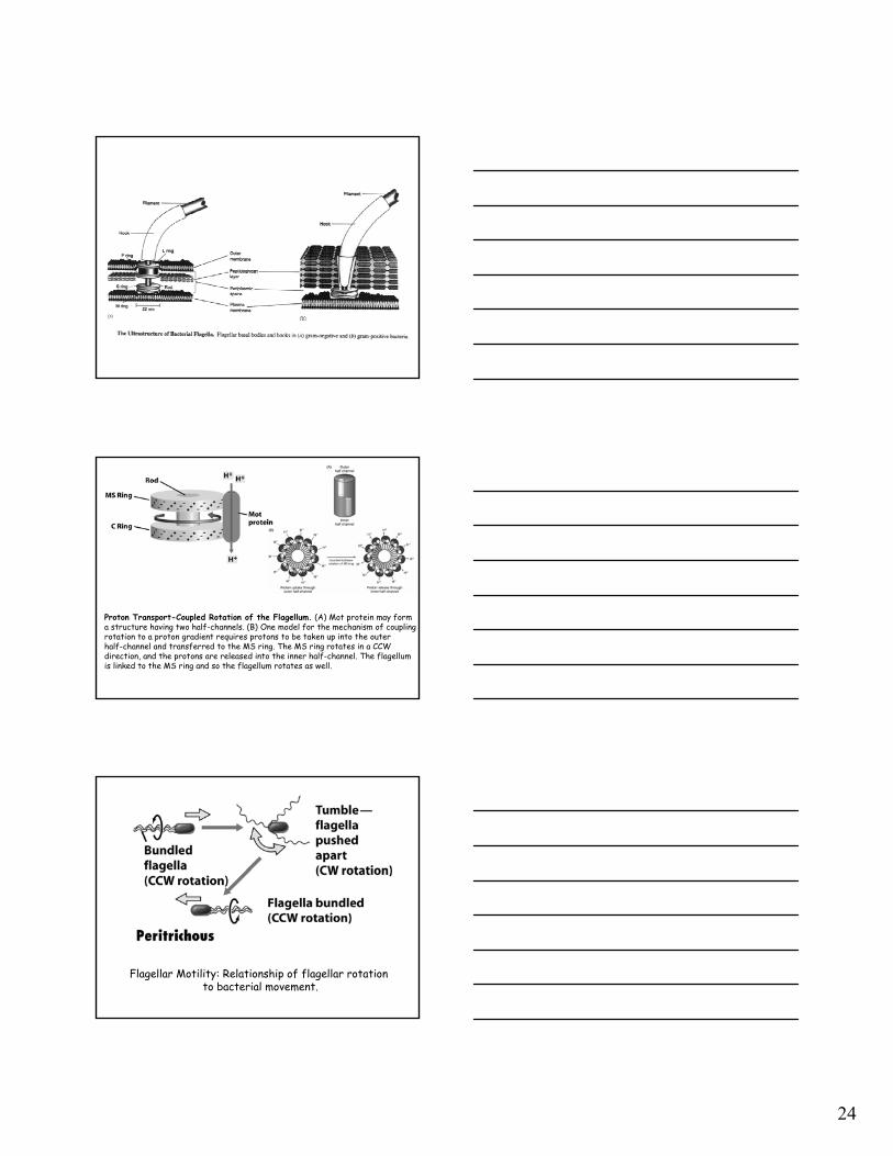
Storage of PHB
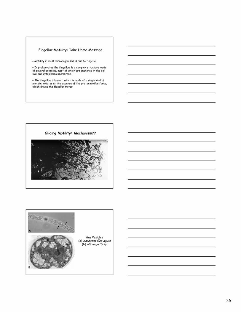
Sulfur globules inside the purple sulfur bacterium Isochromatium buderi
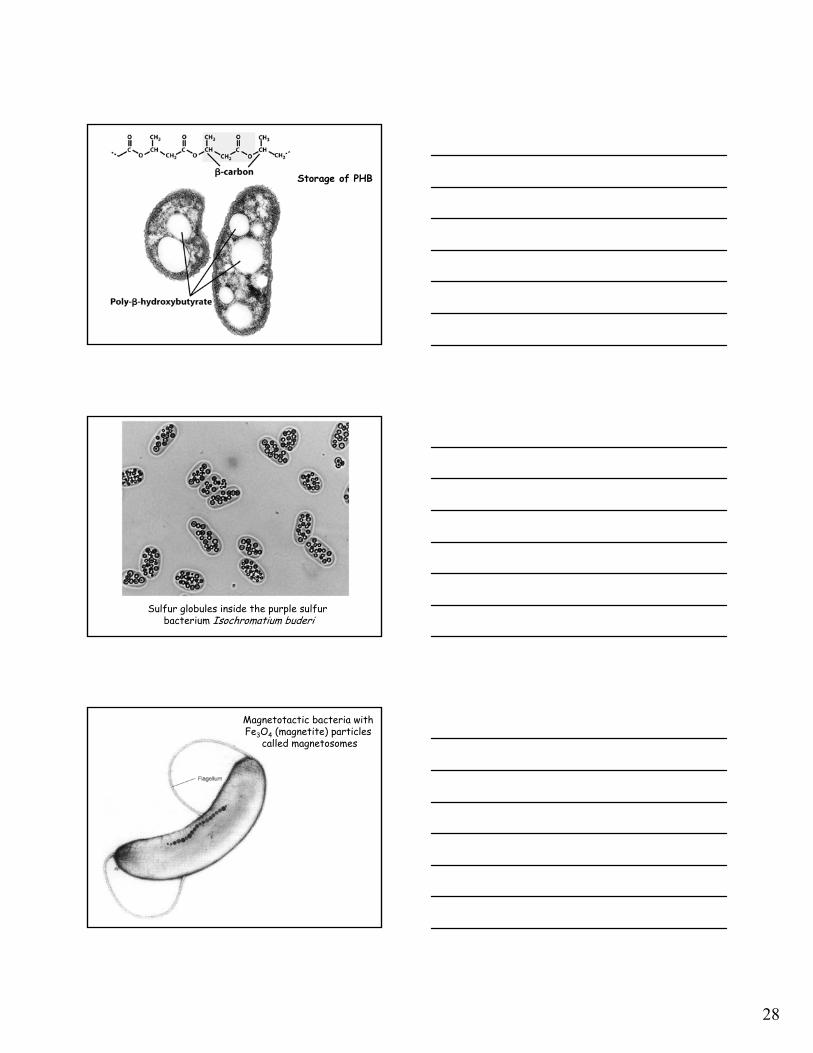
Magnetotactic bacteria with Fe3O4 (magnetite) particles
called magnetosomes

29
EM of Salmonella typhi
“Sex” Pili used in bacterial conjugation of E. coli cells