combining non-invasive transcranial brain stimulation with ... articles research/027.pdf · 2...
TRANSCRIPT
1Q1
2
3
4Q2
56789101112
13
1 4
151617
1819202122
23
24
25
4344454647
4849
50
51
52
53
54
55
56
57
58
59
60
61
NeuroImage xxx (2016) xxx–xxx
YNIMG-12937; No. of pages: 16; 4C: 2, 4, 11
Contents lists available at ScienceDirect
NeuroImage
j ourna l homepage: www.e lsev ie r .com/ locate /yn img
Combining non-invasive transcranial brain stimulation withneuroimaging and electrophysiology: Current approaches andfuture perspectives
RO
OF
Til Ole Bergmann a,b,c, Anke Karabanov d, Gesa Hartwigsen c,e, Axel Thielscher d,f,g, Hartwig Roman Siebner d,h,⁎a Department of Neurology and Stroke, and Hertie Institute for Clinical Brain Research, University of Tübingen, Tübingen, Germanyb Institute for Medical Psychology and Behavioral Neurobiology, University of Tübingen, Tübingen, Germanyc Department of Psychology, Christian-Albrechts-University, Kiel, Germanyd Danish Research Centre for Magnetic Resonance, Centre for Functional and Diagnostic Imaging and Research, Copenhagen University Hospital Hvidovre, Hvidovre, Denmarke Department of Neuropsychology, Max Planck Institute for Human Cognitive and Brain Sciences, Leipzig, Germanyf Department of Electrical Engineering, Technical University of Denmark, Kgs. Lyngby, Denmarkg Max Planck Institute for Human Cognitive and Brain Sciences, Leipzig, Germanyh Department of Neurology, Copenhagen University Hospital Bispebjerg, Copenhagen, Denmark
P⁎ Corresponding author at: Danish Research Centre forFunctional and Diagnostic Imaging and Research, CopHvidovre, Hvidovre, Denmark.
E-mail address: [email protected] (H.R. Siebner).
http://dx.doi.org/10.1016/j.neuroimage.2016.02.0121053-8119/© 2016 Published by Elsevier Inc.
Please cite this article as: Bergmann, T.O., et aCurrent approaches and future persp..., Neur
a b s t r a c t
a r t i c l e i n f oArticle history:Accepted 7 February 2016Available online xxxx
26
27
28
29
30
31
32
33
34
35
36
37
38
39
404142
RRECTEDNon-invasive transcranial brain stimulation (NTBS) techniques such as transcranial magnetic stimulation (TMS)
and transcranial current stimulation (TCS) are important tools in human systems and cognitive neuroscience be-cause they are able to reveal the relevance of certain brain structures or neuronal activity patterns for a givenbrain function. It is nowadays feasible to combine NTBS, either consecutively or concurrently, with a variety ofneuroimaging and electrophysiological techniques. Here we discuss what kind of information can be gainedfrom combined approaches, which often are technically demanding. We argue that the benefit from this combi-nation is twofold. Firstly, neuroimaging and electrophysiology can inform subsequent NTBS, providing the re-quired information to optimize where, when, and how to stimulate the brain. Information can be achievedboth before and during the NTBS experiment, requiring consecutive and concurrent applications, respectively.Secondly, neuroimaging and electrophysiology can provide the readout for neural changes induced by NTBS.Again, using either concurrent or consecutive applications, both “online” NTBS effects immediately followingthe stimulation and “offline” NTBS effects outlasting plasticity-inducing NTBS protocols can be assessed. Finally,both strategies can be combined to close the loop betweenmeasuring andmodulating brain activity bymeans ofclosed-loop brain state-dependent NTBS. In this paper, we will provide a conceptual framework, emphasizingprincipal strategies and highlighting promising future directions to exploit the benefits of combining NTBSwith neuroimaging or electrophysiology.
© 2016 Published by Elsevier Inc.
Keywords:Non-invasive transcranial brain stimulation(NTBS)Transcranial current stimulation (TCS)Transcranial magnetic stimulation (TMS)NeuroimagingElectrophysiologyClosed-loop
62
63
64
65
66
67
68
69Q3
70
71
72
UNCO
Introduction
Non-invasive transcranial brain stimulation (NTBS) plays a pivotalrole in human systems and cognitive neuroscience as it can reveal therelevance of certain brain structures or neuronal activity patterns for agiven cognitive or motor function, especially when used in conjunctionwith neuroimaging and electrophysiology. Brain mapping techniquesare correlational in nature. They can associate specific temporo-spatialactivity patterns with certain cognitive functions, but cannot demon-strate their actual relevance. In contrast, NTBS can be used to interferewith or enhance a specific neuronal pattern (e.g. entrain an oscillation)
73
74
75
76
77
Magnetic Resonance, Centre forenhagen University Hospital
l., Combining non-invasive traoImage (2016), http://dx.doi
to show that it is necessary (though may be not sufficient) for a certainbrain function, rather than being a mere epiphenomenon.
Classical transcranial electric stimulation (TES) uses brief high-voltage currents which are applied via local bipolar scalp electrodemontages (Merton andMorton, 1980). TES is a very painful stimulationtechnique and hardly used in modern cognitive neuroscience research(Rossini et al., 2015). Since 1985, TES has been replaced by painlessNTBS techniques (section “A primer on non-invasive transcranialbrain stimulation (NTBS)”): first transcranial magnetic stimulation(TMS) (Barker et al., 1985), then later transcranial direct current stimu-lation (TDCS) (Nitsche and Paulus, 2000) and transcranial alternatingcurrent stimulation (TACS) (Antal et al., 2008), together referred to astranscranial current stimulation (TCS). Both TMS and TCS nowadayscan be combined with a variety of neuroimaging and electrophysiolog-ical techniques, either consecutively or concurrently (section“Combining NTBS with neuroimaging and electrophysiology”). The
nscranial brain stimulationwith neuroimaging and electrophysiology:.org/10.1016/j.neuroimage.2016.02.012
78
79
80
81
82
83
84
85
86
87
88
89
90
91
92
93
94
95
96
97
98
99
100
101
102
103
104
105
106
107
108
109
110
111
112
113
114
115
116
117
118
119
120
121
122
123
2 T.O. Bergmann et al. / NeuroImage xxx (2016) xxx–xxx
benefit for NTBS is twofold. Firstly, neuroimaging and electrophysiologycan inform subsequent NTBS (section “Neuroimaging andelectrophysiological approaches to inform NTBS”), providing informa-tion about where (section “Where to stimulate?”), when (section“When to stimulate”), and how (section “How to stimulate?”) thebrain should be stimulated. Secondly, neuroimaging and electrophysiol-ogy can provide readouts (i.e. indices ormeasurements) of neuronal ac-tivity, which allow to assess the changes caused by NTBS (section“When to stimulate”). “Online” brain mapping offers an immediatereadout of acute NTBS effects that arise during or seconds after the ap-plication of NTBS (section “Consecutive application to read out after-effects induced by offline NTBS”). “Offline” brain mapping is performedafter a plasticity-inducing NTBS protocol to capture neuromodulatoryafter-effects that outlast NTBS for minutes to hours (section“Concurrent application to read out immediate effects of onlineNTBS”). Both strategies can be combined to close the loop betweenmeasuring and modulating brain activity by means of closed-loopbrain state-dependent brain stimulation (section “Closing the loopwith brain-state dependent non-invasive transcranial brain stimula-tion”). Here we present a conceptual framework for combining NTBSwith neuroimaging and electrophysiology, emphasizing principal strat-egies and highlighting promising future directions. Importantly, TCSand TMS are not discussed in isolation, but rather contrasted and com-pared in each of the sections. Due to space restrictions, we do not
UNCO
RRECT
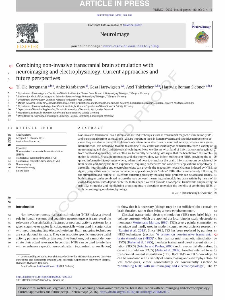
Fig. 1. Principal experimental approaches using NTBS. The “Online” NTBS approach applies Nperception, or brain activity. Depending on the applied NTBS technique and the very stimulatby applying stimuli that are strong enough to evoke direct neural output (i.e., synaptic activitycessing, or (c) to modulate the level or timing of spontaneous or task-related neuronal activityThe “Offline”NTBS approach applies conditioningprotocols that can induce long-termpotentiatcertain brain region before a task or brain mapping (e.g. fMRI). Task performance and brain acplasticity on human brain function.
Please cite this article as: Bergmann, T.O., et al., Combining non-invasive traCurrent approaches and future persp..., NeuroImage (2016), http://dx.doi
RO
OF
provide an exhaustive technical review of the state-of-the-art for com-bining NTBS and neuroimaging/electrophysiology (Siebner et al.,2009a).
A primer on non-invasive transcranial brain stimulation (NTBS)
NTBS can be given “offline” or “online”with respect to a task or brainmapping (Fig. 1): (1) The “Offline” approach applies conditioning NTBSprotocols that can induce long-term potentiation (LTP)-like or long-term depression (LTD)-like plasticity and hereby facilitate or inhibit acertain brain region before a task or brain mapping. Task performanceand brain activity measurements after NTBS are used as readouts to un-cover the consequences of NTBS-induced plasticity on human brainfunction (Siebner et al., 2009a). (2) The “Online” approach appliesNTBS during a task or neuroimaging to measure its immediate impacton brain function or activity. Depending on the applied NTBS techniqueand the very stimulation parameters chosen, online NTBS can be used(a) to quantify local network properties by applying stimuli that arestrong enough to evoke direct neural output (i.e., synaptic activity),(b) to interfere with ongoing spontaneous neural activity or task-related neuronal processing, or (c) to modulate the level or timing ofspontaneous or task-related neuronal activity.
All NTBS techniques produce electrical stimulation of neurons in thebrain, yet these techniques rely on different mechanisms of action. This
ED P
TBS during a task or neuroimaging to measure its immediate consequences for behavior,ion parameters chosen, online NTBS can be used (a) to quantify local network properties), (b) to interfere with ongoing spontaneous neural activity or task-related neuronal pro-(see section “A primer on non-invasive transcranial brain stimulation (NTBS)” for details).ion (LTP)-like or long-termdepression (LTD)-like plasticity andhereby facilitate or inhibit ativity measurements are used as readouts to uncover the consequences of NTBS-induced
nscranial brain stimulationwith neuroimaging and electrophysiology:.org/10.1016/j.neuroimage.2016.02.012
T
124
125
126
127
128
129
130
131
132
133
134
135
136
137
138
139
140
141
142
143
144
145
146
147
148
149
150
151
152
153
154
155
156
157
158
159
160
161
162
163
164
165
166
167
168
169
170
171
172
173
174
175
176
177
178
179
180
181
182
183
184
185
186
187
188
189
190
191
192
193
194
195
196
197
198
199
200
201
202
203
204
205
206
207
208
209
210
211
212
213
214
215
216
217
218
219
220
221
222
223
224
225
226
227
228
229
230
231
232
233
234
235
236
237
238
239
240
241
242
243
244
245
246
247
248
249
3T.O. Bergmann et al. / NeuroImage xxx (2016) xxx–xxx
UNCO
RREC
has strong conceptual implications with regard to their use in cognitiveneuroscience: TCS passes the electric current directly through scalp andskull whereas TMS “bypasses” these electric obstacles by producing amagnetic field that induces an electric current in the brain tissue. TMSand TCS also differ in their neurophysiological mechanism of action:The steep, high-amplitude currents induced by TMS (and TES) areable to fully depolarize the axonal membrane of cortical neurons andthereby trigger action potentials. In contrast, the comparably weak cur-rents of TCS affect the neurons' membrane potential more subtly. TCS isthought to shift themembrane potential causing a slight depolarizationor hyperpolarization. The TCS-induced shift in membrane potential in-creases or decreases the likelihood of spontaneous neuronal firing(Bindman et al., 1964; Paulus et al., 2013). TMS and TCS also differ sub-stantially in terms of the effectively stimulated brain volume, with rele-vant implications for their spatial selectivity (Opitz et al., 2015;Thielscher et al., 2011) and the specific information needed to configurean effective stimulation protocol.
TDCS (Nitsche and Paulus, 2000), TACS (Antal et al., 2008), and TMS(Barker et al., 1985; Walsh and Cowey, 2000) complement each other,as these techniques have different strengths and weaknesses. Whilethese can induce bidirectional plasticity using the offline approach,they do substantially differ regarding their online application. BecauseTMS can excite neurons in a suprathreshold fashion, it is well suited toactually evoke neuronal activity, allowing to quantify network proper-ties such as excitability and connectivity or to interfere with ongoingspontaneous or task-related neuronal activity (Siebner et al., 2009b).In contrast, TCS is primarily suited tomodulate both the level and timingof spontaneous or task-related neuronal activity (Reato et al., 2013). Thedisruptive effect of TMS on neuronal information processing, which iskey to the “interference” approach, is not fully understood but probablyinvolves a combination of the followingmechanisms: (i) degradation ofthe signal-to-noise ratio by evoking random neuronal excitation, (ii)aligned GABAergic inhibition via feedback inhibition or direct stimula-tion of inhibitory interneurons, (iii) decrease of entropy and thereby in-formation capacity due to the resulting neuronal synchronization(Siebner et al., 2009b; Walsh and Cowey, 2000). In any case, the goalof the interference approach is to transiently induce a so-called “virtuallesion.”Here TMS-induced neurostimulation causes a functional pertur-bation of the stimulated region affecting ongoing neural processing for afew tens to hundreds of milliseconds (Pascual-Leone et al., 2000). Incontrast, the “modulation” approach aims not to acutely disrupt butrather to gently shape the neuronal activity profile, gently biasing thestimulated brain region towards a specific working mode while leavingit principally intact. The goal of this approach is to increase or decreaseeither the level (e.g. with TDCS) or the timing (e.g. with TACS) of inter-nally generated excitatory or inhibitory activity patterns. TMS and TCStherefore often require different neuroimaging or electrophysiologicalreadouts to have their effects adequately assessed (section“Neuroimaging and electrophysiology as readout for NTBS effects”)and their combination with different neuroimaging and electrophysio-logical techniques poses specific technical challenges (Siebner et al.,2009a).
Combining NTBS with neuroimaging and electrophysiology
Many non-invasive neuroimaging and electrophysiological tech-niques are available tomapdifferent aspects of brain structure and func-tion at varying levels of spatial and temporal resolution (Bandettini,2009). These techniques can be combined with NTBS in a consecutivefashion to assess the offline effects of NTBS after stimulation. Giventhe limited time of the induced after-effects, recordings should start assoon as possible after the end of the plasticity inducing NTBS protocol.This requires a swift transfer of the participants from theNTBS laborato-ry to the imaging facility. At the same time, it might be important not tounintentionally interfere with the induced NTBS effects during this in-terval, avoiding unnecessary activation of targeted brain regions of
Please cite this article as: Bergmann, T.O., et al., Combining non-invasive traCurrent approaches and future persp..., NeuroImage (2016), http://dx.doi
ED P
RO
OF
interest, e.g., muscle activation (Siebner et al., 2003), speech produc-tion/comprehension (Hartwigsen et al., 2013), or visuospatial attention(Marshall et al., 2015b). Therefore, NTBS is often applied within or closeto the MR scanner room or wheelchairs are used for the transfer whilethe experimenter tries to maintain a relaxed atmosphere and avoidoverly strong effort or excitement of the participant.
In contrast, the concurrent application of NTBS during the recordingof neuroimaging or electrophysiological data is complicated by a varietyof NTBS-related artefacts. The nature and severity of these artefacts dif-fers markedly between the various NTBS-Imaging combinations. A de-tailed description is beyond the scope of this paper. Comprehensivereviews on these issues can be found elsewhere (Siebner et al., 2009a)and specific advice is available on how to prevent or remove those arte-facts for TMS–EEG (Herring et al., 2015; Ilmoniemi and Kicic, 2010;Korhonen et al., 2011; Mäki and Ilmoniemi, 2010; Mutanen et al.,2013; Rogasch et al., 2014), TMS–fMRI (Bestmann et al., 2008a), TMS–EEG–fMRI (Peters et al., 2013), TMS–NIRS (Parks, 2013), TCS–fMRI(Saiote et al., 2013), or TCS–MEG (Marshall et al., 2015a; Neulinget al., 2015; Soekadar et al., 2013).
While we will mainly discuss the manifold benefits neuroimagingand electrophysiology provide for both optimizing and assessingNTBS, there are caseswhereNTBS results informneuroimaging analysis.For example, pre-surgical neuronavigated TMS mapping has been suc-cessfully used to provide seed points for fibre tracking based on diffu-sion weighted imaging (DWI) data when reconstructing thecorticospinal tract (Frey et al., 2012) as well as language-relevant path-ways (Sollmann et al., 2015). Future scenarios may even extend to thecombination of DWI with dual-coil TMS approaches, informing mea-sures of structural by those of effective connectivity.
Neuroimaging and electrophysiological approaches to inform NTBS
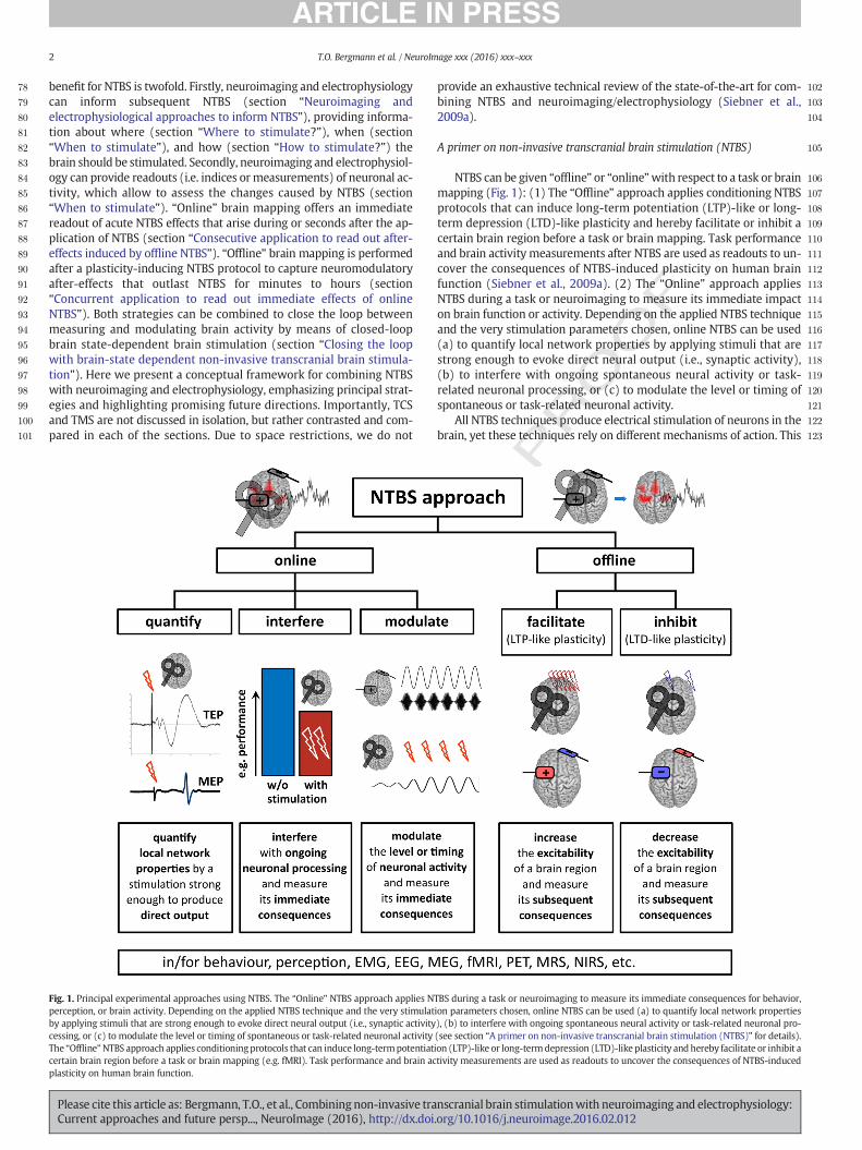
Neuroimaging and electrophysiological measures can guide NTBS,providing information about where, when and how to apply the stimu-lation (Fig. 2). This information can be exploited to improve the preci-sion and efficacy of NTBS protocols. The delay between extraction ofthe required information from neuroimaging or electrophysiology andapplication of NTBS can vary considerably. Information with highinter- but low intra-subject variability, for instance about brain struc-ture, can be acquired before the start of the NTBS experiment, some-times even in a separate session, allowing recordings and NTBS to beconducted in a consecutive fashion. However, information with highintra-subject variability, for instance about the functional brain state,need to be determined immediately before stimulation onset or eventraced continuously for repeated stimulation trials. The latter requiresa concurrentmapping approach duringwhich functional brainmappingand the application of NTBS are interleaved.
Where to stimulate?
Which information is required to optimize the spatial accuracy ofNTBS? A certain degree of spatial information is always necessary toapply NTBS to the brain region of interest, no matter whether NTBS isused in an offline or online approach. For TMS, not only the position ofthe TMS coil relative to the target site (precision of a few millimetres)but also its orientation relative to the orientation of the cortical gyri(precision of a few degrees) is of crucial importance to induce a currentof appropriate strength and orientation in the targeted brain tissue,while leaving regions of no interest as unaffected as possible. For TCS,the placement of the electrodes on the scalp and the orientation of theinduced current flow determine the optimal electrode montage. Twosteps are necessary for spatial optimization of NTBS. First, the precise lo-cation of the target site needs to be localized within the brain of eachsubject. Thereafter, the position and orientation of the stimulation de-vices, the TMS coil or the TCS electrodemontage needs to be determinedto apply a current of optimal intensity and orientation to the target site.
nscranial brain stimulationwith neuroimaging and electrophysiology:.org/10.1016/j.neuroimage.2016.02.012
ECTED P
RO
OF
250
251
252
253
254
255
256
257
258
259
260
261
262
263
264
265Q4
266
267
268
269
270
271
272
273
274
275
276
277
278
279
280
281
282
283
284
285
286
287
288
289
290
291
292
293
294
295
296
297
298
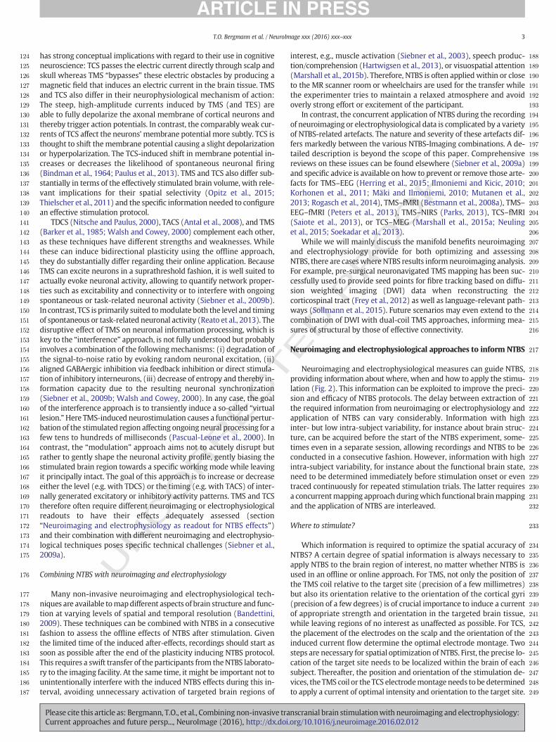
Fig. 2. Neuroimaging and electrophysiological measures may inform NTBS about where, when, and how to stimulate. This information can be exploited to improve the precision andefficacy of NTBS protocols and to enable the investigation of NTBS effects in a brain state-dependent manner by relating the NTBS effects to the spontaneous or task-related neuronalactivity at the time of stimulation.
4 T.O. Bergmann et al. / NeuroImage xxx (2016) xxx–xxx
UNCO
RRConsecutive application to derive spatial information
The target site and optimal position of the NTBS device are relativelystable entities. Therefore, neuroimaging or electrophysiological map-ping can be performed before starting an NTBS experiment to derivethe necessary spatial information. The choice to target a certain regionis primarily motivated by the research question and commonly basedon the existing literature. Yet the precise location of the target site inan individual brain still remains to be determined due to substantialinter-subject anatomical variability. Several approaches are listed herein the order of increasing precision and experimental power (Sacket al., 2009; Sparing et al., 2008).
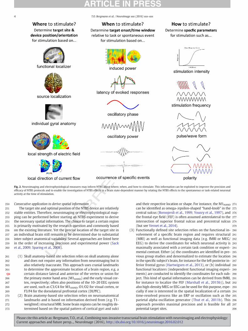
(1) Skull anatomy-based site selection relies on skull anatomy aloneand does not require any information from neuroimaging but isalso relatively inaccurate. This approach uses cranial landmarksto determine the approximate location of a brain region, e.g. acertain distance lateral and anterior of the vertex or union forthe primary motor hand area (M1HAND) and the early visual cor-tex, respectively; often also positions of the 10–20 EEG systemare used, such as C3/C4 for M1HAND, O1/O2 for visual cortex, orF3/F4 for the dorsolateral prefrontal cortex (DLPFC).
(2) Brain anatomy-based site selection relies on neuroanatomicallandmarks and is based on information derived from (e.g. T1-weighted) structuralMRI. Some brain regions can be roughly de-termined based on the spatial pattern of cortical gyri and sulci
Please cite this article as: Bergmann, T.O., et al., Combining non-invasive traCurrent approaches and future persp..., NeuroImage (2016), http://dx.doi
and their respective location or shape. For instance, the M1HANDcan be identified as omega-/epsilon-shaped “hand-knob” in thecentral sulcus (Boroojerdi et al., 1999; Yousry et al., 1997), andthe frontal eye field (FEF) is often assumed anterolateral to theintersection of superior frontal sulcus and precentral sulcus(but see Vernet et al., 2014).
(3) Functionally defined site selection relies on the functional in-volvement of a specific brain region and requires structural(MRI) as well as functional imaging data (e.g. fMRI or MEG/EEG) to derive the coordinates for which neuronal activity ismaximally associated with a certain task condition or experi-mental contrast. Either (a) the coordinates are identified in pre-vious group studies and denormalized to estimate the locationin the specific subject's brain, for instance for the left posterior in-ferior frontal gyrus (Hartwigsen et al., 2013), or (b) individualfunctional localizers (independent functional imaging experi-ments) are conducted to identify the coordinates for each sub-ject. This kind of spatial information can be derived from fMRI,for instance to localize the FEF (Marshall et al., 2015b)), butalso high-densityMEG or EEG can be used for this purpose, espe-cially if one is interested in the spatial localization of a certaintime-resolved process like an ERP or oscillation such as theparietal alpha oscillation generator (Thut et al., 2011b). Thisapproach provides maximal precision and is feasible for allpotential target sites.
nscranial brain stimulationwith neuroimaging and electrophysiology:.org/10.1016/j.neuroimage.2016.02.012
T
299
300
301
302
303
304
305
306
307
308
309
310
311
312
313
314
315
316
317
318
319
320
321
322
323
324
325
326
327
328
329
330
331
332
333
334
335
336
337
338
339
340
341
342
343
344
345
346
347
348
349
350
351
352
353
354
355
356
357
358
359
360
361
362
363
364
365
366
367
368
369
370
371
372
373
374
375
376
377
378
379
380
381
382
383
384
385
386
387
388
389
390
391
392
393
394
395
396
397
398
399
400
401
402
403
404
405
406
407
408
409
410
411
412
413
414
415
416
417
418
419
420
421
422
423
424
425
426
5T.O. Bergmann et al. / NeuroImage xxx (2016) xxx–xxx
UNCO
RREC
(4) Stimulation-based site selection relies on the causal demonstra-tion of a certain brain region being involved in a specific functionand requires an NTBS technique like TMS, which can be used toeither quantify the output of the target brain structure or to inter-fere with the function of the target brain structure. When usingthe quantification approach, the coil position and orientationcausing the maximal (or most consistent) output of the targetstructure, e.g. the amplitude of the motor evoked potential(MEP) forM1HAND or phosphene perception for the visual cortex,is considered the optimal spatial configuration. Following the in-terference approach, the coil position and orientation producingthemaximally disruptive effects is considered optimal, e.g. whenfunctionally localizing Broca's area by inducing speech arrest(Rogic et al., 2014) or to localize the FEF by saccade delay (Roet al., 2002). In fact, stimulation-based localization is the only ap-proach providing proof that the NTBS application actuallyreaches the neuron populations of interest. It is therefore the ap-proach of choice for quantification or interference, whenever areliable behavioral readout is available. However, this is onlythe case for very few brain regions.
After determining the target site, the NTBS device has yet to be posi-tioned correctly to effectively stimulate neurons at the target site,whichis particularly critical for TMS. MR-based frameless stereotacticneuronavigation enables precise positioning of the TMS coils relativeto any desired stereotactic coordinate of interest. Another major advan-tage is that frameless neuronavigation secures the maintenance of aconstant coil position and orientation within and across experimentalsessions (Schonfeldt-Lecuona et al., 2005), particularly important forconcurrent TMS–EEG approaches (Lioumis et al., 2009). However, tonavigate the stimulation coil into an optimal position, additional infor-mation is needed regarding the optimal direction of the induced currentflow in the target region. For TMS, themain direction of induced currentflow in the brain tissue is roughly opposite to the direction of currentflow in the coil (Kammer et al., 2001b),making the orientation (incl. ro-tation and tilt) of the coil the major variable to be controlled byneuronavigation. For TCS, the direction of current flow in the brain tis-sue has to be controlled by the specific location of the two ormore stim-ulation electrodes.
For a few brain regions, the optimal direction of current flow hasbeen determined by systematic experimental variation: for M1HAND, aposterolateral-to-anteromedial direction at an angle of approximately45° relative to the mid-sagittal line and thus perpendicular to the cen-tral sulcus most effectively evokes MEPs (Mills et al., 1992), whereasfor the visual cortex on average an anterior-to-posterior direction(Kammer, 1999) or lateral-to-medial direction (Kammer et al., 2001a)most effectively evokes phosphenes (with optimal direction dependingon individual gyral orientation (Kammer et al., 2007)). Notably, thesedirections correspond roughly to the current flow induced by the now“classical” TCS montages for M1HAND (C3 or C4 vs. contralateral fore-head) (Nitsche and Paulus, 2000) and visual cortex (Cz–Oz) for bothTDCS (Antal et al., 2004a, 2006) and 10 Hz TACS (Neuling et al., 2013).Also lateral-to-medial currents seem to be effective for both TMS(Kammer et al., 2001a) and 10 Hz TACS in O1–O2 montage (Zaehleet al., 2010). For most other brain regions, however, robust outcomemeasures for quantification and systematic investigations are lacking.Attempts to systematically determine the optimal current direction ina specific brain region to maximally interfere with a specific cognitivetask, e.g. posteromedial-to-anterolateral for the right PFC to interferewith memory-guided saccades (Hill et al., 2000), are rare but of criticalimportance to reveal consistent effects. Instead, coil positions are oftenarbitrarily chosen or based on previously published successful attempts,resulting in very different coil orientations for the same target sites. Toimprove the effectiveness andprecision of TMS and TCS, amore detailedknowledge is needed both with respect to the actual current
Please cite this article as: Bergmann, T.O., et al., Combining non-invasive traCurrent approaches and future persp..., NeuroImage (2016), http://dx.doi
ED P
RO
OF
distribution in the brain and the relation between current directionand the relevant neuronal elements.
For both TMS and TCS, the distribution of the electric field in thebrain tissue crucially depends on the specific spatial distribution ofbrain tissue classeswith different conductivity values, such as greymat-ter,whitematter and corticospinalfluid (Windhoff et al., 2013). Howev-er, while TMS induces the effective electric current directly in the braintissue bypassing scalp and skull (Opitz et al., 2011), TCS has to pass thecurrent through these structures, rendering them crucial for the currentdistribution in the brain (Opitz et al., 2015). Moreover, TMS induces acomparably focal electricfield forwhichmainly the local gyral geometryhas to be considered (Kammer et al., 2007; Thielscher et al., 2011),whereas TCS produces a widespread electric field extending in varyingdegrees throughout the entire head (Opitz et al., 2015). The use of indi-vidually computed and empirically validated head models would thusmarkedly improve the spatial precision of both TMS and TCS. Whileneuronavigated small high-precision TMS coils (Groppa et al., 2012b)and high-definition TCS (Edwards et al., 2013), MR-informed custom-ized TCS electrode shapes (Cancelli et al., 2015), and a more informedselection of the electrode properties (Saturnino et al., 2015) may in-crease the reliability and spatial resolution of NTBS, a valid estimationof the effective electric field is necessary to ensure stimulation of the de-sired brain region while leaving adjacent structures as unaffected aspossible.
Concurrent application to derive spatial informationCurrently, spatial information is obtained before starting NTBS and
not up-dated over the course of NTBS. Brain networks are dynamicand task-related neuronal activity pattern may slowly change in the af-termath of an intervention or during the course of a learning paradigm.In these cases, spatial shifts in neuronal activity patterns should betracked continuously (e.g. using fMRI or EEG) to be able to spatially ad-just NTBS to the currently relevant brain site. For TMS, this has becomepossible by the use of a robotic arm, which is constantly controlled by aneuronavigation system (Ginhoux et al., 2013; Richter et al., 2010; Toddet al., 2014). While robotic neuronavigation is currently not MRI-compatible (due to ferromagnetic parts and severe space constraintsin the MRI head coil), it is well feasible for concurrent TMS–EEG. Here,it may even automatically track and target the local occurrence of spon-taneous events like sleep spindles, slow oscillations or epileptic activitypatterns. For TCS, a fast relocation of stimulation electrodes is not possi-ble, but multi-channel devices to rapidly switch for each electrode be-tween EEG recording and TCS application mode are available (Ruffiniet al., 2014). Given that stimulators with a high number of channels ora switch-matrix are offered in the future, it might become feasible totarget multiple sites in a single experimental session by switching be-tween different TCS electrode montages while still retaining the spatialfocality that has been demonstrated formulti-channel TCS. Such a setupis also a prerequisite for future attempts to automatically configurecompletely new stimulationmontages based on the current brain-state.
When to stimulate?
Timing is essential for neuronal communication, emphasizing theneed for “temporal neuronavigation” which is as important as spatialneuronavigation for NTBS. For online NTBS, the temporal properties ofneural processing need to be characterized and extracted to determinethe onset and duration of NTBS accordingly. Indeed, the timing ofNTBS determines its ability to successfully (i) quantify network proper-ties in a brain state-dependentmanner, (ii) interfere with ongoing neu-ronal processes, and (iii) modulate neuronal activity in a temporallyspecific manner (cf. section “A primer on non-invasive transcranialbrain stimulation (NTBS)” and Fig. 1). In addition to the temporal prop-erties of neural processing, the temporal precision of the NTBS tech-nique needs to be taken into account. Since TMS has a highertemporal resolution than TCS, the onset of stimulation relative to a
nscranial brain stimulationwith neuroimaging and electrophysiology:.org/10.1016/j.neuroimage.2016.02.012
T
427
428
429
430
431
432
433
434
435
436
437
438
439
440
441
442
443
444
445
446
447
448
449
450
451
452
453
454
455
456
457
458
459
460
461
462
463
464
465
466
467
468
469
470
471
472
473
474
475
476
477
478
479
480
481
482
483
484
485
486
487
488
489
490
491
492
493
494
495
496
497
498
499
500
501
502
503
504
505
506
507
508
509
510
511
512
513
514
515
516
517
518
519
520
521
522
523
524
525
526
527
528
529
530
531
532
533
534
535
536
537
538
539
540
541
542
543
544
545
546
547
548
549
550
551
552
553
6 T.O. Bergmann et al. / NeuroImage xxx (2016) xxx–xxx
UNCO
RREC
task or assessment of brain activity needs to be defined more preciselyfor TMS. Single TMS pulses themselves last for a few hundredmicrosec-onds only and paired-pulse conditioning-test TMS paradigms can revealfunctional interactions between subsequent pulses with sub-millisecond precision (Reis et al., 2008). At the same time, some of theevoked neurophysiological effectsmay last for several hundredmillisec-onds, as do the associated online effects at the behavioral level(Pascual-Leone et al., 2000; Siebner et al., 2009b; Walsh and Cowey,2000). In contrast, the earliest effects reported for TCS so far start at4–5 s after stimulation onset for both TDCS (Nitsche and Paulus, 2000)and TACS (Joundi et al., 2012), which may restrict its temporal resolu-tion. A notable exception is TACSwhere the internal temporal structureof TACS with respect to its oscillatory phase may actually require highlevels of precision as well to be optimally aligned to ongoing intrinsicbrain activity (Brittain et al., 2013).
At first glance, one might assume that the timing of NTBS is less rel-evant for offline NTBS. However, there is converging evidence showingthat the state of the brain is a critical factor determining the efficacy ofNTBS to induce plastic changes in the brain (Karabanov et al., 2015).This implies that the temporal structure of offline NTBS should betuned to the brain state that is most susceptible to the plasticity induc-ing effects of the NTBS protocol (see below).
Consecutive application to derive temporal informationMEG/EEG can be performed before NIBS to reveal when task-related
neuronal processes emerge and disappear in a given subject. Since thetemporal properties of neural processing can vary substantially acrossindividuals, such experiments provide valuable information to individ-ually determine the optimal time point (e.g., for single-pulse TMS) ortime window (e.g., for burst-TMS or brief TCS blocks) of stimulation.The onset or latency of an evoked cortical potential (Thut et al., 2003)or the time course of induced oscillatory power changes may guidethe timing of NTBS. For instance, the lateralized readiness potential(LRP) has been successfully used to determine the timing of stimulationin a single-pulse TMS study. The LRP is a negative scalp potential pre-ceding lateralized handmovements. By varying the timing of TMS rela-tive to the latency of the peak LRP, the time course of cortico-spinalexcitability changes can be traced preceding lateralized motor re-sponses (Verleger et al., 2009, 2010). Moreover, the individual frequen-cy of an oscillation of interest (e.g., the alpha band) has been assessed tofacilitate the efficacy of NTBS to entrain intrinsic cortical oscillations(Klimesch et al., 2003; Thut et al., 2011b; Zaehle et al., 2010).
Concurrent application to derive temporal informationConcurrent brain mapping and stimulation allow to trace the
dynamic expression of the brain state of interest and to time NTBSbased on ongoing intrinsically generated brain activity. Temporalneuronavigation of NTBS allows to characterize the causal impact ofspecific brain states on a specific brain function. Both TMS and TCS canbe performed concurrently with fMRI to study cortical excitability andconnectivity as a function of spontaneous BOLD fluctuations, yet MEGand EEG have a much higher temporal resolution than fMRI. Therefore,electrophysiological techniques are better suited to inform the timing ofNTBS.
Online triggering of NTBS based on the ongoing MEG or EEG is onlyfeasible when the signal-to-noise ratio is sufficiently high and the auto-mated analysis pipeline sufficiently fast. This often applies to ongoingneuronal oscillations or other highly synchronized neural eventswhere power and even instantaneous amplitude and phase can be de-termined online (Bergmann et al., 2012; Hartmann et al., 2011). For in-stance, EEG-triggered TMS of the primary motor cortex has beensuccessfully used during deep sleep to target the up- vs. down-state ofthe ongoing sleep slow oscillation (b1 Hz) and investigate rapid shiftsin cortical excitability as indexed by MEP and TMS-evoked EEG poten-tial (TEP) amplitude (Bergmann et al., 2012). In principle, the high tem-poral resolution of EEG andMEG should allow for targeting sporadic and
Please cite this article as: Bergmann, T.O., et al., Combining non-invasive traCurrent approaches and future persp..., NeuroImage (2016), http://dx.doi
ED P
RO
OF
very transient spontaneous neural events, such as epileptic spikes ortask-evoked cortical potentials. The problem is that these neural eventshave rarely sufficient SNR and averaging across trials is usually neces-sary. However, spatial filters may increase the timing precision for sin-gle trials (De Vos et al., 2012; Salajegheh et al., 2004). A fully or semi-random application of NTBS together with post-hoc trial sorting maysometimes offer a valid alternative when NTBS triggering based on theongoing EEG is not possible. The stimulation procedure itself is easy toperform and results in a good, though random, coverage of differentbrain states during the recording session. Using this approach, visualalpha power and phase preceding the TMS pulse have been shown topredict the probability of occipital TMS to evoke phosphenes (Dugueet al., 2011; Romei et al., 2008). Likewise, the pericentral power of themu-rhythm has been shown to predict the size of the MEP amplitudeevoked with TMS over the motor cortex (Sauseng et al., 2009). A cleardisadvantage of the post-hoc trial sorting approach is that a relativelarge fraction of NTBS is applied during periods of no interest. Further,the random application of NTBS precludes the possibility to repetitivelyand exclusively target a specific brain state. This option is required if theaim is to systematically address the state-dependency of offline NTBSeffects, for instance the impact of alpha power or phase expressed atthe time of NTBS on the ability of NTBS to induce LTP- and LTD-likeplasticity.
How to stimulate?
Stimulation variables such as intensity, frequency and duration havean influence on the neurobiological effects of NTBS. For TMS, the physi-ological impact additionally depends on the pulse form (e.g. mono-phasic or bi-phasic) and the direction of the electrical current inducedin the brain tissue (Pascual-Leone et al., 2000; Walsh and Cowey,2000) and, in case of repetitive TMS (rTMS), the precise temporal pat-tern of pulses (Fitzgerald et al., 2006; Ziemann et al., 2008). Whenusing TCS, electrode design, size and placement, as well as theelectrode-skin resistance and its spatial homogeneity across the elec-trode surface are additional contributing factors, (Nitsche et al., 2008;Saturnino et al., 2015), whereas frequency, waveform and DC offsetare stimulation variables of interest for TACS (Antal and Paulus, 2013;Herrmann et al., 2013). Theoretically, many of the above-mentionedstimulation settings may benefit from information derived from neuro-imaging or electrophysiological measurements. Due to space restric-tions, we will only focus on how neuroimaging and electrophysiologycan be used to optimize stimulation intensity and frequency.
Consecutive application to derive stimulation parameter informationStimulation intensity is certainly among the most essential parame-
ters to be adjusted, as it impacts not only the effectiveness but also thesafety of a certain NTBS protocol (Miranda, 2013; Nitsche et al., 2008;Rossi et al., 2009). Online TMS, for example, may have beneficial or dis-ruptive effects on a cognitive task depending on stimulation intensity,whereas for offline TMS protocols it may determine whether it is facili-tatory or inhibitory (Miniussi et al., 2013). Therefore, proper adjustmentof stimulation intensity to accommodate for individual brain anatomymay reduce the large inter-session and inter-subject variability ob-served for offline NTBS protocols (Karabanov et al., 2015; Li et al.,2015; Ziemann and Siebner, 2015). For TMS, it is currently commonpractice to adjust stimulation intensity relative to the individual restingor active motor threshold (RMT, AMT), determined as the minimal in-tensity required to induce an MEP with 50% probability in the relaxedor pre-contracted muscle, respectively (Groppa et al., 2012a; Rossiniet al., 1994), or the phosphene threshold, determined asminimal inten-sity to evoke a phosphene with 50% probability (Kammer et al., 2001a;Lou et al., 2011). Given that the correlation between MEP and phos-phene threshold is moderate at best (Antal et al., 2004b; Boroojerdi,2002; Deblieck et al., 2008; Gerwig et al., 2003; Stewart et al., 2001), itis questionable whether effective stimulation intensities of one brain
nscranial brain stimulationwith neuroimaging and electrophysiology:.org/10.1016/j.neuroimage.2016.02.012
T
554
555
556
557
558
559
560
561
562
563
564
565
566
567
568
569
570
571
572
573
574
575
576
577
578Q5
579
580
581
582
583
584
585
586
587
588
589
590
591
592
593
594
595
596
597
598
599
600
601
602
603
604
605
606
607
608
609
610
611
612
613
614
615
616
617
618
619
620
621
622
623
624
625
626
627
628
629
630
631
632
633
634
635
636
637
638
639
640
641
642
643
644
645
646
647
648
649
650
651
652
653
654
655
656
657
658
659
660
661
662
663
664
665
666
667
668
669
670
671
672
673
674
675
676
677
678
679
680
681
682
683
684
685
7T.O. Bergmann et al. / NeuroImage xxx (2016) xxx–xxx
UNCO
RREC
region can be generalized to other brain regions. It would be favorableto adjust TMS intensity based on the local responsiveness of the targetregion for the specific coil position/orientation actually used in the ex-periment. When focal online TMS disrupts task performance, one candetermine the individual intensity threshold for inducing a perturbation(Hartwigsen et al., 2010). However, TMS-induced behavioral effects areoften subtle andmight require a large number of trials or uncomfortablyhigh stimulation intensities.
Themagnetic field rapidly decayswith distance, and the distance be-tween the scalp region where NTBS is applied and the targeted cortexvaries considerably across individuals and cortical sites. Scalp-cortexdistance can be derived from structural MRI data and stimulation inten-sity can be adjusted accordingly (Janssen et al., 2014; Stokes et al.,2005). Anatomically precise headmodelsmay allow the inclusion of ad-ditional indices to estimate the required stimulation intensity for indi-vidual participants, target sites and coil positions. Some commerciallyavailable TMS neuronavigation systems feature an estimation of the in-duced electric field, but the underlyingmodels are crude and do not suf-ficiently take into account individual neuroanatomy to actually basedecisions for stimulation intensity on these values. Other anatomical in-dices that have been related to inter-individual variations in responsive-ness to NTBS are local grey matter density, derived from voxel-basedmorphometry (Granert et al., 2011), orwhitematter fractional anisotro-py, derived from diffusion tensor imaging (DTI) (Kloppel et al., 2008).Regional fractional anisotropy has shown to predict RMT (Kloppelet al., 2008), but see Hubers et al. (2012), as well as intra-hemisphericcortico-cortical facilitation (Groppa et al., 2012c).
Since TMS-evoked EEG potentials (TEP) (section “Concurrentapplication to read out immediate effects of online NTBS”) capture thedirect cortical response to TMS, TEP recordings may offer a more gener-ally applicable means to establish a threshold for any cortical region ac-cessible by TMS without the need for motor or perceptual output.However, no valid TEP threshold estimation has been established forthe following reasons. First, SNR is too low on single trial level requiringthe averaging of several tens of evoked responses per intensity step toobtain a reliable estimate. Second, TEPs are strongly confounded byco-evoked auditory and somatosensory potentials caused by the TMSclick sound and the stimulation of cranial muscles and peripheralnerves, respectively (Herring et al., 2015; Nikouline et al., 1999), bothmodulated by intensity as well. This renders an appropriate sham con-dition mandatory, again doubling the number of trials required. Third,TMS-related electrical, mechanical, and biological non-cortical artefacts(like cranial muscle twitches and electrode-electrolyte polarization)often require cumbersome post-processing of the data (Herring et al.,2015; Ilmoniemi and Kicic, 2010; Korhonen et al., 2011; Rogasch et al.,2014). In summary, establishing a valid TEP threshold would require asophisticated experimental and post-processing setup. Further a con-siderably large number of pulses (e.g. 200 pulses, resulting from 100 tri-als × 2 conditions) need to be acquired at each intensity during astaircasing procedure. Advances in data processing algorithms and spa-tial filter techniques may allow online artefact correction and a reliableestimation of TEP waveform and amplitude from a lower number of tri-als, paving theway towards making TEP threshold estimation a feasibleinstrument in the near future.
Stimulation intensity is even more coarsely determined for TCS, be-cause stimulation intensity is usually not individually adjusted like it isthe case for TMS. Given the fundamental impact of stimulation intensityon the size and nature of physiological effects, it is surprising that TCS isstill applied in a one-intensity-fits-all manner without individual ad-justment of stimulation intensity. This may explain why no simple line-ar relationship seems to exist between stimulation intensity and thesize or direction of TCS effects (Bastani and Jaberzadeh, 2012, 2013;Batsikadze et al., 2013; Moliadze et al., 2012). Up till now, a coarse esti-mation of the current density (mA/cm2) at the scalp level is assumed todetermine the effectiveness of TCS (Nitsche et al., 2007; Nitsche andPaulus, 2000). The same default current densities of 0.029 to
Please cite this article as: Bergmann, T.O., et al., Combining non-invasive traCurrent approaches and future persp..., NeuroImage (2016), http://dx.doi
ED P
RO
OF
0.08 mA/cm2 (corresponding to 1 mA intensity with 35 cm2 to 12 cm2
electrodes) are commonly used uniformly across participants and targetsites, because theywere demonstrated to be effective for TCS ofM1HANDon the group level (Nitsche et al., 2008). However, since current densitydistribution within the brain is largely determined by individual andlocal anatomical parameters like bone thickness etc. (Opitz et al.,2015), it is questionable whether current density directly under theelectrode is of any predictive value for the effective current density intarget regions outsideM1, or whether it is merely relevant as safety pa-rameter to avoid skin irritation (Nitsche et al., 2008; Palm et al., 2008).In addition, the current density is not homogeneously across theelectrode-skin interface and is likely also affected by the electrode andgel properties, making the stated values a very coarse estimate(Miranda et al., 2006; Saturnino et al., 2015). As for TMS, algorithms es-timating the induced electric field in the brain based on individual anat-omy are promising avenues to improve proper dosage determination(Windhoff et al., 2013).
Unlike TMS, TCS does not directly evoke action potentials and no re-sponse thresholds can be obtained. However, TDCSmay induce shifts inneural excitability of corticospinal neurons in the primarymotor cortex,which can be assessed online and offline by TMS (Nitsche et al., 2008;Nitsche and Paulus, 2000; Stagg and Nitsche, 2011). A potential solutionat least for the primary motor cortex would thus be to use concurrentTCS–TMS to determine the intensity at which online TCS causes an im-mediate increase or decrease in MEP amplitude. For TDCS of M1HAND,online effects are observed already after approximately 4 s of stimula-tion, as indexed by increased and decreased MEP amplitude measured50ms before the end of 4 s of anodal and cathodal TDCS blocks (relativeto sham), respectively (Nitsche and Paulus, 2000). Notably, online in-creases in MEP amplitude measured 10, 20 and 30 s after the beginningof the first dozen blocks of 30 s anodal TDCS robustly predicted subse-quent offline TDCS effects measured immediately after a total of 20and 40 min of stimulation (Bergmann et al., 2009). Even if the use of a“TMS probe” to determine a TDCS threshold for inducing shifts in corti-cal excitability might work for the motor cortex, this approach cannotbe blindly transferred to other brain regions, where no objective onlinemeasures of cortical excitability (like theMEP) are available. Phosphenethresholds yet provide a well-established readout for visual cortex ex-citability, and facilitatory online effects of 20 Hz TACS have been de-scribed (Kanai et al., 2010). However, while online TDCS effects oncognitive functions associated with other non-motor cortical areashave been reported in numerous studies (Shin et al., 2015), the reliabil-ity of these effects appears to be very low (Horvath et al., 2015), andnone of them appear to be robust enough to provide a behavioralthreshold criterion. Like for TMS, the use of individually computed andempirically validated head models based on structural neuroimagingdata may allow an estimation of individually effective TDCS intensity.This may improve the reliability of TDCS effects (Datta et al., 2012),which has recently been challenged (Horvath et al., 2014, 2015). ForTACS, the minimal effective current density remains to be examinedsystematically. TACS studies report either peak-to-peak or absolute cur-rent amplitudes of the oscillatory current, and intensity effects appear tobe frequency-dependent (Antal and Paulus, 2013).
Stimulation frequency is another crucial variable,which is highly rel-evant for the safety and efficacy of TMS (Rossi et al., 2009) or TACS(Antal and Paulus, 2013; Fertonani et al., 2015; Herrmann et al.,2013). Inmany online TMS experiments, burst of 3–5 pulses at frequen-cies of 10–20 Hz are used, with 5-pulse 10 Hz bursts being the mostcommon configuration (Hamidi et al., 2009). Compared to single-pulse TMS at the same intensity, burst-like online TMS increases andprolongs the interference effect, interfering with ongoing neuronal pro-cesses for about 500ms. It is worth noting that 10Hz corresponds to thefrequency of spontaneous alpha oscillations (Hamidi et al., 2009), pro-posed to gate information processing by means of pulsed inhibition(Jensen and Mazaheri, 2010; Klimesch et al., 2007). Indeed there is evi-dence suggesting that 10 Hz bursts may actually entrain alpha
nscranial brain stimulationwith neuroimaging and electrophysiology:.org/10.1016/j.neuroimage.2016.02.012
T
686
687
688
689
690
691
692
693
694
695
696
697
698
699
700
701
702
703
704
705
706
707
708
709
710
711
712
713
714
715
716
717
718
719
720
721
722
723
724
725
726
727
728
729
730
731
732
733
734
735
736
737
738
739
740
741
742
743
744
745
746
747
748
749
750
751
752
753
754
755
756
757
758
759
760
761
762
763
764
765
766
767
768
769
770
771
772
773
774
775
776
777
778
779
780
781
782
783
784
785
786
787
788
789
790
791
792
793
794
795
796
797
798
799
800
801
802
803
804
805
806
807
808
809
810
811
812
813
814
8 T.O. Bergmann et al. / NeuroImage xxx (2016) xxx–xxx
UNCO
RREC
oscillations (Romei et al., 2010; Thut et al., 2011a, 2011b). Also for con-tinuous offline rTMS, stimulation frequency is crucial as it determineswhether a protocol produces LTD-like inhibition with 1–2 Hz or LTP-like facilitation with N5 Hz, at least in M1HAND (Fitzgerald et al., 2006;Karabanov et al., 2015; Siebner and Rothwell, 2003). For patternedrTMS protocols, such as theta burst stimulation (TBS) or quadro-pulsestimulation (QPS) (Hamada et al., 2007), both the within- and thebetween-burst interval are important, and the neuronal effects evokedat the two frequencies may interact (Goldsworthy et al., 2012; Huanget al., 2005). Again, all these frequencies are reminiscent of correspond-ing spontaneous neuronal oscillations. Slow frequencies around 1 Hzcorrespond to deltawaves and slowoscillations are associatedwith syn-aptic downscaling during sleep (Tononi and Cirelli, 2006),whereas 5 Hzmatches the frequency of theta oscillations, and 30–50Hz bursts at 5 Hzhave a temporal pattern that mimics theta–gamma coupling. Both,theta oscillations and theta–gamma coupling, are tightly associatedwith memory processes in hippocampus and neocortex (Jensen andLisman, 2005).
Given the analogies between the temporal profiles of NTBS protocolsand intrinsic oscillatory patterns, one might hypothesize that rTMS iscapable of recruiting the same mechanisms that spontaneous neuronaloscillations are based on (Herring et al., 2015; Thut et al., 2011a). As thefrequency profile differs both across individuals and brain regions, itmay be beneficial to individually tailor plasticity inducing protocolsbased on the local EEG frequency profile at the target site (Rosanovaet al., 2009) and, importantly, also during the targeted brain state(Bergmann et al., 2012; Massimini et al., 2007). For both offline and on-line TACS, the individual adjustment of stimulation frequency based onfrequency information derived from preceding EEG sessions is alreadycommon practice (Cecere et al., 2015; Neuling et al., 2013; Zaehleet al., 2010). This is not surprising as this method was initially inspiredby the mimicking or entrainment of neuronal oscillations. Studies fol-lowing the same rationale of trying to entrain brain oscillations withrTMS bursts sometimes adjust their simulation frequencies as well(Klimesch et al., 2003; Thut et al., 2011b). This might, however, not al-ways be critical as the transcranial frequency of TMS or TACS tends to“capture” the endogenous one, even if the transcranial frequency devi-ates to some degree from the endogenous one (Helfrich et al., 2014).
Concurrent application to derive stimulation parameter informationNew opportunities may arise when NTBS parameters are adjusted
during the experiment based on concurrent recordings of brain activityor connectivity. This approach would allow to compensate for fluctua-tions in cortical excitability associated with different brain-states, suchas the power and phase of certain neuronal oscillations (Bergmannet al., 2012; Dugue et al., 2011; Sauseng et al., 2009), to make standardNTBS protocols more effective. For example, the intensity of individualTMS pulses during rTMS may be adjusted based on the current powerof the ongoing sensorimotor mu-rhythm, known to influence MEP am-plitude (Sauseng et al., 2009). For low frequency rTMS (e.g. 1 Hz) orpaired associative stimulation (PAS) there is sufficient time betweensubsequent pulses to assess ongoing oscillatory power. However, thisis challenging during high frequency rTMS (e.g. 5 Hz) or theta burststimulation; here, advanced online correction methods (e.g. based onmoving average template subtraction or spatial filtering) remain to bedeveloped to gather sufficient TMS artefact-free EEG signal betweenconsecutive TMS pulses or bursts. While it is yet unknown whetherand how neuronal oscillations modulate the effects of TDCS, instanta-neous adjustment of current intensity based on concurrent EEG record-ing is feasible (Mangia et al., 2014) as long as DC corrections preventsaturation. Also the after-effects of 10 Hz TACS on subsequent alphapower (Neuling et al., 2013; Zaehle et al., 2010) may benefit from con-current adjustment of stimulation intensity to overcome decreased effi-cacy of the stimulation during periods of high spontaneous alpha power(Neuling et al., 2013). As also the frequency of e.g. induced gammaoscil-lations depends on specific stimulus characteristics (Ray and Maunsell,
Please cite this article as: Bergmann, T.O., et al., Combining non-invasive traCurrent approaches and future persp..., NeuroImage (2016), http://dx.doi
ED P
RO
OF
2010; van Pelt and Fries, 2013), the stimulation frequency of rTMS orTACS targeting these oscillations could be adjusted accordingly continu-ously or in a trial-wise fashion to remain maximally effective. Impor-tantly, a novel approach using amplitude-modulated TACS (Witkowskiet al., 2015) enables online removal of TACS artefacts and thus allowsfor continuous assessment of oscillatory power (see section“Concurrent application to read out immediate effects of online NTBS”for details).
Neuroimaging and electrophysiology as readout for NTBS effects
Neuroimaging and electrophysiologicalmeasures can also be used tocapture either the changes in neuronal activity related to the after-effects following plasticity-inducing NTBS protocols (offline approach)or the neuronal activity (facilitation versus inhibition) immediatelyevoked or induced by NTBS (online approach). When combining NTBSwith neuroimaging and electrophysiology, it is of critical importanceto carefully select the most appropriate readout for the neuronal effectof the specific NTBS protocol at hand. However, given the limitedknowledge of the very mechanisms underlying some of the observedNTBS effects, this may first require a series of methodological studiesto empirically determine appropriate readouts (Table 1).
Two different rationales motivate combined NTBS-neuroimaging orNTBS-electrophysiology studies. (i) Neuroimaging or electrophysiologycan reveal how NTBS impacts on task-related or spontaneous brain ac-tivity and hereby causes online interference or offline effects, such as fa-cilitation or inhibition of a brain region. Here, the NTBS effect is alreadytaken for granted and brainmapping serves as a tool to study the brain'sresponsiveness during a specific state. (ii) Neuroimaging or electro-physiology in conjunction with NTBS can also be exploited to advancethe understanding regarding a specific NTBS technique itself. In thiscase, the very nature of NTBS effects itself is the object of investigation.Here, brain mapping is employed to disclose the underlying neuronalmechanisms of a given NTBS technique, its effects on neural activityand functional connectivity, or the relevance of certain stimulationparameters.
Once brain mapping readouts have been identified and validated,they may qualify as functional probe to test new NTBS protocols. Thiswould be particularly useful when spatial information about the appro-priate TMS coil orientation or TCS electrode montage is lacking. Indeedthere is a high demand for such brain mapping readouts when NTBS istargeting less established cortical sites or when other stimulation pa-rameters such as intensity or frequency have to be optimized. Unfortu-nately, robust imaging and electrophysiological readouts for NTBSeffects are scarce. Moreover, measures of cortical excitability such asthe MEP amplitude and NTBS-induced changes in the BOLD signal donot necessarily reveal congruent results (Turi et al., 2012). Hence, onlineas well as offline NTBS effects are often investigated in a purely explor-atory fashion, giving rise to post-hoc interpretation of the wide-spreadactivation patterns observed in fMRI studies, or the effects extendingacross channel, time, and frequency dimensions in MEG or EEG studies.
Combined NTBS-fMRI studies often reveal widespread stimulationeffects, involving interconnected brain regions (Hartwigsen et al.,2013; Turi et al., 2012; Volman et al., 2011). These “remote”NTBS effectsmay occur in neighbouring cortical regions close to the targeted cortexas well as in distant cortical and subcortical areas via intra- or inter-hemispheric connections (Bestmann et al., 2003; Polania et al., 2012).While it is plausible that remote regions are affected as a result from ac-tivation spread via axonal projections from the stimulated cortex (socalled network effects), sometimes only remote effects can be observedbut no immediate effects at the target site itself (Antal et al., 2011). Notonly are these findings difficult to interpret, they also challenge the as-sumed origin of remote effects in general. At closer inspection, a lack ofNTBS effects at the target site itself can have two potential reasons. First,the site has been effectively stimulated but the readout is either not sen-sitive in general for the induced neuronal effects or it is lacking the
nscranial brain stimulationwith neuroimaging and electrophysiology:.org/10.1016/j.neuroimage.2016.02.012
RECTED P
RO
OF
815
816
817
818
819
820
821
822
823
824
825
826
827
828
829
830
831
832
833
834
835
836
837
838
839
840
841
842
843
844
845
846
847
848
849
850
851
852
853
854
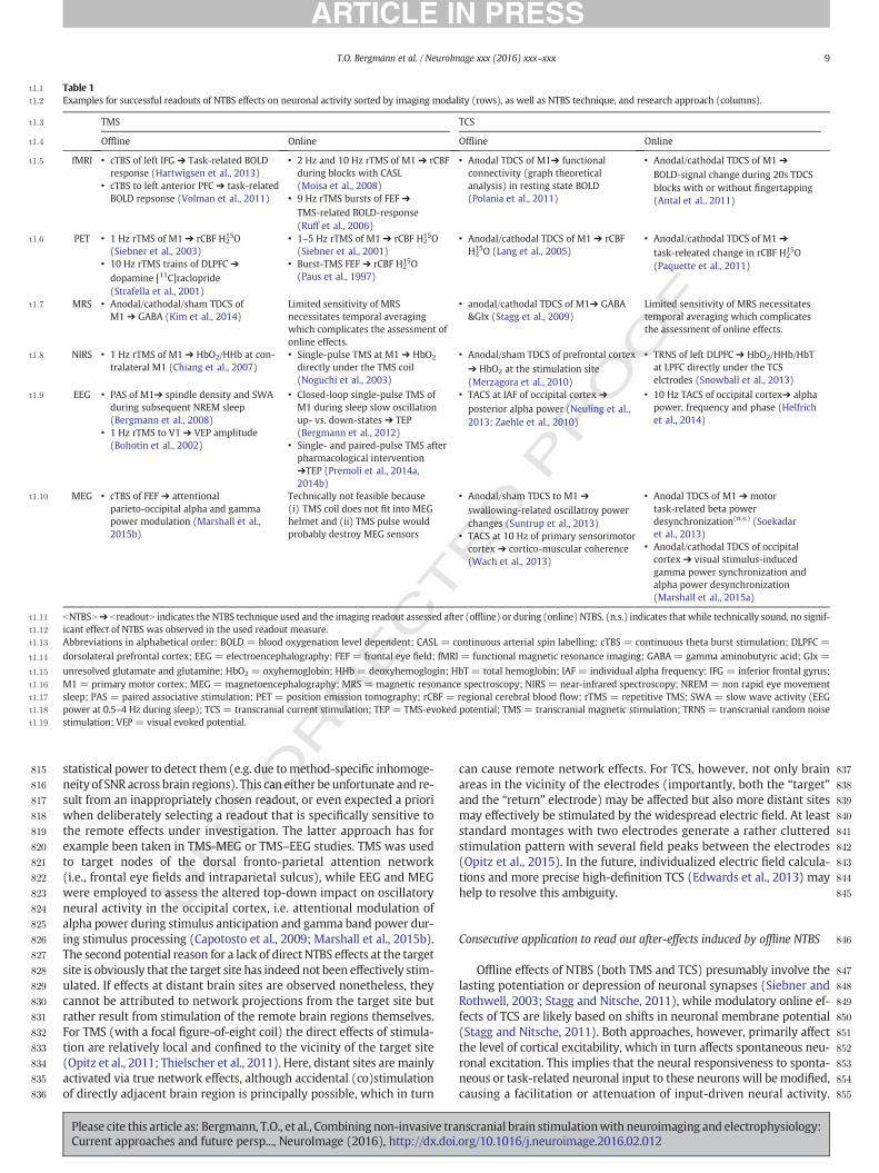
855
t1:1 Table 1t1:2 Examples for successful readouts of NTBS effects on neuronal activity sorted by imaging modality (rows), as well as NTBS technique, and research approach (columns).
t1:3 TMS TCS
t1:4 Offline Online Offline Online
t1:5 fMRI • cTBS of left IFG ➔ Task-related BOLDresponse (Hartwigsen et al., 2013)
• cTBS to left anterior PFC ➔ task-relatedBOLD repsonse (Volman et al., 2011)
• 2 Hz and 10 Hz rTMS of M1 ➔ rCBFduring blocks with CASL(Moisa et al., 2008)
• 9 Hz rTMS bursts of FEF ➔
TMS-related BOLD-response(Ruff et al., 2006)
• Anodal TDCS of M1➔ functionalconnectivity (graph theoreticalanalysis) in resting state BOLD(Polania et al., 2011)
• Anodal/cathodal TDCS of M1 ➔
BOLD-signal change during 20s TDCSblocks with or without fingertapping(Antal et al., 2011)
t1:6 PET • 1 Hz rTMS of M1 ➔ rCBF H215O
(Siebner et al., 2003)• 10 Hz rTMS trains of DLPFC ➔
dopamine [11C]raclopride(Strafella et al., 2001)
• 1–5 Hz rTMS of M1 ➔ rCBF H215O
(Siebner et al., 2001)• Burst-TMS FEF ➔ rCBF H2
15O(Paus et al., 1997)
• Anodal/cathodal TDCS of M1 ➔ rCBFH215O (Lang et al., 2005)
• Anodal/cathodal TDCS of M1 ➔
task-releated change in rCBF H215O
(Paquette et al., 2011)
t1:7 MRS • Anodal/cathodal/sham TDCS ofM1 ➔ GABA (Kim et al., 2014)
Limited sensitivity of MRSnecessitates temporal averagingwhich complicates the assessment ofonline effects.
• anodal/cathodal TDCS of M1➔ GABA&Glx (Stagg et al., 2009)
Limited sensitivity of MRS necessitatestemporal averaging which complicatesthe assessment of online effects.
t1:8 NIRS • 1 Hz rTMS of M1 ➔ HbO2/HHb at con-tralateral M1 (Chiang et al., 2007)
• Single-pulse TMS at M1 ➔ HbO2
directly under the TMS coil(Noguchi et al., 2003)
• Anodal/sham TDCS of prefrontal cortex➔ HbO2 at the stimulation site(Merzagora et al., 2010)
• TRNS of left DLPFC ➔ HbO2/HHb/HbTat LPFC directly under the TCSelctrodes (Snowball et al., 2013)
t1:9 EEG • PAS of M1➔ spindle density and SWAduring subsequent NREM sleep(Bergmann et al., 2008)
• 1 Hz rTMS to V1 ➔ VEP amplitude(Bohotin et al., 2002)
• Closed-loop single-pulse TMS ofM1 during sleep slow oscillationup- vs. down-states ➔ TEP(Bergmann et al., 2012)
• Single- and paired-pulse TMS afterpharmacological intervention➔TEP (Premoli et al., 2014a,2014b)
• TACS at IAF of occipital cortex ➔
posterior alpha power (Neuling et al.,2013; Zaehle et al., 2010)
• 10 Hz TACS of occipital cortex➔ alphapower, frequency and phase (Helfrichet al., 2014)
t1:10 MEG • cTBS of FEF ➔ attentionalparieto-occipital alpha and gammapower modulation (Marshall et al.,2015b)
Technically not feasible because(i) TMS coil does not fit into MEGhelmet and (ii) TMS pulse wouldprobably destroy MEG sensors
• Anodal/sham TDCS to M1 ➔
swallowing-related oscillatroy powerchanges (Suntrup et al., 2013)
• TACS at 10 Hz of primary sensorimotorcortex ➔ cortico-muscular coherence(Wach et al., 2013)
• Anodal TDCS of M1 ➔ motortask-related beta powerdesynchronization(n.s.) (Soekadaret al., 2013)
• Anodal/cathodal TDCS of occipitalcortex ➔ visual stimulus-inducedgamma power synchronization andalpha power desynchronization(Marshall et al., 2015a)
t1:11 bNTBSN➔ breadoutN indicates the NTBS technique used and the imaging readout assessed after (offline) or during (online) NTBS. (n.s.) indicates that while technically sound, no signif-t1:12 icant effect of NTBS was observed in the used readout measure.t1:13 Abbreviations in alphabetical order: BOLD = blood oxygenation level dependent; CASL = continuous arterial spin labelling; cTBS = continuous theta burst stimulation; DLPFC =
t1:14 dorsolateral prefrontal cortex; EEG = electroencephalography; FEF = frontal eye field; fMRI = functional magnetic resonance imaging; GABA = gamma aminobutyric acid; Glx =
t1:15 unresolved glutamate and glutamine; HbO2 = oxyhemoglobin; HHb = deoxyhemoglogin; HbT = total hemoglobin; IAF = individual alpha frequency; IFG = inferior frontal gyrus;t1:16 M1 = primary motor cortex; MEG = magnetoencephalography; MRS = magnetic resonance spectroscopy; NIRS = near-infrared spectroscopy; NREM = non rapid eye movementt1:17 sleep; PAS = paired associative stimulation; PET = position emission tomography; rCBF = regional cerebral blood flow; rTMS = repetitive TMS; SWA = slow wave activity (EEGt1:18 power at 0.5–4 Hz during sleep); TCS = transcranial current stimulation; TEP = TMS-evoked potential; TMS = transcranial magnetic stimulation; TRNS = transcranial random noiset1:19 stimulation; VEP = visual evoked potential.
9T.O. Bergmann et al. / NeuroImage xxx (2016) xxx–xxx
UNCO
R
statistical power to detect them (e.g. due tomethod-specific inhomoge-neity of SNR across brain regions). This can either be unfortunate and re-sult from an inappropriately chosen readout, or even expected a prioriwhen deliberately selecting a readout that is specifically sensitive tothe remote effects under investigation. The latter approach has forexample been taken in TMS-MEG or TMS–EEG studies. TMS was usedto target nodes of the dorsal fronto-parietal attention network(i.e., frontal eye fields and intraparietal sulcus), while EEG and MEGwere employed to assess the altered top-down impact on oscillatoryneural activity in the occipital cortex, i.e. attentional modulation ofalpha power during stimulus anticipation and gamma band power dur-ing stimulus processing (Capotosto et al., 2009; Marshall et al., 2015b).The second potential reason for a lack of direct NTBS effects at the targetsite is obviously that the target site has indeed not been effectively stim-ulated. If effects at distant brain sites are observed nonetheless, theycannot be attributed to network projections from the target site butrather result from stimulation of the remote brain regions themselves.For TMS (with a focal figure-of-eight coil) the direct effects of stimula-tion are relatively local and confined to the vicinity of the target site(Opitz et al., 2011; Thielscher et al., 2011). Here, distant sites are mainlyactivated via true network effects, although accidental (co)stimulationof directly adjacent brain region is principally possible, which in turn
Please cite this article as: Bergmann, T.O., et al., Combining non-invasive traCurrent approaches and future persp..., NeuroImage (2016), http://dx.doi
can cause remote network effects. For TCS, however, not only brainareas in the vicinity of the electrodes (importantly, both the “target”and the “return” electrode) may be affected but also more distant sitesmay effectively be stimulated by the widespread electric field. At leaststandard montages with two electrodes generate a rather clutteredstimulation pattern with several field peaks between the electrodes(Opitz et al., 2015). In the future, individualized electric field calcula-tions and more precise high-definition TCS (Edwards et al., 2013) mayhelp to resolve this ambiguity.
Consecutive application to read out after-effects induced by offline NTBS
Offline effects of NTBS (both TMS and TCS) presumably involve thelasting potentiation or depression of neuronal synapses (Siebner andRothwell, 2003; Stagg and Nitsche, 2011), while modulatory online ef-fects of TCS are likely based on shifts in neuronal membrane potential(Stagg and Nitsche, 2011). Both approaches, however, primarily affectthe level of cortical excitability, which in turn affects spontaneous neu-ronal excitation. This implies that the neural responsiveness to sponta-neous or task-related neuronal input to these neurons will bemodified,causing a facilitation or attenuation of input-driven neural activity.
nscranial brain stimulationwith neuroimaging and electrophysiology:.org/10.1016/j.neuroimage.2016.02.012
T
856
857
858
859
860
861
862
863
864
865
866
867
868
869
870
871
872
873
874
875
876
877
878
879
880
881
882
883
884
885
886
887
888
889
890
891
892
893
894
895
896
897
898
899
900
901
902
903
904
905
906
907
908
909
910
911
912
913
914
915
916
917
918
919
920
921
922
923
924
925
926
927
928
929
930
931
932
933
934
935
936
937
938
939
940
941
942
943
944
945
946
947
948
949
950
951
952
953
954
955
956
957
958
959
960
961
962
963
964
965
966
967
968
969
970
971
972
973
974
975
976
977
978
979
980
981
982
983
984
985
986
10 T.O. Bergmann et al. / NeuroImage xxx (2016) xxx–xxx
UNCO
RREC
Likewise, the outgoing activity will be modified, resulting in increasedor decreased output levels.
Some neuroimaging readouts may be particularly suited for the as-sessment of NTBS offline effects, as they are sensitive to the functionalconsequences in termsof neural integration at the network level follow-ing the induction of synaptic plasticity. They indicate either changedmetabolic demands by means of regional cerebral blood flow (rCBF)via position emission tomography (PET) (Siebner et al., 2003), near-infrared spectroscopy (NIRS) (Chiang et al., 2007) and continuous arte-rial spin labelling (CASL) (Orosz et al., 2012), altered neurotransmittersynthesis and binding via PET (Strafella et al., 2001), and relative con-centrations of neurotransmitters such as glutamate and GABA via mag-netic resonance spectroscopy (MRS) (Kim et al., 2014). Other readoutsrequire some kind of input to actively drive and challenge themodulat-ed neuronal system in order to reliably capture the functional changesinduced by NTBS. This input may be generated experimentally bymeans of exogenous sensory stimuli that trigger task-related BOLD-responses (Hartwigsen et al., 2013; Volman et al., 2011), ERPs(Bohotin et al., 2002) or oscillatory power modulations (Marshallet al., 2015b). Alternatively, this input may be spontaneously generatedwithin the modulated network or connected regions, as reflected inBOLD network connectivity (Polania et al., 2011) or spontaneous EEGoscillations (Bergmann et al., 2008; Huber et al., 2007; Neuling et al.,2013; Vossen et al., 2015; Zaehle et al., 2010). In fact, MEG, EEG andfMRI provide a large variety of readouts to assess the after-effects ofrTMS protocols, which indicate facilitatory and suppressive effects onneural excitability, activity, or connectivity (Thut and Pascual-Leone,2010). In principle, these readouts should be equally sensitive to detectafter-effects induced by TMS and TCS, as both NTBS techniques are as-sumed to rely on LTP- and LTD-like synaptic plasticity (Paulus et al.,2013).
As noted above, the plastic changes in neural activity induced by dif-ferent NTBS protocolsmay not be restricted to the stimulated area itself.In this context, measures of functional connectivity, defined as the tem-poral covariance between remote neurophysiological events or effectiveconnectivity, defined as the causal influence one remote neuronal eventexerts over another (Friston, 2002), are particularly suited to capturerapid NTBS-induced functional reorganization and adaptive plasticityon the network level by investigating the interaction between differentnetwork nodes at rest or during a specific task (for a recent review seeHartwigsen et al., 2015). Changes in effective connectivity followingoffline TMS have been studied during speech production using dynamiccausal modelling (DCM) (e.g. Hartwigsen et al., 2013) and during actionselection using psychophysiological interactions (PPI) (e.g. Ward et al.,2010). Changes in functional connectivity following offline TDCS havebeen demonstrated using a graph theoretical approach, showing thatthe after-effects of TDCS over M1 during subsequent resting-statefMRI mimicked training-induced changes in the motor network(Polania et al., 2011). Particularly, anodal TDCS increased the functionalconnectivity between left sensorimotor andmotor-related cortical areasbut decreased functional coupling between sensorimotor and contralat-eral motor regions.
Moreover, NTBS-induced changes in task-related neural activitymay also be related to the observed modulation in task performance(e.g., Andoh and Paus, 2011; Andoh and Zatorre, 2013; Hartwigsenet al., 2013; Ward et al., 2010). However, in some cases, it remains un-clear whether the behavioral changes induced by different NTBS proto-cols are related to themodulation of neural activity in the targeted brainregion or in remote connected areas (Hartwigsen, 2014). Indeed,studies employing effective connectivity analyses of fMRI data afterTMS-induced perturbation over a key region within a specific networkrevealed changes in the functional drive between different networknodes (Hartwigsen et al., 2013;Herz et al., 2014). Thesefindings suggestthat distributed network effects rather than the modulation of theneural activity at the stimulated cortical area itself might mediate theNTBS-induced changes on the behavioral level during some tasks.
Please cite this article as: Bergmann, T.O., et al., Combining non-invasive traCurrent approaches and future persp..., NeuroImage (2016), http://dx.doi
ED P
RO
OF
Concurrent application to read out immediate effects of online NTBS
Online single-pulse or burst-TMS triggers action potentials and con-sequently trans-synaptic neuronal excitation, making it possible to di-rectly assess the neuronal response to the stimulation itself. For onlineTMS–fMRI, this can mainly be achieved by measuring the immediateBOLD-response to TMS (Bestmann et al., 2005; Ruff et al., 2006). Typi-cally, short bursts of high-frequency TMS are givenwhich allows neuralexcitation to sum up and facilitate the detection of neural activationwith interleaved fMRI. Technical advances in online TMS–fMRI mightrender it possible to reliably assess hemodynamic responses evokedby single TMS pulses in future studies (Navarro de Lara et al., 2014). Ex-perimental manipulations of stimulus intensity and the brain state (e.g.level of attention or task complexity) have been successfully used tomap the online effects of TMS on effective connectivity between thestimulated cortex and other brain regions (Bestmann et al., 2008b;Moisa et al., 2012; Ruff et al., 2006).
For online TMS–EEG, one needs to select the appropriate readoutdepending on the effects of interest at the neuronal level. EEG read-outs of the brain response to TMS are commonly derived in one orthe other way from the TMS-evoked potential (TEP), i.e. the EEG po-tential time-locked to the TMS-pulse (Bonato et al., 2006; Ilmoniemiet al., 1997; Paus et al., 2001). After TMS-artefact correction and av-eraging, the TEP can be analyzed like an ordinary sensory ERP, e.g. byquantifying amplitude and latency of specific components. For in-stance, the N45 and N100 of the TEP evoked in M1HAND have been re-lated to GABA-A- and GABA-B-receptor specific activation,respectively (Premoli et al., 2014a, 2014b). Moreover, frequencyand power of TMS-evoked oscillations can be assessed by means ofwavelet or moving window Fourier analyses, transforming the aver-age TEP into a time-frequency representation (TFR) (Rosanova et al.,2009). Note that this should not be confused with the averaging ofsingle-trial TFRs, which captures a mixture of both phase-locked(i.e. evoked) and non-phase-locked (i.e. induced) oscillatorypower. Additionally, more complex and integrated readouts can bederived, reflecting the state of the thalamo-cortical system (as wellas the level of consciousness). Examples for such readouts are globalmean field power (Esser et al., 2006), significant current densityphase locking, and significant current scattering (Casali et al., 2010).
Online effects of TCS are much harder to quantify as they do notdirectly evoke action potentials but rather modulate the level(TDCS) or timing (TACS) of cortical excitability. Polarization inducedchanges in neural excitability may well affect the level of ongoingpostsynaptic neural activity. If these changes alter the summed neu-ral activity in a brain region, this might bemapped by fMRI. However,some TCS applications might not modify the summed neural activityand therefore fail to show concurrent changes in the BOLD signal.Online TDCS of M1HAND for example does not induce any concurrentchanges in BOLD-signal during 20 s stimulation blocks (Antal et al.,2011), and 10 s blocks of TACS of the visual cortex at individualalpha frequency did not result in a TACS-related BOLD decrease(Vosskuhl et al., 2015) as expected from previous concurrent EEG–fMRI work on alpha oscillations (e.g. Moosmann et al., 2003;Scheeringa et al., 2011). Task-free BOLD-fMRI may thus not be themeasure of choice to directly assess the neuronal online effects ofTDCS, unless specific resting-state connectivity measures are applied(Polania et al., 2011). However, the TDCS induced change in neuralexcitability may acutely change the input–output curves of neuronsin the stimulated brain regions. By shifting the neurons' restingmembrane potential towards depolarization and thus closer to firingthreshold, TDCS increases the likelihood that spontaneous synapticinput will cause neuronal firing. Conversely, shifting the restingmembrane potential towards hyperpolarization has the opposite ef-fect. This may result in a corresponding change of regional activityand inter-regional connectivity during concurrent fMRI. For in-stance, TDCS of sensorimotor cortex acutely modulated the cortical
nscranial brain stimulationwith neuroimaging and electrophysiology:.org/10.1016/j.neuroimage.2016.02.012
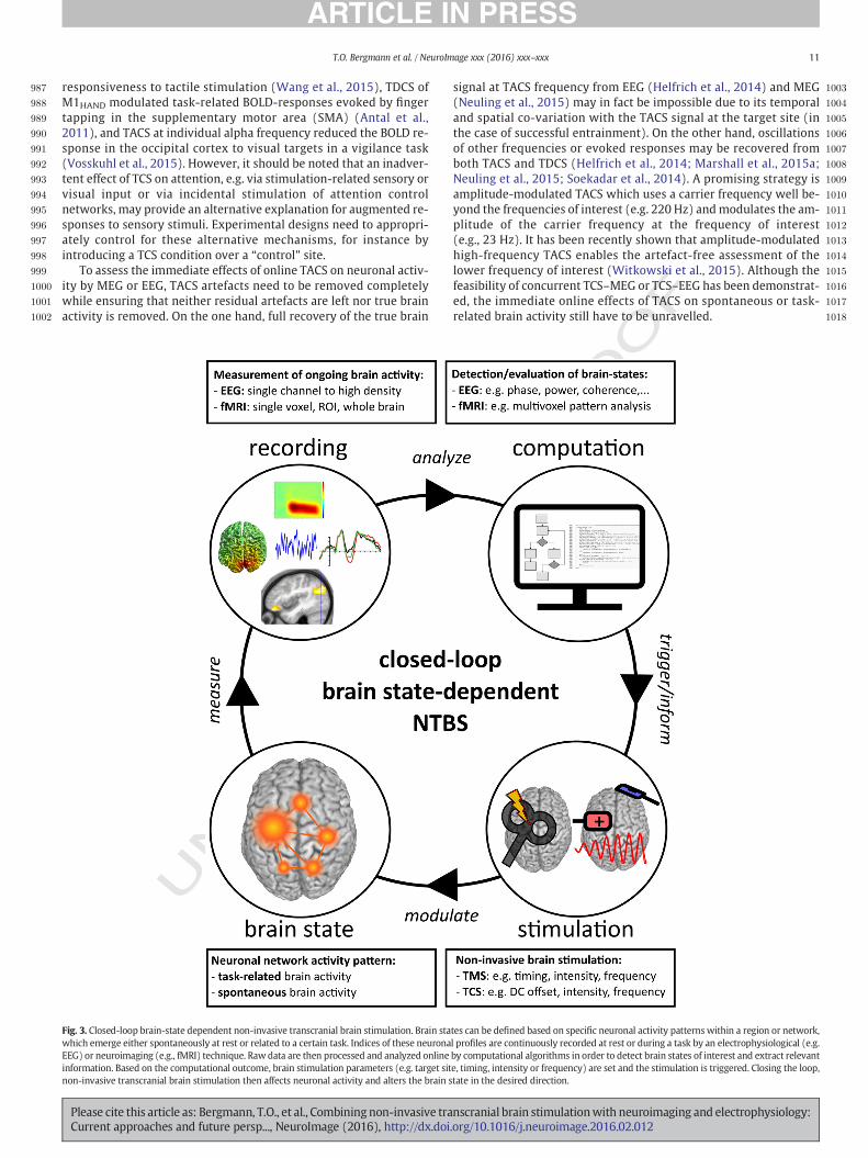
987
988
989
990
991
992
993
994
995
996
997
998
999
1000
1001
1002
1003
1004
1005
1006
1007
1008
1009
1010
1011
1012
1013
1014
1015
1016
1017
1018
11T.O. Bergmann et al. / NeuroImage xxx (2016) xxx–xxx
responsiveness to tactile stimulation (Wang et al., 2015), TDCS ofM1HAND modulated task-related BOLD-responses evoked by fingertapping in the supplementary motor area (SMA) (Antal et al.,2011), and TACS at individual alpha frequency reduced the BOLD re-sponse in the occipital cortex to visual targets in a vigilance task(Vosskuhl et al., 2015). However, it should be noted that an inadver-tent effect of TCS on attention, e.g. via stimulation-related sensory orvisual input or via incidental stimulation of attention controlnetworks, may provide an alternative explanation for augmented re-sponses to sensory stimuli. Experimental designs need to appropri-ately control for these alternative mechanisms, for instance byintroducing a TCS condition over a “control” site.
To assess the immediate effects of online TACS on neuronal activ-ity by MEG or EEG, TACS artefacts need to be removed completelywhile ensuring that neither residual artefacts are left nor true brainactivity is removed. On the one hand, full recovery of the true brain
UNCO
RRECT
Fig. 3. Closed-loop brain-state dependent non-invasive transcranial brain stimulation. Brain stawhich emerge either spontaneously at rest or related to a certain task. Indices of these neuronaEEG) or neuroimaging (e.g., fMRI) technique. Raw data are then processed and analyzed onlineinformation. Based on the computational outcome, brain stimulation parameters (e.g. target sitnon-invasive transcranial brain stimulation then affects neuronal activity and alters the brain s
Please cite this article as: Bergmann, T.O., et al., Combining non-invasive traCurrent approaches and future persp..., NeuroImage (2016), http://dx.doi
F
signal at TACS frequency from EEG (Helfrich et al., 2014) and MEG(Neuling et al., 2015) may in fact be impossible due to its temporaland spatial co-variation with the TACS signal at the target site (inthe case of successful entrainment). On the other hand, oscillationsof other frequencies or evoked responses may be recovered fromboth TACS and TDCS (Helfrich et al., 2014; Marshall et al., 2015a;Neuling et al., 2015; Soekadar et al., 2014). A promising strategy isamplitude-modulated TACS which uses a carrier frequency well be-yond the frequencies of interest (e.g. 220 Hz) andmodulates the am-plitude of the carrier frequency at the frequency of interest(e.g., 23 Hz). It has been recently shown that amplitude-modulatedhigh-frequency TACS enables the artefact-free assessment of thelower frequency of interest (Witkowski et al., 2015). Although thefeasibility of concurrent TCS–MEG or TCS–EEG has been demonstrat-ed, the immediate online effects of TACS on spontaneous or task-related brain activity still have to be unravelled.
ED P
RO
O
tes can be defined based on specific neuronal activity patterns within a region or network,l profiles are continuously recorded at rest or during a task by an electrophysiological (e.g.by computational algorithms in order to detect brain states of interest and extract relevante, timing, intensity or frequency) are set and the stimulation is triggered. Closing the loop,tate in the desired direction.
nscranial brain stimulationwith neuroimaging and electrophysiology:.org/10.1016/j.neuroimage.2016.02.012
T
1019
1020
1021
1022
1023
1024
1025
1026
1027
1028
1029
1030
1031
1032
1033
1034
1035
1036
1037
1038
1039
1040
1041
1042
1043
1044
1045
1046
1047
1048
1049
1050
1051
1052
1053
1054
1055
1056
1057
1058
1059
1060
1061
1062
1063
1064
1065
1066
1067
1068
1069
1070
1071
1072
1073
1074
1075
1076
1077
1078
1079
1080
1081
1082
1083
1084
1085
1086
1087
1088
1089
1090
1091
1092
1093
1094
1095
1096
1097
1098
1099
1100
1101
1102
1103
1104
1105
1106
1107
1108
1109
1110
1111
1112
1113
1114
1115
1116
1117
1118
1119
1120
1121
1122
1123
1124
1125
1126
1127
1128
1129
1130
1131
1132
1133
1134
1135
1136
1137
1138
1139
1140
1141
1142
1143
1144
1145
1146
1147
1148
1149
12 T.O. Bergmann et al. / NeuroImage xxx (2016) xxx–xxx
UNCO
RREC
Closing the loop with brain-state dependent non-invasive transcra-nial brain stimulation
Neuroimaging and electrophysiology can be employed to providespatial, temporal and parameter-specific information for adjustingand optimizing NTBS application (section “Neuroimaging andelectrophysiology approaches to inform NTBS”) or to capture theneural effects that are induced by NTBS at the regional and systemslevel (section “Neuroimaging and electrophysiology as readout forNTBS effects”). These approaches can be merged into a brain–NTBSinterface approach.When NTBS and neuroimaging or electrophysiol-ogy is applied concurrently and both strategies are iteratively com-bined within the same experimental setup, the stage is set forclosed-loop brain state-dependent NTBS (Fig. 3). A closed-loop sys-tem in the strict sense iteratively controls a certain parameter of asystem via a control signal to reach and maintain a predefined set-point of that parameter while trying to reduce errors (i.e. deviationsfrom that set-point) by monitoring the parameter (feedback) andadjusting the control signal accordingly. Applied to online NTBS,concurrent neuroimaging or electrophysiology traces a certainbrain state and the dynamic expression of that brain state is usedto iteratively adjust spatial, temporal or other variables (such as in-tensity or frequency) of NTBS in order to suppress, facilitate, ormain-tain that brain state with NTBS.
What defines a brain state? In the context of brain state-dependentNTBS, we use the term in its broadest sense. Extending the definitionby Zagha and McCormick (2014), we define brain state as a recurringset of neuronal conditions, also referred to as dynamic circuit motif(Womelsdorf et al., 2014), which is stable for a behaviorally (orneuro-computationally) significant period of time. These brain statescan be described along multiple spatial and temporal scales and canbe indexed by a variety of both physiological and behavioral measures.Although the general term brain state suggests a global (i.e. brain-wide)condition, it does in practice often refer to anything ranging from thestate of specific neuron populations and local excitatory/inhibitory cir-cuits over large-scale brain networks to the entire thalamocortical sys-tem (although in the former cases, neuronal state or network statewould be more appropriate terms). Along the temporal dimension,these states can change slowly over days or even years (e.g. in the con-text of brain maturation or post-stroke cortical reorganization), varyacross minutes to hours (e.g. related to certain sleep stages or vigilancestates (Gervasoni et al., 2004), or the recent history of learning-relatedplasticity (Karabanov et al., 2015) or sensory adaptation (Silvantoet al., 2008)), or even fluctuate in the range of seconds to milliseconds.Rapidly fluctuating brain states reflect themomentary degree of neuro-nal (de)synchronization in a network (Zagha and McCormick, 2014) orthe phase of a single oscillatory cycle comprising excitatory and inhibi-tory periods (Destexhe et al., 2007). Importantly, brain states are undercontrol of both subcortical ascending neuromodulatory systems as wellas thalamo-cortical and cortico-cortical projections, giving rise to mod-ulations at various levels of spatial and temporal specificity (Harris,2013; Harris and Thiele, 2011; Lee and Dan, 2012; Zagha andMcCormick, 2014). Especially the level of neuronal synchronization,reflected in the power of network-specific neuronal oscillations (e.g.the 8–12Hz alpha band), has proven as useful indicator, if not constitut-ing element, of the level of vigilance or selective attention which fluctu-ates spontaneously but is also modulated in a task-related manner(Harris and Thiele, 2011; Jensen et al., 2011; Ros et al., 2014).
Brain states can be indexed non-invasively by a plethora of differentmeasures, either indirectly via associated changes in behavioral (e.g. ac-curacy and reaction time on attention, vigilance, or working memorytasks) and peripheral electrophysiological readouts (e.g. muscle activi-ty, heart rate, or skin conductance), or more directly via metabolic indi-ces (e.g. perfusion or neurotransmitter turnover) and readouts ofongoing neuronal activity in the brain via MEG, EEG (e.g. oscillatorypower or phase) or fMRI (e.g. resting state connectivity). Most
Please cite this article as: Bergmann, T.O., et al., Combining non-invasive traCurrent approaches and future persp..., NeuroImage (2016), http://dx.doi
ED P
RO
OF
demanding, but may be also most promising for closed-loop NTBS arethe very transient functional brain states thatfluctuate at the rate ofmil-liseconds to minutes and can only be monitored electrophysiologicallywith methods that offer very high temporal resolution such as EEGandMEG. Theymay be defined based on topographical EEGmicrostates(Khanna et al., 2015), certain rule-based classification schemes (e.g.sleep stages in polysomnography (Iber et al., 2007)), oscillatory powerof specified frequency bands (e.g. 8–12Hz alpha power), the occurrenceof certain neuroelectric events (e.g. sleep spindles or epileptic spikes),or the phase of ongoing oscillations (e.g. sleep slow oscillations).
Closed-loop applications that use invasive brain stimulation tech-niques for clinical treatment purposes are already in use (Berenyiet al., 2012; Cagnan et al., 2013; Rosin et al., 2011; Sun and Morrell,2014), whereas closed-loop NTBS protocols in the strict sense, basedon direct readout of brain activity are currently under development.When extending the readout to peripheral measures, the most elegantexample for closed-loop NTBS so far is the application of TACS toM1HAND at the individual tremor frequency in patients with Parkinson'sdisease to suppress tremor amplitude, presumably by means of phase-cancellation between spontaneous and induced oscillatory neuronal ac-tivity (Brittain et al., 2013): Informed by the ongoing tremor activity,the phase of TACS was constantly adjusted to maintain the optimalphase-delay between TACS and the ongoing tremor rhythm as deter-mined from simultaneous actigraphy measures.
Closed-loop paradigms for concurrent TMS–fMRI setups may be-come relevant if the dynamics of the brain-state of interest have aslow time constant such as slow fluctuations in resting-state connectiv-ity, because they are matching the slow temporal properties of theBOLD-signal. Potential advantages of fMRI-based readouts are the highspatial resolution and the higher sensitivity to subcortical structuresdue to a more uniform spatial sensitivity profile and point-spread func-tion) as compared toMEG or EEG. Conversely,MEG or EEG is required toreadout more transient brain states, dynamically fluctuating at higherfrequencies. So far no actual closed-loop but only informed open-loopNTBS-M/EEG paradigms have been published. Informed open-loopNTBS-M/EEG exploits knowledge about the current expression of agiven brain state to inform NTBS in a feed-forward manner, but doesnot require rapid online feed-back and iterative evaluation of immedi-ate NTBS effects (like the TEP) but merely inform NTBS in a feed-forward manner. For example, EEG-triggered TMS to the up- anddown-states of the sleep slow oscillation was temporally informed bythe ongoing EEG, while a refractory period of about 3 s was deliberatelyused to avoid continuous triggering of TMS by the previous slow-oscillation like TEP (Bergmann et al., 2012). In contrast, continuous trig-gering was actively employed in a slow oscillation up-state-triggeredclosed-loop auditory stimulation paradigm to actively drive slow oscil-lations (Ngo et al., 2013, 2015).
Depending on the stimulation site (Mutanen et al., 2013) a majorchallenge are the various NTBS-related artefacts in MEG and EEG. Theremoval of these artefacts usually requires thorough andmanually guid-ed offline analysis involving spatial filter and template subtraction tech-niques (Helfrich et al., 2014; Hernandez-Pavon et al., 2012; Ilmoniemiand Kicic, 2010; Marshall et al., 2015a; Neuling et al., 2015; Siebneret al., 2009a; Soekadar et al., 2013) to avoid corruption of the readout,especially when targeting transient spectral power changes (Walteret al., 2012). For TCS, amplitude-modulated TACS at higher carrier fre-quencies enables the immediate online assessment of lower oscillatoryfrequencies of interest without any additional processing steps(Witkowski et al., 2015), thus being highly suitable for closed-loop ap-proaches. For TMS, proper sham control is needed to control for theneu-ral effects of auditory and somatosensory input associated with NTBS,even at subliminal stimulation intensities (Herring et al., 2015).
The computational effort to remove artefacts to quickly recover thereadout of interest depends on the very brain state of interest. For exam-ple, to adjust the phase, frequency or intensity of ongoing TACS it is nec-essary to obtain reliable online recordings of phase, frequency or power
nscranial brain stimulationwith neuroimaging and electrophysiology:.org/10.1016/j.neuroimage.2016.02.012
T
1150
1151
1152
1153
1154
1155
1156
1157
1158
1159
1160
1161
1162
1163
1164
1165
1166
1167
1168
1169
1170
1171
1172
1173
1174
1175
1176
1177
1178
1179
1180
1181
1182
1183
1184
1185
1186
1187
1188
1189
1190
1191
1192
1193
1194
1195
1196
1197
1198
1199
1200
1201
1202
1203
1204
1205
1206
1207
1208
1209
1210
121112121213121412151216121712181219122012211222122312241225122612271228122912301231123212331234123512361237123812391240124112421243124412451246124712481249125012511252
13T.O. Bergmann et al. / NeuroImage xxx (2016) xxx–xxx
of a spontaneous oscillation, in analogy to the tremor-informedclosed-loop paradigm discussed above (Brittain et al., 2013). Hencethe TACS artefacts need to be removed online with high precisionto uncover the underlying true neuronal activity, which was not pos-sible so far (cf. section “Concurrent application to read outimmediate effects of online NTBS”), or amplitude modulation needsto be employed (Witkowski et al., 2015). For TMS, a closed-loop ex-ample would be to trigger single pulses based on the phase of a cer-tain TEP component evoked by the previous pulse to constantly drive(or entrain) a certain TMS-locked oscillation, which is currently donewith fixed, though often individually adjusted, frequencies (Thutet al., 2011b). Alternatively, intensity of an ongoing rTMS protocolcould be constantly adapted based on the amplitude of the individualTEPs to adjust for changes in excitability due to the ongoing build-upof after-effects during stimulation. Also an individual terminationcriterion for rTMS protocols is conceivable based on such a functionalreadout provided that spatial filtering techniques can offer sufficientSNR for single trial analyses. The technical equipment and computa-tional power required for real-time analyses of M/EEG and online ad-justment of NTBS are already available. It is therefore up to the NTBScommunity to face the methodological challenges associated withclosed-loop NTBS in the strict sense and develop those paradigmsas a tool for systems and cognitive neuroscience. Technical feasibilityis necessary but not sufficient for closed-loop NTBS protocols to beeffective. It is crucial to select the appropriate brain state of interestas well as the appropriate imaging and electrophysiological indicessupposed to measure it. Another challenge is to design NTBS proto-cols that are capable of modulating the brain state of interest in thedesired direction. Presumably, a closed-loop interference with spe-cific brain states will be easier to accomplish than their facilitation.The latter may first be achieved by targeting well-described brainstates (e.g. specific oscillatory patterns) for which the neurophysio-logical underpinnings are sufficiently understood, allowing for ahypothesis-driven approach (Horschig et al., 2014).
12531254125512561257125812591260126112621263126412651266126712681269127012711272127312741275127612771278127912801281
UNCO
RRECConclusion
NTBS strongly benefits from the information accessible via brainmapping techniques. Firstly, neuroimaging and electrophysiologycan optimize NTBS with regard to where, when and how to stimu-late. While information with high inter- but low intra-subject vari-ability can be derived before NTBS, those fluctuating over timewithin a subject, such as the current of brain state, need to be moni-tored continuously to enable informed open-loop NTBS. Secondly,neuroimaging and electrophysiology can provide appropriate read-outs for either immediate (online) or subsequent (offline) effects ofNTBS on neuronal activity, depending on whether applied concur-rently or consecutively. For closed-loop NTBS in the strict sense,NTBS can be informed about the momentary brain state by concur-rent readouts of neuronal activity and be adjusted accordingly to in-terfere with or modulate neuronal activity to reach or maintain thedesired brain state. Future lines of research should further developthese combined paradigms to increase the precision and effective-ness of NTBS for systems neuroscientific research and to boost thetherapeutic potential of NTBS.
1282128312841285128612871288128912901291129212931294
Acknowledgments
H.R.S. received financial support from the Lundbeck Foundation[Grant of Excellence “Mapping, Modulation and Modeling the Controlof Actions”; grant number R59-A5399]. H.R.S. and A.T. received fundingfrom the Novo Nordisk Foundation Interdisciplinary Synergy Program2014 [“Biophysically adjusted state-informed cortex stimulation (BA-SICS); grant number NNF14OC0011413].
Please cite this article as: Bergmann, T.O., et al., Combining non-invasive traCurrent approaches and future persp..., NeuroImage (2016), http://dx.doi
ED P
RO
OF
References
Andoh, J., Paus, T., 2011. Combining functional neuroimaging with off-line brain stimula-tion: modulation of task-related activity in language areas. J. Cogn. Neurosci. 23,349–361.
Andoh, J., Zatorre, R.J., 2013. Mapping interhemispheric connectivity using functional MRIafter transcranial magnetic stimulation on the human auditory cortex. NeuroImage79, 162–171.
Antal, A., Paulus, W., 2013. Transcranial alternating current stimulation (tACS). Front.Hum. Neurosci. 7, 317.
Antal, A., Kincses, T.Z., Nitsche, M.A., Bartfai, O., Paulus, W., 2004a. Excitability changes in-duced in the human primary visual cortex by transcranial direct current stimulation:direct electrophysiological evidence. Invest. Ophthalmol. Vis. Sci. 45, 702–707.
Antal, A., Nitsche, M.A., Kincses, T.Z., Lampe, C., Paulus, W., 2004b. No correlation betweenmoving phosphene and motor thresholds: a transcranial magnetic stimulation study.Neuroreport 15, 297–302.
Antal, A., Nitsche, M.A., Paulus, W., 2006. Transcranial direct current stimulation and thevisual cortex. Brain Res. Bull. 68, 459–463.
Antal, A., Boros, K., Poreisz, C., Chaieb, L., Terney, D., Paulus, W., 2008. Comparatively weakafter-effects of transcranial alternating current stimulation (tACS) on cortical excit-ability in humans. Brain Stimul. 1, 97–105.
Antal, A., Polania, R., Schmidt-Samoa, C., Dechent, P., Paulus, W., 2011. Transcranial directcurrent stimulation over the primary motor cortex during fMRI. NeuroImage 55,590–596.
Bandettini, P.A., 2009. What's new in neuroimaging methods? Ann. N. Y. Acad. Sci. 1156,260–293.
Barker, A.T., Jalinous, R., Freeston, I.L., 1985. Non-invasive magnetic stimulation of humanmotor cortex. Lancet 1, 1106–1107.
Bastani, A., Jaberzadeh, S., 2012. Does anodal transcranial direct current stimulation en-hance excitability of the motor cortex and motor function in healthy individualsand subjects with stroke: a systematic review and meta-analysis. Clin. Neurophysiol.123, 644–657.
Bastani, A., Jaberzadeh, S., 2013. Differential modulation of corticospinal excitability bydifferent current densities of anodal transcranial direct current stimulation. PLoSOne 8, e72254.
Batsikadze, G., Moliadze, V., Paulus, W., Kuo, M.F., Nitsche, M.A., 2013. Partially non-linearstimulation intensity-dependent effects of direct current stimulation onmotor cortexexcitability in humans. J. Physiol. 591, 1987–2000.
Berenyi, A., Belluscio, M., Mao, D., Buzsaki, G., 2012. Closed-loop control of epilepsy bytranscranial electrical stimulation. Science 337, 735–737.
Bergmann, T.O., Molle, M., Marshall, L., Kaya-Yildiz, L., Born, J., Siebner, H.R., 2008. A localsignature of LTP- and LTD-like plasticity in human NREM sleep. Eur. J. Neurosci. 27,2241–2249.
Bergmann, T.O., Groppa, S., Seeger, M., Mölle, M., Marshall, L., Siebner, H.R., 2009. Acutechanges in motor cortical excitability during slow oscillatory and constant anodaltranscranial direct current stimulation. J. Neurophysiol. 102, 2303–2311.
Bergmann, T.O., Molle, M., Schmidt, M.A., Lindner, C., Marshall, L., Born, J., Siebner,H.R., 2012. EEG-guided transcranial magnetic stimulation reveals rapid shifts inmotor cortical excitability during the human sleep slow oscillation. J. Neurosci.32, 243–253.
Bestmann, S., Baudewig, J., Siebner, H.R., Rothwell, J.C., Frahm, J., 2003. Subthresholdhigh-frequency TMS of human primary motor cortex modulates interconnectedfrontal motor areas as detected by interleaved fMRI-TMS. NeuroImage 20,1685–1696.
Bestmann, S., Baudewig, J., Siebner, H.R., Rothwell, J.C., Frahm, J., 2005. BOLD MRI re-sponses to repetitive TMS over human dorsal premotor cortex. NeuroImage 28,22–29.
Bestmann, S., Ruff, C.C., Blankenburg, F., Weiskopf, N., Driver, J., Rothwell, J.C., 2008a.Map-ping causal interregional influences with concurrent TMS–fMRI. Exp. Brain Res. 191,383–402.
Bestmann, S., Swayne, O., Blankenburg, F., Ruff, C.C., Haggard, P., Weiskopf, N., Josephs, O.,Driver, J., Rothwell, J.C., Ward, N.S., 2008b. Dorsal premotor cortex exerts state-dependent causal influences on activity in contralateral primary motor and dorsalpremotor cortex. Cereb. Cortex 18, 1281–1291.
Bindman, L.J., Lippold, O.C., Redfearn, J.W., 1964. The action of brief polarizing currents onthe cerebral cortex of the rat (1) during current flow and (2) in the production oflong-lasting after-effects. J. Physiol. 172, 369–382.
Bohotin, V., Fumal, A., Vandenheede, M., Gerard, P., Bohotin, C., Maertens de Noordhout,A., Schoenen, J., 2002. Effects of repetitive transcranial magnetic stimulation on visualevoked potentials in migraine. Brain 125, 912–922.
Bonato, C., Miniussi, C., Rossini, P.M., 2006. Transcranial magnetic stimulation and corticalevoked potentials: a TMS/EEG co-registration study. Clin. Neurophysiol. 117,1699–1707.
Boroojerdi, B., 2002. Pharmacologic influences on TMS effects. J. Clin. Neurophysiol. 19,255–271.
Boroojerdi, B., Foltys, H., Krings, T., Spetzger, U., Thron, A., Topper, R., 1999. Localization ofthe motor hand area using transcranial magnetic stimulation and functional magnet-ic resonance imaging. Clin. Neurophysiol. 110, 699–704.
Brittain, J.S., Probert-Smith, P., Aziz, T.Z., Brown, P., 2013. Tremor suppression by rhythmictranscranial current stimulation. Curr. Biol. 23, 436–440.
Cagnan, H., Brittain, J.S., Little, S., Foltynie, T., Limousin, P., Zrinzo, L., Hariz, M., Joint, C.,Fitzgerald, J., Green, A.L., Aziz, T., Brown, P., 2013. Phase dependent modulation oftremor amplitude in essential tremor through thalamic stimulation. Brain 136,3062–3075.
Cancelli, A., Cottone, C., Di Giorgio, M., Carducci, F., Tecchio, F., 2015. Personalizing theelectrode to neuromodulate an extended cortical region. Brain Stimul. 8, 555–560.
nscranial brain stimulationwith neuroimaging and electrophysiology:.org/10.1016/j.neuroimage.2016.02.012
T
12951296129712981299130013011302130313041305130613071308130913101311131213131314131513161317131813191320132113221323132413251326132713281329133013311332133313341335133613371338133913401341134213431344134513461347134813491350135113521353135413551356135713581359136013611362136313641365136613671368136913701371137213731374137513761377137813791380
13811382138313841385138613871388138913901391139213931394139513961397139813991400140114021403140414051406140714081409141014111412141314141415141614171418141914201421142214231424142514261427142814291430143114321433143414351436143714381439144014411442144314441445144614471448144914501451145214531454145514561457145814591460146114621463146414651466
14 T.O. Bergmann et al. / NeuroImage xxx (2016) xxx–xxx
UNCO
RREC
Capotosto, P., Babiloni, C., Romani, G.L., Corbetta, M., 2009. Frontoparietal cortex controlsspatial attention through modulation of anticipatory alpha rhythms. J. Neurosci. 29,5863–5872.
Casali, A.G., Casarotto, S., Rosanova, M., Mariotti, M., Massimini, M., 2010. General indicesto characterize the electrical response of the cerebral cortex to TMS. NeuroImage 49,1459–1468.
Cecere, R., Rees, G., Romei, V., 2015. Individual differences in alpha frequency drivecrossmodal illusory perception. Curr. Biol. 25, 231–235.
Chiang, T.C., Vaithianathan, T., Leung, T., Lavidor, M., Walsh, V., Delpy, D.T., 2007. Elevatedhaemoglobin levels in the motor cortex following 1 Hz transcranial magnetic stimu-lation: a preliminary study. Exp. Brain Res. 181, 555–560.
Datta, A., Truong, D., Minhas, P., Parra, L.C., Bikson, M., 2012. Inter-individual variationduring transcranial direct current stimulation and normalization of dose using MRI-derived computational models. Front Psychiatry 3, 91.
De Vos, M., Thorne, J.D., Yovel, G., Debener, S., 2012. Let's face it, from trial to trial: com-paring procedures for N170 single-trial estimation. NeuroImage 63, 1196–1202.
Deblieck, C., Thompson, B., Iacoboni, M., Wu, A.D., 2008. Correlation between motor andphosphene thresholds: a transcranial magnetic stimulation study. Hum. BrainMapp. 29, 662–670.
Destexhe, A., Hughes, S.W., Rudolph, M., Crunelli, V., 2007. Are corticothalamic 'up' statesfragments of wakefulness? Trends Neurosci. 30, 334–342.
Dugue, L., Marque, P., Vanrullen, R., 2011. The phase of ongoing oscillations mediates thecausal relation between brain excitation and visual perception. J. Neurosci. 31,11889–11893.
Edwards, D., Cortes, M., Datta, A., Minhas, P., Wassermann, E.M., Bikson, M., 2013. Physi-ological and modeling evidence for focal transcranial electrical brain stimulation inhumans: a basis for high-definition tDCS. NeuroImage 74, 266–275.
Esser, S.K., Huber, R., Massimini, M., Peterson, M.J., Ferrarelli, F., Tononi, G., 2006. A directdemonstration of cortical LTP in humans: a combined TMS/EEG study. Brain Res. Bull.69, 86–94.
Fertonani, A., Ferrari, C., Miniussi, C., 2015. What do you feel if i apply transcranial electricstimulation? Safety, sensations and secondary induced effects. Clin. Neurophysiol.
Fitzgerald, P.B., Fountain, S., Daskalakis, Z.J., 2006. A comprehensive review of the effectsof rTMS on motor cortical excitability and inhibition. Clin. Neurophysiol. 117,2584–2596.
Frey, D., Strack, V., Wiener, E., Jussen, D., Vajkoczy, P., Picht, T., 2012. A new approach forcorticospinal tract reconstruction based on navigated transcranial stimulation andstandardized fractional anisotropy values. NeuroImage 62, 1600–1609.
Friston, K., 2002. Functional integration and inference in the brain. Prog. Neurobiol. 68,113–143.
Gervasoni, D., Lin, S.C., Ribeiro, S., Soares, E.S., Pantoja, J., Nicolelis, M.A., 2004. Global fore-brain dynamics predict rat behavioral states and their transitions. J. Neurosci. 24,11137–11147.
Gerwig, M., Kastrup, O., Meyer, B.U., Niehaus, L., 2003. Evaluation of cortical excitability bymotor and phosphene thresholds in transcranial magnetic stimulation. J. Neurol. Sci.215, 75–78.
Ginhoux, R., Renaud, P., Zorn, L., Goffin, L., Bayle, B., Foucher, J., Lamy, J., Armspach, J.P., deMathelin,M., 2013. A custom robot for transcranial magnetic stimulation:first assess-ment on healthy subjects. Conf. Proc. IEEE Eng. Med. Biol. Soc. 2013, 5352–5355.
Goldsworthy, M.R., Pitcher, J.B., Ridding, M.C., 2012. A comparison of two different contin-uous theta burst stimulation paradigms applied to the human primary motor cortex.Clin. Neurophysiol. 123, 2256–2263.
Granert, O., Peller, M., Gaser, C., Groppa, S., Hallett, M., Knutzen, A., Deuschl, G., Zeuner,K.E., Siebner, H.R., 2011. Manual activity shapes structure and function in contralater-al human motor hand area. NeuroImage 54, 32–41.
Groppa, S., Oliviero, A., Eisen, A., Quartarone, A., Cohen, L.G., Mall, V., Kaelin-Lang, A.,Mima, T., Rossi, S., Thickbroom, G.W., Rossini, P.M., Ziemann, U., Valls-Sole, J.,Siebner, H.R., 2012a. A practical guide to diagnostic transcranial magnetic stimula-tion: report of an IFCN committee. Clin. Neurophysiol. 123, 858–882.
Groppa, S., Schlaak, B.H., Munchau, A., Werner-Petroll, N., Dunnweber, J., Baumer, T., vanNuenen, B.F., Siebner, H.R., 2012b. The human dorsal premotor cortex facilitates theexcitability of ipsilateral primary motor cortex via a short latency cortico-corticalroute. Hum. Brain Mapp. 33, 419–430.
Groppa, S., Werner-Petroll, N., Munchau, A., Deuschl, G., Ruschworth, M.F., Siebner, H.R.,2012c. A novel dual-site transcranial magnetic stimulation paradigm to probe fast fa-cilitatory inputs from ipsilateral dorsal premotor cortex to primary motor cortex.NeuroImage 62, 500–509.
Hamada, M., Hanajima, R., Terao, Y., Arai, N., Furubayashi, T., Inomata-Terada, S., Yugeta,A., Matsumoto, H., Shirota, Y., Ugawa, Y., 2007. Quadro-pulse stimulation is more ef-fective than paired-pulse stimulation for plasticity induction of the humanmotor cor-tex. Clin. Neurophysiol. 118, 2672–2682.
Hamidi, M., Slagter, H.A., Tononi, G., Postle, B.R., 2009. Repetitive transcranial magneticstimulation affects behavior by biasing endogenous cortical oscillations. Front. Integr.Neurosci. 3, 14.
Harris, K.D., 2013. Top-down control of cortical state. Neuron 79, 408–410.Harris, K.D., Thiele, A., 2011. Cortical state and attention. Nat. Rev. Neurosci. 12, 509–523.Hartmann, T., Schulz, H., Weisz, N., 2011. Probing of brain states in real-time: introducing
the ConSole environment. Front. Psychol. 2.Hartwigsen, G., 2014. The neurophysiology of language: insights from non-invasive brain
stimulation in the healthy human brain. Brain Lang.Hartwigsen, G., Baumgaertner, A., Price, C.J., Koehnke, M., Ulmer, S., Siebner, H.R., 2010.
Phonological decisions require both the left and right supramarginal gyri. Proc.Natl. Acad. Sci. U. S. A. 107, 16494–16499.
Hartwigsen, G., Saur, D., Price, C.J., Ulmer, S., Baumgaertner, A., Siebner, H.R., 2013. Pertur-bation of the left inferior frontal gyrus triggers adaptive plasticity in the right homol-ogous area during speech production. Proc. Natl. Acad. Sci. U. S. A. 110, 16402–16407.
Please cite this article as: Bergmann, T.O., et al., Combining non-invasive traCurrent approaches and future persp..., NeuroImage (2016), http://dx.doi
ED P
RO
OF
Hartwigsen, G., Bergmann, T.O., Herz, D.M., Angstmann, S., Karabanov, A., Raffin, E.,Thielscher, A., Siebner, H.R., 2015. Modeling the effects of noninvasive transcranialbrain stimulation at the biophysical, network, and cognitive level. Prog. Brain Res.222, 261–287.
Helfrich, R.F., Schneider, T.R., Rach, S., Trautmann-Lengsfeld, S.A., Engel, A.K., Herrmann,C.S., 2014. Entrainment of brain oscillations by transcranial alternating current stim-ulation. Curr. Biol. 24, 333–339.
Hernandez-Pavon, J.C., Metsomaa, J., Mutanen, T., Stenroos, M., Maki, H., Ilmoniemi, R.J.,Sarvas, J., 2012. Uncovering neural independent components from highly artifactualTMS-evoked EEG data. J. Neurosci. Methods 209, 144–157.
Herring, J.D., Thut, G., Jensen, O., Bergmann, T.O., 2015. Attention modulates TMS-lockedalpha oscillations in the visual cortex. J. Neurosci. 35, 14435–14447.
Herrmann, C.S., Rach, S., Neuling, T., Struber, D., 2013. Transcranial alternating currentstimulation: a review of the underlying mechanisms and modulation of cognitiveprocesses. Front. Hum. Neurosci. 7, 279.
Herz, D.M., Christensen, M.S., Bruggemann, N., Hulme, O.J., Ridderinkhof, K.R., Madsen,K.H., Siebner, H.R., 2014.Motivational tuning of fronto-subthalamic connectivity facil-itates control of action impulses. J. Neurosci. 34, 3210–3217.
Hill, A.C., Davey, N.J., Kennard, C., 2000. Current orientation induced by magnetic stimula-tion influences a cognitive task. Neuroreport 11, 3257–3259.
Horschig, J.M., Zumer, J.M., Bahramisharif, A., 2014. Hypothesis-driven methods to aug-ment human cognition by optimizing cortical oscillations. Front. Syst. Neurosci. 8,119.
Horvath, J.C., Forte, J.D., Carter, O., 2014. Evidence that transcranial direct current stimula-tion (tDCS) generates little-to-no reliable neurophysiologic effect beyondMEP ampli-tude modulation in healthy human subjects: A systematic review. Neuropsychologia66C, 213–236.
Horvath, J.C., Forte, J.D., Carter, O., 2015. Quantitative review finds no evidence of cogni-tive effects in healthy populations from single-session transcranial direct currentstimulation (tDCS). Brain Stimul. 8, 535–550.
Huang, Y.Z., Edwards, M.J., Rounis, E., Bhatia, K.P., Rothwell, J.C., 2005. Theta burst stimu-lation of the human motor cortex. Neuron 45, 201–206.
Huber, R., Esser, S.K., Ferrarelli, F., Massimini, M., Peterson, M.J., Tononi, G., 2007. TMS-induced cortical potentiation during wakefulness locally increases slow wave activityduring sleep. PLoS One 2, e276.
Hubers, A., Klein, J.C., Kang, J.S., Hilker, R., Ziemann, U., 2012. The relationship betweenTMS measures of functional properties and DTI measures of microstructure of thecorticospinal tract. Brain Stimul. 5, 297–304.
Iber, C., Ancoli-Israel, S., Chesson, A., Quan, S.F., 2007. The AASMManual for the Scoring ofSleep and Associated Events: Rules, Terminology and Technical Specifications.American Academy Of Sleep Medicine, Westchester, IL.
Ilmoniemi, R.J., Kicic, D., 2010. Methodology for combined TMS and EEG. Brain Topogr. 22,233–248.
Ilmoniemi, R.J., Virtanen, J., Ruohonen, J., Karhu, J., Aronen, H.J., Naatanen, R., Katila, T.,1997. Neuronal responses to magnetic stimulation reveal cortical reactivity and con-nectivity. Neuroreport 8, 3537–3540.
Janssen, A.M., Oostendorp, T.F., Stegeman, D.F., 2014. The effect of local anatomy on theelectric field induced by TMS: evaluation at 14 different target sites. Med. Biol. Eng.Comput. 52, 873–883.
Jensen, O., Lisman, J.E., 2005. Hippocampal sequence-encoding driven by a cortical multi-item working memory buffer. Trends Neurosci. 28, 67–72.
Jensen, O., Mazaheri, A., 2010. Shaping functional architecture by oscillatory alpha activi-ty: gating by inhibition. Front. Hum. Neurosci. 4, 186.
Jensen, O., Bahramisharif, A., Oostenveld, R., Klanke, S., Hadjipapas, A., Okazaki, Y., VanGerven, M., 2011. Using brain-computer interfaces and brain-state dependent stimu-lation as a tool in cognitive neuroscience. Front. Psychol. 2.
Joundi, R.A., Jenkinson, N., Brittain, J.S., Aziz, T.Z., Brown, P., 2012. Driving oscillatory activ-ity in the human cortex enhances motor performance. Curr. Biol. 22, 403–407.
Kammer, T., 1999. Phosphenes and transient scotomas induced by magnetic stimulationof the occipital lobe: their topographic relationship. Neuropsychologia 37, 191–198.
Kammer, T., Beck, S., Erb, M., Grodd, W., 2001a. The influence of current direction onphosphene thresholds evoked by transcranial magnetic stimulation. Clin.Neurophysiol. 112, 2015–2021.
Kammer, T., Beck, S., Thielscher, A., Laubis-Herrmann, U., Topka, H., 2001b. Motor thresholdsin humans: a transcranial magnetic stimulation study comparing different pulse wave-forms, current directions and stimulator types. Clin. Neurophysiol. 112, 250–258.
Kammer, T., Vorwerg, M., Herrnberger, B., 2007. Anisotropy in the visual cortex investigatedby neuronavigated transcranial magnetic stimulation. NeuroImage 36, 313–321.
Kanai, R., Paulus, W., Walsh, V., 2010. Transcranial alternating current stimulation (tACS)modulates cortical excitability as assessed by TMS-induced phosphene thresholds.Clin. Neurophysiol. 121, 1551–1554.
Karabanov, A., Ziemann, U., Hamada, M., George, M.S., Quartarone, A., Classen, J.,Massimini, M., Rothwell, J., Siebner, H.R., 2015. Consensus paper: probing homeostat-ic plasticity of human cortex with non-invasive transcranial brain stimulation. BrainStimul. 8, 442–454.
Khanna, A., Pascual-Leone, A., Michel, C.M., Farzan, F., 2015. Microstates in resting-stateEEG: current status and future directions. Neurosci. Biobehav. Rev. 49C, 105–113.
Kim, S., Stephenson, M.C., Morris, P.G., Jackson, S.R., 2014. tDCS-induced alterations inGABA concentration within primary motor cortex predict motor learning andmotor memory: a 7 T magnetic resonance spectroscopy study. NeuroImage 99,237–243.
Klimesch, W., Sauseng, P., Gerloff, C., 2003. Enhancing cognitive performance with repet-itive transcranial magnetic stimulation at human individual alpha frequency. Eur.J. Neurosci. 17, 1129–1133.
Klimesch, W., Sauseng, P., Hanslmayr, S., 2007. EEG alpha oscillations: the inhibition-timing hypothesis. Brain Res. Rev. 53, 63–88.
nscranial brain stimulationwith neuroimaging and electrophysiology:.org/10.1016/j.neuroimage.2016.02.012
T
14671468146914701471147214731474147514761477147814791480148114821483148414851486148714881489149014911492149314941495149614971498149915001501150215031504150515061507150815091510151115121513151415151516151715181519152015211522152315241525152615271528152915301531153215331534153515361537153815391540154115421543154415451546154715481549155015511552
15531554155515561557155815591560156115621563156415651566156715681569157015711572157315741575157615771578157915801581158215831584158515861587158815891590159115921593159415951596159715981599160016011602160316041605160616071608160916101611161216131614161516161617161816191620162116221623162416251626162716281629163016311632163316341635163616371638
15T.O. Bergmann et al. / NeuroImage xxx (2016) xxx–xxx
UNCO
RREC
Kloppel, S., Baumer, T., Kroeger, J., Koch, M.A., Buchel, C., Munchau, A., Siebner, H.R., 2008.The cortical motor threshold reflects microstructural properties of cerebral whitematter. NeuroImage 40, 1782–1791.
Korhonen, R.J., Hernandez-Pavon, J.C., Metsomaa, J., Maki, H., Ilmoniemi, R.J., Sarvas, J., 2011.Removal of large muscle artifacts from transcranial magnetic stimulation-evoked EEGby independent component analysis. Med. Biol. Eng. Comput. 49, 397–407.
Lang, N., Siebner, H.R., Ward, N.S., Lee, L., Nitsche, M.A., Paulus, W., Rothwell, J.C., Lemon,R.N., Frackowiak, R.S., 2005. How does transcranial DC stimulation of the primarymotor cortex alter regional neuronal activity in the human brain? Eur. J. Neurosci.22, 495–504.
Lee, S.H., Dan, Y., 2012. Neuromodulation of brain states. Neuron 76, 209–222.Li, L.M., Uehara, K., Hanakawa, T., 2015. The contribution of interindividual factors to var-
iability of response in transcranial direct current stimulation studies. Front. Cell.Neurosci. 9, 181.
Lioumis, P., Kicic, D., Savolainen, P., Makela, J.P., Kahkonen, S., 2009. Reproducibility ofTMS-evoked EEG responses. Hum. Brain Mapp. 30, 1387–1396.
Lou, A.R., Madsen, K.H., Paulson, O.B., Julian, H.O., Prause, J.U., Siebner, H.R., Kjaer, T.W.,2011. Monocular visual deprivation suppresses excitability in adult human visual cor-tex. Cereb. Cortex 21, 2876–2882.
Mäki, H., Ilmoniemi, R.J., 2010. Projecting out muscle artifacts from TMS-evoked EEG.NeuroImage.
Mangia, A.L., Pirini, M., Cappello, A., 2014. Transcranial direct current stimulation and powerspectral parameters: a tDCS/EEG co-registration study. Front. Hum. Neurosci. 8, 601.
Marshall, T.R., Esterer, S., Herring, J.D., Bergmann, T.O., Jensen, O., 2015a. On the relation-ship between cortical excitability and visual oscillatory responses — a concurrenttDCS-MEG study. NeuroImage.
Marshall, T.R., O'Shea, J., Jensen, O., Bergmann, T.O., 2015b. Frontal eye fields control at-tentional modulation of alpha and gamma oscillations in contralateraloccipitoparietal cortex. J. Neurosci. 35, 1638–1647.
Massimini, M., Ferrarelli, F., Esser, S.K., Riedner, B.A., Huber, R., Murphy, M., Peterson, M.J.,Tononi, G., 2007. Triggering sleep slow waves by transcranial magnetic stimulation.Proc. Natl. Acad. Sci. U. S. A. 104, 8496–8501.
Merton, P.A., Morton, H.B., 1980. Stimulation of the cerebral cortex in the intact humansubject. Nature 285, 227.
Merzagora, A.C., Foffani, G., Panyavin, I., Mordillo-Mateos, L., Aguilar, J., Onaral, B., Oliviero,A., 2010. Prefrontal hemodynamic changes produced by anodal direct current stimu-lation. NeuroImage 49, 2304–2310.
Mills, K.R., Boniface, S.J., Schubert, M., 1992. Magnetic brain stimulation with a double coil:the importance of coil orientation. Electroencephalogr. Clin. Neurophysiol. 85, 17–21.
Miniussi, C., Harris, J.A., Ruzzoli, M., 2013. Modelling non-invasive brain stimulation incognitive neuroscience. Neurosci. Biobehav. Rev. 37, 1702–1712.
Miranda, P.C., 2013. Physics of effects of transcranial brain stimulation. Handb. Clin.Neurol. 116, 353–366.
Miranda, P.C., Lomarev, M., Hallett, M., 2006. Modeling the current distribution duringtranscranial direct current stimulation. Clin. Neurophysiol. 117, 1623–1629.
Moisa, M., Pohmann, R., Uludag, K., Thielscher, A., 2008. Interleaved TMS/CASL: a motorcortex study. Brain Stimulation (Abstract) 1, 290–291, Special Issue: Proceedingsfrom the Third International Conference on TMS and tDCS, October 291–294,Göttingen, Germany.
Moisa, M., Siebner, H.R., Pohmann, R., Thielscher, A., 2012. Uncovering a context-specificconnectional fingerprint of human dorsal premotor cortex. J. Neurosci. 32, 7244–7252.
Moliadze, V., Atalay, D., Antal, A., Paulus, W., 2012. Close to threshold transcranial electri-cal stimulation preferentially activates inhibitory networks before switching to exci-tation with higher intensities. Brain Stimul. 5, 505–511.
Moosmann, M., Ritter, P., Krastel, I., Brink, A., Thees, S., Blankenburg, F., Taskin, B., Obrig,H., Villringer, A., 2003. Correlates of alpha rhythm in functional magnetic resonanceimaging and near infrared spectroscopy. NeuroImage 20, 145–158.
Mutanen, T., Maki, H., Ilmoniemi, R.J., 2013. The effect of stimulus parameters on TMS–EEG muscle artifacts. Brain Stimul. 6, 371–376.
Navarro de Lara, L.I., Windischberger, C., Kuehne, A., Woletz, M., Sieg, J., Bestmann, S.,Weiskopf, N., Strasser, B., Moser, E., Laistler, E., 2014. A novel coil array for combinedTMS/fMRI experiments at 3 T. Magn. Reson. Med.
Neuling, T., Rach, S., Herrmann, C.S., 2013. Orchestrating neuronal networks: sustainedafter-effects of transcranial alternating current stimulation depend upon brain states.Front. Hum. Neurosci. 7, 161.
Neuling, T., Ruhnau, P., Fusca, M., Demarchi, G., Herrmann, C.S., Weisz, N., 2015. Friends,not foes: magnetoencephalography as a tool to uncover brain dynamics during trans-cranial alternating current stimulation. NeuroImage 118, 406–413.
Ngo, H.V., Martinetz, T., Born, J., Molle, M., 2013. Auditory closed-loop stimulation of thesleep slow oscillation enhances memory. Neuron 78, 545–553.
Ngo, H.V., Miedema, A., Faude, I., Martinetz, T., Molle, M., Born, J., 2015. Driving sleep slowoscillations by auditory closed-loop stimulation-a self-limiting process. J. Neurosci.35, 6630–6638.
Nikouline, V., Ruohonen, J., Ilmoniemi, R.J., 1999. The role of the coil click in TMS assessedwith simultaneous EEG. Clin. Neurophysiol. 110, 1325–1328.
Nitsche, M.A., Paulus, W., 2000. Excitability changes induced in the human motor cortexby weak transcranial direct current stimulation. J. Physiol. 527 (Pt 3), 633–639.
Nitsche, M.A., Doemkes, S., Karakose, T., Antal, A., Liebetanz, D., Lang, N., Tergau, F., Paulus,W., 2007. Shaping the effects of transcranial direct current stimulation of the humanmotor cortex. J. Neurophysiol. 97, 3109–3117.
Nitsche, M., Cohen, L.G., Wassermann, E.M., Priori, A., Lang, N., Antal, A., Paulus, W.,Hummel, F., Boggio, P.S., Fregni, F., Pascual-Leone, A., 2008. Transcranial direct currentstimulation: state of the art 2008. Brain Stimul. 1, 206–223.
Noguchi, Y., Watanabe, E., Sakai, K.L., 2003. An event-related optical topography study ofcortical activation induced by single-pulse transcranial magnetic stimulation.NeuroImage 19, 156–162.
Please cite this article as: Bergmann, T.O., et al., Combining non-invasive traCurrent approaches and future persp..., NeuroImage (2016), http://dx.doi
ED P
RO
OF
Opitz, A., Windhoff, M., Heidemann, R.M., Turner, R., Thielscher, A., 2011. How the braintissue shapes the electric field induced by transcranial magnetic stimulation.NeuroImage 58, 849–859.
Opitz, A., Paulus, W., Will, S., Antunes, A., Thielscher, A., 2015. Determinants of the electricfield during transcranial direct current stimulation. NeuroImage 109, 140–150.
Orosz, A., Jann, K., Wirth, M., Wiest, R., Dierks, T., Federspiel, A., 2012. Theta burst TMS in-creases cerebral blood flow in the primary motor cortex during motor performanceas assessed by arterial spin labeling (ASL). NeuroImage 61, 599–605.
Palm, U., Keeser, D., Schiller, C., Fintescu, Z., Nitsche, M., Reisinger, E., Padberg, F., 2008.Skin lesions after treatment with transcranial direct current stimulation (tDCS).Brain Stimul. 1, 386–387.
Paquette, C., Sidel,M., Radinska, B.A., Soucy, J.P., Thiel, A., 2011. Bilateral transcranial directcurrent stimulation modulates activation-induced regional blood flow changes dur-ing voluntary movement. J. Cereb. Blood Flow Metab. 31, 2086–2095.
Parks, N.A., 2013. Concurrent application of TMS and near-infrared optical imaging: meth-odological considerations and potential artifacts. Front. Hum. Neurosci. 7, 592.
Pascual-Leone, A., Walsh, V., Rothwell, J., 2000. Transcranial magnetic stimulation in cog-nitive neuroscience–virtual lesion, chronometry, and functional connectivity. Curr.Opin. Neurobiol. 10, 232–237.
Paulus, W., Peterchev, A.V., Ridding, M., 2013. Transcranial electric and magnetic stimula-tion: technique and paradigms. Handb. Clin. Neurol. 116, 329–342.
Paus, T., Jech, R., Thompson, C.J., Comeau, R., Peters, T., Evans, A.C., 1997. Transcranialmagnetic stimulation during positron emission tomography: a new method forstudying connectivity of the human cerebral cortex. J. Neurosci. 17, 3178–3184.
Paus, T., Sipila, P.K., Strafella, A.P., 2001. Synchronization of neuronal activity in the humanprimary motor cortex by transcranial magnetic stimulation: an EEG study.J. Neurophysiol. 86, 1983–1990.
Peters, J.C., Reithler, J., Schuhmann, T., de Graaf, T., Uludag, K., Goebel, R., Sack, A.T., 2013.On the feasibility of concurrent human TMS–EEG–fMRI measurements.J. Neurophysiol. 109, 1214–1227.
Polania, R., Paulus, W., Antal, A., Nitsche, M.A., 2011. Introducing graph theory to track forneuroplastic alterations in the resting human brain: a transcranial direct currentstimulation study. NeuroImage 54, 2287–2296.
Polania, R., Paulus, W., Nitsche, M.A., 2012. Modulating cortico-striatal and thalamo-cortical functional connectivity with transcranial direct current stimulation. Hum.Brain Mapp. 33, 2499–2508.
Premoli, I., Castellanos, N., Rivolta, D., Belardinelli, P., Bajo, R., Zipser, C., Espenhahn, S.,Heidegger, T., Muller-Dahlhaus, F., Ziemann, U., 2014a. TMS–EEG signatures ofGABAergic neurotransmission in the human cortex. J. Neurosci. 34, 5603–5612.
Premoli, I., Rivolta, D., Espenhahn, S., Castellanos, N., Belardinelli, P., Ziemann, U.,Muller-Dahlhaus, F., 2014b. Characterization of GABAB-receptor mediated neu-rotransmission in the human cortex by paired-pulse TMS–EEG. NeuroImage103C, 152–162.
Ray, S., Maunsell, J.H., 2010. Differences in gamma frequencies across visual cortex restricttheir possible use in computation. Neuron 67, 885–896.
Reato, D., Rahman, A., Bikson, M., Parra, L.C., 2013. Effects of weak transcranial alternatingcurrent stimulation on brain activity-a review of known mechanisms from animalstudies. Front. Hum. Neurosci. 7, 687.
Reis, J., Swayne, O.B., Vandermeeren, Y., Camus, M., Dimyan, M.A., Harris-Love, M., Perez,M.A., Ragert, P., Rothwell, J.C., Cohen, L.G., 2008. Contribution of transcranial magneticstimulation to the understanding of cortical mechanisms involved in motor control.J. Physiol. 586, 325–351.
Richter, L., Bruder, R., Schlaefer, A., Schweikard, A., 2010. Towards direct head navigationfor robot-guided transcranial magnetic stimulation using 3D laserscans: idea, setupand feasibility. Conf. Proc. IEEE Eng. Med. Biol. Soc. 2010, 2283–2286.
Ro, T., Farne, A., Chang, E., 2002. Locating the human frontal eye fields with transcranialmagnetic stimulation. J. Clin. Exp. Neuropsychol. 24, 930–940.
Rogasch, N.C., Thomson, R.H., Farzan, F., Fitzgibbon, B.M., Bailey, N.W., Hernandez-Pavon,J.C., Daskalakis, Z.J., Fitzgerald, P.B., 2014. Removing artifacts from TMS–EEG record-ings using independent component analysis: importance for assessing prefrontaland motor cortex network properties. NeuroImage.
Rogic, M., Deletis, V., Fernandez-Conejero, I., 2014. Inducing transient language disrup-tions by mapping of Broca's area with modified patterned repetitive transcranialmagnetic stimulation protocol. J. Neurosurg. 120, 1033–1041.
Romei, V., Brodbeck, V., Michel, C., Amedi, A., Pascual-Leone, A., Thut, G., 2008. Spontane-ous fluctuations in posterior alpha-band EEG activity reflect variability in excitabilityof human visual areas. Cereb. Cortex 18, 2010–2018.
Romei, V., Gross, J., Thut, G., 2010. On the role of prestimulus alpha rhythms over occipito-parietal areas in visual input regulation: correlation or causation? J. Neurosci. 30,8692–8697.
Ros, T., B, J.B., Lanius, R.A., Vuilleumier, P., 2014. Tuning pathological brain oscillationswith neurofeedback: a systems neuroscience framework. Front. Hum. Neurosci. 8,1008.
Rosanova, M., Casali, A., Bellina, V., Resta, F., Mariotti, M., Massimini, M., 2009. Natural fre-quencies of human corticothalamic circuits. J. Neurosci. 29, 7679–7685.
Rosin, B., Slovik, M., Mitelman, R., Rivlin-Etzion, M., Haber, S.N., Israel, Z., Vaadia, E., Berg-man, H., 2011. Closed-loop deep brain stimulation is Superior in ameliorating parkin-sonism. Neuron 72, 370–384.
Rossi, S., Hallett, M., Rossini, P.M., Pascual-Leone, A., 2009. Safety, ethical considerations,and application guidelines for the use of transcranial magnetic stimulation in clinicalpractice and research. Clin. Neurophysiol. 120, 2008–2039.
Rossini, P.M., Barker, A.T., Berardelli, A., Caramia, M.D., Caruso, G., Cracco, R.Q.,Dimitrijevic, M.R., Hallett, M., Katayama, Y., Lucking, C.H., et al., 1994. Non-invasiveelectrical and magnetic stimulation of the brain, spinal cord and roots: basic princi-ples and procedures for routine clinical application. Report of an IFCN committee.Electroencephalogr. Clin. Neurophysiol. 91, 79–92.
nscranial brain stimulationwith neuroimaging and electrophysiology:.org/10.1016/j.neuroimage.2016.02.012
T
163916401641164216431644164516461647164816491650165116521653165416551656165716581659166016611662166316641665166616671668166916701671167216731674167516761677167816791680168116821683168416851686168716881689169016911692169316941695169616971698169917001701170217031704170517061707170817091710171117121713171417151716171717181719172017211722
17231724172517261727172817291730173117321733173417351736173717381739174017411742174317441745174617471748174917501751175217531754175517561757175817591760176117621763176417651766176717681769177017711772177317741775177617771778177917801781178217831784178517861787178817891790179117921793179417951796179717981799180018011802180318041805
1806
16 T.O. Bergmann et al. / NeuroImage xxx (2016) xxx–xxx
UNCO
RREC
Rossini, P.M., Burke, D., Chen, R., Cohen, L.G., Daskalakis, Z., Di Iorio, R., Di Lazzaro, V.,Ferreri, F., Fitzgerald, P.B., George, M.S., Hallett, M., Lefaucheur, J.P., Langguth, B.,Matsumoto, H., Miniussi, C., Nitsche, M.A., Pascual-Leone, A., Paulus, W., Rossi, S.,Rothwell, J.C., Siebner, H.R., Ugawa, Y., Walsh, V., Ziemann, U., 2015. Non-invasiveelectrical and magnetic stimulation of the brain, spinal cord, roots and peripheralnerves: basic principles and procedures for routine clinical and research application.An updated report from an I.F.C.N. Committee. Clin. Neurophysiol. 126, 1071–1107.
Ruff, C.C., Blankenburg, F., Bjoertomt, O., Bestmann, S., Freeman, E., Haynes, J.D., Rees, G.,Josephs, O., Deichmann, R., Driver, J., 2006. Concurrent TMS–fMRI and psychophysics re-veal frontal influences on human retinotopic visual cortex. Curr. Biol. 16, 1479–1488.
Ruffini, G., Fox, M.D., Ripolles, O., Miranda, P.C., Pascual-Leone, A., 2014. Optimization ofmultifocal transcranial current stimulation for weighted cortical pattern targetingfrom realistic modeling of electric fields. NeuroImage 89, 216–225.
Sack, A.T., Cohen Kadosh, R., Schuhmann, T., Moerel, M., Walsh, V., Goebel, R., 2009. Opti-mizing functional accuracy of TMS in cognitive studies: a comparison of methods.J. Cogn. Neurosci. 21, 207–221.
Saiote, C., Turi, Z., Paulus, W., Antal, A., 2013. Combining functional magnetic resonanceimaging with transcranial electrical stimulation. Front. Hum. Neurosci. 7, 435.
Salajegheh, A., Link, A., Elster, C., Burghoff, M., Sander, T., Trahms, L., Poeppel, D., 2004.Systematic latency variation of the auditory evoked M100: from average to single-trial data. NeuroImage 23, 288–295.
Saturnino, G.B., Antunes, A., Thielscher, A., 2015. On the importance of electrode parame-ters for shaping electric field patterns generated by tDCS. NeuroImage 120, 25–35.
Sauseng, P., Klimesch, W., Gerloff, C., Hummel, F.C., 2009. Spontaneous locally restrictedEEG alpha activity determines cortical excitability in the motor cortex.Neuropsychologia 47, 284–288.
Scheeringa, R., Fries, P., Petersson, K.M., Oostenveld, R., Grothe, I., Norris, D.G., Hagoort, P.,Bastiaansen, M.C., 2011. Neuronal dynamics underlying high- and low-frequency EEGoscillations contribute independently to the human BOLD signal. Neuron 69, 572–583.
Schonfeldt-Lecuona, C., Thielscher, A., Freudenmann, R.W., Kron, M., Spitzer, M., Herwig,U., 2005. Accuracy of stereotaxic positioning of transcranial magnetic stimulation.Brain Topogr. 17, 253–259.
Shin, Y.I., Foerster, A., Nitsche, M.A., 2015. Transcranial direct current stimulation(tDCS) — application in neuropsychology. Neuropsychologia 69, 154–175.
Siebner, H.R., Rothwell, J., 2003. Transcranial magnetic stimulation: new insights into rep-resentational cortical plasticity. Exp. Brain Res. 148, 1–16.
Siebner, H.R., Takano, B., Peinemann, A., Schwaiger, M., Conrad, B., Drzezga, A., 2001. Con-tinuous transcranial magnetic stimulation during positron emission tomography: asuitable tool for imaging regional excitability of the human cortex. NeuroImage 14,883–890.
Siebner, H.R., Filipovic, S.R., Rowe, J.B., Cordivari, C., Gerschlager, W., Rothwell, J.C.,Frackowiak, R.S., Bhatia, K.P., 2003. Patients with focal arm dystonia have increasedsensitivity to slow-frequency repetitive TMS of the dorsal premotor cortex. Brain126, 2710–2725.
Siebner, H.R., Bergmann, T.O., Bestmann, S., Massimini, M., Johansen-Berg, H., Mochizuki,H., Bohning, D.E., Boorman, E.D., Groppa, S., Miniussi, C., Pascual-Leone, A., Huber, R.,Taylor, P.C., Ilmoniemi, R.J., De Gennaro, L., Strafella, A.P., Kähkönen, S., Klöppel, S.,Frisoni, G.B., George, M.S., Hallett, M., Brandt, S.A., Rushworth, M.F., Ziemann, U.,Rothwell, J.C., Ward, N., Cohen, L.G., Baudewig, J., Paus, T., Ugawa, Y., Rossini, P.M.,2009a. Consensus paper: combining transcranial stimulation with neuroimaging.Brain Stimul. 2, 58–80.
Siebner, H.R., Hartwigsen, G., Kassuba, T., Rothwell, J.C., 2009b. How does transcranialmagnetic stimulation modify neuronal activity in the brain? implications for studiesof cognition. Cortex 45, 1035–1042.
Silvanto, J., Muggleton, N., Walsh, V., 2008. State-dependency in brain stimulation studiesof perception and cognition. Trends Cogn. Sci. 12, 447–454.
Snowball, A., Tachtsidis, I., Popescu, T., Thompson, J., Delazer, M., Zamarian, L., Zhu, T.,Cohen Kadosh, R., 2013. Long-term enhancement of brain function and cognitionusing cognitive training and brain stimulation. Curr. Biol. 23, 987–992.
Soekadar, S.R., Witkowski, M., Cossio, E.G., Birbaumer, N., Robinson, S.E., Cohen, L.G., 2013.In vivo assessment of human brain oscillations during application of transcranialelectric currents. Nat. Commun. 4, 2032.
Soekadar, S.R., Witkowski, M., Birbaumer, N., Cohen, L.G., 2014. Enhancing Hebbian learn-ing to control brain oscillatory activity. Cereb. Cortex.
Sollmann, N., Giglhuber, K., Tussis, L., Meyer, B., Ringel, F., Krieg, S.M., 2015. nTMS-basedDTI fiber tracking for language pathways correlates with language function and apha-sia — a case report. Clin. Neurol. Neurosurg. 136, 25–28.
Sparing, R., Buelte, D., Meister, I.G., Paus, T., Fink, G.R., 2008. Transcranial magnetic stim-ulation and the challenge of coil placement: a comparison of conventional and ste-reotaxic neuronavigational strategies. Hum. Brain Mapp. 29, 82–96.
Stagg, C.J., Nitsche, M.A., 2011. Physiological basis of transcranial direct current stimula-tion. Neuroscientist 17, 37–53.
Stagg, C.J., Best, J.G., Stephenson, M.C., O'Shea, J., Wylezinska, M., Kincses, Z.T., Morris, P.G.,Matthews, P.M., Johansen-Berg, H., 2009. Polarity-sensitive modulation of corticalneurotransmitters by transcranial stimulation. J. Neurosci. 29, 5202–5206.
Stewart, L.M.,Walsh, V., Rothwell, J.C., 2001.Motor and phosphene thresholds: a transcra-nial magnetic stimulation correlation study. Neuropsychologia 39, 415–419.
Stokes, M.G., Chambers, C.D., Gould, I.C., Henderson, T.R., Janko, N.E., Allen, N.B.,Mattingley, J.B., 2005. Simple metric for scaling motor threshold based on scalp-cortex distance: application to studies using transcranial magnetic stimulation.J. Neurophysiol. 94, 4520–4527.
Strafella, A.P., Paus, T., Barrett, J., Dagher, A., 2001. Repetitive transcranial magnetic stim-ulation of the human prefrontal cortex induces dopamine release in the caudate nu-cleus. J. Neurosci. 21, RC157.
Please cite this article as: Bergmann, T.O., et al., Combining non-invasive traCurrent approaches and future persp..., NeuroImage (2016), http://dx.doi
ED P
RO
OF
Sun, F.T., Morrell, M.J., 2014. Closed-loop neurostimulation: the clinical experience.Neurotherapeutics 11, 553–563.
Suntrup, S., Teismann, I., Wollbrink, A., Winkels, M., Warnecke, T., Floel, A., Pantev, C.,Dziewas, R., 2013. Magnetoencephalographic evidence for the modulation of corticalswallowing processing by transcranial direct current stimulation. NeuroImage 83,346–354.
Thielscher, A., Opitz, A., Windhoff, M., 2011. Impact of the gyral geometry on the electricfield induced by transcranial magnetic stimulation. NeuroImage 54, 234–243.
Thut, G., Pascual-Leone, A., 2010. A review of combined TMS–EEG studies to characterizelasting effects of repetitive TMS and assess their usefulness in cognitive and clinicalneuroscience. Brain Topogr. 22, 219–232.
Thut, G., Northoff, G., Ives, J.R., Kamitani, Y., Pfennig, A., Kampmann, F., Schomer, D.L.,Pascual-Leone, A., 2003. Effects of single-pulse transcranial magnetic stimulation(TMS) on functional brain activity: a combined event-related TMS and evoked poten-tial study. Clin. Neurophysiol. 114, 2071–2080.
Thut, G., Schyns, P.G., Gross, J., 2011a. Entrainment of perceptually relevant brain oscillationsby non-invasive rhythmic stimulation of the human brain. Front. Psychol. 2, 170.
Thut, G., Veniero, D., Romei, V., Miniussi, C., Schyns, P., Gross, J., 2011b. Rhythmic TMScauses local entrainment of natural oscillatory signatures. Curr. Biol. 21, 1176–1185.
Todd, G.D., Abdellatif, A., Sabouni, A., 2014. BRAIN initiative: transcranial magnetic stimula-tion automation and calibration. Conf. Proc. IEEE Eng. Med. Biol. Soc. 2014, 502–505.
Tononi, G., Cirelli, C., 2006. Sleep function and synaptic homeostasis. Sleep Med. Rev. 10,49–62.
Turi, Z., Paulus, W., Antal, A., 2012. Functional neuroimaging and transcranial electricalstimulation. Clin. EEG Neurosci. 43, 200–208.
van Pelt, S., Fries, P., 2013. Visual stimulus eccentricity affects human gamma peak fre-quency. NeuroImage 78, 439–447.
Verleger, R., Kuniecki, M., Moller, F., Fritzmannova, M., Siebner, H.R., 2009. On how themotor cortices resolve an inter-hemispheric response conflict: an event-related EEGpotential-guided TMS study of the flankers task. Eur. J. Neurosci. 30, 318–326.
Verleger, R., Hagenah, J., Weiss, M., Ewers, T., Heberlein, I., Pramstaller, P.P., Siebner, H.R.,Klein, C., 2010. Responsiveness to distracting stimuli, though increased in Parkinson'sdisease, is decreased in asymptomatic PINK1 and parkin mutation carriers.Neuropsychologia 48, 467–476.
Vernet, M., Quentin, R., Chanes, L., Mitsumasu, A., Valero-Cabre, A., 2014. Frontal eye field,where art thou? anatomy, function, and non-invasivemanipulation of frontal regionsinvolved in eye movements and associated cognitive operations. Front. Integr.Neurosci. 8, 66.
Volman, I., Roelofs, K., Koch, S., Verhagen, L., Toni, I., 2011. Anterior prefrontal cortex inhi-bition impairs control over social emotional actions. Curr. Biol. 21, 1766–1770.
Vossen, A., Gross, J., Thut, G., 2015. Alpha Power Increase after transcranial alternatingcurrent stimulation at Alpha Frequency (alpha-tACS) reflects plastic changes ratherThan Entrainment. Brain Stimul. 8, 499–508.
Vosskuhl, J., Huster, R.J., Herrmann, C.S., 2015. BOLD signal effects of transcranial alternat-ing current stimulation (tACS) in the alpha range: a concurrent tACS-fMRI study.NeuroImage.
Wach, C., Krause, V., Moliadze, V., Paulus, W., Schnitzler, A., Pollok, B., 2013. The effect of10 Hz transcranial alternating current stimulation (tACS) on corticomuscular coher-ence. Front. Hum. Neurosci. 7, 511.
Walsh, V., Cowey, A., 2000. Transcranial magnetic stimulation and cognitive neuroscience.Nat. Rev. Neurosci. 1, 73–79.
Walter, A., Murguialday, A.R., Rosenstiel, W., Birbaumer, N., Bogdan, M., 2012. Coupling BCIand cortical stimulation for brain-state-dependent stimulation: methods for spectral es-timation in the presence of stimulation after-effects. Front Neural Circ. 6, 87.
Wang, Y., Hao, Y., Zhou, J., Fried, P.J., Wang, X., Zhang, J., Fang, J., Pascual-Leone, A., Manor,B., 2015. Direct current stimulation over the human sensorimotor cortex modulatesthe brain's hemodynamic response to tactile stimulation. Eur. J. Neurosci. 42,1933–1940.
Ward, N.S., Bestmann, S., Hartwigsen, G., Weiss, M.M., Christensen, L.O., Frackowiak, R.S.,Rothwell, J.C., Siebner, H.R., 2010. Low-frequency transcranial magnetic stimulationover left dorsal premotor cortex improves the dynamic control of visuospatiallycued actions. J. Neurosci. 30, 9216–9223.
Windhoff, M., Opitz, A., Thielscher, A., 2013. Electric field calculations in brain stimulationbased on finite elements: an optimized processing pipeline for the generation andusage of accurate individual head models. Hum. Brain Mapp. 34, 923–935.
Witkowski, M., Cossio, E.G., Chander, B.S., Braun, C., Birbaumer, N., Robinson, S.E.,Soekadar, S.R., 2015. Mapping entrained brain oscillations during transcranial alter-nating current stimulation (tACS). NeuroImage.
Womelsdorf, T., Valiante, T.A., Sahin, N.T., Miller, K.J., Tiesinga, P., 2014. Dynamic circuitmotifs underlying rhythmic gain control, gating and integration. Nat. Neurosci. 17,1031–1039.
Yousry, T.A., Schmid, U.D., Alkadhi, H., Schmidt, D., Peraud, A., Buettner, A., Winkler, P.,1997. Localization of the motor hand area to a knob on the precentral gyrus. A newlandmark. Brain 120 (Pt 1), 141–157.
Zaehle, T., Rach, S., Herrmann, C.S., 2010. Transcranial alternating current stimulation en-hances individual alpha activity in human EEG. PLoS One 5, e13766.
Zagha, E., McCormick, D.A., 2014. Neural control of brain state. Curr. Opin. Neurobiol. 29,178–186.
Ziemann, U., Siebner, H.R., 2015. Inter-subject and inter-session variability of plasticity in-duction by non-invasive brain stimulation: boon or bane? Brain Stimul. 8, 662–663.
Ziemann, U., Paulus, W., Nitsche, M.A., Pascual-Leone, A., Byblow, W.D., Berardelli, A.,Siebner, H.R., Classen, J., Cohen, L.G., Rothwell, J.C., 2008. Consensus: motor cortexplasticity protocols. Brain Stimul. 1, 164–182.
nscranial brain stimulationwith neuroimaging and electrophysiology:.org/10.1016/j.neuroimage.2016.02.012