colonoscopic surveillance for prevention of colorectal
TRANSCRIPT

Issue date: March 2011
NICE clinical guideline 118 Developed by the Centre for Clinical Practice at NICE
Colonoscopic surveillance for prevention of colorectal cancer in people with ulcerative colitis, Crohn’s disease or adenomas

NICE clinical guideline 118 Colonoscopic surveillance Ordering information You can download the following documents from www.nice.org.uk/guidance/CG118
A quick reference guide – a summary of the recommendations for healthcare professionals.
‘Understanding NICE guidance’ – a summary for patients and carers.
The full guideline – all the recommendations, details of how they were developed, and reviews of the evidence they were based on.
For printed copies of the quick reference guide or ‘Understanding NICE guidance’, phone NICE publications on 0845 003 7783 or email [email protected] and quote:
N2353 (quick reference guide)
N2354 (‘Understanding NICE guidance’).
NICE clinical guidelines are recommendations about the treatment and care of people with specific diseases and conditions in the NHS in England and Wales.
This guidance represents the view of NICE, which was arrived at after careful consideration of the evidence available. Healthcare professionals are expected to take it fully into account when exercising their clinical judgement. However, the guidance does not override the individual responsibility of healthcare professionals to make decisions appropriate to the circumstances of the individual patient, in consultation with the patient and/or guardian or carer, and informed by the summary of product characteristics of any drugs they are considering.
Implementation of this guidance is the responsibility of local commissioners and/or providers. Commissioners and providers are reminded that it is their responsibility to implement the guidance, in their local context, in light of their duties to avoid unlawful discrimination and to have regard to promoting equality of opportunity. Nothing in this guidance should be interpreted in a way that would be inconsistent with compliance with those duties.
National Institute for Health and Clinical Excellence
MidCity Place 71 High Holborn London WC1V 6NA
www.nice.org.uk
© National Institute for Health and Clinical Excellence, 2011. All rights reserved. This material may be freely reproduced for educational and not-for-profit purposes. No reproduction by or for commercial organisations, or for commercial purposes, is allowed without the express written permission of NICE.

Contents
Disclaimer ........................................................................................................ 3
Introduction ...................................................................................................... 4
Patient-centred care ......................................................................................... 5
1 Summary .................................................................................................. 7
1.1 List of all recommendations ................................................................ 7
1.2 Care pathways .................................................................................. 12
1.3 Overview ........................................................................................... 15
2 How this guideline was developed .......................................................... 15
2.1 Introduction ....................................................................................... 15
2.2 Clinical effectiveness of colonoscopic surveillance compared with no surveillance ................................................................................................ 16
2.3 Colonoscopic surveillance techniques .............................................. 36
2.4 Conventional colonoscopy compared with chromoscopy.................. 44
2.5 Initiation and frequency of surveillance ............................................. 54
2.6 Providing information and support .................................................... 88
3 Research recommendations ................................................................... 97
3.1 Surveillance programmes for people at increased risk of colorectal cancer ........................................................................................................ 97
3.2 Natural history of progression to colorectal cancer in people at increased risk ............................................................................................. 97
3.3 Effectiveness of biomarkers for determining level of risk of colorectal cancer ........................................................................................................ 98
3.4 Adenoma types and risk of colorectal cancer ................................... 98
4 Other versions of this guideline ............................................................... 98
5 Related NICE guidance .......................................................................... 99
6 Updating the guideline ............................................................................ 99
7 References, glossary and abbreviations ............................................... 100
7.1 References ..................................................................................... 100
7.2 Glossary .......................................................................................... 109
7.3 Abbreviations .................................................................................. 112
8 Contributors .......................................................................................... 113
8.1 The Guideline Development Group ................................................ 113
8.2 The short clinical guidelines technical team .................................... 114
8.3 The Guideline Review Panel ........................................................... 115
8.4 Declarations of interest ................................................................... 115
8.5 Authorship and citation ................................................................... 115
NHS Evidence has accredited the process used by the Centre for Clinical Practice at NICE to produce guidelines. Accreditation is valid for 3 years from April 2010 and is applicable to guidance produced using the processes described in NICE’s ‘The guidelines manual’ (2009). More information on accreditation can be viewed at www.evidence.nhs.uk

NICE clinical guideline 118 – Colonoscopic surveillance 3
Disclaimer
NICE clinical guidelines are recommendations about the treatment and care of
people with specific diseases and conditions in the NHS in England and
Wales.
This guidance represents the view of NICE, which was arrived at after careful
consideration of the evidence available. Healthcare professionals are
expected to take it fully into account when exercising their clinical judgement.
However, the guidance does not override the individual responsibility of
healthcare professionals to make decisions appropriate to the circumstances
of the individual patient, in consultation with the patient and/or guardian or
carer.
Implementation of this guidance is the responsibility of local commissioners
and/or providers. Commissioners and providers are reminded that it is their
responsibility to implement the guidance, in their local context, in light of their
duties to avoid unlawful discrimination and to have regard to promoting
equality of opportunity. Nothing in this guidance should be interpreted in a way
that would be inconsistent with compliance with those duties.
This clinical guideline incorporates the following NICE guidance:
Computed tomographic colonography (virtual colonoscopy). NICE
interventional procedure guidance 129 (2005).

NICE clinical guideline 118 – Colonoscopic surveillance 4
Introduction
Adults with inflammatory bowel disease (IBD, which covers ulcerative colitis
and Crohn's disease) or with adenomas have a higher risk of developing
colorectal cancer than the general population. Colorectal cancer is the third
most common cancer in the UK, with approximately 32,300 new cases
diagnosed and 14,000 deaths in England and Wales each year. Around half of
the people diagnosed with colorectal cancer survive for at least 5 years after
diagnosis.
The prevalence of ulcerative colitis is approximately 100–200 per 100,000 and
the annual incidence is 10–20 per 100,000. The risk of developing colorectal
cancer for people with ulcerative colitis is estimated as 2% after 10 years, 8%
after 20 years and 18% after 30 years of disease.
The prevalence of Crohn's disease is approximately 50–100 per 100,000 and
the annual incidence is 5–10 per 100,000. The risk of developing colorectal
cancer for people with Crohn's disease is considered to be similar to that for
people with ulcerative colitis with the same extent of colonic involvement.
Colonoscopic surveillance in people with IBD or adenomas can detect any
problems early and potentially prevent progression to colorectal cancer. For
people who are not in these high-risk groups, the NHS Bowel Cancer
Screening Programme
(www.cancerscreening.nhs.uk/bowel/publications/nhsbcsp-guidance-note-
01.html) offers screening using faecal occult blood testing every 2 years to all
men and women aged 60–74 years. People undergoing colonoscopic
surveillance are not generally offered screening as part of the Bowel Cancer
Screening programme.
The British Society of Gastroenterology (BSG) issued guidelines for
colonoscopic surveillance for people who have had adenomas removed and
for people with IBD (Atkin and Saunders 2002; Eaden and Mayberry 2002;
updated by Cairns et al. 2010). NICE has developed this short clinical
guideline on the use of colonoscopic surveillance because of variations in

NICE clinical guideline 118 – Colonoscopic surveillance 5
clinical practice. Some members of the NICE Guideline Development Group
(GDG) were also members of the group that developed the BSG guidelines.
The evidence-based recommendations and algorithms developed in the NICE
guideline are broadly consistent with those in the 2010 BSG guidelines. Both
guidelines used a similar evidence base, with the exception of health
economics evidence, which was not considered for the BSG guidelines.
However, there are some differences between the two guidelines because the
processes and methods used to develop each guideline were different.
Throughout this guideline, the term ‘adenomas’ is used. However, other terms
have been used in the clinical studies included in the evidence review, for
example ‘polyps’ or ‘adenomatous polyps’.
Patient-centred care
This guideline offers best practice advice on the use of colonoscopic
surveillance in adults with inflammatory bowel disease (IBD, which covers
ulcerative colitis and Crohn's disease) or adenomas.
Treatment and care should take into account patients’ needs and preferences.
People with IBD or adenomas should have the opportunity to make informed
decisions about their care and treatment, in partnership with their healthcare
professionals. If patients do not have the capacity to make decisions,
healthcare professionals should follow the Department of Health's advice on
consent (available from www.dh.gov.uk/consent) and the code of practice that
accompanies the Mental Capacity Act (summary available from
www.publicguardian.gov.uk). In Wales, healthcare professionals should follow
advice on consent from the Welsh Assembly Government (available from
www.wales.nhs.uk/consent).
Good communication between healthcare professionals and patients is
essential. It should be supported by evidence-based written information
tailored to the patient’s needs. Treatment and care, and the information
patients are given about it, should be culturally appropriate. It should also be

NICE clinical guideline 118 – Colonoscopic surveillance 6
accessible to people with additional needs such as physical, sensory or
learning disabilities, and to people who do not speak or read English.
If the patient agrees, families and carers should have the opportunity to be
involved in decisions about treatment and care.
Families and carers should also be given the information and support they
need.

NICE clinical guideline 118 – Colonoscopic surveillance 7
1 Summary
1.1 List of all recommendations
People with inflammatory bowel disease
1.1.1 Offer colonoscopic surveillance to people with inflammatory bowel
disease (IBD) whose symptoms started 10 years ago and who
have:
ulcerative colitis (but not proctitis alone) or
Crohn’s colitis involving more than one segment of colon.
1.1.2 Offer a baseline colonoscopy with chromoscopy and targeted
biopsy of any abnormal areas to people with IBD who are being
considered for colonoscopic surveillance to determine their risk of
developing colorectal cancer (see table 1).
Table 1 Risk of developing colorectal cancer in people with IBD
Low risk:
extensive but quiescent ulcerative colitis or
extensive but quiescent Crohn’s colitis or
left-sided ulcerative colitis (but not proctitis alone) or Crohn’s colitis of a similar extent.
Intermediate risk:
extensive ulcerative or Crohn’s colitis with mild active inflammation that has been confirmed endoscopically or histologically or
post-inflammatory polyps or
family history of colorectal cancer in a first-degree relative aged 50 years or over.
High risk:
extensive ulcerative or Crohn’s colitis with moderate or severe active inflammation that has been confirmed endoscopically or histologically or
primary sclerosing cholangitis (including after liver transplant) or
colonic stricture in the past 5 years or
any grade of dysplasia in the past 5 years or
family history of colorectal cancer in a first-degree relative aged under 50 years.

NICE clinical guideline 118 – Colonoscopic surveillance 8
1.1.3 Offer colonoscopic surveillance to people with IBD as defined in
1.1.1 based on their risk of developing colorectal cancer (see table
1), determined at the last complete colonoscopy:
Low risk: offer colonoscopy at 5 years.
Intermediate risk: offer colonoscopy at 3 years.
High risk: offer colonoscopy at 1 year.
1.1.4 For people with IBD who have been offered colonoscopic
surveillance, continue to use colonoscopy with chromoscopy as the
method of surveillance.
1.1.5 Offer a repeat colonoscopy with chromoscopy if any colonoscopy is
incomplete. Consider whether a more experienced colonoscopist is
needed.
People with adenomas
1.1.6 Consider colonoscopic surveillance for people who have had
adenomas removed and are at low risk of developing colorectal
cancer (see table 2).
1.1.7 Offer colonoscopic surveillance to people who have had adenomas
removed and are at intermediate or high risk of developing
colorectal cancer (see table 2).
1.1.8 Use the findings at adenoma removal to determine people’s risk of
developing colorectal cancer (see table 2).

NICE clinical guideline 118 – Colonoscopic surveillance 9
Table 2 Risk of developing colorectal cancer in people with adenomas
Low risk:
one or two adenomas smaller than 10 mm.
Intermediate risk:
three or four adenomas smaller than 10 mm or
one or two adenomas if one is 10 mm or larger.
High risk:
five or more adenomas smaller than 10 mm or
three or more adenomas if one is 10 mm or larger.
1.1.9 Offer the appropriate colonoscopic surveillance strategy to people
with adenomas based on their risk of developing colorectal cancer
as determined at initial adenoma removal (see table 2).
Low risk: consider colonoscopy at 5 years:
if the colonoscopy is negative (that is, no adenomas are
found) stop surveillance
if low risk, consider the next colonoscopy at 5 years (with
follow-up surveillance as for low risk)
if intermediate risk, offer the next colonoscopy at 3 years (with
follow-up surveillance as for intermediate risk)
if high risk, offer the next colonoscopy at 1 year (with follow-
up surveillance as for high risk).
Intermediate risk: offer colonoscopy at 3 years:
if the colonoscopy is negative, offer the next colonoscopy at
3 years. Stop surveillance if there is a further negative result
if low or intermediate risk, offer the next colonoscopy at
3 years (with follow-up surveillance as for intermediate risk)
if high risk, offer the next colonoscopy at 1 year (with follow-
up surveillance as for high risk).

NICE clinical guideline 118 – Colonoscopic surveillance 10
High risk: offer colonoscopy at 1 year.
if the colonoscopy is negative, or low or intermediate risk,
offer the next colonoscopy at 3 years (with follow-up
surveillance as for intermediate risk)
if high risk, offer the next colonoscopy at 1 year (with follow-
up surveillance as for high risk).
1.1.10 Offer a repeat colonoscopy if any colonoscopy is incomplete.
Consider whether a more experienced colonoscopist is needed.
1.1.11 Consider computed tomographic colonography1 (CTC) as a single
examination if colonoscopy is not clinically appropriate (for
example, because of comorbidity or because colonoscopy cannot
be tolerated).
1.1.12 Consider double contrast barium enema as a single examination if
CTC is not available or not appropriate.
1.1.13 Consider CTC or double contrast barium enema for ongoing
surveillance if colonoscopy remains clinically inappropriate, but
discuss the risks and benefits with the person and their family or
carers.
Providing information and support
1.1.14 Discuss the potential benefits, limitations and risks with people who
are considering colonoscopic surveillance including:
early detection and prevention of colorectal cancer and
quality of life and psychological outcomes.
1.1.15 Inform people who have been offered colonoscopy, CTC, or barium
enema about the procedure, including:
bowel preparation
1 Computed tomographic colonography (virtual colonoscopy). NICE interventional procedure
guidance 129 (2005).

NICE clinical guideline 118 – Colonoscopic surveillance 11
impact on everyday activities
sedation
potential discomfort
risk of perforation and bleeding.
1.1.16 After receiving the results of each surveillance test, discuss the
potential benefits, limitations and risks of ongoing surveillance.
Base a decision to stop surveillance on potential benefits for the
person, their preferences and any comorbidities. Make the decision
jointly with the person, and if appropriate, their family or carers.
1.1.17 If there are any findings at surveillance that need treatment or
referral, discuss the options with the person, and if appropriate,
their family or carers.
1.1.18 Throughout the surveillance programme, give the person and their
family or carers the opportunity to discuss any issues with a
healthcare professional. Information should be provided in a variety
of formats tailored to the person’s needs and should include
illustrations.

NICE clinical guideline 118 – Colonoscopic surveillance 12
1.2 Care pathways
The care pathways are reproduced from the quick reference guide for the
guideline, which is available at
www.nice.org.uk/guidance/CG118/QuickRefGuide.

NICE clinical guideline 118 – Colonoscopic surveillance 13

NICE clinical guideline 118 – Colonoscopic surveillance 14

NICE clinical guideline 118 – Colonoscopic surveillance 15
1.3 Overview
1.3.1 Colonoscopic surveillance for colorectal cancer in high-
risk groups: inflammatory bowel disease and adenomas
Colonoscopic surveillance in people at high risk of developing colorectal
cancer can detect precancerous changes early on and potentially prevent
progression to colorectal cancer. It can also identify invasive cancer early.
However, in clinical practice there is variation in when colonoscopic
surveillance starts and how frequently it is offered to people at high risk. This
NICE short clinical guideline aims to improve the care of people with IBD or
adenomas at high risk of developing colorectal cancer by making evidence-
based recommendations on the use of colonoscopic surveillance.
1.3.2 Who this guideline is for
This guideline is for healthcare professionals who provide care for people at
high risk of developing colorectal cancer in primary and secondary care
settings. The target population is adults with IBD (ulcerative colitis or Crohn's
colitis) or with adenomas in the colon or rectum.
2 How this guideline was developed
2.1 Introduction
‘Colonoscopic surveillance for prevention of colorectal cancer in people with
ulcerative colitis, Crohn’s disease and adenomas (NICE clinical guideline XX)
is a NICE short clinical guideline. For a full explanation of how this type of
guideline is developed, see 'The guidelines manual' (2009) at
www.nice.org.uk/GuidelinesManual’
The eligibility criteria for including studies were developed with the help of the
GDG using a questionnaire (see appendix 3). For this guideline, colonoscopic
surveillance was considered as an intervention. The results from the included
studies are presented in ‘Grading of recommendations, assessment,
development and evaluation’ (GRADE) profiles and evidence statements.
GRADE profiles were modified to allow for evidence from both randomised

NICE clinical guideline 118 – Colonoscopic surveillance 16
controlled trials (RCTs) and observational studies to be presented together for
the same outcomes.
For each review question, the evidence sections are split. The evidence for
people with IBD is presented first, followed by the evidence for people with
adenomas.
2.2 Clinical effectiveness of colonoscopic surveillance
compared with no surveillance
2.2.1 Review question
Is colonoscopic surveillance for prevention and/or early detection of colorectal
cancer in adults with IBD or adenomas clinically effective compared with no
surveillance?
Clinical effectiveness of colonoscopic surveillance compared with no
surveillance in people with IBD
2.2.2 Evidence review
A total of 9688 articles were found by systematic searches, of which 6533
were unique articles. An additional two articles were identified from references
in reviews and one article was found by the GDG. Overall, limited evidence
was available; only four studies met the eligibility criteria (for the review
protocol and inclusion and exclusion criteria, see appendices 2 and 4) and
examined the effectiveness of colonoscopic surveillance compared with no
surveillance. There were three primary studies (Choi et al. 1993; Lashner et
al. 1990; Lutgens et al. 2009) and one Cochrane systematic review (Collins et
al. 2006).
The aim of the Cochrane review was to assess the effectiveness of cancer
surveillance programmes in reducing the mortality rate from colorectal cancer
in patients with ulcerative colitis and colonic Crohn’s disease. The Cochrane
review included three primary studies: two studies (Choi et al. 1993; Lashner
et al. 1990) compared colonoscopic surveillance with no surveillance. The
other study (Karlén et al. 1998) compared colonoscopic surveillance with no

NICE clinical guideline 118 – Colonoscopic surveillance 17
surveillance in people who had one, two or more surveillance colonoscopies
and is considered in this guideline in section 2.5. Another study (Velayos et al.
2006) also examined the effect of the number of surveillance colonoscopies
on progression to colorectal cancer and is considered in this guideline in
section 2.5. The review assessed the three studies using a validated scale
developed by Downs and Black (1998)2 and all studies were scored as ‘high
quality’. The authors of the Cochrane review concluded that there was no
clear evidence that colonoscopic surveillance prolonged survival in people
with extensive colitis (ulcerative colitis or Crohn’s colitis). They reported the
evidence suggested that colorectal cancer tends to be detected at an earlier
stage in people who are undergoing surveillance and these people therefore
have a better prognosis. But lead-time bias (the period between early
detection of disease and the time of its usual clinical presentation) could
contribute substantially to this apparent benefit.
The other primary study identified (Lutgens et al. 2009) showed a significant
difference in the 5-year cancer-related mortality rate in people undergoing
surveillance compared with those not having surveillance.
The characteristics of the three primary studies are summarised in table 1 and
the evidence is reviewed in GRADE profile 1. The GRADE assessment of
quality, which is by outcome and not by study, is different to the quality
assessments in the Cochrane review because different methods have been
used.
The GDG agreed that a 10% improvement in the long-term clinical outcomes
would be significant and this percentage was used for the imprecision
calculation. Detailed evidence tables for the included studies are given in
appendix 6.
2 Downs and Black’s (1998) checklist can be used for both randomised and non-randomised
studies. The criteria for assessment include an overall score for study quality and a profile of scores for the quality of reporting, internal validity (bias and confounding), power and external validity.

NICE clinical guideline 118 – Colonoscopic surveillance 18
Table 1: Summary of study characteristics for the three primary studies
Study
Parameters Choi et al. (1993) Lashner et al. (1990)
Lutgens et al. (2009)
Population People with ulcerative colitis of at least 8 years’ duration and extension of disease proximal to the sigmoid colon (n = 50)
People with extensive ulcerative colitis (defined as continued disease from any point proximal to the splenic flexure to the distal rectum) of at least 9 years’ duration (n = 186)
People with IBD; 89 with ulcerative colitis, 59 with Crohn’s disease and 1 with indeterminate colitis. For the surveillance group, surveillance started after a median of 14.3 (standard 8) years after diagnosis of IBD (n = 149 total)
Intervention Surveillance with biopsies every 2 years (every 3 years in the early years of the programme) after negative results on two consecutive annual examinations
People had 4.2 ± 3.0 (range 1–16) colonoscopies during the study period at a mean of 17.0 years after symptom onset
At least one or more surveillance colonoscopies at regular intervals (every 1–3 years) to detect neoplasia; four random biopsies taken every 10 cm in addition to targeted biopsies of suspicious areas
Comparator No surveillance No surveillance No surveillance
Outcomes used for GRADE profile
Stage of carcinoma (early and advanced) detected, 5-year overall survival and overall mortality
Number of colectomies, indication for colectomy, cancer detection rate and overall mortality
Stage of carcinoma (early and advanced) detected, 5-year overall survival, overall mortality and 5-year colorectal cancer-related mortality
IBD: inflammatory bowel disease

NICE clinical guideline 118 – Colonoscopic surveillance 19
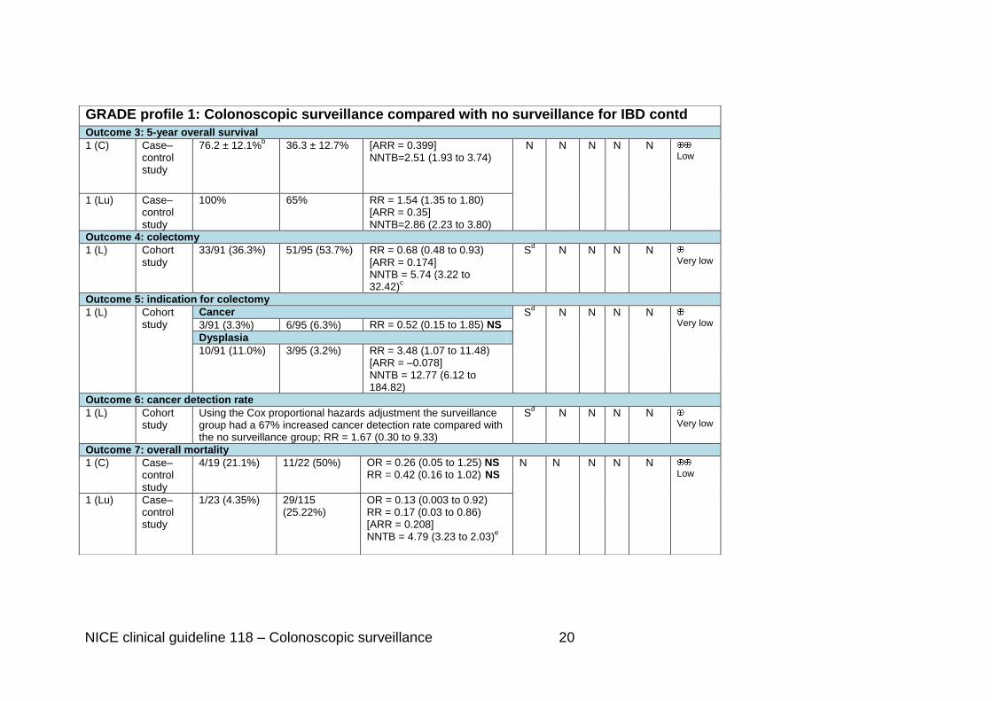
GRADE profile 1: Colonoscopic surveillance compared with no surveillance for IBD
No. of studies
Design Colonoscopic surveillance
No colonoscopic surveillance
OR/RR (95% CI)
[ARR]
NNTB (95% CI)
Lim
itati
on
s
Inc
on
sis
ten
cy
Ind
irectn
ess
Imp
recis
ion
Oth
er
co
ns
ide
rati
on
s Quality
Outcome 1: detected carcinoma at early stage (Duke’s stage A or B; AJCC stage 0 or 1)
1 (C) Case–control study
Dukes’ stage A or B OR = 5.42 (1.14 to 28.95); RR = 1.93 (1.15 to 3.51) [ARR = 0.38] NNTB = 2.63 (1.62 to 13.11)
N N N N N Low
15/19 (79.0%) 9/22 (40.9%)
1 (Lu) Case–control study
AJCC stage 0 or 1 OR = 3.39 (1.21 to 9.45) RR = 2.14 (1.24 to 3.43) [ARR = 0.28] NNTB = 3.60 (2.08 to 14.90)
12/23 (52.2%) 28/115a
(24.3%)
Outcome 2: detected carcinoma at advanced stage (Duke’s stage C or D; AJCC stage 3B–C and 4)
1 (C) Case–control study
Dukes’ stage C or D OR = 0.18 (0.03 to 0.88) RR = 0.36 (0.14 to 0.83) [ARR = 0.38] NNTB = 2.63 (1.62 to 13.11)
N N N N N Low
4/19 (21.1%) 13/22 (59.1%)
1 (Lu) Case–control study
AJCC stage 3B–C and 4 OR = 0.29 (0.07 to 0.97) RR = 0.42 (0.16 to 0.92) [ARR = 0.243] NNTB = 4.12 (2.56 to 35.39)
4/23 (17.4%) 48/115 (41.7%)
NICE clinical guideline 118 – Colonoscopic surveillance 20
GRADE profile 1: Colonoscopic surveillance compared with no surveillance for IBD contd
Outcome 3: 5-year overall survival
1 (C) Case–control study
76.2 ± 12.1%b 36.3 ± 12.7% [ARR = 0.399]
NNTB=2.51 (1.93 to 3.74) N N N N N
Low
1 (Lu) Case–control study
100% 65% RR = 1.54 (1.35 to 1.80) [ARR = 0.35] NNTB=2.86 (2.23 to 3.80)
Outcome 4: colectomy
1 (L) Cohort study
33/91 (36.3%) 51/95 (53.7%) RR = 0.68 (0.48 to 0.93) [ARR = 0.174] NNTB = 5.74 (3.22 to 32.42)
c
Sd N N N N
Very low
Outcome 5: indication for colectomy
1 (L) Cohort study
Cancer Sd N N N N
Very low 3/91 (3.3%) 6/95 (6.3%) RR = 0.52 (0.15 to 1.85) NS
Dysplasia
10/91 (11.0%) 3/95 (3.2%) RR = 3.48 (1.07 to 11.48) [ARR = –0.078] NNTB = 12.77 (6.12 to 184.82)
Outcome 6: cancer detection rate
1 (L) Cohort study
Using the Cox proportional hazards adjustment the surveillance group had a 67% increased cancer detection rate compared with the no surveillance group; RR = 1.67 (0.30 to 9.33)
Sd N N N N
Very low
Outcome 7: overall mortality
1 (C) Case–control study
4/19 (21.1%) 11/22 (50%) OR = 0.26 (0.05 to 1.25) NS RR = 0.42 (0.16 to 1.02) NS
N N N N N Low
1 (Lu) Case–control study
1/23 (4.35%) 29/115 (25.22%)
OR = 0.13 (0.003 to 0.92) RR = 0.17 (0.03 to 0.86) [ARR = 0.208] NNTB = 4.79 (3.23 to 2.03)
e

NICE clinical guideline 118 – Colonoscopic surveillance 21
GRADE profile 1: Colonoscopic surveillance compared with no surveillance for IBD contd.
1 (L) Cohort study
6/91(6.6%) 14/95 (14.7%) RR = 0.45 (0.18 to 1.07) NSf S
d N N N N
Very low
Outcome 8: 5-year CRC-related mortality
1 (Lu) Case–control study
0% 26% [ARR = 0.26 (0.18 to 0.35)] NNTB = 3.85 (2.83 to 5.44)
N N N N N Low
AJCC: American Joint Committee on Cancer; ARR: absolute risk reduction; (C): Choi et al. (1993); CI: confidence interval; CRC: colorectal cancer; IBD: inflammatory bowel disease; (L): Lashner et al. (1990); (Lu): Lutgens et al. (2009); N: not serious; NNTB/H: number needed to treat to benefit/harm; NS: not significant; OR: odds ratio; RR: relative risk; S: serious; VS: very serious; U: upgrade
All evidence found was for people with extensive colitis (ulcerative or Crohn’s colitis) at least 8–10 years after onset of symptoms.
a Lutgens et al. (2009): the tumour stages could not be found for 11 people and so 115 instead of 126 people were studied.
b Choi et al. (1993): the 5-year overall survival rate was 77.2 ± 10.1% for the surveillance group but changed to 76.2 ± 12.1% after
adjusting for (removing) the people in whom colorectal cancer was detected without the surveillance programme. c Lashner et al. (1990): using the Cox proportional hazards model for adjustment, the surveillance group had 47% reduction in
colectomy rate compared with the no surveillance group; RR = 0.53 (0.34 to 0.83). d
Downgraded to serious because some people not receiving surveillance could have had surveillance outside the study. e Lutgens et al. (2009): when the 11 people with simultaneous IBD and CRC diagnosis were excluded.
f Lashner et al. (1990): using the Cox proportional hazards model for adjustment, the surveillance group had 61% reduction in mortality compared with the no surveillance group; RR = 0.39 (0.15 to 1.00).

NICE clinical guideline 118 – Colonoscopic surveillance 22
2.2.3 Evidence statements (see GRADE profile 1)
2.2.3.1 Low quality evidence showed that colonoscopic surveillance
statistically significantly increased the probability of detecting
cancer at an earlier stage. There was a corresponding statistically
significant decrease in the probability of detecting cancer at a later
stage.
2.2.3.2 Low quality evidence found the 5-year overall survival rate to be
statistically significantly higher for the surveillance group.
2.2.3.3 Very low quality evidence showed a statistically significantly lower
rate of colectomy in the surveillance group.
2.2.3.4 Very low quality evidence showed that cancer was a more frequent
indication for colectomy in the no surveillance group compared with
the surveillance group, but the difference was not significant.
2.2.3.5 Very low quality evidence showed that dysplasia was a more
frequent indication for colectomy in the surveillance group
compared with the no surveillance group. This difference was
statistically significant.
2.2.3.6 Very low quality evidence found a statistically significantly
increased cancer detection rate in the surveillance group compared
with the no surveillance group after adjustment for covariates by
the Cox proportional hazards model.
2.2.3.7 Low quality evidence showed a tendency for a higher overall
mortality rate for the no surveillance group compared with the
surveillance group.
2.2.3.8 Low quality evidence found the 5-year colorectal cancer related
mortality rate to be statistically significantly higher for the no
surveillance group compared with the surveillance group.

NICE clinical guideline 118 – Colonoscopic surveillance 23
2.2.4 Health economic modelling
No cost-effectiveness studies were found that specifically examined
colonoscopic surveillance for the prevention of colorectal cancer in people
with IBD. However, three studies were found that examined colonoscopic
surveillance in people with ulcerative colitis (Nguyen et al. 2009; Provenzale
et al. 1995; Delco et al. 2000). All three studies explored approaches to
modelling strategies such as decision tree versus Markov models, and when
applicable, informed the model structure. Given the absence of any
appropriate analysis that addressed the decision problem directly, a new cost-
effectiveness model was developed based on the views of the GDG and
clinical data available at the time of guideline development.
A major component of the model is the natural history of dysplasia, because
dysplasia is a precancerous marker for colorectal cancer. During the evidence
review, the presence of any grade of dysplasia in the past 5 years was found
to be one of the risk factors for the high-risk group (please see the evidence to
recommendations section 2.2.5). Therefore, the model could only determine
the cost effectiveness of surveillance for the high-risk group because
dysplasia was only included as a risk factor in this group (please see the care
pathway, people with IBD). Furthermore, it was not possible to construct an
additional cost-effectiveness model or carry out an economic evaluation for
the low- and intermediate-risk groups because of the lack of natural history
data.
The model included men and women aged 30–85 who had flat dysplastic
lesions (that is, non-resectable low- or high-grade dysplasia) who had
declined surgery. The analysis was run over a 55-year time horizon (cycle
length 3 months) and examined the use of colonoscopic surveillance
compared with no surveillance. Evidence that colonoscopic surveillance was
effective required a reduction in colorectal cancer related mortality.
The model split the single state of dysplasia into two mutually exclusive states
of low-grade and high-grade dysplasia. Similarly, the single colorectal cancer
state was broken down into four mutually exclusive states of Dukes’ A, Dukes’

NICE clinical guideline 118 – Colonoscopic surveillance 24
B, Dukes’ C and Dukes’ D colorectal cancer. Any other cause of mortality was
considered in all states in the model (see figure 1).
Figure 1: Colonoscopic surveillance model for people with IBD in the
high-risk group
LGD: low-grade dysplasia; HGD: high-grade dysplasia; DA: Dukes’ A; DB: Dukes’ B; DC:
Dukes’ C; DD: Dukes’ D; CRC: colorectal cancer
Colonoscopic surveillance is recommended every year in the high-risk group
(every fourth cycle in the model) and it was assumed that colonoscopy was
carried out at the beginning of the scheduled cycle. In the model, the
development of colorectal cancer could be sequential, that is, progress from
low-grade to high-grade dysplasia to cancer, or from low-grade dysplasia
directly to colorectal cancer because some people do not progress through a
detectable phase of high-grade dysplasia. People with high-grade dysplasia
could also progress directly to colorectal cancer and were assumed not to
regress to low-grade dysplasia. Progression to colorectal cancer could occur
either asymptomatically or symptomatically between the scheduled
surveillance colonoscopies. Over time, if people in the three risk groups had
no evidence of progression they would remain in the same health state.

NICE clinical guideline 118 – Colonoscopic surveillance 25
The natural history of the progression of IBD to colorectal cancer is unknown.
Therefore, the probabilities of moving from one health state to another were
based on a published clinical study that examined colonoscopic surveillance
for colorectal cancer in UK patients with ulcerative colitis (Rutter et al. 2006)
and from a published cost-effectiveness study by Tappenden et al. (2004).
The transition probabilities from both studies were calculated using a
Bayesian dirichlet method. Details are presented in appendix 7. The model
assumed there were no complications from colonoscopy – although
perforation and bleeding are serious risks, they occur infrequently and were
assumed to be negligible.
Utility values (quality of life benefits) were not available for all the health
states. Several studies reported utility values obtained from a disease-specific
questionnaire (the Inflammatory Bowel Disease Questionnaire). However
these values could not be used for calculating quality-adjusted life years
(QALYs) because they did not report the values on a 0–1 scale, which is the
format for generic questionnaires. Therefore, the utility values for people with
low- and high-grade dysplasia were taken from a study of people with Crohn’s
disease (based on disease severity using a time trade off method; Gregor et
al. 1997). The GDG confirmed that this approach was acceptable; a person
with low-grade dysplasia has a lower quality of life than a person in the
general population and a person with high-grade dysplasia has a lower quality
of life than a person with low-grade dysplasia. Stage-specific utility values for
people with colorectal cancer were obtained from Ness et al. (1999).
Colonoscopic surveillance costs were obtained from NHS reference costs and
the GDG. The costs for the lifetime stage-specific treatment of colorectal
cancer were uplifted to incorporate the relevant NICE guidance published
since 2004 (personal communication with Paul Tappenden and Hazel Pilgrim,
8 April 2010). Full details of utility values and costs are presented in appendix
7.
Both deterministic (base case using only point estimates) and probabilistic
sensitivity analyses (using a range of values and simulations to take into
account uncertainty) were conducted to examine cost effectiveness.

NICE clinical guideline 118 – Colonoscopic surveillance 26
The overall deterministic results are presented in table 2 and more detailed
results are given in appendix 7.
Table 2: Deterministic analysis over a 55-year period
QALYs Cost (£) Incremental QALYs
Incremental cost (£) ICER (£)
No surveillance 16.42 2320.44
Surveillance – high-risk group only 17.19 15,785.13 0.77 13,464.69 17,557.32
QALY: quality-adjusted life year; ICER: incremental cost-effectiveness ratio
The analysis suggested that surveillance for the high-risk group is cost
effective.
The overall probabilistic sensitivity analysis results are presented in table 3
and more detailed results are given in appendix 7.
Table 3: Probabilistic sensitivity analysis over a 55-year period
QALYs Costs (£) Incremental QALYs
Incremental costs (£) ICER (£)
Probability of being cost effective at £20,000 per QALY gained (%)
No surveillance 13.04 7368.92 – – – –
Surveillance – high-risk group only
14.64 16,316.82 1.61 8947.90 5571.44 99
QALY: quality-adjusted life year; ICER: incremental cost-effectiveness ratio
The incremental cost-effectiveness ratio (ICER) from the probabilistic
sensitivity analysis was lower than the ICER from the deterministic sensitivity
analysis. This suggests that there may be a high degree of uncertainty
associated with some model parameters, which resulted in a large change in
the ICER. However, in spite of the uncertainty the probabilistic sensitivity
analysis suggests that there is a 99% probability that colonoscopic

NICE clinical guideline 118 – Colonoscopic surveillance 27
surveillance for the high-risk group (among the three risk groups) with IBD is
cost effective at the usual threshold of £20,000 per QALY gained.
2.2.5 Evidence to recommendations
The GDG considered that although the quality of the evidence was very low to
low, there was still evidence in favour of colonoscopic surveillance compared
with no surveillance for people with IBD. The GDG also felt that it would not
be possible to find RCT evidence for this review question because it would be
unethical to randomise people with IBD to have no colonoscopic surveillance.
The GDG also considered that the evidence obtained was sufficient to make
recommendations in favour of colonoscopic surveillance, and that because of
the similar colorectal cancer risk in ulcerative colitis and Crohn's colitis (Choi
and Zelig 1994) recommendations could be made for Crohn's colitis despite
most of the evidence being for people with ulcerative colitis. There was also
some discussion about the evidence potentially showing lead-time bias, with
early detection achieved because of colonoscopic surveillance. This would
improve 5-year survival but not overall survival. However, Lutgens et al.
(2009) showed a significant difference in the 5-year cancer-related mortality
rates in people undergoing surveillance compared with no surveillance, which
does not support the effect of lead-time bias.
The health economic modelling indicated that colonoscopic surveillance is a
cost-effective use of resources for people with any grade of dysplasia in the
past 5 years. Because people with any grade of dysplasia share a similar risk
of developing colorectal cancer as people in the ‘high-risk’ group, specifically
those who have extensive ulcerative or Crohn’s colitis with moderate or
severe active inflammation that has been confirmed histologically, or primary
sclerosing cholangitis (including after liver transplant), or colonic stricture in
the past 5 years, or a family history of colorectal cancer in a first-degree
relative aged under 50 years, the cost-effectiveness results could be
extrapolated to the high-risk group. The GDG acknowledged that, given the
quality of the data and the number of simplifying assumptions in the model,
the results were exploratory. The assumptions covering compliance and
complications would increase uncertainty in the ICERs and could potentially

NICE clinical guideline 118 – Colonoscopic surveillance 28
increase them. The GDG also felt that because all the studies included for this
review question looked at people who had IBD for at least 10 years, it would
be appropriate only to offer surveillance to people 10 years after symptom
onset.
2.2.6 Recommendations
Recommendation 1.1.1
Offer colonoscopic surveillance to people with inflammatory bowel disease
(IBD) whose symptoms started 10 years ago and who have:
ulcerative colitis (but not proctitis alone) or
Crohn’s colitis involving more than one segment of colon.
Clinical effectiveness of colonoscopic surveillance compared with no
surveillance in people with adenomas
2.2.7 Evidence review
A total of 9688 articles were found by systematic searches, of which 6533
were unique articles. Overall, two studies met the eligibility criteria (for the
review protocol and inclusion and exclusion criteria, see appendices 2 and 4)
and examined the effectiveness of colonoscopic surveillance compared with
no surveillance. The two studies were initially considered to be relevant, but
were later considered by the GDG not to provide relevant evidence of the
benefits of colonoscopic surveillance. In Thiis-Evensen (1999a) people had
flexible sigmoidoscopy, and on discovering polyps, they were offered
colonoscopic polypectomy. In Jorgensen (1993) an indirect comparison was
made. Mortality rates were compared in people offered colonoscopic
surveillance and in people who had died from colorectal cancer in Denmark,
with data taken from the cancer registry.
Therefore, no evidence meeting the eligibility criteria was identified for this
group.

NICE clinical guideline 118 – Colonoscopic surveillance 29
2.2.8 Evidence statement
2.2.8.1 There was no high-quality evidence for or against colonoscopic
surveillance for the prevention and early detection of colorectal
cancer after adenoma removal.
2.2.9 Health economic modelling
A search for cost-effectiveness studies found no directly relevant studies for
colonoscopic surveillance and one related analysis (Tappenden et al. 2004).
There was no direct evidence demonstrating the clinical effectiveness of
colonoscopic surveillance after adenoma removal in reducing colorectal
cancer mortality. However, one observational study reported a 70–90% lower
than expected incidence of colorectal cancer in people undergoing
colonoscopic surveillance compared with the reference populations (Winawer
et al. 1993a). A full systematic review of the literature was not possible
because of time constraints. Existing economic models, including screening
and surveillance, were examined. Information about the natural history of
undetected colorectal cancer, the related probabilities of progressing through
undiagnosed cancer states and the probabilities of clinical presentation by
cancer stage were obtained from Tappenden et al. (2004). Tappenden et al.
obtained estimates from two sources: the National Polyp Study (Winawer et
al. 1993a) and calibrating their model against published incidence and
mortality data. Because these data were based on the model constructed by
Tappenden et al. (2004) and not directly on clinical evidence, they were highly
dependent on the structure of the model. This means that transferring these
estimates to another model may have resulted in inconsistencies and
uncertainty.
A Markov model was developed based on Tappenden et al. (2004) and is
presented in figure 2. It included 50-year old men and women who had
adenomas removed at baseline colonoscopy. The analysis was run over a 50-
year time horizon. In the model, three strategies were examined: no
surveillance, surveillance in all risk groups (low-, intermediate- and high-risk
groups), and surveillance in intermediate- and high-risk groups only. Detection

NICE clinical guideline 118 – Colonoscopic surveillance 30
rates of early cancer (Dukes’ A and Dukes’ B colorectal cancer) leading to
mortality from the disease were considered using lifetime treatment costs for
colorectal cancer in each strategy.
In the model, colonoscopic surveillance after adenoma removal is consistent
with the current BSG guidelines (Cairns et al. 2010). The person’s risk state is
determined after the baseline colonoscopy and is based on the number and
size of adenomas removed. People are offered colonoscopic surveillance
based on their risk state, as follows:
Low risk: consider colonoscopy at 5 years:
if the colonoscopy is negative (that is, no adenomas are found) stop
surveillance
if low risk, consider the next colonoscopy at 5 years (with follow-up
surveillance as for low risk)
if intermediate risk, offer the next colonoscopy at 3 years (with follow-up
surveillance as for intermediate risk)
if high risk, offer the next colonoscopy at 1 year (with follow-up
surveillance as for high risk).
Intermediate risk: offer colonoscopy at 3 years:
if the colonoscopy is negative offer the next colonoscopy at 3 years.
Stop surveillance if there is a further negative result
if low or intermediate risk, offer the next colonoscopy at 3 years (with
follow-up surveillance as for intermediate risk)
if high risk, offer the next colonoscopy at 1 year (with follow-up
surveillance as for high risk).
High risk: offer colonoscopy at 1 year.
if the colonoscopy is negative, or low or intermediate risk, offer the next
colonoscopy at 3 years (with follow-up surveillance as for intermediate
risk)
if high risk, offer the next colonoscopy at 1 year (with follow-up
surveillance as for high risk).

NICE clinical guideline 118 – Colonoscopic surveillance 31
Figure 2: Colonoscopic surveillance model for people with adenomas
NAA AAi DA DB DC DD
Other Cause Mortality
Exit surveillance CRCMortality
AAh
NAA: non-advanced adenoma, low risk, AAi: advanced adenoma, intermediate risk; AAh: advanced adenoma, high risk; DA: Dukes’ A; DB: Dukes’ B; DC: Dukes’ C; DD: Dukes’ D; CRC: colorectal cancer
In the model people were grouped into a finite number of Markov states, and
all events or progression are represented as transitions from one state to
another with a certain probability. Estimated transition probabilities were
assumed to be constant with the exception of age-related adenoma incidence
(Tappenden et al. 2004) and age-specific mortality rates, which were taken
from published government sources. The effectiveness of colonoscopic
surveillance was modelled as an intervention under near-perfect conditions to
determine whether colonoscopic surveillance using colonoscopy for the early
detection of adenomas and colorectal cancer is clinically and cost effective
compared with no surveillance.
The three diagnostic states in the model, low, intermediate and high risk, differ
only in terms of the surveillance offered. Movement between diagnostic states
is only possible through surveillance using tunnel states or symptomatic
presentation of colorectal cancer. According to surveillance criteria people can
drop out of surveillance and can be assumed to return to UK population
norms. Any newly developed adenomas will be removed during surveillance.
If any lesions are confirmed to be malignant, the surveillance programme will
be stopped and the person referred for appropriate diagnosis and treatment.
Empirical evidence strongly suggests that people with a history of adenomas
are more likely to develop them in the future than people who have never had

NICE clinical guideline 118 – Colonoscopic surveillance 32
adenomas (Winawer et al. 1993a). It was assumed in the model that all
colorectal cancers arise from pre-existing adenomas, and the GDG
considered this assumption to be appropriate. For the purpose of the
guideline, when comparing a surveillance programme with no surveillance, the
sensitivity and specificity of colonoscopy were assumed to be 100% for
adenoma detection. This was agreed with the GDG. Utility values for health
states and treatment were obtained from published studies. Data on stage-
specific utility values for colorectal cancer were limited and no EQ-5D values
were available. Utility values were assessed in relation to the stage of cancer
and treatment (Ness et al. 1999, 2000). The GDG agreed with the assumption
that the utility values for people who are cancer free or have undiagnosed
(asymptomatic) cancer would be the same as those for the general
population. Surveillance costs were obtained from NHS reference costs.
Costs for the stage-specific lifetime treatment of colorectal cancer were based
on Tappenden et al. (2004) (personal communication with Paul Tappenden
and Hazel Pilgrim, 8 April 2010). Full details of the utility values and costs are
presented in appendix 8.
The overall deterministic results are presented in table 4 and more detailed
results are given in appendix 8. These results indicate that the most cost-
effective surveillance strategy is to include surveillance for low-, intermediate-
and high-risk groups with an ICER below the usual threshold (£20,000 per
QALY gained). Surveillance of only the intermediate- and high-risk groups
was associated with an ICER below £20,000 per QALY gained and the
incremental costs were lower than the strategy that includes the low-risk
group, but the potential gain in QALYs was also lower. However, these results
are highly sensitive to the natural history data, which were extrapolated from
another model and are highly uncertain. Therefore, the results should be
interpreted with caution.

NICE clinical guideline 118 – Colonoscopic surveillance 33
Table 4: Deterministic analysis over a 50-year period
QALYs
Costs (£)
Incremental QALYs
Incremental costs (£) ICER (£)
No surveillance 16.11 641.06 – – –
Colonoscopic surveillance in intermediate- and high-risk groups
16.16 841.54 0.05 200.49 4235.75
Colonoscopic surveillance in all risk groups
16.26 1177.03
0.15 535.97 3669.70
QALY: quality-adjusted life year; ICER: incremental cost-effectiveness ratio; all risk groups: low-, intermediate- and high-risk groups.
The overall probabilistic sensitivity analysis results are presented in table 5
and more detailed results are given in appendix 8. The analysis shows that
the results are consistent with the deterministic analysis.
Table 5: Probabilistic sensitivity analysis over a 50-year period
QALYs Costs (£)
Incremental QALYs
Incremental costs (£) ICER (£)
Probability of being cost effective at £20,000 per QALY gained
No surveillance 16.12 562.91 – – – –
Colonoscopic surveillance in intermediate- and high-risk groups
16.17 786.25 0.04 223.33 5298.03 78
Colonoscopic surveillance in all risk groups
16.25 1167.77 0.13 604.85 4626.57 81
QALY: quality-adjusted life year; ICER: incremental cost-effectiveness ratio; all risk groups: low-, intermediate- and high-risk groups.
The probabilistic sensitivity analysis suggests that the probability of
colonoscopic surveillance being cost effective at £20,000 per QALY gained is
78% in the intermediate- and high-risk groups only and 81% in all risk groups
(including the low-risk group).

NICE clinical guideline 118 – Colonoscopic surveillance 34
The GDG acknowledged that, given the quality of the data and the number of
simplifying assumptions in the model, the results were exploratory. The
assumptions covering compliance and complications would result in an
increase in uncertainty in the ICERs and could potentially increase them.
Details of the cost-effectiveness analysis are discussed in appendix 8.
2.2.10 Evidence to recommendations
Because of the lack of evidence, the GDG made recommendations based on
experience, and the colorectal cancer incidence and overall mortality reported
in Thiis-Evensen (1999a) and Jorgensen (1993). These articles showed that
the risk of colorectal cancer in people with adenomas in the low-risk group is
similar to that of the general population. For evidence on the risk categories
used in the recommendation, see section 2.5.7.
The GDG noted that carrying out colonoscopic surveillance in all risk groups
was the most cost-effective strategy according to the deterministic and
probabilistic sensitivity analysis results. The GDG noted that the modelling did
not consider issues around compliance, colonoscopy-related complications or
the sensitivity or specificity of colonoscopy. However, the GDG did not
consider that these factors would increase the results beyond the thresholds
that are considered cost effective. The GDG discussed the uncertainty around
the clinical benefits of surveillance in the low-risk group. The GDG considered
the balance of the potential risks of perforation and bleeding associated with
colonoscopy and removal of adenomas and the benefits of detecting
colorectal cancer in the low-risk population. The GDG considered that there
were potential groups of people in the low-risk group who could benefit from
surveillance. Therefore, the GDG concluded that surveillance should be
considered in this group. However the GDG highlighted that clinical judgement
should be used when considering people’s comorbidities and the potential
risks of bleeding and perforation for each colonoscopy.

NICE clinical guideline 118 – Colonoscopic surveillance 35
2.2.11 Recommendations
Recommendation 1.1.6
Consider colonoscopic surveillance for people who have had adenomas
removed and are at low risk of developing colorectal cancer (see table 2).
Recommendation 1.1.7
Offer colonoscopic surveillance to people who have had adenomas removed
and are at intermediate or high risk of developing colorectal cancer (see table
2).
Table 2 Risk of developing colorectal cancer in people with adenomas
Low risk:
one or two adenomas smaller than 10 mm.
Intermediate risk:
three or four adenomas smaller than 10 mm or
one or two adenomas if one is 10 mm or larger.
High risk:
five or more adenomas smaller than 10 mm or
three or more adenomas if one is 10 mm or larger.

NICE clinical guideline 118 – Colonoscopic surveillance 36
2.3 Colonoscopic surveillance techniques
2.3.1 Review question
Which colonoscopic surveillance technique (conventional colonoscopy or
chromoscopy) for prevention and/or early detection of colorectal cancer in
adults with IBD or adenomas is more clinically effective compared with other
methods of surveillance (flexible sigmoidoscopy, double-contrast barium
enema, computed tomographic [CT] colonography, tri-modal imaging [high-
resolution white light endoscopy, narrow-band imaging, and auto-fluorescence
imaging])?
Colonoscopic surveillance techniques in people with IBD
2.3.2 Evidence review
A total of 14,701 articles were found by systematic searches, of which 9544
were unique articles. The full text was ordered for 108 articles. One study
(Dekker et al. 2007) met the eligibility criteria (for the review protocol and
inclusion and exclusion criteria, see appendices 2 and 4).
The characteristics of the primary study are summarised in table 6 and the
evidence is reviewed in GRADE profile 2.
Table 6: Summary of study characteristics
Study Population Study characteristics
Outcomes used for GRADE profile
Dekker et al. (2007)
Forty-two patients with ulcerative colitis of long duration (mean duration 21 ± 8.6 years). The study group comprised 31 men and 11 women with a mean age ± SD of 50 ± 11.2 years
Prospective RCT: cross-over study design (that is, the 42 patients in the study received both procedures)
Detection of neoplastic lesion with narrow-band imaging compared with conventional colonoscopy
RCT: randomised controlled trial; SD: standard deviation

NICE clinical guideline 118 – Colonoscopic surveillance 37
GRADE profile 2: Conventional colonoscopy compared with narrow-band imaging
No. of studies
Design Conventional colonoscopy
Other technique
SN
Lim
ita
tio
ns
Inc
on
sis
ten
cy
Ind
ire
ctn
es
s
Imp
rec
isio
n
Oth
er
co
ns
ide
rati
on
s
Quality
NBI versus conventional colonoscopy for inflammatory bowel disease
Primary outcome:
1 (D) RCT (the 42 patients in the study received both procedures)
7/42
(19%)
8/42
(17%)
SN for NBI = 67%
N N N N S Moderatea
(D): Dekker et al. (2007); N: not serious; NBI: narrow-band imaging; RCT: randomised controlled trial; S: serious; SN: sensitivity
a The study did not contain a predefined sample size and therefore included only 42 people. A first-generation
prototype NBI system with an experimental light source was used.
2.3.3 Evidence statements (see GRADE profile 2)
2.3.3.1 Moderate quality evidence comparing narrow-band imaging with
conventional colonoscopy in people with ulcerative colitis of long
duration showed no significant difference in the number of
neoplastic lesions detected between the two techniques.
2.3.4 Health economic modelling
No health economic modelling was undertaken for this review question.
2.3.5 Evidence to recommendations
The GDG agreed that the Dekker et al. (2007) study was underpowered, that
is, the sample size was small and not a true representation of people with
IBD. In addition, narrow-band imaging is not routinely used for colonoscopic
surveillance in the UK. Therefore the GDG considered that it was not possible
to recommend narrow-band imaging for people with IBD.
2.3.6 Recommendations
No recommendations were made on the use of other surveillance methods for
people with IBD (see Evidence to recommendations for details).

NICE clinical guideline 118 – Colonoscopic surveillance 38
Colonoscopic surveillance techniques in people with adenomas
2.3.7 Evidence review
A total of 14,701 articles were found by systematic searches, of which 9544
were unique articles. The full text was ordered for 108 articles. Two primary
studies (Rex et al. 1995, Winawer et al. 2000) and two systematic reviews
(Van den Broek et al. 2009, Mulhall et al. 2005) that looked at the
effectiveness of conventional colonoscopy compared with narrow-band
imaging, double-contrast barium enema, CT colonography and flexible
sigmoidoscopy for surveillance for adenomas met the inclusion and exclusion
criteria (for the review protocol and inclusion and exclusion criteria, see
appendices 2 and 4).
The characteristics of the included studies are summarised in table 7 and the
evidence is reviewed in GRADE profile 3. The forest plots for the meta-
analysis of outcomes and a detailed evidence table for the two systematic
reviews are given in appendix 6.

NICE clinical guideline 118 – Colonoscopic surveillance 39
Table 7: Summary of study characteristics
Study Population Study characteristics
Outcomes used for GRADE profile
Van den Broek et al. (2009)
A pooled result of 537 people undergoing NBI compared with 536 people having conventional colonoscopy
Systematic review of three RCTs: NBI compared with conventional colonoscopy (white light endoscopy)
Detection and removal of adenomas with NBI compared with conventional colonoscopy
Rex et al. (1995) 149 people aged at least 40 years (mean age 63) with symptoms suggestive of colonic disease
RCT comparing flexible sigmoidoscopy plus double contrast barium enema with colonoscopy
Adenoma detection
Mulhall et al. (2005)
Prospective studies of adults undergoing CTC after full bowel preparation, with colonoscopy as the gold standard.
33 studies provided data on 6393 people
Systematic review and meta-analysis of CTC
Diagnostic efficacy of CTC in detecting adenomas, pooled sensitivity and specificity for polyp detection
Winawer et al. (2000)
973 people underwent one or more surveillance colonoscopies. In 580 of these people, 862 paired surveillance colonoscopies and double-contrast barium enema were performed
Controlled trial comparing colonoscopy and double-contrast barium enema with colonoscopy without barium enema
Adenoma detection
CTC: computed tomographic colonography; NBI: narrow-band imaging; RCT: randomised controlled trial

NICE clinical guideline 118 – Colonoscopic surveillance 40
GRADE profile 3: Conventional colonoscopy compared with double-contrast barium enema, flexible sigmoidoscopy, narrow-band imaging and CT colonography
No. of studies Design Conventional colonoscopy
Other technique
OR (95% CI)
Sensitivity
Specificity
p value
Lim
ita
tio
ns
Inc
on
sis
ten
cy
Ind
irec
tne
ss
Imp
rec
isio
n
Oth
er
co
ns
ide
rati
on
s
Quality
NBI versus conventional colonoscopy for adenomas
Primary outcome: detection and removal of adenomas
1 (V) Systematic review/meta-analysis
236/537
(44%)
219/536
(41%)
OR = 1.19 (95% CI 0.86 to 1.64) NS
N N N N N High
FSIG plus DCBE versus conventional colonoscopy for adenomas
Primary outcome: adenoma detection
1 (R) RCT 23/75
(31%)
13/74 (18%) OR = 2.07 (95% CI 0.90 to 4.92) NS
N N N N S Moderateb
CTC versus conventional colonoscopy for adenomas
Primary outcome: adenoma detection
1 (M) Systematic review/meta analysis
33 studies providing data on 6393 people
Pooled sensitivity for CTC = 70% (95% CI 53% to 87%).
Pooled specificity for CTC = 86% (95% CI 84% to 88%; p = 0.001). Sensitivity and specificity increased as polyp size increased.
N N N N S Moderatec

NICE clinical guideline 118 – Colonoscopic surveillance 41
GRADE profile 3 contd.
DCBE versus conventional colonoscopy for adenomas
Primary outcome: adenoma detection
1 (W) Controlled trial
Polyps were detected in 392 of the 862 colonoscopic examinations (45%); adenomas were detected in 242 colonoscopic examinations (28%). Findings on barium enema were positive in 222 of the 862 paired examinations (26%) and in 139 (35%) of the 392 colonoscopic examinations in which one or more polyps were detected.
N N N N S Low
CI: confidence interval; CTC: computed tomographic colonography; DCBE: double-contrast barium enema; FSIG: flexible sigmoidoscopy; IBD: inflammatory bowel disease; (M): Mulhall et al. (2005); N: not serious; NBI: narrow-band imaging; NS: not significant; OR: odds ratio; (R): Rex et al. (1995); RCT:
randomised controlled trial; S: serious; (V): Van den Broek et al. (2009); (W): Winawer et al. (2000)
a The study did not contain a predefined sample size and therefore included only 42 people. A first-generation prototype NBI system with an experimental light
source was used.
b Downgraded based on small sample size.
c Eighteen of the studies used colonoscopy as the gold standard. Eleven studies used segmental unblinded colonoscopy or optimised colonoscopy.

NICE clinical guideline 118 – Colonoscopic surveillance 42
2.3.8 Evidence statements (see GRADE profile 3)
2.3.8.1 High quality evidence comparing narrow-band imaging with
colonoscopy (white light endoscopy) to detect adenomas showed
that narrow-band imaging did not significantly improve the detection
of adenomas.
2.3.8.2 Moderate quality evidence showed a non-significant two-fold
increase in adenoma detection rate with conventional colonoscopy
compared with flexible sigmoidoscopy plus double-contrast barium
enema.
2.3.8.3 Moderate quality evidence showed that computed tomographic
(CT) colonography was highly specific for polyps larger than 9 mm.
This evidence also showed that sensitivity for CT colonography
increased with polyp size.
2.3.8.4 Low quality evidence showed that colonoscopic examination
detected more polyps than double-contrast barium enema. Half of
these polyps were adenomas, and the remainder were primarily
normal mucosal tags, with some hyperplastic polyps.
2.3.9 Health economic modelling
No health economic modelling was undertaken for this review question.
2.3.10 Evidence to recommendations
Overall, the evidence was considered to be limited. The GDG agreed that the
Rex (1995) study was underpowered, that is, the sample size was small and
not a true representation of people with adenomas.
The GDG agreed that conventional colonoscopy (high-resolution white-light
endoscopy) should be used for routine colonoscopic surveillance in people
with adenomas because of its increased detection rate of adenomas
compared with other techniques.
The GDG noted that there was ongoing research comparing CT colonography
with conventional colonoscopy. It considered that when conventional

NICE clinical guideline 118 – Colonoscopic surveillance 43
colonoscopy was contraindicated (for example if it was inappropriate because
of comorbidity or could not be tolerated) or incomplete, CT colonography
should be considered as an alternative for a single examination. This was
because of the high level of specificity of CT colonography for large polyps
(larger than 9 mm). Double-contrast barium enema should be considered
when CT colonograpy is not appropriate.
The GDG noted that there was no evidence to support the safety and efficacy
of CT colonography as a routine surveillance technique. Because CT
colonography and double-contrast barium enema were not evaluated in the
reviewed evidence, the GDG considered that any decision about using these
methods as alternative surveillance techniques should be made on a case-by-
case basis.
The GDG also recognised the significant inter-operator variability of
colonoscopy. It recommended that if a colonoscopy is incomplete, a repeat
colonoscopy should be undertaken, with a more experienced colonoscopist if
appropriate.

NICE clinical guideline 118 – Colonoscopic surveillance 44
2.3.11 Recommendations
Recommendation 1.1.10
Offer a repeat colonoscopy if any colonoscopy is incomplete. Consider
whether a more experienced colonoscopist is needed.
Recommendation 1.1.11
Consider computed tomographic colonography3 (CTC) as a single
examination if colonoscopy is not clinically appropriate (for example, because
of comorbidity or because colonoscopy cannot be tolerated).
Recommendation 1.1.12
Consider double contrast barium enema as a single examination if CTC is not
available or not appropriate.
Recommendation 1.1.13
Consider CTC or double contrast barium enema for ongoing surveillance if
colonoscopy remains clinically inappropriate, but discuss the risks and
benefits with the person and their family or carers.
2.4 Conventional colonoscopy compared with
chromoscopy
2.4.1 Review question
Is colonoscopic surveillance with a dye (chromoscopy) for prevention and/or
early detection of colorectal cancer clinically effective compared with
colonoscopic surveillance without a dye (conventional colonoscopy)?
3 Computed tomographic colonography (virtual colonoscopy). NICE interventional procedure
guidance 129 (2005).

NICE clinical guideline 118 – Colonoscopic surveillance 45
Conventional colonoscopy compared with chromoscopy in people with
IBD
2.4.2 Evidence review
A total of 14,701 articles were found by systematic searches, of which 9544
were unique articles. The full text was ordered for 23 articles. Only four
studies examined the effectiveness of chromoscopy compared with
conventional colonoscopy for IBD and met the eligibility criteria (for the review
protocol and inclusion and exclusion criteria, see appendices 2 and 4). The
four primary studies were Kiesslich et al. (2003, 2007), Marion et al. (2008)
and Rutter et al. (2004a).
The characteristics of the included primary studies are summarised in table 8
and the evidence reviewed in GRADE profile 4. For this review question the
histopathology of the adenomas at presentation could not be determined by
the surveillance technique, so the numbers and types of lesions were used
because of their relevance to the long-term clinical outcomes. The GDG
agreed that any statistically significant improvement in detection would be
used for the imprecision calculation.
The forest plots for the meta-analysis of outcomes and the detailed evidence
tables for the included studies are given in appendix 6. The meta-analysis of
the dichotomous outcomes used the pooled odds ratio calculated by the
Mantel-Haenszel fixed-effects model because the heterogeneity was less than
50%. Subgroup analysis was performed when appropriate.

NICE clinical guideline 118 – Colonoscopic surveillance 46
Table 8: Summary of study characteristics
Study Population Intervention Comparator Outcomes used for GRADE profile
Kiesslich et al. (2003) RCT
People with clinically inactive, ulcerative colitis (of at least 8 years’ duration), N = 165
Chromoscopy using 0.1% methylene blue, n = 84
Conventional colonoscopy, using conventional video colonoscopy, n = 81
Total number of neoplastic lesions, number of LGD, HGD and flat neoplastic lesions detected, and number of people with neoplastic lesions
Kiesslich et al. (2007) RCT
People with clinically inactive, ulcerative colitis (of at least 8 years’ duration), N = 161; 8 people were excluded because of insufficient bowel preparation, therefore N=153
Chromoscopy using 0.1% methylene blue with endomicroscopy n = 80
Conventional colonoscopy, using conventional video colonoscopy, n = 73
Total number of neoplastic lesions, number of LGD, HGD and flat neoplastic lesions detected and number of people with neoplastic lesions
Marion et al. (2008) Back-to-back controlled trial
People with extensive ulcerative colitis (at least left sided, n = 79) or Crohn's colitis (at least one third of the colon, n = 23), N = 102
Chromoscopy using 0.1% methylene blue, n = 102
Conventional colonoscopy, n = 102, targeted and random
Total number of neoplastic lesions, number of LGD, HGD and flat neoplastic lesions detected and number of people with neoplastic lesions
Rutter et al. (2004a) Back-to-back controlled trial
People with extensive ulcerative colitis of long duration, N = 100
Chromoscopy with 0.1% indigo carmine, n = 100
Conventional colonoscopy, n = 100, targeted and random
Total number of neoplastic lesions, number of LGD lesions detected and number of people with neoplastic lesions
RCT: randomised controlled trial; HGD: high-grade dysplasia; LGD: low-grade dysplasia

NICE clinical guideline 118 – Colonoscopic surveillance 47
GRADE profile 4: Chromoscopy compared with conventional colonoscopy for IBD
No. of studies
Design Chromoscopy Conventional colonoscopy
OR M-H, fixed (95%CI)
Lim
itati
on
s
Inc
on
sis
ten
cy
Ind
irectn
ess
Imp
recis
ion
Oth
er
co
ns
ide
rati
on
Quality
Outcome 1: Total number of people with intra-epithelial neoplasia detected
4a RCT/CT 48/366
(13.11%) 23/356 (6.46%)
OR = 2.21 (1.31 to 3.74) N N N N N High
CI: confidence interval; CT: controlled trial; M-H fixed: Mantel-Haenszel fixed-effects model; N: not serious; OR: odds ratio; RCT: randomised controlled trial
a Kiesslich et al. (2003, 2007), Marion et al. (2008) and Rutter et al. (2004a)

NICE clinical guideline 118 – Colonoscopic surveillance 48
2.4.3 Evidence statements (see GRADE profile 4) 1
2.4.3.1 High quality evidence showed that chromoscopy detected 2
statistically significantly more intra-epithelial dysplastic lesions in 3
people with extensive colitis (at least 8 years’ duration) compared 4
with conventional colonoscopy. 5
2.4.4 Health economic modelling 6
No health economic modelling was undertaken for this review question. 7
2.4.5 Evidence to recommendations 8
See section 2.5.2 for the evidence on risk categories. 9
The GDG felt that the high quality evidence supported the use of 10
chromoscopy compared with conventional colonoscopy. The GDG discussed 11
that using chromoscopy instead of colonoscopy would increase the procedure 12
time, and in practice, four mapping biopsies (to determine the extent of 13
inflammation) and on average one targeted biopsy would be taken when using 14
chromoscopy. The GDG noted that no health economic analysis was 15
conducted for this question and there may be additional costs from the use of 16
dye and the extra time needed for the procedure. However, the GDG also 17
acknowledged that fewer biopsies would be needed, which could lead to 18
reduced pathology cost and time. Therefore, the costs could potentially 19
equalise. In addition, it noted that currently the NHS uses the same health 20
resource group (HRG) code ‘FZ26A Endoscopic or Intermediate Large 21
Intestine Procedure 19 years and over’ for both procedures (Department of 22
Health 2010). The GDG considered that the significantly increased detection 23
rate made chromoscopy the favoured method for colonoscopic surveillance in 24
people with IBD. It also recognised the significant inter-operator variability in 25
colonoscopy, and recommended that if any colonoscopy was incomplete, a 26
repeat colonoscopy should be undertaken with a more experienced 27
colonoscopist if appropriate. 28

NICE clinical guideline 118 – Colonoscopic surveillance 49
2.4.6 Recommendation 1
Recommendation 1.1.2
Offer a baseline colonoscopy with chromoscopy, and targeted biopsy of any
abnormal areas to people with IBD who are being considered for colonoscopic
surveillance to determine their risk of developing colorectal cancer (see table
1).
Table 1 Risk of developing colorectal cancer in people with IBD
Low risk:
extensive but quiescent ulcerative colitis or
extensive but quiescent Crohn’s colitis or
left-sided ulcerative colitis (but not proctitis alone) or Crohn’s colitis of a similar extent.
Intermediate risk:
extensive ulcerative or Crohn’s colitis with mild active inflammation that has been confirmed endoscopically or histologically or
post-inflammatory polyps or
family history of colorectal cancer in a first-degree relative aged 50 years or over.
High risk:
extensive ulcerative or Crohn’s colitis with moderate or severe active inflammation that has been confirmed endoscopically or histologically or
primary sclerosing cholangitis (including after liver transplant) or
colonic stricture in the past 5 years or
any grade of dysplasia in the past 5 years or
family history of colorectal cancer in a first-degree relative aged under 50 years.
Recommendation 1.1.4
For people with IBD who have been offered colonoscopic surveillance,
continue to use colonoscopy with chromoscopy as the method of surveillance.
.Recommendation 1.1.5
Offer a repeat colonoscopy with chromoscopy if any colonoscopy is
incomplete. Consider whether a more experienced colonoscopist is needed.
2
3

NICE clinical guideline 118 – Colonoscopic surveillance 50
Conventional colonoscopy compared with chromoscopy in people with 1
adenomas 2
2.4.7 Evidence review 3
A total of 14,701 articles were found by systematic searches, of which 9544 4
were unique articles. The full text was ordered for 23 articles. One Cochrane 5
systematic review that looked at the effectiveness of chromoscopy compared 6
with conventional colonoscopic surveillance for polyps met the eligibility 7
criteria (for the review protocol and inclusion and exclusion criteria, see 8
appendices 2 and 4). 9
The Cochrane review (Brown et al. 2007) included four studies (Brooker et al. 10
2002; Hurlstone et al. 2004; Lapalus et al. 2006; Le Rhun et al. 2006). The 11
aim of the review was to determine whether chromoscopy increased the 12
detection rate of polyps and neoplastic lesions during endoscopic examination 13
of the colon and rectum. The Hurlstone et al. (2004) study was not included in 14
the analysis by the technical team after discussion with the GDG and advice 15
from the editors of the journal because there was some uncertainty about the 16
methods used. 17
The characteristics of the included studies are summarised in table 9 and the 18
evidence is reviewed in GRADE profile 5. For this review question the stage at 19
presentation could not be determined by the surveillance technique, so the 20
number and size of adenomas were used because of their relevance to the 21
long-term clinical outcomes. The GDG agreed that any statistically significant 22
improvement in detection would be used for the imprecision calculation. 23
The forest plots for the meta-analysis of outcomes and a detailed evidence 24
table for the systematic review are given in appendix 6. The meta-analysis of 25
the dichotomous outcomes used the pooled odds ratio calculated by the 26
Mantel-Haenszel method and the meta-analysis of the continuous outcomes 27
used the inverse variance method. The fixed-effects model was used when 28
the heterogeneity was less than 50% and the random-effects model was used 29
when the heterogeneity was greater than 50%. 30

NICE clinical guideline 118 – Colonoscopic surveillance 51
Table 9: Summary of study characteristics 1
Study Population Intervention Comparator Outcomes used for GRADE profile
Brown et al. (2007)
included Brooker et al. (2002), Hurlstone et al. (2004), Lapalus et al. (2006), and Le Rhun et al. (2006)
Brooker et al. (2002)
People enrolled at consultation before colonoscopy who had an indication for colonoscopy and who were at high risk for colorectal cancer (personal history of adenoma, with or without first-degree family history), N = 259
Chromoscopy with 0.1% indigo carmine, n = 124
Conventional colonoscopy, n = 135
Total number of polyps detected by location, total number of neoplastic lesions detected by location, number of diminutive neoplastic lesions detected
Lapalus et al. (2006)
People enrolled at consultation before colonoscopy who had an indication for colonoscopy and who were at high risk for colorectal cancer (personal history of adenoma, with or without first-degree family history), N = 292
Conventional colonoscopy followed by pan-colonic chromoscopy using indigo carmine with high-resolution imaging, n = 146
Conventional colonoscopy, two passes, n = 146
Le Rhun et al. (2006)
People referred to four centres over 18-month period with: known polyps on surveillance programme; family history on screening programme; older than 60 years with symptoms, N = 203
Chromoscopy using 0.4% indigo carmine, with high-resolution imaging, n = 103
Conventional colonoscopy, n = 100

NICE clinical guideline 118 – Colonoscopic surveillance 52
GRADE profile 5: Chromoscopy compared with conventional colonoscopy for adenomas
No. of studies
Design Chromoscopy
N: total pooled study population in this arm
Conventional colonoscopy
N: total pooled study population in this arm
WMD (95%CI)
IV fixed/ random
OR (95%CI)
M-H fixed/ random
Lim
itati
on
s
Inc
on
sis
ten
cy
Ind
irectn
ess
Imp
recis
ion
Oth
er
co
ns
ide
rati
on
s
Quality
Outcome1: total number of polyps detected – IV random
3a RCT 369 380 WMD = 0.81 (0.35 to 1.26) N N N N N High
Outcome 2: total number of polyps detected in proximal colon – IV random
2b RCT 270 281 WMD = 0.55
(0.07 to 1.03) N N N N N High
Outcome 3: total number of polyps detected in distal colon – IV fixed
2c RCT 270 281 WMD = 0.37
(0.20 to 0.54) N N N N N High
Outcome 4: total number of neoplastic lesions detected – IV random
3d RCT/CT 369 380 WMD = 0.33
(–0.04 to 0.71) NS N N N S
h N Moderate
Outcome 5: total number of neoplastic lesions detected in proximal colon – IV random
2e RCT/CT 270 281 WMD = 0.33
(–0.05 to 0.71) NS N N N S
h N Moderate
Outcome 6: total number of neoplastic lesions detected in distal colon – IV fixed
2f RCT/CT 270 281 WMD = 0.09
(–0.08 to 0.26) NS N N N S
h N Moderate
Outcome 7: total number of diminutive neoplastic lesions detected – IV random
3g RCT/CT 369 380 WMD = 0.28
(0.08 to 0.47) N N N N N High
CI: confidence interval; CT: controlled trial; IV: inverse variance method; N: not serious; NS: not statistically significant; OR: odds ratio; RCT:
randomised controlled trial; S: serious; VS: very serious; WMD: weighed mean difference
a Brooker et al. (2002), Lapalus et al. (2006) and Le Rhun et al. (2004)
b Brooker et al. (2002) and Lapalus et al. (2006)
c Brooker et al. (2002) and Lapalus et al. (2006)
d Brooker et al. (2002), Lapalus et al. (2006) and Le Rhun et al. (2004)
e Brooker et al. (2002) and Lapalus et al. (2006)
f Brooker et al. (2002) and Lapalus et al. (2006)
g Brooker et al. (2002), Lapalus et al. (2006) and Le Rhun et al. (2004)
h The outcomes were downgraded for imprecision as there no significant difference between the two arms.

NICE clinical guideline 118 – Colonoscopic surveillance 53
2.4.8 Evidence statements (see GRADE profile 5)
2.4.8.1 High quality evidence showed that chromoscopy detected
statistically significantly more polyps than conventional
colonoscopy.
2.4.8.2 High quality evidence showed that chromoscopy detected
statistically significantly more polyps in the proximal colon than
conventional colonoscopy.
2.4.8.3 High quality evidence showed that chromoscopy detected
statistically significantly more polyps in the distal colon than
conventional colonoscopy.
2.4.8.4 Moderate quality evidence showed that there was no statistically
significant difference in the number of neoplastic lesions detected
by chromoscopy compared with conventional colonoscopy.
2.4.8.5 Moderate quality evidence showed that there was no statistically
significant difference in the number of neoplastic lesions detected
in the proximal colon by chromoscopy compared with conventional
colonoscopy.
2.4.8.6 Moderate quality evidence showed that there was no statistically
significant difference in the number of neoplastic lesions detected
in the distal colon by chromoscopy compared with conventional
colonoscopy.
2.4.8.7 High quality evidence showed that chromoscopy detected
statistically significantly more diminutive neoplastic lesions than
conventional colonoscopy.
2.4.9 Health economic modelling
No health economic modelling was undertaken for this review question.
NICE clinical guideline 118 – Colonoscopic surveillance 54
2.4.10 Evidence to recommendations
The GDG agreed that there was increased detection of adenomas and
neoplastic lesions using chromoscopy compared with conventional
colonoscopy. However, the GDG felt that the additional adenomas and
neoplastic lesions found are generally not pre-malignant and there are likely to
be additional costs from the use of the dye and the additional time needed for
the procedure. While the GDG understood that the total cost difference was
likely to be small, it considered that the limited clinical benefit did not justify a
change in practice. The number of people undergoing surveillance after
adenoma removal is much larger than the number of people with IBD on
surveillance programmes, therefore the benefit needed to be significant to be
clinically important.
2.4.11 Recommendations
No recommendations were made on the use of chromoscopy for people with
adenomas (see Evidence to recommendations for details).
2.5 Initiation and frequency of surveillance
2.5.1 Review question
When should colonoscopic surveillance be started and what should be the
frequency of surveillance?
Initiation and frequency of surveillance in people with IBD
2.5.2 Evidence review
A total of 14,701 articles were found by systematic searches, of which 9544
were unique articles. The full text was ordered for 62 articles and only six met
the eligibility criteria (for the review protocol, inclusion and exclusion criteria,
see appendices 2 and 4). Only limited evidence was available and there was
no direct evidence for specific surveillance schemes for the different
subgroups for people with IBD. Of the included studies, four were primary
studies (Karlén et al. 1998; Manning et al. 1987; Odze et al. 20044; Rutter et
4 Manning et al. 1987 and Odze et al. 2004 were subsequently excluded.
NICE clinical guideline 118 – Colonoscopic surveillance 55
al. 2006) and two were reviews: one meta-analysis of 116 primary studies
(Eaden et al. 2001) and one meta-analysis of 11 studies, comparing the risk of
colorectal neoplasia in people with ulcerative colitis with and without primary
sclerosing cholangitis (Soetikno et al. 2002). Additionally five primary studies
were suggested by the GDG (Askling et al. 2001; Gupta et al. 2007; Rutter et
al. 2004b, 2004c; Velayos et al. 2006) that were not identified by the
systematic search. The technical team therefore decided to broaden the
search criteria to try and identify other similar relevant prognostic studies that
may have been missed.
Additional searches found 1781 articles (including some duplicates and non-
English language papers). Of these, 130 were assessed as relevant based on
the title and abstract alone. Including the 11 papers already assessed as
relevant, 140 articles in total (1 duplicate) were considered for this question.
Studies that were included in any of the meta-analyses were re-examined to
see if any other relevant outcomes were reported (based on the abstract
alone). This resulted in a total of 173 papers considered as relevant based on
the title and abstract. Details of the 29 included studies and a list of excluded
studies are given in appendix 4. The main reasons for exclusion based on the
title and abstract were papers evaluating the use of DNA techniques
(including gene staining), methylation or microsatellite instability for risk
assessment, or when direct comparisons were not reported.
No direct evidence was identified comparing different surveillance
programmes, including when surveillance is started or its frequency. To
provide evidence on whether such recommendations could be made, the
review question was interpreted as follows: is there any evidence that there
are subgroups of adults with IBD who are at higher risk of developing
colorectal cancer? This makes the underlying assumption that if people are
shown to be at high risk, they would benefit from more intensive surveillance.
The included studies compared the risk of colorectal cancer in adults with IBD.
The characteristics of the studies are summarised in table 10. The evidence
reviewed related to the timing of surveillance and prognostic factors. A
GRADE profile has not yet been developed for prognostic studies, so modified

NICE clinical guideline 118 – Colonoscopic surveillance 56
full GRADE profiles were not completed. Many factors were not consistent
among the studies (for example the clinical and prognostic factors, summary
estimates and quality) and so it was not appropriate to pool any results, or to
present overall estimates, such as means or medians. Therefore the results of
each included study are presented separately and the principles of GRADE
(limitations, inconsistencies, directness) were applied when generating the
evidence statements. Imprecision was not assessed because of the lack of a
summary estimate and there were no agreed methods. Prospective cohort
studies were considered high quality but could move to moderate, low or very
low depending on other factors (Schünemann et al. 2008). Evidence tables for
the included studies are available in appendix 6.
Table 10: Summary of study characteristics
Study Study design Population Eaden et al. (2001) Meta-analysis of 116 studies 24,478 people with ulcerative
colitis 1698 with CRC
Jess et al. (2005) Meta-analysis of 6 studies 6538 people with Crohn’s disease 55 with CRC
Soetikno et al. (2002) Meta-analysis of 11 studies 16,844 people with ulcerative colitis 564 with ulcerative colitis and PSC 560 with CRC, including 60 with ulcerative colitis and PSC
Thomas et al. (2007) Meta-analysis of 20 studies Over 2677 people with ulcerative colitis 508 with LGD 31 with CRC
Askling et al. (2001) Retrospective (assumed) cohort, with nested case–control
19,876 people with ulcerative colitis or Crohn’s disease 143 with CRC
Brentnall et al. (1996) Prospective cohort 45 people with ulcerative colitis 20 with PSC 13 with dysplasia
Broome et al. (1992) Retrospective (assumed) cohort
72 people with ulcerative colitis 5 with PSC 17 with dysplasia, carcinoma, and/or DNA aneuploidy
Broome et al. (1995) Retrospective (assumed) cohort
120 people with ulcerative colitis 40 with PSC and ulcerative colitis 7 with CRC
D'Haens et al. (1993) Retrospective case–control 58 people with ulcerative colitis 29 with PSC 9 with CRC
Ekbom et al. (1990) Retrospective (assumed) cohort
1655 people with Crohn’s disease 12 with CRC

NICE clinical guideline 118 – Colonoscopic surveillance 57
Study Study design Population Florin et al. (2004) Retrospective case–control 384 people with ulcerative
colitis 90 with PSC 8 with CRC
Friedman et al. (2001) Retrospective (assumed) cohort
259 people with Crohn’s disease 5 with CRC
Gilat et al. (1988) Prospective (assumed) cohort 1035 people with ulcerative colitis Number with CRC not reported
Gupta et al. (2007) Retrospective cohort 418 people with ulcerative colitis 65 with any neoplasia 15 progressed to advanced neoplasia
Gyde et al. (1988) Retrospective cohort 823 people with ulcerative colitis 38 with CRC
Hendriksen et al. (1985) Retrospective (assumed) cohort
783 people with ulcerative colitis 7 with colonic cancer
Jess et al. (2006) Retrospective (assumed) cohort
692 people with IBD 29 with colorectal dysplasia
Jess et al. (2007) Retrospective (assumed) cohort, with nested case–control
145 people with IBD 43 with neoplasia
Karlén et al. (1998) Retrospective cohort, with nested case–control
142 people with ulcerative colitis 40 with CRC (deaths)
Kvist et al. (1989) Retrospective (assumed) cohort
759 people with ulcerative colitis 17 with CRC
Langholz et al. (1992) Retrospective (assumed) cohort
161 people with ulcerative colitis 6 with CRC
Lennard-Jones et al. (1990) Prospective cohort 401 people with extensive ulcerative colitis 22 with CRC
Loftus et al. (2005) Prospective cohort (with matched controls)
213 people with IBD/ulcerative colitis 71 with PSC-IBD 11 with CRC
Nuako et al. (1998) FH Retrospective (assumed) case–control
297 people with ulcerative colitis 31 with CRC
Nuako et al. (1998) PSC Prospective (assumed) case–control
342 people with ulcerative colitis 171 with CRC
Rutter et al. (2004b, 2004c) Retrospective case–control 204 people with ulcerative colitis 68 with colorectal neoplasia
Rutter et al. (2006) Retrospective (assumed) cohort
354 people with ulcerative colitis 215 with dysplasia or CRC
Stewenius et al. (1995) Retrospective (assumed) cohort
471 people with ulcerative colitis 9 with CRC
Velayos et al. (2006) Retrospective case–control 356 people with ulcerative colitis 188 with CRC
CRC: colorectal cancer; FH: family history; IBD: inflammatory bowel disease; LGD: low-grade dysplasia; PSC: primary sclerosing cholangitis

NICE clinical guideline 118 – Colonoscopic surveillance 58
Table 11: Summary of frequency of surveillance
Gupta et al. (2007) Jess et al. (2007) Karlen et al. (1998) Velayos et al. (2006) Overall quality
Study design Retrospective cohort Retrospective (assumed) cohort, with nested case–control
Retrospective cohort, with nested case–control
Retrospective case–control
Very low (retrospective and consistency could not be assessed)
Frequency of colonoscopy
HR 1.7 (95% CI 0.9 to 3.0) for association of frequency of colonoscopy (1 or more per year) with any neoplasia HR 3.9 (95% CI 1.3 to 11.4) for advanced neoplasia (univariate only)
Adjusted OR 5.3 (95% CI 1.4 to 20) for colorectal neoplasia if one or more colonoscopic surveillances during the disease course compared with no surveillance
RR 0.29 (95% CI 0.06 to 1.31) for risk of CRC mortality if colonoscopic surveillance ever compared with never. RR 0.43 (95% CI 0.05 to 3.76) for risk of CRC mortality if one colonoscopic surveillance compared with never. RR 0.22 (95% CI 0.03 to 1.74) for risk of CRC mortality if two or more colonoscopic surveillances compared with never.
Adjusted OR 0.4 (95% CI 0.2 to 0.7) for risk of CRC with one or two colonoscopies compared with none Adjusted OR 0.3 (95% CI 0.1 to 0.8) for risk of CRC with two colonoscopies compared with none
HR: hazard ratio; CI: confidence interval; OR: odds ratio; RR: relative risk; CRC: colorectal cancer
Risk of CRC mortality: assessed as very low quality because of limitations in studies (retrospective and consistency could not be assessed). Risk of CRC: assessed as very low quality because of limitations in study (retrospective and consistency could not be assessed). Risk of advanced neoplasia: assessed as very low quality because of limitations in studies (retrospective, concerns over detection bias).

NICE clinical guideline 118 – Colonoscopic surveillance 59
Table 12: Summary of prognostic factors for recurrence or progression of dysplasia or cancer
Type of IBD
Jess et al. (2006) Overall quality
Study design Retrospective (assumed) cohort
Very low (retrospective and consistency could not be assessed) Disease - IBD
HR 0.7 (95% CI 0.2 to 3.0) for risk of recurrence and progression of dysplasia in Crohn’s disease compared with ulcerative colitis
IBD: inflammatory bowel disease; HR: hazard ratio
Gender
Ekbom et al. (1990) Gupta et al. (2007) Gyde et al. (1988) Jess et al. (2006) Overall quality
Study design Retrospective (assumed) cohort
Retrospective cohort Retrospective cohort Retrospective (assumed) cohort
Low (retrospective and no significant inconsistency)
Gender
SIR for CRC 2.8 (95% CI 1.1 to 5.8) in men; 2.1 (0.7 to 4.8) in women. Not direct comparison
HR 1.5 (95% CI 0.9 to 2.4) for association of gender (male) with any neoplasia HR 2.5 (95% CI 0.8 to 7.8) for advanced neoplasia (univariate only)
No difference between RR of CRC in men and women (NS.)
HR 2.8 (95% CI 0.3 to 23) for risk of recurrence and progression of dysplasia in men compared with women
SIR: standardised incidence ratio; CRC: colorectal cancer; HR: hazard ratio; NS: not significant; RR: relative risk;
Age
Friedman et al. (2001) Overall quality
Study design Retrospective (assumed) cohort Very low quality (retrospective, consistency could not be assessed)
Age
Risk of neoplasia (LGD, HGD, CRC) identified on surveillance was higher in people aged over 45 years (p = 0.048) compared with people aged 45 years and younger This remained significant when adjusted for duration of disease
LGD: low-grade dysplasia; HGD: high-grade dysplasia; CRC: colorectal cancer

NICE clinical guideline 118 – Colonoscopic surveillance 60
Age at diagnosis
Brentnall et al. (1996)
Broome et al. (1992)
D'Haens et al. (1993)
Ekbom et al. (1990)
Gupta et al. (2007)
Gyde et al. (1988) Jess et al. (2006) Overall quality
Study design
Prospective cohort
Retrospective (assumed) cohort
Retrospective case–control
Retrospective (assumed) cohort
Retrospective cohort
Retrospective cohort Retrospective (assumed) cohort
Low quality (significant inconsistency, and confounding with age, duration of disease)
Age at diagnosis or onset
No significant association of age at onset of UC with development of dysplasia (indefinite, LGD, HGD) (logistic coefficient –0.03; p = 0.58)
No significant association of age at onset of UC with development of dysplasia and/or DNA aneuploidy (logistic coefficient –0.041; p = 0.153)
OR 1.04 (95% CI 1.00 to 1.08) for association of risk of dysplasia or CRC with age at onset of symptoms in years (conditional logistic regression)
SIR 9.5 (95% CI 3.1 to 23.2) for CRC if aged < 30 years at diagnosis; 1.6 (0.6 to 3.3) if aged 30 years or over. Not direct comparison – compared with the general population.
HR 0.7 (95% CI 0.4 to 1.2) for association of age (< 25 years) with any neoplasia HR 1.6 (95% CI 0.6 to 4.5) for advanced neoplasia (univariate only)
RR 107.1 (observed/expected; 95% CI 55.3 to 187.2) for extensive colitis with age of onset 15–24 years compared with the general population RR 27.9 (observed/expected; 95% CI 15.2 to 46.8) for extensive colitis with age of onset 25–39 years compared with the general population RR 3.3 (observed/expected; 95% CI 0.7 to 9.8) for extensive colitis with age of onset aged 40 and over compared with the general population
HR 0.7 (95% CI 0.2 to 2.9) for risk of recurrence and progression of dysplasia for age of IBD diagnosis at over 40 years compared with 40 years and younger HR 0.7 (95% CI 0.2 to 3.3) for risk of recurrence and progression of dysplasia for age of dysplasia diagnosis at over 50 years compared with 50 years and younger
UC: ulcerative colitis; LGD: low-grade dysplasia; HGD: high-grade dysplasia; OR: odds ratio; CRC: colorectal cancer; SIR: standardised incidence ratio; HR: hazard ratio; CI: confidence interval; RR relative risk; IBD: inflammatory bowel disease

NICE clinical guideline 118 – Colonoscopic surveillance 61
Duration of disease
Brentnall et al. (1996)
Broome et al. (1992)
Gilat et al. (1988)
Gupta et al. (2007)
Gyde et al. (1988)
Kvist et al. (1989)
Langholz et al. (1992)
Lennard-Jones et al. (1990)
Stewenius et al. (1995)
Overall quality
Study design
Prospective cohort
Retrospective (assumed) cohort
Prospective (assumed) cohort
Retrospective cohort
Retrospective cohort
Retrospective (assumed) cohort
Retrospective (assumed) cohort
Prospective cohort
Retrospective (assumed) cohort
Moderate quality (some inconsistency) Duration of
disease
No significant association of duration of disease with development of dysplasia (indefinite, LGD, HGD) (logistic coefficient 0.07; p = 0.35)
Significant association of duration of disease with development of dysplasia and/or DNA aneuploidy (logistic coefficient 0.051; p = 0.038)
Association of duration with risk of CRC included in Eaden et al. (2001) analysis Cumulative incidence of CRC with total colitis 0% at 10 years; 9.3% at 15 years; 13.8% at 20 years
HR 1.6 (95% CI 0.9 to 2.8) for association of duration of disease (> 15 years) with any neoplasia HR 2.0 (95% CI 0.6 to 6.3) for advanced neoplasia (univariate only)
Association of duration with risk of CRC included in Eaden et al. (2001) analysis
Association of duration of disease with CRC risk included in Eaden et al. (2001) analysis
Association of duration with risk of CRC included in Eaden et al. (2001) analysis Cumulative incidence of CRC with extensive disease 1.8% at 25 years
Association of duration of disease with CRC risk included in Eaden et al. (2001) analysis
Association of duration with risk of CRC included in Eaden et al. (2001) analysis Cumulative incidence of CRC with total colitis at diagnosis 5% at 15 years; 8% at 20 years; 8% at 25 years Cumulative incidence of CRC with initial or later total colitis 6% at 15 years; 8% at 20 years; 10% at 25 years
LGD: low-grade dysplasia; HGD: high-grade dysplasia; CRC: colorectal cancer; HR: hazard ratio; CI: confidence interval

NICE clinical guideline 118 – Colonoscopic surveillance 62
Duration of disease contd
Eaden et al. (2001)
Ekbom et al. (1990) Hendriksen et al. (1985)
Lennard-Jones et al. (1990)
Loftus et al. (2005) Rutter et al. (2006) Overall quality
Study design Meta-analysis of 116 studies
Retrospective (assumed) cohort
Retrospective (assumed) cohort
Prospective cohort
Prospective cohort (with matched controls)
Retrospective (assumed) cohort
Moderate quality (some inconsistency)
Duration of disease 0–10 years (all ulcerative colitis)
Cumulative probability of CRC 1.6% (95% CI 1.2 to 2) by 10 years
SIR for CRC 2.5 (95% CI 1.0 to 5.1) for duration of follow-up < 10 years. Not direct comparison – as compared with general population
Cumulative risk of CRC 0.8% (no CI reported) by 10 years
Cumulative risk of dysplasia or CRC at 5 years 33% (95% CI 17 to 46) for PSC-IBD compared with 13% (2 to 21) for ulceratve colitis (p = 0.054) Cumulative risk of CRC at 5 years 14% (95% CI 3 to 25) for PSC-IBD compared with 4% (0 to 10) for ulcerative colitis (p = 0.13)
Cumulative incidence of neoplasia at 10 years 1.5%; 0% for CRC
Duration of disease 11–20 years (all ulcerative colitis)
Cumulative probability of CRC 8.3% (95% CI 4.8 to 11.7) by 20 years
SIR for CRC 2.0 (95% CI 0.4 to 6.0) for duration of follow-up 10–19 years. Not direct comparison – as compared with general population
Cumulative risk of CRC 1.1% (no CI reported) by 15 years, and 1.4% (95% CI 0.7 to 2.8) by 18 years
Cumulative risk of HGD or CRC at 15 years 4%. Cumulative risk of HGD or CRC at 20 years 7%
Cumulative incidence of neoplasia at 20 years 7.7%; 2.5% for CRC
Duration of disease 21–30 years (all ulcerative colitis)
Cumulative probability of CRC 18.4% (95% CI 15.3 to 21.5) by 30 years
SIR for CRC 3.2 (95% CI 0.4 to 11.4) for duration of follow-up of 19 years or more. Not direct comparison – as compared with general population
Cumulative risk of HGD or CRC at 25 years 13%
Cumulative incidence of neoplasia at 30 years 15.8%; 7.6% for CRC
Duration of disease over 30 years (all ulcerative colitis)
Cumulative incidence of neoplasia at 40 years 22.7%; 10.8% for CRC Cumulative incidence of neoplasia at 45 years 27.5%; 13.5% for CRC
CRC: colorectal cancer; CI: confidence interval; SIR: standardised incidence ratio; HGD: high-grade dysplasia; PSC: primary sclerosing cholangitis; IBD: inflammatory bowel disease

NICE clinical guideline 118 – Colonoscopic surveillance 63
Extent of disease
Eaden et al. (2001)
Jess et al. (2005)
Askling et al. (2001)
Ekbom et al. (1990)
Gupta et al. (2007)
Gyde et al. (1988) Hendriksen et al. (1985)
Jess et al. (2006)
Kvist et al. (1989)
Overall quality
Study design
Meta-analysis of 116 studies
Meta-analysis of 6 studies
Retrospective (assumed) cohort, with nested case–control
Retrospective (assumed) cohort
Retrospective cohort
Retrospective cohort
Retrospective (assumed) cohort
Retrospective (assumed) cohort
Retrospective (assumed) cohort
Moderate (some inconsistency)
Extent of disease
Total ulcerative colitis only: Cumulative probability of CRC 2.1% (95% CI 1.0 to 3.2) by 10 years, 8.5% (3.8 to 13.3) by 20 years, 17.8% (8.3 to 27.4) by 30 years
Meta-regression of 4 studies showed no significant influence of disease extent on SIR for CRC. Noted however, that the prevalence was similar across the included studies
RR 3.5 (95% CI 1.2 to 20) of CRC if pancolitis or colorectal Crohn’s disease compared with ulcerative colitis or Crohn’s disease. This did not significantly modify the association with family history of CRC (p = 0.51 interaction)
SIR 1.0 (95% CI 0.1 to 3.4) for risk of CRC if disease confined to the terminal ileum; 3.2 (0.7 to 9.2) for terminal ileum and part of the colon; 5.6 (2.1 to 12.2) for the colon alone; 1.2 (0.0 to 5.9) for other; 4.4 (2.0 to 8.4) for any colonic involvement. Not direct comparison – as compared with the general population
HR 1.1 (95% CI 0.4 to 3.5) for association of extent of disease with any neoplasia. No extensive disease in advanced neoplasia group (univariate only)
RR 19.2 (observed/expected; no CI reported, p = 0.001) of CRC in extensive colitis compared with the general population RR 3.6 (observed/expected; no CI reported, p = 0.01) of CRC in left sided colitis and proctitis compared with the general population
Cumulative risk of CRC not influenced by initial extent of the colon. Cumulative risk after 18 years was 1.3%
HR 0.9 (95% CI 0.2 to 4.6) for risk of recurrence and progression of dysplasia in pancolitis or pure colonic Crohn’s disease compared with other extent
Crude CRC rates for 'left-sided' (proctosigmoiditis and left-sided disease) and universal disease were 'virtually the same' at 3% Time courses for duration of disease in the two groups were 'indistiguishable'
CRC: colorectal cancer; CI: confidence interval; SIR: standardised incidence ratio; HR: hazard ratio; RR: relative risk
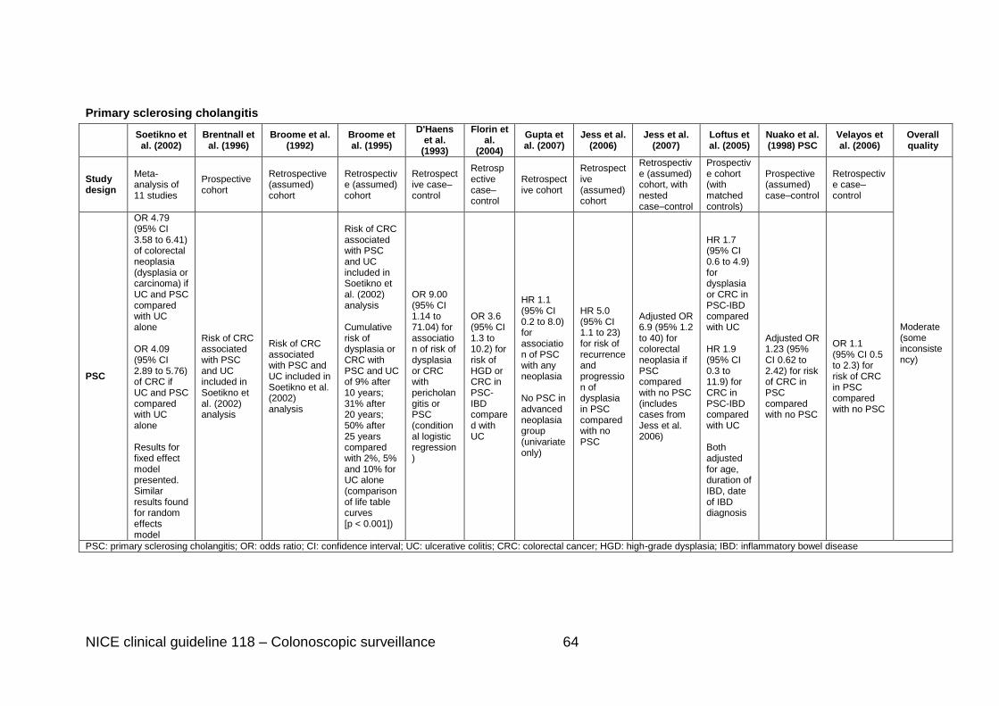
NICE clinical guideline 118 – Colonoscopic surveillance 64
Primary sclerosing cholangitis
Soetikno et al. (2002)
Brentnall et al. (1996)
Broome et al. (1992)
Broome et al. (1995)
D'Haens et al.
(1993)
Florin et al.
(2004)
Gupta et al. (2007)
Jess et al. (2006)
Jess et al. (2007)
Loftus et al. (2005)
Nuako et al. (1998) PSC
Velayos et al. (2006)
Overall quality
Study design
Meta-analysis of 11 studies
Prospective cohort
Retrospective (assumed) cohort
Retrospective (assumed) cohort
Retrospective case–control
Retrospective case–control
Retrospective cohort
Retrospective (assumed) cohort
Retrospective (assumed) cohort, with nested case–control
Prospective cohort (with matched controls)
Prospective (assumed) case–control
Retrospective case–control
Moderate (some inconsistency)
PSC
OR 4.79 (95% CI 3.58 to 6.41) of colorectal neoplasia (dysplasia or carcinoma) if UC and PSC compared with UC alone OR 4.09 (95% CI 2.89 to 5.76) of CRC if UC and PSC compared with UC alone Results for fixed effect model presented. Similar results found for random effects model
Risk of CRC associated with PSC and UC included in Soetikno et al. (2002) analysis
Risk of CRC associated with PSC and UC included in Soetikno et al. (2002) analysis
Risk of CRC associated with PSC and UC included in Soetikno et al. (2002) analysis Cumulative risk of dysplasia or CRC with PSC and UC of 9% after 10 years; 31% after 20 years; 50% after 25 years compared with 2%, 5% and 10% for UC alone (comparison of life table curves [p < 0.001])
OR 9.00 (95% CI 1.14 to 71.04) for association of risk of dysplasia or CRC with pericholangitis or PSC (conditional logistic regression)
OR 3.6 (95% CI 1.3 to 10.2) for risk of HGD or CRC in PSC-IBD compared with UC
HR 1.1 (95% CI 0.2 to 8.0) for association of PSC with any neoplasia No PSC in advanced neoplasia group (univariate only)
HR 5.0 (95% CI 1.1 to 23) for risk of recurrence and progression of dysplasia in PSC compared with no PSC
Adjusted OR 6.9 (95% 1.2 to 40) for colorectal neoplasia if PSC compared with no PSC (includes cases from Jess et al. 2006)
HR 1.7 (95% CI 0.6 to 4.9) for dysplasia or CRC in PSC-IBD compared with UC HR 1.9 (95% CI 0.3 to 11.9) for CRC in PSC-IBD compared with UC Both adjusted for age, duration of IBD, date of IBD diagnosis
Adjusted OR 1.23 (95% CI 0.62 to 2.42) for risk of CRC in PSC compared with no PSC
OR 1.1 (95% CI 0.5 to 2.3) for risk of CRC in PSC compared with no PSC
PSC: primary sclerosing cholangitis; OR: odds ratio; CI: confidence interval; UC: ulcerative colitis; CRC: colorectal cancer; HGD: high-grade dysplasia; IBD: inflammatory bowel disease

NICE clinical guideline 118 – Colonoscopic surveillance 65
Family history
Askling et al. (2001) Jess et al. (2007) Nuako et al. (1998 FH) Velayos et al. (2006) Overall quality
Study design Retrospective (assumed) cohort, with nested case–control
Retrospective (assumed) cohort, with nested case–control
Retrospective (assumed) case–control
Retrospective case–control
Low quality (retrospective and some inconsistency)
At least one first-degree relative with CRC
RR 2.5 (95% CI 1.4 to 4.4) for CRC if family history of CRC compared with no family history of CRC
Adjusted OR 1.4 (95% CI 0.3 to 5.9) for colorectal neoplasia if first-degree relative with CRC compared with no relative with CRC
Adjusted OR 2.31 (95% CI 1.03 to 5.18) for CRC in family history compared with no family history. Adjusted for sex, age, and year of ulcerative colitis diagnosis
Adjusted OR 3.7 (95% CI 1.0 to 13.2) for risk of CRC in family history compared with no family history
Relative aged < 50 at diagnosis of CRC
RR 9.2 (95% CI 3.7 to 23) for CRC if relative aged < 50 at diagnosis of CRC compared with no family history of CRC
Relative aged ≥ 50
at diagnosis of CRC
RR 1.7 (95% CI 0.8 to 3.4) for CRC if relative aged ≥ 50 at diagnosis of CRC compared with no family history of CRC
CRC: colorectal cancer; RR: relative risk; CI: confidence interval; OR: odds ratio

NICE clinical guideline 118 – Colonoscopic surveillance 66
Severity of inflammation
Gupta et al. (2007) Jess et al. (2007) Rutter et al. (2004 b and c) Overall quality
Study design Retrospective cohort Retrospective (assumed) cohort, with nested case–control
Retrospective case–control
Low quality (retrospective, some inconsistency)
inflammation score (mean)
HR 1.4 (95% CI 0.9 to 2.3) for association of inflammation with any neoplasia HR 3.0 (95% CI 1.4 to 6.3) for advanced neoplasia Remained significant for advanced neoplasia when adjusted for frequency of colonoscopy
Adjusted OR 1.3 (95% CI 0.6 to 2.9) for association of mean macroscopic inflammation score with colorectal neoplasia Adjusted OR 0.7 (95% CI 0.3 to 1.5) for association of mean microscopic inflammation score with colorectal neoplasia
Adjusted OR 4.69 (95% CI 2.10 to 10.48) for association between histological inflammation score and colorectal neoplasia
Inflammation score (cumulative mean)
HR 1.7 (95% CI 0.9 to 3.1) for association of inflammation with any neoplasia HR 3.4 (95% CI 1.1 to 10.4) for advanced neoplasia Similar results when adjusted for frequency of colonoscopy
Inflammation score (maximum)
HR 1.0 (95% CI 0.7 to 1.5) for association of inflammation with any neoplasia HR 2.2 (95% CI 1.2 to 4.2) for advanced neoplasia Similar results when adjusted for frequency of colonoscopy
Location of dysplasia
Jess et al. (2006) Overall quality
Study design Retrospective (assumed) cohort Very low quality (retrospective and consistency could not be assessed) Location of dysplasia
HR 5.4 (95% CI 1.0 to 28) for risk of recurrence and progression of dysplasia in dysplasia distal to splenic flexure compared with proximal
HR: hazard ratio; CI: confidence interval; OR: odds ratio

NICE clinical guideline 118 – Colonoscopic surveillance 67
Dysplasia at diagnosis
Thomas et al. (2007) Overall quality
Study design Meta-analysis of 20 studies
Moderate quality Progression of LGD to CRC OR 9.0 (95% CI 4.0 to 20.5) of CRC if LGD diagnosis compared with no dysplasia. Meta-regression showed no significant effect of duration of disease on CRC risk (p = 0.57)
Progression of LGD to HGD or CRC OR 11.9 (95% CI 5.2 to 27) of HGD or CRC if LGD diagnosis compared with no dysplasia
Colonic appearance
Rutter et al. (2004 b and c) Overall quality
Study design Retrospective case–control Very low quality (retrospective and consistency could not be assessed) Colonoscopic appearance
OR 0.38 (95% CI 0.19 to 0.73) for risk of CRC of normal appearance compared with abnormal appearance
Post-inflammatory polyps
Rutter et al. (2004 b and c) Velayos et al. (2006) Overall quality
Study design Retrospective case–control Retrospective case–control
Very low quality (retrospective) Post-inflammatory polyps
OR 2.29 (95% CI 1.28 to 4.11) for risk of CRC with post-inflammatory polyps compared with no polyps
Adjusted OR 2.5 (95% CI 1.4 to 4.6) for risk of CRC with pseudopolyps compared with none
Colonic stricture
Rutter et al. (2004 b and c)
Overall quality
Study design Retrospective case–control Very low quality (retrospective and consistency could not be assessed) Colonic stricture OR 4.62 (95% CI 1.03 to 20.8) for risk of CRC with colonic stricture compared with no stricture
CI: confidence interval; CRC: colorectal cancer; FH: family history; HR: hazard ratio; HGD: high-grade dysplasia; LGD: low-grade dysplasia; NS: non-significant; PSC: primary sclerosing cholangitis; RR: relative risk; SIR: standardised incidence ratio

NICE clinical guideline 118 – Colonoscopic surveillance 68
2.5.3 Evidence statements
Frequency of surveillance (see table 11)
2.5.3.1 Very low quality evidence (one study) showed that an increased
number of surveillance colonoscopies was associated with a lower
risk of colorectal cancer mortality (although this was not significant).
2.5.3.2 Very low quality evidence (one study) showed a decreased risk for
colorectal cancer with an increased number of surveillance
colonoscopies.
2.5.3.3 Very low quality evidence (two studies) showed an increased risk
for advanced neoplasia with an increased number of surveillance
colonoscopies (possible detection bias).
Prognostic factors (see table 12)
2.5.3.4 Very low quality evidence (one study) showed that the risk of
recurrence or progression of dysplasia was no different for people
with Crohn’s disease or ulcerative colitis.
2.5.3.5 Low quality evidence (four studies) showed that the risk of
neoplasia (incidence, recurrence, progression) was no different for
men or women.
2.5.3.6 Very low quality evidence (one study) showed that the risk of
identifying neoplasia was higher in people aged over 45 years,
even when adjusted for duration of IBD.
2.5.3.7 Low quality evidence (seven studies) showed that the risk of
dysplasia or colorectal cancer increased with a lower age at
diagnosis.
2.5.3.8 Moderate quality evidence (14 studies) showed that the risk of
dysplasia or colorectal cancer increased with duration of
inflammatory bowel disease.

NICE clinical guideline 118 – Colonoscopic surveillance 69
2.5.3.9 Moderate quality evidence (nine studies) showed that people with
extensive or total colitis had a higher risk of dysplasia than those
without extensive or total colitis; this increased for people with a
younger age at diagnosis.
2.5.3.10 Moderate quality evidence (nine studies) showed that people with
primary sclerosing cholangitis had a higher risk of neoplasia than
those without primary sclerosing cholangitis.
2.5.3.11 Low quality evidence (four studies) showed that a family history of
colorectal cancer was associated with an increased risk of
colorectal cancer (which increased if the relative was younger than
50 years).
2.5.3.12 Low quality evidence (three studies) showed that increased
inflammation was a predictor of neoplasia.
2.5.3.13 Very low quality evidence (one study) showed that the risk of
recurrence and progression of dysplasia was higher if located
distally compared with proximally.
2.5.3.14 Moderate quality evidence (one meta-analysis) showed that low-
grade dysplasia was associated with progression to high-grade
dysplasia and colorectal cancer compared with no dysplasia.
2.5.3.15 Very low quality evidence (one study) showed that a normal
appearance at colonoscopy was associated with a lower risk of
colorectal cancer.
2.5.3.16 Very low quality evidence (two studies) showed that post-
inflammatory polyps were associated with a higher risk of colorectal
cancer.
2.5.3.17 Very low quality evidence (one study) showed that colonic stricture
was associated with a higher risk of colorectal cancer.

NICE clinical guideline 118 – Colonoscopic surveillance 70
2.5.4 Health economic modelling
No health economic modelling was undertaken for this review question.
2.5.5 Evidence to recommendations
Because there was no direct evidence for surveillance schemes for different
subgroups of the IBD population, the GDG made recommendations based on
the risk of people with IBD developing colorectal cancer, taking into account
the significant risk factors from the available evidence. Although the evidence
showed that the risk of colorectal cancer increased with a lower age at
diagnosis of IBD, this could be confounded by duration of disease or overall
age, so recommendations were based on the duration of disease. This was
consistent with the recommendation to start surveillance 10 years after onset
of symptoms. The GDG felt that there were differences in the incidence of
colorectal cancer by disease duration between the Eaden et al. (2001) meta-
analysis and the Rutter et al. (2006) study. However, people taking part in the
latter study were on surveillance and therefore the Eaden et al. (2001) figures
were considered to be more realistic. The GDG also felt that a detailed look at
severity of inflammation was necessary because it is a precursor to dysplasia.
It felt that using a validated score for describing inflammation would be useful,
as used in the Gupta et al. (2007) study. The GDG also considered that there
is sufficient agreement internationally that proctitis does not increase
colorectal cancer risk and therefore people with proctitis do not need
surveillance.
Apart from the duration, extent and severity of the disease, having a family
history of colorectal cancer was an important prognostic factor for neoplasia.
The presence of dysplasia at diagnosis was a significant factor for progression
to high-grade dysplasia and colorectal cancer and was therefore included as a
risk factor in the high-risk group. Although the evidence for colonic stricture
was very low quality, the GDG considered that strictures are an indication of
malignancy or severe inflammation, so further surveillance and investigation
would be warranted.

NICE clinical guideline 118 – Colonoscopic surveillance 71
Although there was a reasonable amount of evidence evaluating different risk
factors, no one study compared all of the factors directly. The GDG therefore
stratified the risk groups in the recommendations based on their knowledge
and expertise.
Because there was no direct evidence for timings of surveillance for the
different risk groups, these were determined by GDG consensus. The GDG
felt strongly that before entering the surveillance programme a confirmed
histological diagnosis was essential. The GDG also stated that any resectable
lesion found should be removed endoscopically. For people with flat
dysplastic lesions, surgery should be offered but if declined these people
should remain on surveillance in the high-risk group. However, management
of dysplasia was outside the scope of this guideline, so a recommendation
was made that people should be referred for further investigations or
treatment if there are any findings at surveillance. The GDG also made a
recommendation about stopping surveillance. This was based on its expertise
and knowledge.
2.5.6 Recommendations
Recommendation 1.1.2
Offer a baseline colonoscopy with chromoscopy and targeted biopsy of any
abnormal areas to people with IBD who are being considered for colonoscopic
surveillance to determine their risk of developing colorectal cancer (see table
1).
Table 1 Risk of developing colorectal cancer in people with IBD
Low risk:
extensive but quiescent ulcerative colitis or
extensive but quiescent Crohn’s colitis or
left-sided ulcerative colitis (but not proctitis alone) or Crohn’s colitis of a similar extent.
Intermediate risk:
extensive ulcerative or Crohn’s colitis with mild active inflammation that has been confirmed endoscopically or histologically or
post-inflammatory polyps or
family history of colorectal cancer in a first-degree relative aged 50 years or over.

NICE clinical guideline 118 – Colonoscopic surveillance 72
High risk:
extensive ulcerative or Crohn’s colitis with moderate or severe active inflammation that has been confirmed endoscopically or histologically or
primary sclerosing cholangitis (including after liver transplant) or
colonic stricture in the past 5 years or
any grade of dysplasia in the past 5 years or
family history of colorectal cancer in a first-degree relative aged under 50 years.
Recommendation 1.1.3
Offer colonoscopic surveillance to people with IBD as defined in 1.1.1 based
on their risk of developing colorectal cancer (see table 1), determined at the
last complete colonoscopy:
Low risk: offer colonoscopy at 5 years.
Intermediate risk: offer colonoscopy at 3 years.
High risk: offer colonoscopy at 1 year.
Recommendation 1.1.14
Discuss the potential benefits, limitations and risks with people who are
considering colonoscopic surveillance including:
early detection and prevention of colorectal cancer and
quality of life and psychological outcomes.
Recommendation 1.1.16
After receiving the results of each surveillance test, discuss the potential
benefits, limitations and risks of ongoing surveillance. Base a decision to stop
surveillance on potential benefits for the person, their preferences and any
comorbidities. Make the decision jointly with the person, and if appropriate,
their family or carers..
Recommendation 1.1.17
If there are any findings at surveillance that need treatment or referral, discuss
the options with the person, and if appropriate, their family or carers.

NICE clinical guideline 118 – Colonoscopic surveillance 73
Initiation and frequency of surveillance in people with adenomas
2.5.7 Evidence review
A total of 14,701 articles were found by systematic searches, of which 9544
were unique articles. The full text was ordered for 62 articles and a further four
articles were identified through manual reference searching. Only six articles
met the eligibility criteria (for the review protocol, inclusion and exclusion
criteria, see appendices 2 and 4). Of these, two were meta-analyses of
primary studies (Martinez et al. 2009; Saini et al. 2006) and four were primary
studies that were not included in the meta-analyses or the outcomes of
interest were not reported (Kronborg et al. 2006; Lieberman et al. 2007, 2008;
Nusko et al. 2002). The Martinez et al. (2009) meta-analysis included the data
from the Lieberman et al. (2000) study and additional data up to June 2005.
The updated data from Lieberman et al. (2007) and the prevalence study of
advanced histology in smaller adenomas (Lieberman et al. 2008) were not
included in the meta-analysis, but were included in our analysis. The Saini et
al. (2006) systematic review included the Nusko et al. (2002) study, but only
for the outcome of risk factors for recurrent adenomas. Nusko et al. (2002)
was included in our analysis for two additional outcomes: risk factors and time
taken for the development of advanced metachronous adenomas (defined as
larger than 10 mm, or with high-grade dysplasia or with invasive cancer).
The characteristics of the 6 included studies are summarised in table 13 and
the evidence reviewed in GRADE profiles 6 and 7. Detailed evidence tables
for the included studies are available in appendix 6.

NICE clinical guideline 118 – Colonoscopic surveillance 74
Table 13: Summary of study characteristics
Study Aim Study type Population Prognostic factors or surveillance programmes
Outcomes used for GRADE profile
Kronborg et al. (2006)
To ‘measure the outcome resulting from extension of intervals between colonoscopies as measured by risk of new neoplasia as well as complications in two patient groups believed to carry different risks of new neoplasia’
Three randomised long-term surveillance trials
20 years of surveillance of people with previously diagnosed adenomas, N = 671, 73, and 200
1. Surveillance group A: 24 months 2. Surveillance group B: 48 months 3. Surveillance group C: 6 months 4. Surveillance group D: 12 months 5. Surveillance group E: 12 months 6. Surveillance group F: 24 months
Recurrence risk of new adenomas, advanced adenomas and progression to colorectal cancer
Lieberman et al. (2007)
To ‘determine the 5.5-year cumulative incidence rate of advanced neoplasia in patients with and without neoplasia at the baseline screening colonoscopy’ and ‘whether there is an association between baseline endoscopic findings and subsequent risk of advanced neoplasia’
Observational cohort, with nested randomised comparison
5 years of surveillance of people with previously diagnosed polyps, N = 3121
Histopathology of the index polyp: 1. with 1 or 2 tubular
adenomas <10 mm 2. with 3 or more
tubular adenomas <10 mm
3. with tubular adenomas >10 mm
4. with villous adenomas
5. with adenomas with high-grade dysplasia
Risk of new neoplasia, high-grade dysplasia and colorectal cancer by histopathology of index
Lieberman et al. (2008)
To compare ‘proportions of advanced histologic features’ and to ‘determine whether there were
Observational cohort
People undergoing colonoscopic surveillance with
1. Histopathology of the index polyp
2. Location of the index
Prevalence of advanced histology and its association

NICE clinical guideline 118 – Colonoscopic surveillance 75
Study Aim Study type Population Prognostic factors or surveillance programmes
Outcomes used for GRADE profile
any risk factors for advanced histology in each patient group’
largest index polyp being less than 10 mm, in 2005, N = 5977
polyp with the distal colon
Martinez et al. (2009)
To ‘estimate absolute risks of metachronous advanced adenoma, colorectal cancer, and their combination (advanced colorectal neoplasia) and to identify patient characteristics and adenoma features that are associated independently with risk of these outcomes’
Meta-analysis from selected studies (not systematically identified)
Meta-analysis of 8 studies (6 RCTs) for people undergoing surveillance after polypectomy. Median follow-up period of 47.2 months and N = 10,021
Risk factors considered: 1. age 2. sex 3. race 4. family history of
colorectal cancer 5. smoking status 6. body mass index 7. previous polyps 8. number of adenomas 9. location of polyps 10. size of largest
adenoma 11. adenomas histology 12. high-grade dysplasia
Risk factors for advanced metachronous neoplasia
Nusko et al. (2002)
To ‘identify predictive variables’ and whether ‘these risk factors of advanced pathology could also be used to predict the likely time interval to the development of metachronous adenomas of advanced pathology’
Observational cohort
People undergoing surveillance after polypectomy, N = 1159
Risk factors considered: 1. size of largest
adenoma 2. parental history of
colorectal cancer 3. histological type 4. dysplasia 5. location of adenomas 6. multiplicity
Risk factors and time taken for progression to advanced metachronous adenomas
Saini et al. To evaluate the incidence of Systematic Criterion for inclusion 14 studies, reported a Risk factors for

NICE clinical guideline 118 – Colonoscopic surveillance 76
Study Aim Study type Population Prognostic factors or surveillance programmes
Outcomes used for GRADE profile
(2006) advanced adenomas at 3-year surveillance colonoscopy in high- and low-risk groups. Also to determine associated risk factors
review and meta-analysis
was people with a personal history of adenomas, N = 10,009 (but differs by analysis because of study inclusion)
total of 6 risk factors: 1. number of adenomas 2. size of largest
adenoma 3. patient age 4. tubulovillous/villous
features or severe dysplasia
5. advanced adenoma 6. adenoma in the
proximal colon
recurrent advanced adenomas

NICE clinical guideline 118 – Colonoscopic surveillance 77
Two studies included in the meta-analyses were RCTs comparing different
surveillance frequencies (Lund et al. 2001; Winawer et al. 1993b). Lund et al.
compared six surveillance strategies for people with colonic adenomas:
colonoscopy every 2 years or every 5 years, or flexible sigmoidoscopy every
year, every 2 years, or every 5 years. The authors concluded that ‘a
surveillance interval of 5 years was as effective as shorter intervals in terms of
cancer prevention’. However, it should be noted that the trial was stopped
early because of lower rates of adenoma recurrence than expected, and
therefore the trial was not powered to detect differences among the
surveillance strategies. Winawer et al. compared two surveillance strategies in
people with adenomatous polyps: two colonoscopies 1 and 3 years after
adenoma removal and one colonoscopy 3 years after adenoma removal. The
results showed that the relative risk of detecting any adenomas with two
colonoscopies compared with one was 1.3 (95% confidence interval [CI] 1.1 to
1.6). However, the detection rate of advanced adenomas was the same for
both strategies (3.3%; relative risk 1.0; 95% CI 0.5 to 2.2).

NICE clinical guideline 118 – Colonoscopic surveillance 78
GRADE profile 6: When and at what frequency should colonoscopic surveillance be offered to people with adenomas? Frequency of surveillance
Quality assessment Summary of findings
Study Design
Lim
ita
tio
ns
Inc
on
sis
ten
cy
Dir
ec
tne
ss
Imp
rec
isio
n Other
considerations
RR (95% CI)
Quality
Surveillance groups
Detection of new adenomas by surveillance group
Kronborg et al. (2006)
RCT S* N N S† None B: 48 months (n = 340) vs
A: 24 months (n = 331) D: 12 months (n = 32) vs C: 6 months (n = 42)
F: 24 months (n = 103) vs E: 12 months (n = 97)
Low
RR = 0.88 (0.69 to 1.12) NS
RR = 0.82 (0.43 to 1.52) NS
RR = 0.88 (0.57 to 1.34) NS
Winawer et al. (1993b)
RCT N N S¥ N None 2-exam: 1 and 3 years (n = 338) vs 1-exam: 3 years (n = 428) Moderate
RR = 1.3 (1.1 to 1.6) p = 0.006
Detection of new advanceda adenomas by surveillance group
Kronborg et al. (2006)
RCT S* N N S† None B: 48 months (n = 340) vs
A: 24 months (n = 331) D: 12 months (n = 32) vs C: 6 months (n = 42)
F: 24 months (n = 103) vs E: 12 months (n = 97)
Low
RR = 1.15 (0.61 to 2.15) NS
RR = 3.12 (0.87 to 14.50) NS
RR = 0.97 (0.40 to 2.35) NS
Winawer et al. (1993b)
RCT N N S¥ N None 2-exam: 1 and 3 years (n = 338) vs 1-exam: 3 years (n = 428) Moderate
RR =1.0 (0.5 to 2.2) NS
Detection of colorectal cancer by surveillance group
Kronborg et al. (2006)
RCT S* N N S† None B: 48 months (n = 340) vs
A: 24 months (n = 331) D: 12 months (n = 32) vs C: 6 months (n = 42)
F: 24 months (n = 103) vs E: 12 months (n = 97)
Low
RR = 6.22 (1.06 to 117)
c
None identified RR = 1.93 (0.38 to 13.94) NS
Lund et al. (2001)
RCT Sb N N N/A Underpo
wered for difference
Authors concluded that ‘a surveillance interval of five years was as effective as shorter intervals in terms of cancer prevention’. No comparisons reported because of the lack of power.
Very low

NICE clinical guideline 118 – Colonoscopic surveillance 79
between strategies
Adverse events
Kronborg et al. (2006)
RCT S* N N N None B: 48 months (n = 340) vs A: 24 months (n = 331)
D: 12 months (n = 32) vs C: 6 months (n = 42)
F: 24 months (n = 103) vs E: 12 months (n = 97)
Moderate
7 total, 6 during surveillance. Perforation at initial colonoscopy seen in group A was fatal (septicemia). A: 2 diagnostic perforations and 2 therapeutic perforations; B: 1 diagnostic perforation and 1 polypectomy syndrome
2 total (1 diagnostic perforation and 1 polypectomy syndrome) both in group C. None seen in D
2 total, one diagnostic perforation seen in each group
CI: confidence interval; N: not serious; NS: not significant; RCT: randomised controlled trial; RR: relative risk; S: serious
Surveillance group A: 24 months, surveillance group B: 48 months, surveillance group C: 6 months, surveillance group D: 12 months, surveillance group E: 12 months, surveillance group F: 24 months * The study was randomised by random numbers but no details of concealment or blinding of pathologists is mentioned. ¥
The population under surveillance was not stratified by risk for different strategies. † The 95% confidence intervals did not give statistically or clinically significant results.
a The advanced adenomas were defined as those with severe dysplasia or being at least 10 mm in diameter or villous.
b The trial was stopped early because of the low rate of adenoma recurrence.
c Therefore there is an increased risk for cancer progression (RR = 6.22) if surveillance is done after 48 months instead of 24 months.

NICE clinical guideline 118 – Colonoscopic surveillance 80
GRADE profile 7: When and at what frequency should colonoscopic surveillance be offered to people with adenomas? Determining significant predictors
Quality assessment Summary of findings
Study Design
Lim
ita
tio
ns
Inc
on
sis
ten
cy
Dir
ec
tne
ss
Imp
rec
isio
n
Other considerations
R; RR; OR (95% CI) Quality
Risk of new neoplasia by histopathology of the polyps at index colonoscopy
Lieberman et al. (2007)
Multi-centre registry
S* N N N None Compared with no neoplasia at baseline: 1 or 2 tubular adenomas <10 mm: RR = 1.92 (0.83 to 4.42) NS
Very low
≥3 tubular adenomas <10 mm: RR = 5.01 (2.10 to 11.96)
Tubular adenoma >10 mm: RR = 6.40 (2.74 to14.94)
Villous adenoma: RR = 6.05 (2.48 to 14.71)
High-grade dysplasia: RR = 6.87 (2.61 to18.07)
Risk of high-grade dysplasia or cancer by histopathology of the polyps at index colonoscopy
Lieberman et al. (2007)
Multi-centre registry
S* N N N None Rates per 1000 person-years of follow-up no neoplasia at baseline: R = 0.7 (0 to 2.0) NS
Very low
1.5 with tubular adenomas <10 mm (0 to 2.9) NS
>10 mm tubular: R = 6.4 (0 to 13.5) NS
Villous adenomas: R = 6.2 (0 to 14.7) NS
HGD: R = 26.0 (3.2 to 48.8) vs no neoplasia at baseline: RR = 7.23 (2.81 to 18.17)
Prevalence of advanced histology (defined as an adenoma with villous or serrated histology, HGD, or an invasive cancer) in 2005
Lieberman et al. (2008)
Multi-centre registry
S* N N N Sensitivity analysis done for misclassification
a for
prevalence
1–5 mm group: 1.7% (1.2 to 2.0) Very low
6–9 mm group: 6.6% (4.6 to 11.7)
>10 mm group: 30.6% (29.2 to 40.0)
Distal location’s associated with advanced histology in 2005
Lieberman et al. (2008)
Multi-centre registry
S* N N N None 6–9 mm group (p = 0.04) Very low
>10 mm group (p = 0.002)
Risk factors for advanced metachronous neoplasia (advanced adenomasb and invasive cancer)
Martinez et al. (2009)
Meta-analysis of 8 studies (6
Sc N N N Patient level data
used and confounders adjusted
Older age (p < 0.0001 for trend) Low
Male sex: OR = 1.40 (1.19 to 1.65)
Number and size of previous adenomas (p < 0.0001 for trend)

NICE clinical guideline 118 – Colonoscopic surveillance 81
Quality assessment Summary of findings
Study Design
Lim
ita
tio
ns
Inc
on
sis
ten
cy
Dir
ec
tne
ss
Imp
rec
isio
n
Other considerations
R; RR; OR (95% CI) Quality
RCTs) by multivariate logistic regression
Presence of villous features: OR = 1.28 (1.07 to 1.52)
Proximal location: OR = 1.68 (1.43 to 1.98)
Risk factors for advanced metachronous adenomas (defined as larger than 10 mm or with HGD or invasive carcinoma)
Nusko et al. (2002)
Single centre registry, prospective single cohort
Sd N N N Adjusted by
multivariate logistic regression
Considering only patients with tubular adenomas at index: adenoma size (p < 0.0001)
Moderate
Multiplicity of adenomas at index (p = 0.021)
Parental history of colorectal carcinoma (p = 0.017)
An interactive effect between size and sex (p = 0.00392): male patients with large adenomas had a significantly higher risk than others
Time taken for advanced metachronous adenomas (defined as larger than 10 mm or with HGD or invasive carcinoma) to develop over time
Nusko et al. (2002)
Single centre registry, prospective single cohort
Se N N N 1000 Bootstrap
samples done for sensitivity analyses and confounders adjusted by multivariate logistic regression
Prp Low riskf High risk
g Moderate
5% 10.4 years ( 4.1 to 13.2) 0.5 years (0.1 to 1.6)
10% 12.2 years (10.1 to 15.2) 6.1 years (3.2 to 11.5)
20% 16.2 years (10.5 to 19.2) 15.6 years (11.5 to 18.2)
Risk factors for recurrent advanced adenomas (defined as adenomas ≥1 cm, villous histological features, or with cancer) based on adenomas at index colonoscopy
Saini et al. (2006) Systematic review
N N N N None RF RR RD H Moderate
Number and size of adenomas
>3 vs 1 or 2
2.52 (1.07 to 5.97)
5% (1% to 10%)
p < 0.001
Histological diagnosis
tubulovillous/villous vs tubular
1.26 (0.95 to 1.66) NS
2% (–1% to 4%) NS
p > 0.2
Dysplasi HGD vs no HGD

NICE clinical guideline 118 – Colonoscopic surveillance 82
Quality assessment Summary of findings
Study Design
Lim
ita
tio
ns
Inc
on
sis
ten
cy
Dir
ec
tne
ss
Imp
rec
isio
n
Other considerations
R; RR; OR (95% CI) Quality
a 1.84 (1.06 to 3.19)
4% (0 to 8%)
p > 0.2 NS
CI: confidence interval; H: heterogeneity; HGD: high-grade dysplasia; N: not serious; NS: not significant; OR: odds ratio; Prp: proportion of patients expected to develop
advanced metachronous adenomas; R: risk; RD: risk difference; RF: risk factor; RR: relative risk; S: serious * The study did not adjust for confounders. a The sensitivity analysis was done to determine how misclassification of polyp size would impact the outcome. The analysis assumed that polyps were either overestimated in
size by 1 mm (for example, a 10-mm polyp is reclassified as 9 mm) or underestimated (a 9-mm polyp is reclassified as 10 mm). b The advanced adenomas were defined as those that had one or more of the following features: 10 mm in diameter or larger, presence of high-grade dysplasia, or greater than
25% villous features (also classified as tubulovillous or villous histology). c The study combined randomised and non-randomised studies together.
d The study only had a single arm cohort.
e The study only had a single arm cohort.
f People at low risk were defined as: no parental history of colorectal carcinoma and with only small (<10 mm) tubular adenomas at index.
g People at high risk were defined as: those with multiple or large adenomas, tubulovillous or villous adenomas, or a parental history of colorectal cancer.

NICE clinical guideline 118 – Colonoscopic surveillance 83
2.5.8 Evidence statements (see GRADE profiles 6 and 7)
2.5.8.1 Low to moderate quality evidence showed that the detection of new
adenomas was higher at 1 and 3 years compared with 3 years
alone.
2.5.8.2 Low to moderate quality evidence showed that the detection of new
advanced adenomas tended to be the same at different
surveillance frequencies.
2.5.8.3 Very low to low quality evidence showed that the detection of
colorectal cancer was higher at 4 years compared with 2 years.
2.5.8.4 Moderate quality evidence showed adverse events of perforations
and polypectomy syndrome during follow-up at 6–48 months.
2.5.8.5 Very low quality evidence showed that having at least three tubular
adenomas smaller than 10 mm, or tubular adenomas larger than
10 mm, or villous adenomas or high-grade dysplasia at baseline
colonoscopy were significant predictors for risk of new neoplasia.
2.5.8.6 Very low quality evidence showed that having high-grade dysplasia
compared with no neoplasia at baseline colonoscopy was a
significant predictor for high-grade dysplasia or colorectal cancer in
the future.
2.5.8.7 Very low quality evidence that studied the risk associated with
small adenomas and distal location showed that the prevalence of
advanced histology5 increased with the size of the polyp.
2.5.8.8 Very low quality evidence on the risk associated with small
adenomas and distal location showed that the prevalence of
advanced histology in the distal colon increased with polyp size and
was statistically significant in the 6–9 mm and >10 mm groups.
5 Advanced histology was defined as an adenoma with villous or serrated histology, high-
grade dysplasia, or an invasive cancer.

NICE clinical guideline 118 – Colonoscopic surveillance 84
2.5.8.9 Low quality evidence showed that being older, being male, an
increase in the number and size of previous adenomas, the
presence of villous features and proximal location at baseline
colonoscopy were significant predictors for advanced
metachronous neoplasia (advanced adenomas and invasive
cancer).
2.5.8.10 Moderate quality evidence showed that increased adenoma size,
multiplicity of adenomas, parental history of colorectal cancer and
an interactive effect between adenoma size and sex (male) were
significant predictors for advanced metachronous adenomas6. Men
with large adenomas had a significantly higher risk than other
people.
2.5.8.11 Moderate quality evidence showed that the time taken for
advanced metachronous adenomas to develop in 5% of people at
low risk7 was10.4 years, in 10% it was12.2 years and in 20% it was
16.2 years.
2.5.8.12 Moderate quality evidence showed that the time taken for
advanced metachronous adenomas to develop in 5% of people at
high risk8 was 0.5 years, in 10% it was 6.1 years and in 20% it was
15.6 years.
2.5.8.13 Moderate quality evidence showed that the risk for recurrent
advanced adenomas9 increased with increasing number and size
of adenomas at baseline colonoscopy.
6 Advanced metachronous adenomas were defined as larger than 10 mm or with high-grade
dysplasia or invasive carcinoma. 7 People at low risk were defined as: no parental history of colorectal carcinoma and with only
small (<10 mm) tubular adenomas at index colonoscopy. 8 People at high risk were defined as: those with multiple or large adenomas, tubulovillous or
villous adenomas, or a parental history of colorectal carcinoma. 9 Advanced adenomas were defined as adenomas ≥1 cm, villous histological features, or with
cancer.

NICE clinical guideline 118 – Colonoscopic surveillance 85
2.5.9 Evidence to recommendations
Because there was no direct evidence for surveillance strategies for the
different subgroups of the population who had adenomas removed previously,
the GDG made recommendations based on their risk of developing colorectal
cancer, taking into account the significant risk factors from the available
evidence. The GDG felt that there was enough evidence to stratify people who
had previously had adenomas removed according to their risk of developing
advanced neoplasia (advanced adenoma and colorectal cancer). It felt that
the frequency of surveillance should be based on the risk assessment. The
GDG felt that the evidence showed that only the number and size of the
adenomas removed at baseline colonoscopy were consistent significant
predictors for neoplasia and therefore should determine the risk state. Villous
histology was also a significant predictor for advanced neoplasia, although the
confidence intervals were wide (odds ratio 1.28, 95% CI 1.07 to 1.52)
(Martinez et al. 2009). However, the GDG considered that because
pathologists’ classification of villous histology tends to vary, particularly for
small biopsies, including this predictor could lead to wide variation in referral
rates for colonoscopy. The GDG also stated that all adenomas detected
during colonoscopic surveillance should be removed endoscopically.
Because there was very limited direct evidence on frequency of surveillance
for different risk groups (Lund et al. 2001), the timing of surveillance was
based on evidence relating to the incidence of advanced adenomas and
colorectal cancer and risk for the disease as described above, and also RCTs
with different surveillance frequencies without risk stratification (Kronborg et
al. 2006; Winawer et al. 1993b).
Results from ongoing research on the long-term safety of people at low risk
not having surveillance are expected to be reported in the next 2 years (noted
in Cairns et al. 2010). This will give valuable evidence for the future.
The GDG also made a recommendation about stopping surveillance. This was
based on its expertise and knowledge.

NICE clinical guideline 118 – Colonoscopic surveillance 86
2.5.10 Recommendations
Recommendation 1.1.6
Consider colonoscopic surveillance for people who have had adenomas
removed and are at low risk of developing colorectal cancer (see table 2).
Recommendation 1.1.7
Offer colonoscopic surveillance to people who have had adenomas removed
and are at intermediate or high risk of developing colorectal cancer (see table
2).
Recommendation 1.1.8
Use the findings at adenoma removal to determine people’s risk of developing
colorectal cancer (see table 2).
Table 2 Risk of developing colorectal cancer in people with adenomas
Low risk:
one or two adenomas smaller than 10 mm.
Intermediate risk:
three or four adenomas smaller than 10 mm or
one or two adenomas if one is10 mm or larger.
High risk:
five or more adenomas smaller than 10 mm or
three or more adenomas if one is 10 mm or larger.
Recommendation 1.1.9
Offer the appropriate colonoscopic surveillance strategy to people with
adenomas based on their risk of developing colorectal cancer as determined
at initial adenoma removal (see table 2).
Low risk: consider colonoscopy at 5 years:
if the colonoscopy is negative (that is, no adenomas are found) stop
surveillance

NICE clinical guideline 118 – Colonoscopic surveillance 87
-if low risk, consider the next colonoscopy at 5 years (with follow-up
surveillance as for low risk)
if intermediate risk, offer the next colonoscopy at 3 years (with follow-
up surveillance as for intermediate risk)
if high risk, offer the next colonoscopy at 1 year (with follow-up
surveillance as for high risk).
Intermediate risk: offer colonoscopy at 3 years:
- if the colonoscopy is negative, offer the next colonoscopy at 3 years.
Stop surveillance if there is a further negative result
- if low or intermediate risk, offer the next colonoscopy at 3 years (with
follow-up surveillance as for intermediate risk)
- if high risk, offer the next colonoscopy at 1 year (with follow-up
surveillance as for high risk).
High risk: offer colonoscopy at 1 year:
if the colonoscopy is negative, or low or intermediate risk, offer the
next colonoscopy at 3 years (with follow-up surveillance as for
intermediate risk)
if high risk, offer the next colonoscopy at 1 year (with follow-up
surveillance as for high risk).
Recommendation 1.1.16
After receiving the results of each surveillance test, discuss the potential
benefits, limitations and risks of ongoing surveillance. Base a decision to stop
surveillance on potential benefits for the person, their preferences and any
comorbidities. Make the decision jointly with the person, and if appropriate,
their family or carers.

NICE clinical guideline 118 – Colonoscopic surveillance 88
2.6 Providing information and support
2.6.1 Review question
What are the information and support needs of people, or the carers of
people, undergoing or considering undergoing colonoscopic surveillance?
2.6.2 Evidence review
A total of 1910 articles were found by systematic searches, of which 28 were
unique articles. Full text was ordered for these articles and only seven met the
eligibility criteria (for the review protocol, inclusion and exclusion criteria, see
appendices 2 and 4). Thematic analysis was used to analyse these seven
studies to adequately answer the review question.
The characteristics of the included studies are summarised in table 14 and
detailed evidence tables are available in appendix 6.
The seven studies are:
Rutter et al. (2006): a 58-question self-administered postal questionnaire
with an 85.4% response rate.
Thiis-Evensen et al. (1999b): a postal questionnaire studying the
psychological effect of attending a screening programme to detect and
remove colorectal polyps.
Sheikh et al. (2004): a questionnaire design study to determine people’s
screening preferences.
Brotherstone et al. (2006): a study examining the effectiveness of visual
illustrations in improving people’s understanding of the preventative aim of
flexible sigmoidoscopy screening.
Makoul et al. (2009): a pretest–post-test study assessing a multimedia
patient education programme that provides information about colorectal
cancer and screening.
Sequist et al. (2009): a RCT promoting colorectal cancer screening. The
screening options in this study also looked at faecal occult blood testing.

NICE clinical guideline 118 – Colonoscopic surveillance 89
Miles et al. (2009): a postal survey examining the psychological impact of
being assigned to colonoscopic surveillance after detection of
adenomatous polyps.

NICE clinical guideline 118 – Colonoscopic surveillance 90
Table 14: Thematic analysis
People’s experience of the procedure
Rutter et al. (2006) 39% of the respondents found bowel preparation difficult to take
60.2% of the respondents found their last colonoscopy comfortable or very comfortable
People expressed less discomfort with more experienced colonoscopists (r = 0.20, p = 0.0007)
There was a correlation between comfort and pethidine dose (r = 0.16, p = 0.007, i.e. those with more discomfort were given more pethidine)
Thiis-Evensen et al. (1999b)
When asked if they found the colonoscopic examination uncomfortable, 50% said no, 45% found it moderately uncomfortable and 5% found it very uncomfortable
Rutter et al. (2006) 16.4% of the respondents experienced abdominal pain (attributed to the procedure) in the week after their last colonoscopy, of which 3.7% stated that the pain interfered with everyday activities. Post-procedural pain was strongly related to the Hospital Anxiety and Depression Scale anxiety score (p < 0.0001) but not with the drug doses used during the procedure. Five patients (1.7%) reported complications after previous colonoscopies
People’s preference
Sheikh et al. (2004) Of people who had had a previous colonoscopy, 55% preferred this method for repeat screening, compared with only 30% of people who had never had a colonoscopy (p = 0.017)
Of people who had had a previous sigmoidoscopy, 53% preferred this method for repeat screening, compared with only 33% of people who had never had a sigmoidoscopy, although the differences were not statistically significant
Thiis-Evensen et al. (1999b)
When asked if they would attend a repeat examination in 5 years’ time, 90% said yes, 2% said no and 7.6% were not sure
Information given
Rutter et al. (2006) 91.4% described the information given as easy to understand, 2.6% thought it was difficult and 6.1% could not remember being given information
Rutter et al. (2006)
When asked about the amount of information they had received about the surveillance programme, 83.8% thought they had received the right amount of information, 16.2% thought they had received too little, and no one thought they had received too much
65.5% reported being content with their current involvement, whereas 34.2% preferred to be more involved and only 0.4% wished to be less involved
Brotherstone et al. (2006)
In the written information group, 57% had a good understanding of the aims of the test, while in the group who were sent written information and illustrations, 84% had a good understanding
The addition of the illustrations resulted in significantly better understanding (OR = 3.75; CI 1.16 to 12.09; p = 0.027) which remained significant after controlling for age, gender and socioeconomic status (OR = 10.85; CI 1.72 to 68.43; p = 0.011)
Makoul et al. (2009) A pretest–post-test multimedia patient education programme on colorectal cancer screening, which used graphics and audio, led to a significant increase in the knowledge of flexible sigmoidoscopy (from 11.5% to 53.0%; p < 0.001) and colonoscopy (from 23.3% to

NICE clinical guideline 118 – Colonoscopic surveillance 91
57.0%; p < 0.001)
More than 90% of people wanted to discuss colorectal cancer with their doctors after the education programme
Surveillance programme
Rutter et al. (2006) 97.8% of people felt that surveillance was important for them
96.4% thought that the surveillance programme gave them reassurance, while 3.6% stated that the programme made them more anxious
When asked about the effect of the surveillance programme on reducing risk of colorectal cancer, 1.8% believed it completely removed the risk, 67.9% believed it greatly reduced the risk, 24.4% believed it moderately reduced the risk, and 5.9% believed it slightly reduced the risk
Makoul et al. (2009) A multimedia pretest–posttest patient education programme led to a significant increase in the number of people willing to undergo colorectal cancer screening with flexible sigmoidoscopy (from 54.1% to 78.1%; p < 0.001) and colonoscopy (from 64.8% to 84.4%; p < 0.001)
Sequist et al. (2009)
People who received mailings about colorectal cancer screening were significantly more likely to complete screening than those who did not (44.0% vs 38.1%; p < 0.001)
Detection of adenomas tended to be greater among people who received mailings compared with the control group (5.7% vs 5.2%; p = 0.10)
Psychological impact of surveillance
Thiis-Evensen et al. (1999b)
The scores for both Goldberg’s General Health Questionnaire (GHQ-28) and the Hospital Anxiety and Depression Scale were lower, indicating a lower level of psychiatric morbidity among those attending the examination than the controls
Miles et al. (2009) People offered surveillance reported lower psychological distress and anxiety than those with either no polyps (p < 0.05) or lower risk polyps (p < 0.01). The surveillance group also reported more positive emotional benefits of screening than the other outcome groups. Post-screening bowel cancer worry and bowel symptoms were higher in people assigned to surveillance, but both declined over time, reaching levels observed in either one or both of the other two groups found to have polyps, suggesting these results were a consequence of polyp detection rather than surveillance
CI: confidence interval; OR: odds ratio
NICE clinical guideline 118 – Colonoscopic surveillance 92
2.6.3 Evidence statements (see table 14)
2.6.3.1 There is limited evidence describing people’s experience of
colonoscopy:
39% found bowel preparation difficult to take.
50% did not find the examination uncomfortable, 45% found it
moderately uncomfortable and 5% found it very uncomfortable.
People expressed less discomfort with a more experienced
colonoscopist and with sedation.
2.6.3.2 There is limited evidence describing people’s preference:
55% of those who had had a previous colonoscopy preferred
this method for repeat screening, compared with only 30% of
those who had never had a colonoscopy.
53% of those who had had a previous sigmoidoscopy preferred
this method for repeat screening, compared with only 33% of
those who had never had a sigmoidoscopy, although the
differences were not statistically significant.
When asked if they would attend a repeat examination in
5 years’ time, 90% said yes, 2% said no and 7.6% were not
sure.
2.6.3.3 There is limited evidence describing the amount of information
given and how the information improved people’s understanding:
57% in the written information group had a good understanding
of the aims of the screening test, while in the group who were
sent written information and illustrations, 84% had a good
understanding.
The addition of the illustrations resulted in significantly better
understanding, even after controlling for age, sex and
socioeconomic status.
A pretest–post-test multimedia patient education programme on
colorectal cancer screening using graphics and audio led to a

NICE clinical guideline 118 – Colonoscopic surveillance 93
significant increase in the knowledge of flexible sigmoidoscopy
and colonoscopy.
More than 90% of people wanted to discuss colorectal cancer
with their doctors after the education programme.
When asked about the amount of information they had received
about the surveillance programme, 83.8% thought they had
received the right amount of information.
91.4% described the information given as easy to understand
and 2.6% thought it was difficult.
2.6.3.4 There is limited evidence describing the benefits, risks and uptake
of a surveillance programme:
People who received mailings about colorectal cancer screening
were significantly more likely to undergo screening than those
who did not.
Detection of adenomas tended to be greater among people who
received mailings compared with the control group.
The multimedia pretest–post-test patient education programme
led to a significant increase in the number of people willing to
undergo colorectal cancer screening with flexible sigmoidoscopy
and colonoscopy.
97.8% of people felt that surveillance was important for them.
96.4% thought that the surveillance programme gave them
reassurance, while 3.6% stated that the programme made them
more anxious.
When asked about the effect of the surveillance programme on
reducing the risk of colorectal cancer, 67.9% believed it greatly
reduced the risk.
2.6.3.5 Two papers described the psychological impact of surveillance:
A lower level of psychiatric morbidity was noticed among people
attending the screening examination than in the control group.

NICE clinical guideline 118 – Colonoscopic surveillance 94
People offered surveillance reported lower psychological
distress and anxiety than people with either no polyps or lower
risk polyps. The surveillance group also reported more positive
emotional benefits of screening than the other outcome groups.
2.6.4 Health economic modelling
No health economic modelling was undertaken for this review question.
2.6.5 Evidence to recommendations
The patient experts on the GDG drew on their personal experience and that of
patient groups to inform the evidence to recommendations. They considered
that the figure ‘39% finding bowel preparation difficult to take’ was low and
would have expected a higher number of people to have reported discomfort
during bowel preparation. They suggested that the phrase ‘difficult to take’
could be more accurately described as ‘unpleasant’ because people describe
discomfort felt before, during and after the procedure. This includes bloating
and abdominal cramps.
The patient experts advised that people should be told to expect discomfort
during the procedures (which include bowel preparation, colonoscopy, flexible
sigmoidoscopy) and that they may not be able to undertake normal day-to-day
activities after bowel preparation. They also noted that sedation and an
experienced colonoscopist help to reduce discomfort.
The patient experts agreed with the evidence (Sequist et al. 2009; Makoul et
al. 2009; Rutter et al. 2006) that giving adequate information in a way that
people understand improves the uptake, knowledge and understanding of
colonoscopic surveillance. People should also be given the opportunity to
speak to a consultant.
The patient experts also pointed out that being on a surveillance programme
does not have a negative psychological impact. However, the benefits as well
as the risks should be properly explained to people considering colonoscopic
surveillance.

NICE clinical guideline 118 – Colonoscopic surveillance 95
The GDG advised that some of the evidence provided may not be
generalisable to all people with IBD and/or adenomas and this should be
considered when reading the evidence. Recognising the limitations of the
evidence and using the experience of the GDG members, recommendations
were made on information provision for people considering colonoscopic
surveillance.
The GDG also advised that information and support for people considering
colonoscopic surveillance should be offered before surveillance and should
continue during the surveillance programme.

NICE clinical guideline 118 – Colonoscopic surveillance 96
2.6.6 Recommendations
Recommendation 1.1.14
Discuss the potential benefits, limitations and risks with people who are
considering colonoscopic surveillance including:
early detection and prevention of colorectal cancer and
quality of life and psychological outcomes.
Recommendation 1.1.15
Inform people who have been offered colonoscopy, CTC, or barium enema
about the procedure, including:
bowel preparation
impact on everyday activities
sedation
potential discomfort
risk of perforation and bleeding.
Recommendation 1.1.16
After receiving the results of each surveillance test, discuss the potential
benefits, limitations and risks of ongoing surveillance. Base a decision to stop
surveillance on potential benefits for the person, their preferences and any
comorbidities. Make the decision jointly with the person, and if appropriate,
their family or carers.
Recommendation 1.1.17
If there are any findings at surveillance that need treatment or referral, discuss
the options with the person, and if appropriate, their family or carers.
Recommendation 1.1.18
Throughout the surveillance programme, give the person and their family or
carers the opportunity to discuss any issues with a healthcare professional.
Information should be provided in a variety of formats tailored to the person’s
needs and should include illustrations.

NICE clinical guideline 118 – Colonoscopic surveillance 97
3 Research recommendations
We have made the following recommendations for research, based on our
review of the evidence, to improve NICE guidance and patient care in the
future.
3.1 Surveillance programmes for people at increased risk
of colorectal cancer
How effective are colonoscopic surveillance programmes in improving overall
survival and cancer-related survival in people at increased risk of colorectal
cancer?
Why this is important
There is no evidence from RCTs on the effectiveness of colonoscopic
surveillance programmes in improving survival in people at increased risk of
colorectal cancer. Although there is some observational evidence in people
with IBD, there is no evidence in people after adenoma removal. RCTs should
be undertaken to determine the comparative effect of different surveillance
programmes on survival (preferably with a follow-up of 5 years and longer)
and quality of life in people at increased risk of colorectal cancer because of
IBD or adenomas. Such trials should also assess any differential effects
associated with risk category (as defined in this guideline).
3.2 Natural history of progression to colorectal cancer in
people at increased risk
What is the natural history of progression to colorectal cancer in people with
IBD or adenomas?
Why this is important
There is very limited evidence on the natural history of progression to
colorectal cancer, and how factors such as extent of disease, grade of
dysplasia and adenoma-related factors affect progression. Long-term studies

NICE clinical guideline 118 – Colonoscopic surveillance 98
(ideally with a follow-up of 20 years or longer) should be conducted to
determine the natural history of colorectal cancer in people with IBD or
adenomas.
3.3 Effectiveness of biomarkers for determining level of
risk of colorectal cancer
Which biomarkers, including epigenic and genetic markers, are predictors of
colorectal cancer? How should these be used to improve risk stratification?
Why this is important
There is no high quality evidence on the predictive value of biomarkers,
including epigenic and genetic markers, for colorectal cancer in people with
IBD or adenomas. Research should be undertaken to identify the biomarkers
that are predictive of colorectal cancer, if any can improve levels of early
detection, and how they can be used to improve risk stratification.
3.4 Adenoma types and risk of colorectal cancer
Does the risk of colorectal cancer depend on the type of adenoma?
Why this is important
There is no high quality evidence on the association between risk of colorectal
cancer and some adenoma types (sessile, hyperplastic non-adenomatous).
Research should be undertaken to determine if the level of risk of colorectal
cancer depends on the adenoma type.
4 Other versions of this guideline
This is the full guideline. It contains details of the methods and evidence used
to develop the guideline. It is available from our website
(www.nice.org.uk/guidance/CG118/FullGuidance).
Quick reference guide
A quick reference guide for healthcare professionals is available from
www.nice.org.uk/guidance/CG118/QuickRefGuide

NICE clinical guideline 118 – Colonoscopic surveillance 99
For printed copies, phone NICE publications on 0845 003 7783 or email
[email protected] (quote reference number N2353).
‘Understanding NICE guidance’
A summary for patients and carers (‘Understanding NICE guidance’) is
available from www.nice.org.uk/guidance/CG118/PublicInfo
For printed copies, phone NICE publications on 0845 003 7783 or email
[email protected] (quote reference number N2354).
We encourage NHS and voluntary sector organisations to use text from this
booklet in their own information about colonoscopic surveillance in people with
IBD and adenomas.
5 Related NICE guidance
Published
Improving outcomes in colorectal cancer. NICE cancer service guidance
(2004). Available from www.nice.org.uk/guidance/CSGCC
Wireless capsule endoscopy for investigation of the small bowel. NICE
interventional procedure guidance 101 (2004). Available from
www.nice.org.uk/guidance/IPG101
Under development
NICE is developing the following guidance (details available from
www.nice.org.uk):
Diagnosis and management of colorectal cancer. NICE clinical guideline.
Publication expected October 2011.
The management of Crohn’s disease. NICE clinical guideline. Publication
expected December 2012.
6 Updating the guideline
NICE clinical guidelines are updated so that recommendations take into
account important new information. New evidence is checked 3 years after
publication, and healthcare professionals and patients are asked for their

NICE clinical guideline 118 – Colonoscopic surveillance 100
views; we use this information to decide whether all or part of a guideline
needs updating. If important new evidence is published at other times, we
may decide to do a more rapid update of some recommendations.
7 References, glossary and abbreviations
7.1 References
Askling J, Dickman PW, Karlén P et al. (2001) Family history as a risk factor
for colorectal cancer in inflammatory bowel disease. Gastroenterology 120 (6):
1356–62
Atkin WS, Saunders BP (2002) Surveillance guidelines after removal of
colorectal adenomatous polyps. Gut 51(Suppl. V): v6–v9
Brentnall TA, Haggitt RC, Rabinovitch PS et al. (1996) Risk and natural
history of colonic neoplasia in patients with primary sclerosing cholangitis and
ulcerative colitis. Gastroenterology 110: 331–8
Brooker JC, Saunders BP, Shah SG et al. (2002) Total colonic dye-spray
increases the detection of diminutive adenomas during routine colonoscopy: a
randomized controlled trial. Gastrointestinal Endoscopy 56: 333–8
Broome U, Lindberg G, Lofberg R (1992) Primary sclerosing cholangitis in
ulcerative colitis – a risk factor for the development of dysplasia and DNA
aneuploidy? Gastroenterology 102: 1877–80
Broome U, Lofberg R, Veress B et al. (1995) Primary sclerosing cholangitis
and ulcerative colitis: evidence for increased neoplastic potential. Hepatology
22: 1404–8
Brotherstone H, Miles A, Robb KA et al. (2006) The impact of illustrations on
public understanding of the aim of cancer screening. Patient Education and
Counseling 63: 328–35
Brown SR, Baraza W, Hurlstone P (2007) Chromoscopy versus conventional
endoscopy for the detection of polyps in the colon and rectum [review; 36
refs]. Cochrane Database of Systematic Reviews CD006439
NICE clinical guideline 118 – Colonoscopic surveillance 101
Cairns SR, Scholefield JH, Steele RJ et al. and the British Society of
Gastroenterology and the Association of Coloproctology for Great Britain and
Ireland (2010) Guideline for colorectal cancer screening and surveillance in
moderate and high risk groups (update from 2002). Gut 59: 666–90
Choi PM, Nugent FW, Schoetz DJ et al. (1993) Colonoscopic surveillance
reduces mortality from colorectal cancer in ulcerative colitis. Gastroenterology
105: 418–24
Choi PM, Zelig MP (1994) Similarity of colorectal cancer in Crohn's disease
and ulcerative colitis: implications for carcinogenesis and prevention. Gut 35:
950–4
Collins PD, Mpofu C, Watson AJ et al. (2006) Strategies for detecting colon
cancer and/or dysplasia in patients with inflammatory bowel disease [update
of Cochrane Database of Systematic Reviews 2004 issue 2: CD000279;
PMID: 15106148] [review; 90 refs]. Cochrane Database of Systematic
Reviews CD000279
Dekker E, van den Broek FJC, Reitsma JB et al. (2007) Narrow-band imaging
compared with conventional colonoscopy for the detection of dysplasia in
patients with longstanding ulcerative colitis. Endoscopy 39: 216–21
Department of Health (2010) National Schedule of Reference Costs 2008–09
for NHS Trusts and PCTs combined. Available from:
www.dh.gov.uk/en/Publicationsandstatistics/Publications/PublicationsPolicyAn
dGuidance/DH_111591
Downs SH, Black N (1998) The feasibility of creating a checklist for the
assessment of the methodological quality both of randomised and non-
randomised studies of health care interventions. Journal of Epidemiology and
Community Health 52: 377–84
Eaden JA, Abrams KR, Mayberry JF (2001) The risk of colorectal cancer in
ulcerative colitis: a meta-analysis. Gut 48: 526–35

NICE clinical guideline 118 – Colonoscopic surveillance 102
Florin TH, Pandeya N, Radford-Smith GL (2004) Epidemiology of
appendicectomy in primary sclerosing cholangitis and ulcerative colitis: its
influence on the clinical behaviour of these diseases. Gut 53: 973–9
Friedman S, Rubin PH, Bodian C et al. (2001) Screening and surveillance
colonoscopy in chronic Crohn's colitis. Gastroenterology 120: 820–6
Gilat T, Fireman Z, Grossman A et al. (1988) Colorectal cancer in patients
with ulcerative colitis. A population study in central Israel. Gastroenterology
94: 870–7
Gupta RB, Harpaz N, Itzkowitz S et al. (2007) Histologic inflammation is a risk
factor for progression to colorectal neoplasia in ulcerative colitis: a cohort
study. Gastroenterology 133: 1099–105
Gyde SN, Prior P, Allan RN et al. (1988) Colorectal cancer in ulcerative colitis:
a cohort study of primary referrals from three centres. Gut 29: 206–17
Hendriksen C, Kreiner S, Binder V (1985) Long term prognosis in ulcerative
colitis – based on results from a regional patient group from the county of
Copenhagen. Gut 26: 158–63
Jess T, Gamborg M, Matzen P et al. (2005) Increased risk of intestinal cancer
in Crohn's disease: a meta-analysis of population-based cohort studies.
American Journal of Gastroenterology 100: 2724–9
Jess T, Loftus EV Jr, Velayos FS et al. (2006) Incidence and prognosis of
colorectal dysplasia in inflammatory bowel disease: a population-based study
from Olmsted County, Minnesota. Inflammatory Bowel Diseases 12: 669–76
Jess T, Loftus EV Jr, Velayos FS et al. (2007) Risk factors for colorectal
neoplasia in inflammatory bowel disease: a nested case–control study from
Copenhagen county, Denmark and Olmsted county, Minnesota. American
Journal of Gastroenterology 102: 829–36

NICE clinical guideline 118 – Colonoscopic surveillance 103
Jørgensen OD, Kronborg O, Fenger C (1993) The Funen Adenoma Follow-up
Study. Incidence and death from colorectal carcinoma in an adenoma
surveillance program. Scandinavian Journal of Gastroenterology 28: 869–74
Jørgensen OD, Kronborg O, Fenger C et al. (2007) Influence of long-term
colonoscopic surveillance on incidence of colorectal cancer and death from
the disease in patients with precursors (adenomas). Acta Oncologica 46: 355–
60
Karlén P, Kornfeld D, Brostrom O et al. (1998) Is colonoscopic surveillance
reducing colorectal cancer mortality in ulcerative colitis? A population based
case control study. Gut 42: 711–4
Kiesslich R, Fritsch J, Holtmann M et al. (2003) Methylene blue-aided
chromoendoscopy for the detection of intraepithelial neoplasia and colon
cancer in ulcerative colitis. Gastroenterology 124: 880–8
Kiesslich R, Goetz M, Lammersdorf K et al. (2007) Chromoscopy-guided
endomicroscopy increases the diagnostic yield of intraepithelial neoplasia in
ulcerative colitis. Gastroenterology 132: 874–82
Kronborg O, Jorgensen OD, Fenger C et al. (2006) Three randomized long-
term surveillance trials in patients with sporadic colorectal adenomas.
Scandinavian Journal of Gastroenterology 41: 737–43
Kvist N, Jacobsen O, Kvist HK et al. (1989) Malignancy in ulcerative colitis.
Scandinavian Journal of Gastroenterology 24: 497–506
Langholz E, Munkholm P, Davidsen M et al. (1992) Colorectal cancer risk and
mortality in patients with ulcerative colitis. Gastroenterology 103: 1444–51
Lapalus M-G, Helbert T, Napoleon B et al. (2006) Does chromoendoscopy
with structure enhancement improve the colonoscopic adenoma detection
rate? Endoscopy 38: 444–8

NICE clinical guideline 118 – Colonoscopic surveillance 104
Lashner BA, Kane SV, Hanauer SB (1990) Colon cancer surveillance in
chronic ulcerative colitis: historical cohort study. American Journal of
Gastroenterology 85: 1083–7
Le Rhun M, Coron E, Parlier D et al. (2006) High resolution colonoscopy with
chromoscopy versus standard colonoscopy for the detection of colonic
neoplasia: a randomized study. Clinical Gastroenterology and Hepatology 4:
349–54
Lennard-Jones JE, Melville DM, Morson BC et al. (1990) Precancer and
cancer in extensive ulcerative colitis: findings among 401 patients over 22
years. Gut 31: 800–6
Lieberman DA, Moravec M, Holub J et al. (2008) Polyp size and advanced
histology in patients undergoing colonoscopy screening: implications for CT
colonography [abstract]. Gastroenterology 135 (4): 1100–5
Lieberman DA, Weiss DG, Harford WV et al. (2007) Five-year colon
surveillance after screening colonoscopy [see comment]. Gastroenterology
133: 1077–85
Loftus EV Jr, Harewood GC, Loftus CG et al. (2005) PSC-IBD: a unique form
of inflammatory bowel disease associated with primary sclerosing cholangitis.
Gut 54: 91–6
Lund JN, Scholefield JH, Grainge MJ et al. (2001) Risks, costs, and
compliance limit colorectal adenoma surveillance: lessons from a randomised
trial. Gut 49: 91–96
Makoul G, Cameron KA, Baker DW et al. (2009) A multimedia patient
education program on colorectal cancer screening increases knowledge and
willingness to consider screening among Hispanic/Latino patients. Patient
Education and Counseling 76: 220–6
Manning AP, Bulgim OR, Dixon MF et al. (1987) Screening by colonoscopy
for colonic epithelial dysplasia in inflammatory bowel disease. Gut 28: 1489–
94

NICE clinical guideline 118 – Colonoscopic surveillance 105
Marion JF, Waye JD, Present DH et al. (2008) Chromoendoscopy-targeted
biopsies are superior to standard colonoscopic surveillance for detecting
dysplasia in inflammatory bowel disease patients: a prospective endoscopic
trial. American Journal of Gastroenterology 103: 2342–9
Martinez ME, Baron JA, Lieberman DA et al. (2009) A pooled analysis of
advanced colorectal neoplasia diagnoses after colonoscopic polypectomy.
Gastroenterology 136 (3):832–41
Miles A, Atkin WS, Kralj-Hans I et al. (2009) The psychological impact of
being offered surveillance colonoscopy following attendance at colorectal
screening using flexible sigmoidoscopy. Journal of Medical Screening 16:
124–30
Mulhall BP, Veerappan GR, Jackson JL (2005) Meta-analysis: computed
tomographic colonography. Annals of Internal Medicine 142: 635–50
Nuako KW, Ahlquist DA, Mahoney DW et al. (1998) Familial predisposition for
colorectal cancer in chronic ulcerative colitis: a case–control study.
Gastroenterology 115: 1079–83
Nuako KW, Ahlquist DA, Sandborn WJ et al. (1998) Primary sclerosing
cholangitis and colorectal carcinoma in patients with chronic ulcerative colitis:
a case–control study. Cancer 82: 822–6
Nusko G, Mansmann U, Kirchner T et al. (2002) Risk related surveillance
following colorectal polypectomy Gut 51: 424–8
Odze RD, Farraye FA, Hecht JL et al. (2004) Long-term follow-up after
polypectomy treatment for adenoma-like dysplastic lesions in ulcerative colitis
[abstract]. Clinical Gastroenterology and Hepatology: the official clinical
practice journal of the American Gastroenterological Association 2 (7): 534–41
Ransohoff DF, Lang CA, Kuo HS (1991) Colonoscopic surveillance after
polypectomy: considerations of cost effectiveness. Annals of Internal Medicine
114 (3): 177–82

NICE clinical guideline 118 – Colonoscopic surveillance 106
Rex DK, Helbig CC (2007) High yields of small and flat adenomas with high-
definition colonoscopes using either white light or narrow band imaging.
Gastroenterology 133: 42–7
Rutter MD, Saunders BP, Schofield G et al. (2004a) Pancolonic indigo
carmine dye spraying for the detection of dysplasia in ulcerative colitis [see
comment]. Gut 53: 256–60
Rutter M, Saunders B, Wilkinson K et al. (2004b) Severity of inflammation is a
risk factor for colorectal neoplasia in ulcerative colitis. Gastroenterology 126:
451–9
Rutter MD, Saunders BP, Wilkinson KH et al. (2004c) Cancer surveillance in
longstandinq ulcerative colitis: endoscopic appearances help predict cancer
risk. Gut 53: 1813–6
Rutter MD, Saunders BP, Wilkinson KH et al. (2006a) Intangible costs and
benefits of ulcerative colitis surveillance: a patient survey. Diseases of the
Colon and Rectum 49: 1177–83
Rutter MD, Saunders BP, Wilkinson KH et al. (2006b) Thirty-year analysis of a
colonoscopic surveillance program for neoplasia in ulcerative colitis.
Gastroenterology 130: 1030–8
Saini SD, Kim HM, Schoenfeld P (2006) Incidence of advanced adenomas at
surveillance colonoscopy in patients with a personal history of colon
adenomas: a meta-analysis and systematic review. Gastrointestinal
Endoscopy 64: 614–26
Schunemann HJ, Oxman AD, Brozek J et al. (2008) Grading quality of
evidence and strength of recommendations for diagnostic tests and strategies.
BMJ 336: 1106–10
Sequist TD, Zaslavsky AM, Marshall R et al. (2009) Patient and physician
reminders to promote colorectal cancer screening: a randomized controlled
trial. Archives of Internal Medicine 169: 364–71
NICE clinical guideline 118 – Colonoscopic surveillance 107
Sheikh RA, Kapre S, Calof OM et al. (2004) Screening preferences for
colorectal cancer: a patient demographic study. Southern Medical Journal 97:
224–30
Soetikno RM, Lin OS, Heidenreich PA et al. (2002) Increased risk of colorectal
neoplasia in patients with primary sclerosing cholangitis and ulcerative colitis:
a meta-analysis. Gastrointestinal Endoscopy 56: 48–54
Stewenius J, Adnerhill I, Anderson H et al. (1995) Incidence of colorectal
cancer and all cause mortality in non-selected patients with ulcerative colitis
and indeterminate colitis in Malmo, Sweden. International Journal of
Colorectal Disease 10: 117–22
Tappenden P, Eggington S, Nixon R et al. (2004) Colorectal cancer screening
options appraisal: cost-effectiveness, cost-utility and resource impact of
alternative screening options for colorectal cancer. Report to the English
Bowel Cancer Screening Work Group.
Thiis-Evensen E, Hoff GS, Sauar J et al. (1999a) Population-based
surveillance by colonoscopy: effect on the incidence of colorectal cancer:
Telemark Polyp Study I. Scandinavian Journal of Gastroenterology 34: 414–
20
Thiis-Evensen E, Wilhelmsen I, Hoff GS et al. (1999b) The psychologic effect
of attending a screening program for colorectal polyps. Scandinavian Journal
of Gastroenterology 34: 103–9
Thomas T, Abrams KA, Robinson RJ et al. (2007) Meta-analysis: cancer risk
of low-grade dysplasia in chronic ulcerative colitis. Alimentary Pharmacology
and Therapeutics 25: 657–68
van den Broek FJ, Reitsma JB, Curvers WL et al. (2009) Systematic review of
narrow-band imaging for the detection and differentiation of neoplastic and
nonneoplastic lesions in the colon. Gastrointestinal Endoscopy 69(1): 124–35

NICE clinical guideline 118 – Colonoscopic surveillance 108
Velayos FS, Loftus J, Jess T et al. (2006) Predictive and protective factors
associated with colorectal cancer in ulcerative colitis: a case–control study.
Gastroenterology 130: 1941–9
Winawer SJ, Zauber AG, Ho MN et al. (1993a) Prevention of colorectal cancer
by colonoscopic polypectomy. The New England Journal of Medicine 329:
1977–1981
Winawer SJ, Zauber AG, O'Brien MJ et al. (1993b) Randomized comparison
of surveillance intervals after colonoscopic removal of newly diagnosed
adenomatous polyps. New England Journal of Medicine 328: 901–6
Winawer SJ, Stewart ET, Zauber AG et al. (2000) A comparison of
colonoscopy and double-contrast barium enema for surveillance after
polypectomy. New England Journal of Medicine 342: 1766–72

NICE clinical guideline 118 – Colonoscopic surveillance 109
7.2 Glossary
Adenoma
A benign tumour of a glandular structure or of glandular origin.
Baseline colonoscopy
A colonoscopic examination in which measurements are taken (after a run-in
period where applicable). The results of subsequent colonoscopies can be
compared with the baseline colonoscopy.
Bowel preparation
The use of various laxatives to clear out the bowel in preparation for lower
gastrointestinal surgery or other bowel investigations, for example
colonoscopy or barium enema.
Chromoscopy
Application of dyes onto the surface of the mucosal lining to enhance mucosal
irregularities.
Colitis
Inflammation of the part of the large intestine (colon) that extends from the
caecum to the rectum.
Colonoscopy
The endoscopic examination of the large intestine (colon) and the distal part
of the small bowel.
Computed tomographic colonography
A medical imaging procedure that uses X-rays and computers to produce two-
and three-dimensional images of the large intestine (colon) from the lowest
part, the rectum, all the way to the lower end of the small intestine. The
procedure is used to diagnose colon and bowel disease, including polyps,
diverticulosis and cancer.

NICE clinical guideline 118 – Colonoscopic surveillance 110
Crohn’s disease
Chronic inflammation that typically involves the distal portion of the small
intestine, often spreads to the colon, and is characterised by diarrhoea,
cramping, loss of appetite and weight and local abscesses and scarring.
Diminutive lesion
A very small abnormal change in structure of an organ or part because of
injury or disease.
Drop out (of surveillance)
Stopping surveillance after meeting the exit criteria.
Dysplasia
Abnormal development or growth of tissues, organs or cells. Dysplasia can be
low grade or high grade. High-grade dysplasia increases the chances of
cancer developing and spreading.
Index colonoscopy
A baseline colonoscopy which is used as an indicator or measure.
Inflammation
A local response to cellular injury that is marked by capillary dilation,
leukocytic infiltration, redness, heat, pain, swelling, and often loss of function.
Inflammation helps to eliminate toxic substances and damaged tissue.
Inflammatory bowel disease
A group of inflammatory conditions of the colon and small intestine. In this
guideline, inflammatory bowel disease refers to Crohn's disease and
ulcerative colitis.
Malignant
A growth that tends to spread into nearby normal tissue and travel to other
parts of the body.
NICE clinical guideline 118 – Colonoscopic surveillance 111
Mucosa
The thin layer (mucous membrane) that lines body cavities and passages.
Narrow band imaging
The use of blue and green light at certain wavelengths to examine capillaries,
veins and other tissues. It allows these to be seen more easily in and below
the mucosa, or lining, of the gastrointestinal tract.
Proctitis
Inflammation of the anus and rectum.
Sedation
Inducing a relaxed state, usually by using sedatives.
Sigmoidoscopy
A minimally invasive medical examination of the large intestine from the
rectum through to the last part of the colon. There are two types of
sigmoidoscopy, flexible sigmoidoscopy, which uses a flexible endoscope, and
rigid sigmoidoscopy, which uses a rigid device.

NICE clinical guideline 118 – Colonoscopic surveillance 112
7.3 Abbreviations
Abbreviation Meaning
AA Advanced adenoma
AJCC American Joint Committee on Cancer
ARR Absolute risk reduction
CEAC Cost effectiveness acceptability curve
CI Confidence interval
CRC Colorectal cancer
CT Computed tomography
CTC Computed tomographic colonography
DA Dukes’ A
DB Dukes’ B
DC Dukes’ C
DCBE Double-contrast barium enema
DD Dukes’ D
FH Family history
FSIG Flexible sigmoidoscopy
GDG Guideline Development Group
GRADE Grading of Recommendations Assessment, Development and Evaluation
HGD High-grade dysplasia
HR Hazard ratio
IBD Inflammatory bowel disease
ICER Incremental cost-effectiveness ratio
LGD Low-grade dysplasia
NAA Non-advanced adenoma
NBI Narrow band imaging
NNTB Number needed to treat to benefit
NNTH Number needed to treat to harm
NS Not significant
OR Odds ratio
PSC Primary sclerosing cholangitis
QALY Quality-adjusted life year
RCT Randomised clinical trial
RD Risk difference
RF Risk factor
RR Relative risk
SD Standard deviation
SIR Standardised incidence ratio
UC Ulcerative colitis
WMD Weighted mean difference

NICE clinical guideline 118 – Colonoscopic surveillance 113
8 Contributors
8.1 The Guideline Development Group
Peter Howdle
Emeritus Professor and Chair of the Guideline Development Group
Wendy Atkin
Professor of Epidemiology, Imperial College London, (GDG member February–July 2010)
Matthew Rutter
Consultant Gastroenterologist, University Hospital of North Tees
Janusz Jankowski
Professor of Gastroenterology, University Hospitals of Leicester NHS Trust
Bruce Fox
Consultant Gastrointestinal Radiologist, Derriford Hospital, Devon
Carol Makin
Consultant General and Colorectal Surgeon, Wirral University Teaching Hospital
Julie D’Silva
Consultant Endoscopist, Clinical Lead for Quality Assurance, Endoscopy Services, Rotherham General Hospital NHS Trust
Marco Novelli
Professor of Histopathology, University College Hospital NHS Foundation Trust London
Tracey Cole
Patient member
Elaine Trump
Patient member

NICE clinical guideline 118 – Colonoscopic surveillance 114
8.2 The short clinical guidelines technical team
A short clinical guidelines technical team was responsible for this guideline
throughout its development. It prepared information for the Guideline
Development Group, drafted the guideline and responded to consultation
comments. The following NICE employees made up the technical team for
this guideline.
Beth Shaw
Technical Adviser
Prashanth Kandaswamy
Technical Adviser (Health Economics)
Tarang Sharma
Technical Analyst (September 2009–July 2010)
Jonathan Nyong
Assistant Technical Analyst (October 2009–September 2010)
Kim Jeong
Technical Analyst (Health Economics)
Yaminah Rajput
Assistant Technical Analyst (Health Economics) (November 2009–November 2010)
Victoria Kelly
Project Manager
Michael Heath
Programme Manager
Louise Foster
Information Specialist

NICE clinical guideline 118 – Colonoscopic surveillance 115
8.3 The Guideline Review Panel
The Guideline Review Panel is an independent panel that oversees the
development of the guideline and takes responsibility for monitoring
adherence to NICE guideline development processes. In particular, the panel
ensures that stakeholder comments have been adequately considered and
responded to. The panel includes members from the following perspectives:
primary care, secondary care, lay, public health and industry.
Professor Mike Drummond - Chair
Director, Centre for Health Economics, University of York
Dr Graham Archard
General Practitioner, Dorset
Ms Catherine Arkley
Lay member
Dr David Gillen
Medical Director, Wyeth Pharmaceutical
Dr Ruth Stephenson
Consultant in Anaesthetics Clinical Ethics Lead, NHS Grampian
8.4 Declarations of interest
A full list of all declarations of interest made by this Guideline Development
Group is available on the NICE website (www.nice.org.uk).
8.5 Authorship and citation
Authorship of this document is attributed to the NICE Short Clinical Guidelines
Technical Team and members of the Guideline Development Group under
group authorship.
The guideline should be cited as:

NICE clinical guideline 118 – Colonoscopic surveillance 116
National Institute for Health and Clinical Excellence (2011) Colonoscopic
surveillance for prevention of colorectal cancer in people with ulcerative colits,
Crohn’s disease or adenomas. London: National Institute for Health and
Clinical Excellence. Available from: www.nice.org.uk/guidance/CG118