collection and pre-selection of available data to be used ... · collection and pre-selection of...
TRANSCRIPT

Collection and pre-selection of available data to be used for the risk assess-ment of malachite green residues by JECFA Updated* BfR Expert Opinion No. 007/2008 of 24 August 2007 Residues from the use of malachite green, a compound illegally applied as veterinary drug in aquacultures intended for human consumption, are frequently reported to occur in fish and fish products. Legally, zero tolerance applies to all residues of malachite green including its main metabolite leucomalachite green which is the dominating residue in tissues of exposed fish. In the European Union (EU), a “minimum required performance limit” (MRPL) of 2 µg/kg has been set as action limit for internationally traded food consignments. At the 16th session of Codex Committee on Residues of Veterinary Drugs in Foods (CCRVDF) in Cancun, Mexico (May 8-12, 2006), several countries emphasized the need to conduct a risk assessment for the veterinary drug malachite green because of frequent de-tects in aqua-cultured fish resulting from illegal use. On behalf of Germany, the Federal Insti-tute for Risk Assessment (BfR) proposed and agreed to collect all available data to be used for the preparation of a risk assessment report on malachite green residues by JECFA (Joint FAO/WHO Expert Committee on Food Additives). A first summary report was presented at the 17th session of the CCRVDF in Breckenridge, CO, USA (September 3-7, 2007) and dis-tributed as conference room document (CRD 9) to all delegations*. The preparation of the final report to be submitted to JECFA was requested by CCRVDF and agreed by BfR. In the literature survey, all freely available scientific data (open literature) on malachite green and leucomalachite green has been collected, evaluated and data gaps have been identified. Based on the available data, it appears to be impossible to finally conclude if residues of malachite green or leucomalachite green pose a carcinogenic risk to humans. Current data suggest that malachite green and especially leucomalachite green may be carcinogenic and also provided some evidence that these compounds are in vivo mutagens. At this point, it appears to be unlikely for JECFA to succeed in establishing an ADI/TDI either for malachite green or leucomalachite green. Data may, however, be sufficient to conduct a “case-related” risk assessment on the basis of a “margin of exposure” (MOE) concept to evaluate consumer risks posed by contaminated foodstuffs (imported food). Alternative methods such as a “Threshold of Toxicological Concern” (TTC) concept may also be applicable to derive a risk-based MRL. 1 Introduction Malachite green (MG) is a triphenylmethane dye used to color materials such as polyacrylo-nitrile fibres, silk, leather, and paper products (Srivastava et al., 2004). MG is also used as a veterinary drug applied as topical antiseptic or to treat parasites, fungal infections, and bacte-rial infections in fish and fish eggs (Campbel et al., 2001; Celada et al., 2004; King, 1992; Lanzing, 1963; Leteux, 1972; Morris et al., 2003; Olah and Farkas, 1978; Pickering and Pot-tinger, 1985; Pironer and Jones, 2000; Rahkonen et al., 2002; Schachte et al., 1974; Seng and Seng, 1992; Srivastava and Srivastava, 1978). Other applications are uses as biological stain, gain medium, to detect latent blood in forensic medicine or as pH indicator compound * Codex Alimentarius Commission (2007), Risk Assessment of Malachite Green Residues - Literatu-
re Study (prepared by Germany), Conference Room Document (CRD 9), Agenda Item 9, Joint FAO/WHO Food Standards Programme Codex Committee on Residues of Veterinary Drugs in Foods, Seventeenth Session, Breckenridge, Colorado, United States of America, 3 -7 September 2007.
* updated 7 January 2008 Seite 1 von 29

Bundesinstitut für Risikobewertung
(Akhavan et al., 2002; Atardi et al., 2000; Castella et al., 1997; Catap et al., 2003; Hornsby et al., 2000). Recently, Tripathi et al. (2007) also reported singular cases of illegal coloring of foodstuffs (sweets) with MG. The metabolite leucomalachite green (LMG) is formed by the reduction of MG and persists in the tissues of exposed fish (Plakas et al., 1996). 1.1 Legal situation concerning the use of MG as veterinary drug in the EU MG is not listed in Annex I to III (registered substances) or Annex IV (prohibited substances) of Council Regulation (EEC) No. 2377/90. Legally, zero tolerance applies to all residues of MG and LMG in foodstuffs (Heberer et al., 2007). To our knowledge, MG is currently not reg-istered for use with food producing animals worldwide. Nevertheless, consumers are ex-posed to residues of MG as demonstrated by frequent detects of MG residues in fish and fish products most likely resulting from illegal use. Other contaminations by environmental sources (uses with ornamental fish, production sites or uses in consumer products) should also be additionally considered. In the European Union (EU), a “minimum required perform-ance limit” (MRPL) of 2 µg/kg has been set as action limit for internationally traded food con-signments which will not be rejected below this level. But technical feasibility alone and not potential health risks is the yardstick for the establishment of MRPLs which have never as a rule undergone a risk assessment. Furthermore, any other higher performance methods that are available and have been validated might also be used for the determination of MG or LMG residues. The MRPL is not a legally binding maximum level for the verification of a zero tolerance (Heberer et al., 2007). 2 Results of the literature survey 2.1 Application of MG as veterinary drug MG was found to be highly active against mycoses caused by fungus Saprolegnia infecting fish and fish eggs in commercial aquaculture (Olah and Farkas, 1978; Srivastava and Srivastava, 1978). Additionally, it is also used for the treatment against ichthyophthirius (cili-ate protozoa) in freshwater aquaria (Leteux and Meyer, 1972; Schachte, 1974). Dosage ranges from 100 ppm used for a few seconds as a dip application down to 0.15 ppm used in prolonged treatments of fish cultivated in ponds (Sudova et al., 2007). The advantages of the use of MG are low costs, high efficacy, missing alternatives and its availability which may be due to its multiple-use pattern (Sudova et al., 2007). Data sheets for MG and LMG compiling some chemical information are attached as annex I to this document. 2.2 Reported residues of MG in fish and other aqua cultured products Frequent detects of MG residues in fish and fish products have been reported. Results from routine monitoring investigations at the Institute of Ichthyology in Cuxhaven carried out in 2005 yielded 14 positive detects out of 166 investigated fish tissue samples. The highest residues were measured in caviar of trouts from Sweden (619 µg/kg) (LAVES, 2005a) and in an eel sample from China (3911 µg/kg) (LAVES, 2005b). In 2005, investigations by the Hong Kong Health Department revealed that freshwater fish, crabs and other aquaculture products from China contained MG (Xiaomin, 2005). Hong Kong’s Food & Environmental Hygiene Department confirmed that 11 of 14 eel-based products tested from local supermarkets con-tained high levels of MG up to 4,500 μg/kg (up to 900 μg/kg for freshwater fish). The Hong Kong Health Department also released further test results showing that eight types of fresh-water fish from China also contain malachite green, including grass carp, mandarin carp, milk fish, snakehead fish and California perch (Xiaomin, 2005). Further information about the re-ported residues of MG and LMG can be found in the annex II.
Seite 2 von 29

Bundesinstitut für Risikobewertung
3 Environmental background concentrations In a pilot study conducted by the German Federal Institute for Risk Assessment (BfR) resi-dues of MG and LMG have been detected in eels living downstream from municipal sewage effluents (Schuetze et al., submitted): “Residues of malachite green (MG), a veterinary drug illegally used for the treatment of aqua-cultured fish, have been found in wildlife fish caught from surface waters under the influence of effluents discharged by municipal sewage treat-ment plants (STPs). MG and its metabolite leucomalachite green (LMG) were detected with total concentrations up to 0.765 µg/kg fresh weight in tissues of eels caught from lakes, rivers and a canal in Berlin, Germany. The occurrence of the residues found in 25 out of 45 sam-ples could directly be linked to the presence of discharges by municipal STPs into the receiv-ing surface waters. MG is a multiple-use compound that is also used to color materials. Thus, it seems very likely that the residues of MG found in the eel samples originate from such uses, e.g. by wash off from clothes or paper towels colored with MG. Additional loads from legal uses of MG as veterinary drug for the treatment of ornamental fish (private aquaria) are possible. The results obtained from this study are the first proof of background contamina-tions of a veterinary drug found in samples of fish not intentionally treated with such agents.” However, Schuetze et al. (submitted) also stated that “none of the samples exceeded the MRPL value of 2 µg/kg (for the sum of MG and LMG) set as action limit for internationally traded food consignments by the EU. Thus, an exceedance of this MRPL value in (imported) fish intended for human consumption may not be explained by background contaminations originating from purified municipal sewage effluents.” 4 Analytical methods A number of different methods for the analysis of MG and LMG residues in fish and fish products have been described. This section compiles some general information. More de-tailed information is given in annex III. The most commonly analyzed matrices were:
Filets of various types of fish such as carp, channel catfish, eel, finfish, pangasius, rainbow trout, salmon, tiger shrimps, tilapia, tropical prawn, turbot or victoria perch
Fish eggs and fry Fish plasma Drinking and river water
Most of the methods currently used to analyze MG and LMG residues are based on solvent extraction of fish tissues using acetonitrile or methanol in aqueous buffer solutions (pH 3-4.5). Purification of samples is often carried out by applying a combination of liquid/liquid partitioning and solid phase extraction (SPE) using RP-C18 or cyano cartidges. Often, liquid chromatography with UV/VIS detection (LC-UV/VIS) is used as analytical method for the detection and determination of MG and LMG. For such methods, limits of quantitation (LOQs) range from 0.75 µg/kg (Rushing and Thompson, 1997) to 7.6 µg/kg (Fink and Auch, 1993) when analyzing fish tissue samples. When using LC-VIS, the simultaneous determination of MG and its colorless metabolite LMG is possible by precolumn or postcol-umn oxidation of LMG to MG using an in-liner oxidation reactor containing an appropriate oxidant. Many methods are using pre- or postcolumns filled with lead (IV) oxide (PbO2) and a mixture of lead (IV) oxide and celite as oxidant, respectively. Other alternatives for the oxida-tion of leucometabolites incorporate an in situ oxidation using 2,3-dichloro-5,6-dicyano-1,4-benzoquinone (DDQ).
Seite 3 von 29

Bundesinstitut für Risikobewertung
The most recent and sensitive analytical methods for detection and determination of MG and LMG apply liquid chromatography-mass spectrometry (LC/MS), liquid chromatography-tandem mass spectrometry (LC/MS-MS) or liquid chromatography with detection by time-of-flight mass spectrometry (LC/TOF-MS). The LOQs for LC-MS methods range from 0.15 µg/kg (Turnipseed et al., 2005) to 0.25 µg/kg (Andersen et al., 2005). For methods ap-plying LC-MS/MS the LOQs are between 0.18 µg/kg (Scherpenisse and Bergwerff, 2005) and 1.7 µg/kg (Effkemann, 2005). For the detection by LC-TOF-MS LOQs between 13 and 65 µg/kg have been reported (Herando, 2006). Generally, methods applying LC-MS/MS de-tection are preferable to other methods as they provide both high sensitivity and high selec-tivity for the unequivocal identification and undisturbed quantitation of such trace-level resi-dues in often complex matrices. Other methods for detection and determination of MG and LMG reported are using gas chromatography/mass spectrometry (GC-MS) (Turnipseed et al., 1995), thin layer chro-matography (TLC) (Klein and Edelhäuser, 1988), cavity ring-down absorption mesuarement with integrated flowing liquid-sheet jet (Alexander, 2006), electrochemical methods (Ngamu-kot et al., 2006; Sagar and Smyth, 1994) and analysis using a surface-enhanced Raman microfluidic sensor (Lee et al., 2007). 5 Toxicological Data 5.1 Toxicokinetics (Absorption, Distribution, Metabolism and Excretion) Several studies reported that MG applied to water is rapidly taken up by fish and distributed into all organs. Bauer et al. (1988) demonstrated that about 90 % of the MG absorbed by treated rainbow trouts is stored in muscle tissue in its leucobase form as LMG. They also stated that a lower excretion of LMG is closely related to fish tissue lipid content. Thus, fatty fish also retained more LMG in their tissues. A rapid and extensive metabolism of MG into its reduced form LMG as well as a slow elimination of LMG from the tissues of channel catfish (Ictalurus punctatus) and from juvenile eels (Anguilla Anguilla), respectively, was also re-ported by Plakas et al. (1996) and Bergwerff et al. (2004). Bauer et al. (1988) and Plakas et al. (1996) reported half-lifes for MG and LMG in muscles of fish (rainbow trouts and channel catfish) of only less than 3 and 10-40 days, respectively. In a study by Henderson et al. (1997) several bacterial strains as well as human and other mammalian intestinal microflora were able to metabolize the MG to LMG. Therefore, it was concluded that intestinal micro-flora could also play an important role in the metabolic activation of MG to a potential car-cinogen. 5.2 Acute toxicity LC50 values reported for fish were 30.5 µg/L for bluegills and 383 µg/L for Coho salmon (Bills et al. 1977). LD50 values of 275 and 50 mg/kg body weight (oral) were reported for Wistar rats and NMRI mice, respectively (Clemmensen et al. 1984). Hernando et al. (2007) demonstrated that MG “is very toxic to aquatic organisms”. Evaluated EC50, 30 min value reported for V. fishery was 0.031 mg/L. Furthermore, some of transformation products generated during photolytic degradation of MG were reported to be more toxic to the marine bacteria V. fishery than the parent com-pound (Pérez-Estrada et al., 2007).
Seite 4 von 29

Bundesinstitut für Risikobewertung
5.3 Genotoxicity MG and LMG were tested for their potentials of mutagenicity in several in vitro and in vivo studies (in vitro Ames tests (Schneider et al., 2004), in vitro Comet assay (Fessard et al. 1999), in vitro and in vivo forward mutation assays (Mahudawala et al., 1999; Panandiker et al., 1992; Panandiker et al., 1993, 1994; Rao et al. 1998, 2000), in vivo micronucleus assay (Mittelstaedt et al., 2004), in vivo 32P-post labeling studies for the investigation of DNA adduct formation (Culp et al., 1999, 2002; Sundarrajan et al., 2000; Gupta et al., 2003), in vivo stud-ies to investigate the induction of lac I mutation or cII mutation in the liver of Big Blue rats and Big Blue mice, respectively (Manjanatha, 2004; Mittelstaedt et al., 2004)). A study by Culp et al. (1999) indicated that feeding rodents MG or LMG resulted in a dose-related increase in liver DNA adducts. In a later study using a 32P-postlabeling assay, Culp et al. (2002) ob-served a dose-related DNA adduct in the livers of rats fed 91, 272, and 543 ppm LMG. Culp et al. (2002) concluded that the results from their investigations suggest that the DNA adduct formed in the livers of rats fed LMG has little mutagenic or carcinogenic consequence. Although the results from all these studies are ambiguous, there are some indications for a genotoxic potential of MG and LMG. Nevertheless, it is unclear whether or not the positive results in some of the in vivo studies were caused by direct DNA damage. 5.4 Teratogenicity Teratogenic effects of MG in New Zealand white rabbits were reported (Jorgenson, 1977). However, a dose response could not clearly be established. 5.5 Chronic toxicity and carcinogenicity Based on the results from a preceding 28 days toxicity study (Culp et al., 1999), the National Toxicology Program (NTP, 2005) conducted several two-year feeding studies for MG and LMG, respectively (Culp et al., 2006). Female F344 rats and female B6C3F1 mice were ex-posed to MG chloride in feed for 2 years. Additionally, male and female F344 rats and female B6C3F1 mice were exposed to LMC in feed for 2 years. There was equivocal evidence of carcinogenic activity of MG chloride in female F344/N rats but no evidence of carcinogenic activity of MG chloride in female B6C3F1 mice exposed to 100, 225, or 450 ppm. Equivocal evidence was also derived for the carcinogenic activity of LMG in male and female F344/N rats. Some evidence was concluded regarding the carcinogenic activity of LMG in female B6C3F1 mice. Stammati et al. (2005) also pointed out that the potential toxicity of both MG and LMG re-mains an unsolved problem because observations concerning the LMG toxic action contain a number of contradictions. Thus, in certain instances the strongest tumour response was no-ticed in animals fed with LMG rather then MG, whereas in other studies the former was dem-onstrated as not being genotoxic or carcinogenic (Mittelstaed et al., 2004). It is also not yet clear if the tumorigenicity responses to MG are dissimilar to those elicited by LMG as the two chemicals are known to be readily inter-convertible. 5.6 Other studies De Angelis et al. (2003) and Stammati et al. (2005) conducted studies to ascertain the in vitro toxicity of MG and LMG in two human tumour cell lines (Caco-2 and HEp-2). In contrast to LMG, MG was found to be cytotoxic in tests with two cell lines of human origin. The HEp-2 cells were more sensitive than Caco-2 cells regarding toxic effects caused by MG. It was concluded that MG reduces proliferation capability and impairs mitochondrial activity.
Seite 5 von 29

Bundesinstitut für Risikobewertung
Bose et al. (2005) founded out that MG is able to cause DNA damage, to induct the apop-tosis and G2/M cell cycle arrest and causes elevated phosphorylation of ERK1 (Extracellular Regulated Kinase) and JNK1 (Jun-N-terminal Kinase) in exposed SHE cells. 6 Residue studies Currently, the BfR conducts a study on the uptake of triphenylmethane dyes by fish. In this study, several supervised trials are conducted according to Good Veterinary Practice (GVP) as used for the treatment of ornamental fish. First results were obtained from supervised trials carried out in co-operation with the German Federal Office of Consumer Protection and Food Safety (BVL). In these trials MG, brilliant green (BG) and crystal violet (CV) were indi-vidually administered to carps raised in small ponds. Mean residues of triphenylmethane dyes in filets were 235 µg/kg for BG and 486 µg/kg for LMG, respectively. It was observed that these dyes accumulate better in livers than in filets. Despite of equal levels of treatment, MG was better absorbed than CV. MG residues were accumulated up to 75 % as LMG. CV was converted to 96% into leuco CV. Up to 112 days after the last treatment LMG can still be detected in the samples. Additional data is now also available from a recent article by Su-dova et al. (2007) reporting results for MG residues (sum of MG and LMG) in the muscle tis-sue of various fish species after bath. These results were cited by Sudova et al. (2007) from an article by Mitrowska and Posyniak (2005) that is only available in Polish. 7 Identification of Further Data Gaps Information on practical importance of MG especially in view of its multiple uses (veterinary applications, uses as dye in consumer products or illegal uses in foodstuffs) is missing. Resi-due data from supervised trials conducted according to Good Practice in the Use of the Vete-rinary Drugs (GPVD) is missing. Thus, more information is needed on the uptake, distribu-tion, and bioaccumulation of MG residues and on possible interspecies variations. There is also insufficient information on possible background contaminations (analytical back-ground/noise or environmental background concentrations). Some toxicological data is mis-sing. Mechanistic studies, studies on reproductive toxicity and additional teratogenicity stud-ies are needed. 8 Preliminary conclusions
MG has proven to be highly efficient for the treatment of mycosis in fish eggs and fish from aquaculture.
Consumers are exposed to residues of MG although MG is not registered for use with animals intended for human consumption.
The legal situation is clear but not finally satisfactory both regarding global trade and risk assessment of illegal but occurring residues.
Based on the available data, it appears to be impossible to finally conclude if residues of MG or LMG pose a carcinogenic risk to humans.
However, there is some evidence of carcinogenic activity of LMG in female mice and a non-threshold mechanism cannot be excluded. Based on the available data an ADI/TDI cannot be established either for LMG or MG which is easily reduced to LMG.
This is in line with the summary of the toxicological evaluation report for MG and LMG by the Food Safety Commission of Japan (FSCJ, 2007) and with the conclusions of the as-sessment report entitled “Risk assessment of MG in food” of the National Food Institute of Denmark (Olesen et al., 2007).
Additional toxicological data will be required for a comprehensive risk assessment. Such toxicological data should be derived from mechanistic studies, studies on reproductive toxicity and additional (new) teratogenicity studies.
Seite 6 von 29

Bundesinstitut für Risikobewertung
The literature survey has shown that essential data will usually not be available in the open literature. Such data will only be found in reports from sponsor trials which were not available in this case.
Nevertheless, current data appear to be sufficient to conduct a “case-related” risk as-sessment on the basis of a “margin of exposure” (MOE) concept to evaluate potential consumer risks by contaminated foodstuffs. Such an approach has already previously been applied for the evaluation of MG residues in fish and caviar samples by the Ger-man Federal Institute for Risk Assessment (BfR).
MG is a multiple-use compound. It may be used legally (ornamental fish) or illegally (fish for human consumption) as veterinary drug in aquacultures or as dye in consumer prod-ucts. Even illegal uses as coloring agent in foodstuffs have been reported. Different sources must be considered to calculate the total exposure to MG residues. Thus, a ho-listic risk assessment approach is needed for the evaluation of MG residues.
Additionally, an alternative risk assessment concept such as a “Threshold of Toxicologi-cal Concern” (TTC) may be applicable to derive a risk-based MRL.
As long as such concepts are not accepted, action should be taken to find alternatives to MG for the treatment of mycosis in fish eggs or in fish from aquaculture and to ban the use of MG for the treatment of food producing animals.
9 References:
Akhavan, A., Sohrabpour, M., Sharifzadeh, M. (2002). Preparation of a new chemical radio-chromic film dosimeter. Radiation Physics and Chemistry. 63, 773–775. Alexander, A.J. (2006). Flowing Liquid-Sheet Jet for Cavity Ring-Down Absorption Meas-urements. Anal. Chem., 78, 5597-5600. Allen, J.L., Meinertz, J. R:, Gofus, J.E. (1992). Determination of Malachite Green and ist Leuco Form in Water. Journal of AOAC International, 75 (4), 646-649. Allen, J. L., Gofus, J. E:, Meinertz, J. R. (1994). Determination of malachite green residues in the eggs, fry, and adult muscle tissue of rainbow trout (Oncorhynchus Mykiss). Journal of AOAC International, 77 (3), 553-557. Andersen, W. C., Roybal, J. E., Turnipseed, S. B., (2005). Liquid chromatographic determi-nation of malachite green and leucomalachite green (LMG) residues in salmon with in situ LMG oxidation. Journal of AOAC International 88 (5):1292-1298. Andersen, W. C., Roybal, J. E., Turnipseed, S. B., (2006). Quantitative and confirmatory analyses of malachite green and leucomalachite green residues in fish and shrimp. Journal of Agricultural and Food Chemistry, 54 (13), 4517-4523. Applied Biosystems, (2006). Analysis of Malachite Green and Leucomalachite Green in Aquaculture Samples by LC/MS/MS. Available in the internet at: http://www3.appliedbiosystems.com/cms/groups/psm_marketing/documents/generaldocuments/cms_042555.pdf Attardi, M.E., Porcu, G., Taddei, M. (2000). Malachite green, a valuable reagent to monitor the presence of free COOH on the solid-phase. Tetrahedron Letters, 41, 7391-394.
Seite 7 von 29

Bundesinstitut für Risikobewertung
Bajc, Z., Doganoc, D. Z., Gačnik, K. Š. (2007). Determination of malachite green and leuco-malachite green in trout and carp muscle by liquid chromatography with visible and fluores-cence detection. Slov. Vet. Res., 44(3), 81-90. Barek, J., Berka, A., Nováková, L, Matrka, M. (1976). Indirect determintaion of malachite green with cerium(IV) sulfate. Collection Czechosiov. Chem. Commun., 41, 3546-3554. Bauer, V. K., Dangschat, H., Knöeppler, H. O, Neudegger, J. (1988). Aufnahme und Aus-scheidung von Malachitgrün bei Regenbogenforellen. Arch. Lebensmittelhyg. 39, 85-108. Bergwerff A. A., Scherpenisse P., (2003). Determination of residues of malachite green in aquatic animals. Journal of Chromatography B, 788, 351-359. Bergwerff A. A., Scherpenisse P. (2004). Persistence of residues of malachite green in juve-nile eels (Anguilla anguilla). Aquaculture, 233 (1-4), 55-63. Bills, T.D., Marking, L.L., Chandler, J.H. (1977). Malachite green: its toxicity to aquatic organ-isms, persistence and removal with activated carbon. Investigations in Fish Control 75, 1-6. Bose, B., Motiwale, L., Rao, K.V.K., (2005). DNA damage and G2/M arrest in Syrian hamster embryo cells during Malachite green exposure are associated with elevated phosphorylation of ERK1 and JNK1. Cancer Letters, 230, 260-270. Campbell, R.E., Lilley, J.H., Taukhid, Panyawachira, V., Kanchanakhan, S. (2001). In vitro screening of novel treatments for Aphanomyces invadans. Aquaculture Research, 32, 223-233. Castella, G., Bragulat, M.R., Rubiales, M.V., Cabanes, V J. (1997). Malachite green agar, a new selective medium for Fusarium spp. Mycopathologia, 137, 173-178. Catap, E.S., Lavilla-Pitogo, C.R., Maeno, Y, Traviña, R.D. (2003). Occurrence, histopathol-ogy and experimental transmission of hepatopancreatic parvovirus infection in Penaeus monodon postlarvae. Diseases of aquatic organisms, 57, 11-17. Celada, J.D., Carral, J.M., Saez-Royuela, M., Melendre, P.M, Aguilera, A. (2004). Effects of different antifungal treatments on artificial incubation of the astacid crayfish (Pacifastacus leniusculus Dana) eggs. Aquaculture 239., 249-259. Clemmensen, S., Jensen, J.C., Jensen, N.J., Meyer, O., Olsen, P., Würtzen, G. (1984). To-xicological studies on malachite green: A triphenylmethane dye. Arch. Toxicol. 56, 43-45. Council Regulation (EEC) No. 2377/90 laying down a Community procedure for the estab-lishment of maximum residue limits of veterinary medicinal products in foodstuffs of animal origin. Culp, S.J., Blankenship, L.R., Kusewitt, D.F., Doerge, D.R., Mulligan, L.T., Beland, F.A. (1999) Toxicity and metabolism of malachite green and leucomalachite green during short-term feeding to Fischer 344 rats and B6C3F(1) mice, Chemico-Biological Interactions 122, 153-170. Culp, S.J., Beland, F.A., Heflich, R.H., Benson, R.W., Blankenship, L.R., Webb, P.J., Mellick, P.W., Trotter, R.W., Shelton, S.D., Greenlees, K.J., Manjanatha, M.G. (2002) Mutagenicity and carcinogenicity in relation to DNA adduct formation in rats fed leucomalachite green, Mut. Res. - Fundamental and Molecular Mechanisms of Mutagenesis 506, 55-63.
Seite 8 von 29

Bundesinstitut für Risikobewertung
Culp, S.J., Mellick, P.W., Trotter, R.W., Greenlees, K.J., Kodell, R.L., Beland, F.A. (2006) Carcinogenicity of malachite green chloride and leucomalachite green in B6C3F1 mice and F344 rats, Food Chem. Toxicol. 44, 1204-1212. De Angelis, I., Giuliano Albo, A., Nebbia, C., Stammati, A., Zampaglioni, F., Dacasto, M. (2003) 204 Cytotoxic effects of malachite green in two human cell lines, Toxicol. Lett. 144, 58. Ding, T., Xu, J., Wu, B., Chen, H., Shen, C., Liu, F., Wang, K. (2007). LC-MS/MS Determina-tion of Malachite Green and Leucomalachite Green in Fish Products. Thermo scientific, Ap-plication Note: 385. Doerge, D.R., Chang, H.C., Divi, R.L., Churchewell, M.I., (1998a). Mechanism for inhibition of thyroid peroxidase by leucomalachite green. Chem. Res. Toxicol. 11 (9), 1098-1104. Doerge, D.R., Churchwell, M.I., Gehring, T.A., Pu, Y.M., and Plakas, S.M. (1998b). Analysis of malachite green and metabolites in fish using liquid chromatography atmospheric pressure chemical ionization mass spectrometry. Rapid Commun. Mass Spectrom. 12, 1625-1634. Dowling, G., Mulder, P. P. J., Duffy, C., Regan L., Smyth, M. R., (2007). Confirmatory analy-sis of malachite green, leucomalachite green, crystal violet and leucocrystal violet in salmon by liquid chromatography–tandem mass spectrometry. Analytica Chimica Acta. Edelhäuser, M., Klein, E., (1986). Determination of malachite green residues in edible fish. Deutsche Lebensmittel-Rundschau, 82 (12), 386-389. Effkemann, S., (2007), Simultaneous determination of triphenylmethane dyes in fish applying LC-MS/MS (in German). Oral presentation at a scientific semiar on the “Residue analysis of veterinary drugs” in Berlin, 09-10 October 2007. Fessard, V., Godard, T., Huet, S., Mourot, A., Poul, J.M. (1999). Mutagenicity of malachite green and leucomalachite green in in vitro tests. Journal of Applied Toxicology 19, 421-430. Fink, W., Auch, J., (1993). Determination of malachite green, crystal violet and brilliant green in edible fish by HPLC. Deutsche Lebensmittel-Rundschau, 89 (8), 246-251. Food Standards, Australia New Zealand (2005). Report on a Survey of Chemical Residues in Domestic and Imported Aquacultured Fish. 33 pages. Food Standards Agency (1999). COT statement on surveillance for malachite green and leu-comalachite green in farmed fish (May 1999). 8 pages. available in the internet at: http://www.food.gov.uk/science/ouradvisors/toxicity/statements/cotstatements1999/greenfish Fornier de Violet, P., Belin, C., Nougayrede, P., Marbach, M., (1995). Direct detection of ma-lachite green in tissues of fish by reflectance spectrofluorimetry at 77 K. Analusis, 23, 110-113. FSCJ (2007) Summary of Evaluation Report of Malachite Green and Leucomalachite Green conducted by the Food Safety Commission of Japan, working document. Gupta S., Sundarrajan M., Rao K. V. K. (2003). Tumor promotion by metanil yellow and ma-lachite green during rat hepatocarcinogenesis is associated with dysregulated expression of cell cycle regulatory proteins. Teratogenesis, carcinogenesis, and mutagenesis, 1, 301-312.
Seite 9 von 29
Bundesinstitut für Risikobewertung
Hajee, C.A.J., Haagsma, N., (1995). Simultaneous determination of malachite green and its metabolite leucomalachite green in eel plasma using post-column oxidation. Journal of Chromatography B, 669, 219-227. Halme, K., Lindfors, E., Peltonen, K., (2004). Determination of malachite green residues in rainbow trout muscle with liquid chromatography and liquid chromatography coupled with tandem mass spectrometry. Food Additives and Contaminants, 21 (7),641-648. Halme, K., Lindfors, E., Peltonen, K., (2007). A confirmatory analysis of malachite green residues in rainbow trout with liquid chromatography – electrospray tandem mass spectrome-try. Journal of Chromatography B, 845, 74-79. Heberer, Th., Lahrssen-Wiederholt, M., Schafft, H., Abraham, K., Pzyrembel, H., Henning, K.J., Schauzu, M., Braeunig, J., Goetz, M., Niemann, L., Gundert-Remy, U., Luch, A., Appel, B., Banasiak, U., Boel, G.F., Lampen, A., Wittkowski, R., Hensel, A. (2007) Zero tolerances in food and animal feed - Are there any scientific alternatives? A European point of view on an international controversy. Toxicol. Lett., 175, 118-135. Henderson, A .L., Schmitt, T .C., Heinze, T .M., Cerniglia, C.E. (1997). Reduction of mala-chite green to leucomalachite green by intestinal bacteria. Applied and Environmental Micro-biology 63, 4099-410. Hernando, M. D., Mezcua, M., Suarez-Barcena, J. M., Fernandez-Alba, A. R. (2006). Liquid chromatography with time-of-flight mass spectrometry for simultaneous determination of chemotherapeutant residues in salmon. Analytica Chimica Acta, 562 (2), 176-184. Hernando, M.D., De Vettori, S., Bueno, M.J.M., Fernández-Alba, A.R:, (2007). Toxicity evaluation with Vibrio fischery test of organic chemicals used in aquaculture. Chemosphere 68, 724-730. Hormazabal, V., Steffenak, I., Yndestad, M., (1992). A time and cost-effective assay for the determination of residues of malachite green in fish tissues by HPLC. J. Liquid Chromatogr. 15 (12), 2035-2044. Hornsby, R.L., Jensen, A.J., Olsen, S.C., Thoen, C.O. (2000). Selective media for isolation of Brucella abortus strain RB51. Veterinary Microbiology, 73, 51-60. Jorgenson, T.A. (1977). Teratology study of malachite green in New Zealand white rabbits. Sri International, Final report. King, C.R. (1993). Egg development time and storage for redclaw crayfish Cherax quadri-carinatus von Martens. Aquaculture, 109, 275-280. Klein E., Edelhauser M., (1988). Determination of Malachite Green Residues in Edible Fish by Means of HPLC. Deutsche Lebensmittel-Rundschau, 84 (3), 77-79. Lanzing, W.J.R. (1963.) Observations on Malachite green in Relation to its Application to Fish Diseases. Anonymous, 426-441. LAVES (2005a) Malachitgrün in Forellenkaviar nachgewiesen (in German), press release of the Niedersächsisches Landesamt für Verbraucherschutz und Lebensmittelsicherheit (LA-VES) 03.05.2005, available in the internet at: http://www.laves.niedersachsen.de/master/C10047751_L20_D0_I826_h1.html
Seite 10 von 29

Bundesinstitut für Risikobewertung
LAVES (2005b) „Weihnachts“karpfen: aus niedersächsischen Aquakulturen auf den Fest-tags-Tisch (in German), press release No. 74 of the Niedersächsisches Landesamt für Verbraucherschutz und Lebensmittelsicherheit (LAVES) 22.12.2005, available in the internet at: http://www.laves.niedersachsen.de/master/C15337944_L20_D0_I826_h1.html Lee, K.-C., Wu, L. W., Cai, Z., (2006). Determination of malachite green and leucomalachite green in edible goldfish muscle by liquid chromatography–ion trap mass spectrometry. Jour-nal of Chromatography B, 843, 247-251. Lee, S., Choi, J., Chen, L. , Park, B., Kyong, J. B., Seong, G. H., Choo, J., Lee, Y., Shin, K.-H., Lee, E. K., Joo, S.-W., Lee, K.-H., (2007). Fast and sensitive trace analysis of malachite green using a surface-enhanced Raman microfluidic sensor. Analytica Chimica Acta, 590, 139-144. Leteux F., Meyer F., (1972). Mixtures of malachite green and formalin for controlling Ichthyo-phthirius and other protozoan parasites of fish. The Progressive Fish-Culturist, 34 (1), 21-26. Mahudawala, D.M., Redkar, A.A., Wagh, A., Gladstone, B., Rao, K.V., (1999). Malignant transformation of Syrian hamster embryo (SHE) cells in culture by malachite green: an agent of environmental importance. Indian J. Exp. Biol. 37 (9), 904-918. Manjanatha, M.G., Shelton, S.D., Bishop, M., Shaddock, J. G., Dobrovolsky, V.N., Helfich, R.H., Webb, P.J., Blankenship, L.R., Belang, F.A., Greenless, K.J., Culp, S.J. (2004). Analy-sis of mutations and bone marrow micronuclei in Big Blue ® rats fed leucomalachite green. Mutation research-Fundamental and Molecular Mechanism of Mutagenesis 547 (1-2), 5-18. Matysik, F.-M., (1998a). Potentialities of electrochemical detection in conjunction with non-aqueous capillary electrophoresis. Electrochemica Acta, 43, (23), 3475-3482. Matysik, F.-M., (1998b). Non-aqueous capillary electrophoresis with electrochemical detec-tion. Journal of Chromatography A, 802, 349-354. Meinertz, J.R., Stehly, G.R., Gingerich, W.H., Allen, J.L., (1995). Residues of [14C]-malachite green in eggs and fry of rainbow trout, Oncorhynchus mykiss (Walbaum), after treatment of eggs. Journal of Fish Deseases, 18, 239-247. Milanova, E., Sithole, B.B., (1997). A simple method for estimation of newsprint dyes in efflu-ents and their migration from paper samples. Tappi Journal, 80 (5), 121-128. Mitrowska, K., Posyniak, A., (2004). Determination of malachite green and its metabolite, leucomalachite green, in fish muscle by liquid chromatography. Bull. Vet. Inst. Pulawy, 48, 173-176. Mitrowska K., Posyniak A. (2005): Malachite green: pharmacological and toxicological as-pects and residue control (in Polish). Medycyna Weterynaryjna, 61, 742–745. as cited in: Sudova, E.; Machova J.; Svobodova, Z.; Vesely, T. (2007): Negative effects of malachite green and possibilities of its replacement in the treatment of fish eggs and fish: a review, Veterinarni Medicina, 52, 527-539. Mittelstaedt, R.A., Mei, N., Webb, P.J., Shaddock, J.G., Dobrovolsky, V.N., McGarrity, L.J., Morris, S.M., Chen, T., Beland, F.A., Greenlees, K.J., Heflich, R.H. (2004). Genotoxicity of malachite green and leucomalachite green in Female Big Blue B6C3F1 Mice. Mutation Re-search, 561, 127-138.
Seite 11 von 29

Bundesinstitut für Risikobewertung
Möller A. (2007). Analysis of triphenylmethane dyes (in German). Oral presentation at a scientific semiar on the “Residue analysis of veterinary drugs” in Berlin, 09-10 October 2007. Morris, D.J., Adams, A., Smith, P., Richards, R.H. (2003). Effects of oral treatment with TNP-470 on rainbow trout (Oncorhynchus mykiss) infected with Tetracapsuloides bryosalmonae (Malacosporea), the causative agent of proliferative kidney disease. Aquaculture, 221, 51-64. Ngamukot, P., Charoenraks, T., Chailapakul, O., Motomizu, S., Chuanuwatanakul, S., (2006). Cost-Effective Flow Cell for the Determination of Malachite green and Leucomala-chite Green at a Boron-Doped Diamond Thin-Film Electrode. Analytical Science, 22, 111-116. National Toxicology Program (NTP, 2005). “Toxicology and Carcinogengesis Studies of Ma-lachite Green Chloride and Leucomalachite Green (CAS Nos. 569-64-2 and 129-73-7) in F344/N Rats and B6C3F1 Mice (Feed Studies)” P.O. Box 12233 Research Triangle Park, NC 27709 NTP TR 527 NIH Publication No. 05-4463 U.S. Department of Health and Human Services Public Health Service National Institutes of Health. Olah, J., Farkas, J., (1978). Effect of Temperature, pH, Antibiotics, Formalin and Malachite Green on The Growth and Survival of Sapprolegnia and Achylya Parasitic on Fish. Aquacul-ture, 13, 273-288. Olesen, P.T., Larsen, J.C., Schnipper, A. (2007). Risk Assessment of Malachite Green in Food. Report of the National Food Institute, Technical University of Denmark, 35 pp. Panandiker, A., Fernandes, C., Rao, K.V.K. (1992). The cytotoxic properties of malachite green are associates with the increased demethylase, aryl hydrocarbon hydroxylase and lipid peroxidation in primary cultures of Syrian hamster embryo cells. Cancer Letters, 67, 93-101. Panandiker, A., Fernandes, C., Rao, T.K.G., Rao, K.V.K. (1993). Morphological Transforma-tion of Syrian-hamster embryo cells in primary culture by malachite green correlates well with the evidence for formation of reactive free-radicals. Cancer Letters 74 (1-2), 31-36. Panandiker, A., Maru, G.B., and Rao, K.V.K. (1994). Dose-response effects of malachite green on free radical formation, lipid peroxidation and DNA damage in Syrian hamster emb-ryo cells and their modulation by antioxidants. Carcinogenesis 15, 2445-2448. Pérez-Estrada, L.A., Agüera, A., Hernando, M.D., Malato, S., Fernández-Alba, A.R., (2007). Photodegradation of malachite green under natural sunlight irradiation: Kinetic and toyicity of the transformation products. Chemosphere. article in press. Pickering, A.D., Pottinger, T.G. (1985). Acclimation of The Brown Trout, Salmo Trutta Il., to The Stress of Daily Exposure to Malachite Green. Aquaculture, 44, 145-152. Pironet, F.N., Jones, J.B. (2000). Treatments for ectoparasites and diseases in captive Western Australian dhufish. Aquaculture International 8 (4), 349-361. Plakas, S.M., El Said, K.R., Stehly, R., Roybal, J. E. (1995). Optimization of a Liquid Chro-matophoric Method for Determination of Malachite Green and Its Metabolites in Fish Tissues. Journal of AOAC International, 78 (6), 1388-1394.
Seite 12 von 29

Bundesinstitut für Risikobewertung
Plakas, S.M., El Said, K.R., Stehly, G.R., Gingerich, W.H., Allen, J.L. (1996). Uptake, tissue distribution, and metabolism of malachite green in the channel catfish (Ictalurus punctatus). Canadian Journal of Fisheries and Aquatic Sciences, 53 (6), 1427-1433. Pourreza, N., Elhami, Sh. (2007). Spectrometric determination of malachite green in fish farming water samples after cloud point extraction using noionic surfactant Triton- X 100. Analytica Chimica Acta 596, 62-65. Rahkonen, R., Koski, P. (2002). Post malachite green: Alternative strategies for fungal infections and white spot disease. Bulletin of the European association of fish pathologists, 22 (2), 152-157. Rao, K.V., Mahudawala, D.M., Redkar, A.A. (1998). Malignant transformation of Syrian hamster embryo (SHE) cells in primary culture by malachite green: transformation is associa-ted with abrogation of G2/M checkpoint control. Cell Biology Internation, 22 (718), 581-589. Rao, K.V., Mahudawala, D.M., Redkar, A.A. (2000). Malachite green induced malignant transformation of Syrian hamster embryo (SHE) cells in primary culture: transformation is associated with enhanced expression of altered p53, bcl-2 and decreased sensitivity to apop-tosis. J. Exp. Clin. Cancer Res. 19 (1), 89-98. Rasmussen, R.R. (2007). Restindhold af malakitgrønt i dambrugsfisk. Dansk kemi 88 (1), 39-41, as cited in: Olesen P.T., Larsen J.C., Schnipper A. (2007). Risk Assessment of Malachite Green in Food. Report of the National Food Institute, Technical University of Denmark, 35 pp. Roybal, J. E., Pfenning, A. P., Munns, R. K., Holland, D. C., Hurlbut, J. A., Long, A. R., (1995). Determination of malachite green and its metabolite, leucomalachite green, in catfish (Ictalurus-Punctatus) tissue by liquid-chromatography with visible detection. Journal of AOAC International, 78 (2), 453-457. Rushing, L.G., Thompson, H.C. (1997a). Simultaneous determination of malachite green, gentian violet and their leuco metabolites in catfish or trout tissue by high-performance liquid chromatography with visible detection. Journal of Chromatography B: Biomedical Sciences and Applications, 688 (2), 325-330. Rushing, L. G., Hansen, E. B. (1997b). Confirmation of malachite green, gentian violet and their leuco analogs in catfish and trout tissue by high-performance liquid chromatography utilizing electrochemistry with ultraviolet-visible diode array detection and fluorescence detec-tion. Journal of Chromatography B: Biomedical Sciences and Applications, 700 (1-2), 223-231. Safarik, I., Safarikova, M., (2002). Detection of low concentrations of malachite green and crystal violet in water. Water Research, 36, 196-200. Sagar, K., Smyth, M. R., Wilson, J. G., Mclaughlin, K., (1994). High-Performance Liquid-Chromatographic Determination of the Triphenylmethane Dye, Malachite Green, Using Am-perometric Detection at A Carbon-Fiber Microelectrode. Journal of Chromatography A, 659 (2), 329-336. Schachte, J.H., (1974). A short term treatment of malachite green and formalin for the control of Ichthyophtirius Multifiliis on channel catfish in holding tanks. The Progressive Fish-Culturist, 36 (2), 103-104.
Seite 13 von 29
Bundesinstitut für Risikobewertung
Scherpenisse, P., Bergwerff, A.A. (2005). Determination of residues of malachite green in finfish by liquid chromatography tandem mass spectrometry. Analytica Chimica Acta, 529 (1-2), 173-177. Schneider, K., Hafner, C., Jäger, I. (2004). Mutagenicity of textile dye products. J. Appl. Toxi-col., 24, 83-91. Schuetze, A., Heberer, T., Juergensen, S. (submitted). Occurrence of Residues of the Vete-rinary Drug Malachite Green in Eels Caught Downstream from Municipal Sewage Treatment Plants. Environ. Sci. Technol. Seng, L.K., Seng, L.T. (1992). Treatment of cultured golden snapper, Lutjanus johni Bloch, infected with monogeneans Aquacullure, 106, 1-8. Srivastava, G.C., Srivastava, R.C. (1978). Note on Potential Applicability of Malachite Green Oxalate in Combating Fish-Mycoses. Mycopathologia 64 (3), 169-171. Srivastava, S. (2004). Toxicological effects of malachite green. Aquatic Toxicology 66 (3), 319-329. Stammati, A., Nebbia, C., Angelis, I.D., Albo, A.G., Carletti, M., Rebecchi, C., Zampaglioni, F., Dacasto, M. (2005) Effects of malachite green (MG) and its major metabolite, leucomala-chite green (LMG), in two human cell lines, Toxicol. in Vitro 19, 853-858. Stoev, G., Stoyanov, Al., (2007). Comparison of the reliability of the identification with diode array detector and mass spectrometry. Journal of Chromatography A, 1150, 302-311. Sudova, E.; Machova J.; Svobodova, Z.; Vesely, T. (2007): Negative effects of malachite green and possibilities of its replacement in the treatment of fish eggs and fish: a review, Veterinarni Medicina, 52, 527-539. Sundarajan, M., Fernandis, A.Z., Subrahmanyam, G., Prabhudesai, S., Krishnamurthy, S.C., Rao, K.V.K. (2000). Overexpression of G1/S cyclins and PCNA and their relationship to tyro-sine phosphorylation and dephosphorylation during tumor promotion by metanil yellow and malachite green. Toxicology Letters 116, 119-130. Swarbrick, A., Murby E.J., Hume, P., (1997). Post-column electrochemical oxidation of leuco malachite green for the analysis of rainbow trout flesh using HPLC with absorbance detec-tion. Journal Liq. Chrom.& Technol., 20 (14), 2269-2280. Tarbin, J. A., Barnes, K. A., Bygrave, J., Farrington H. H., (1998). Screening and conforma-tion of triphenylmethane dyes and their leuco metabolites in trout muscle using HPLC-vis and ESP-LC-MS. Analyst, 123, 2567-2571. Tittlemier, S.A., Van De Riet, J., Burns, G., Potter, R., Murphy, C.,. Rourke, W., Pearce, H., Dufresne, G., (2007). Analysis of veterinary drug residues in fish and shrimp composites col-lected during the Canadian Total Diet Study, 1993–2004. Food Additives and Contaminants, 24(1), 14120. Tripathi, M., Khanna, S.K., Das, M., (2007). Surveillance on use of synthetic colours in eat-ables vis a vis Prevention of Food Adulteration Act of India. Food Control 18, 211-219. Turnipseed, S. B., Roybal, J. E., Rupp, H. S., Hurlbut, J. A., Long, A. R., (1995a). Particle-Beam Liquid-Chromatography Mass-Spectrometry of Triphenylmethane Dyes - Application to
Seite 14 von 29
Bundesinstitut für Risikobewertung
Confirmation of Malachite Green in Incurred Catfish Tissue. Journal of Chromatography B-Biomedical Applications, 670 (1), 55-62. Turnipseed, S.B., Roybal, J.E., Hurlbut, J.A., Long, A.R., (1995b). Gas Chroma-tographic/Mass Spectrometric Confirmation of Leucomalachite Green in Catfish (Ictalurus punctatus) Tissue. Journal of AOAC International, 78 (4), 971-977. Turnipseed, S.B., Andersen, W.C., Roybal, J.E. (2005). Determination and Conformation of Malachite Green and Leucomalachite Green Residues in Salmon Using Liquid Chromatog-raphy/Mass Spectrometry with No-Discharge Atmospheric Pressure Chemical Ionisation. Journal of AOAC International, 88 (5), 1312-1317. Turnipseed, S.B., Andersen, W. C., Karbiwnyk, C..M., Roybal, J.E., Miller, K.E., (2006). No-discharge atmospheric pressure chemical ionization: evaluation and application to the analy-sis of animal drug residues in complex matrices. Rapid Communications in Mass Spectrome-try, 20, 1231-1239. Valle, L., Diaz, C., Zanocco, A. L., Richter, P., (2005). Determination of the sum of malachite green and leucomalachite green in salmon muscle by liquid chromatography-atmospheric pressure chemical ionisation-mass spectrometry. Journal of Chromatography A, 1067 (1-2), 101-105. Van de Riet, J. M., Murphy, C. J., Potter, R. A., Burns, B. G., (2005). Determination of mala-chite green and leucomalachite green in a variety of aquacultured products by liquid chroma-tography with tandem mass spectrometry detection Journal of AOAC International,88 (3), 744-749. Wu, X., Zhang, G., Wu, J., Hou, X., Yuan, Z., (2007). Simultaneous determination of mala-chite green, gentian violet and their leuco-metabolites in aquatic products by high-performance liquid chromatography – linear ion trap mass spectrometry. Journal of Chroma-tography A, 1172, 121-126. Xiaomin, Z. (2005) Hong Kong People Fear Poisonous Chinese Fish. The Epoch Times, Aug 31, 2005, available in the internet at: http://en.epochtimes.com/news/5-8-31/31823.html. Xue, Y. J., Yeung, E. S :, (1993). On column double beam laser absorption detection for cap-illary electrophoresis. Analytical Chemistry, 65 (15), 1988-1993. Yang, M.-C., Fang, J.-M., Kuo, T.-F., Wang, D.-M., Hauang, Y.-L., Liu, L.-Y., Chen, P.-H., Chang, T.-H., (2007). Production of Antibodies for Selective Detection of Malachite Green and the Related Triphenylmethane Dyes in Fish and Fishpond Water. Journal of Agricultural and Food Chemistry, 55, 8851-8856. Zhu, K., Wang, P., Lin, Y., Xiao, S., Mei, S. (2007). Simultaneous determination of residues of malachite green, crystal violet and their leuco metabolites in aquatic products by liquid chromatography-tandem mass spectrometry. Se Pu, 25 (1), 66-69.
Seite 15 von 29
Bundesinstitut für Risikobewertung
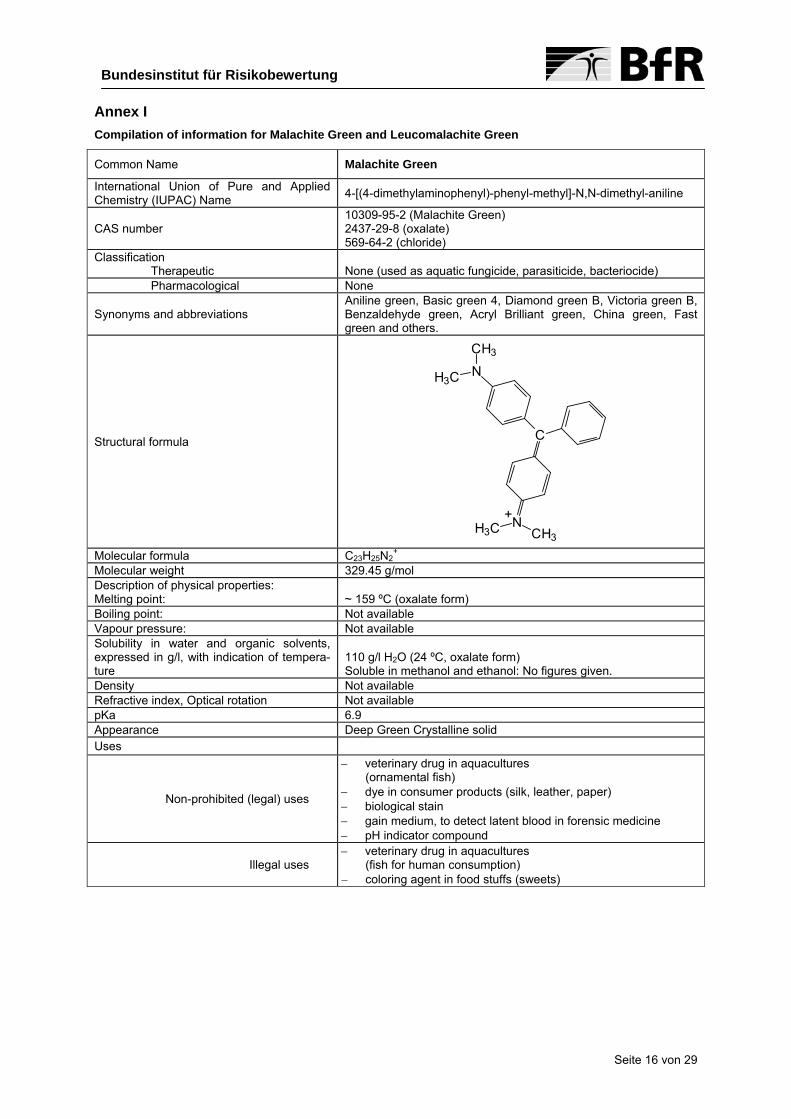
Annex I Compilation of information for Malachite Green and Leucomalachite Green
Common Name Malachite Green
International Union of Pure and Applied Chemistry (IUPAC) Name 4-[(4-dimethylaminophenyl)-phenyl-methyl]-N,N-dimethyl-aniline
CAS number 10309-95-2 (Malachite Green) 2437-29-8 (oxalate) 569-64-2 (chloride)
Classification Therapeutic
None (used as aquatic fungicide, parasiticide, bacteriocide)
Pharmacological None
Synonyms and abbreviations Aniline green, Basic green 4, Diamond green B, Victoria green B, Benzaldehyde green, Acryl Brilliant green, China green, Fast green and others.
Structural formula
+CH3
H3C N
C
N
CH3
H3C
Molecular formula C23H25N2
+ Molecular weight 329.45 g/mol Description of physical properties: Melting point:
~ 159 ºC (oxalate form)
Boiling point: Not available Vapour pressure: Not available Solubility in water and organic solvents, expressed in g/l, with indication of tempera-ture
110 g/l H2O (24 ºC, oxalate form) Soluble in methanol and ethanol: No figures given.
Density Not available Refractive index, Optical rotation Not available pKa 6.9 Appearance Deep Green Crystalline solid Uses
Non-prohibited (legal) uses
− veterinary drug in aquacultures (ornamental fish)
− dye in consumer products (silk, leather, paper) − biological stain − gain medium, to detect latent blood in forensic medicine − pH indicator compound
Illegal uses − veterinary drug in aquacultures
(fish for human consumption) − coloring agent in food stuffs (sweets)
Seite 16 von 29
Seite 17 von 29
Bundesinstitut für Risikobewertung
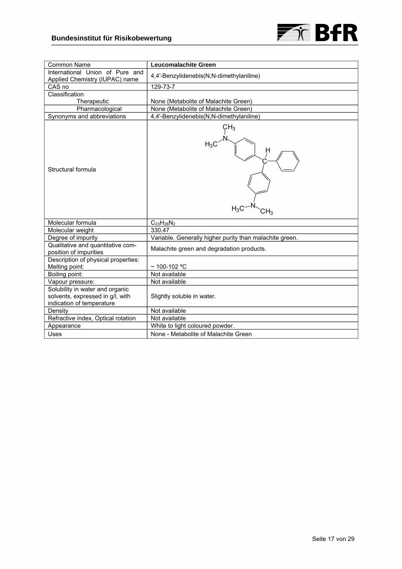
Common Name Leucomalachite Green International Union of Pure and Applied Chemistry (IUPAC) name 4,4′-Benzylidenebis(N,N-dimethylaniline)
CAS no 129-73-7 Classification
Therapeutic None (Metabolite of Malachite Green)
Pharmacological None (Metabolite of Malachite Green) Synonyms and abbreviations 4,4'-Benzylidenebis(N,N-dimethylaniline)
Structural formula CH
N
N
CH3
H3C
H3C CH3 Molecular formula C23H26N2 Molecular weight 330.47 Degree of impurity Variable. Generally higher purity than malachite green. Qualitative and quantitative com-position of impurities Malachite green and degradation products.
Description of physical properties: Melting point:
~ 100-102 ºC
Boiling point: Not available Vapour pressure: Not available Solubility in water and organic solvents, expressed in g/l, with indication of temperature
Slightly soluble in water.
Density Not available Refractive index, Optical rotation Not available Appearance White to light coloured powder. Uses None - Metabolite of Malachite Green

Annex II Overview of reported residues of Malachite Green (MG) and Leucomalachite Green (LMG) in fish and other aqua-cultured products
References Fish types Year Residue concentration [µg/kg] MG LMG Sample origin Detection
2005 5.6 and 6.11 Imported to Denmark
2005 2.72
2003 23 28³ 1991 4 and 51, 4
1989 5-175 not analyzed
Farmed fish
1988 15-2146 not analyzed
Denmark
Eel 2002 1-300 > 100 China
Rasmussen (2007)
Eel, Trouts, Seatrouts 2000 < 47 Denmark
-
2002 0.95 Fresh water trout (caviar) 2003 0.73 Tittlemier et al.
(2007) Shrimp 2002 1.2
Canada LC-MS/MS
LAVES (2005a) Trout (Sweden) 619 - HPLC LAVES (2005b) Eel (China) 3911 - -
Eel-based products 4500 Xiaomin (2005)
Fresh water fish
900 Local supermarket
(Hong Kong) -
1 2 positive samples of 5 samples 2 1 positive sample of 117 samples 3 1 positive sample of 23 samples 4 2 positive samples of 49 samples 5 6 positive samples of 20 samples 6 13 positive samples of 49 samples 7 4 positive samples of 20 samples
Seite 18 von 29

References Fish types Year Residue concentration [µg/kg] MG LMG Sample origin Detection
Trout 24 0.15 Scherpenisse and Bergwerff (2005) Pangasius
7
Local retailer Utrecht LC-MS/MS
Valle et al. (2005) Salmon (muscle) < 0.15-7.08 Chile Pacific Ocean LC-UV/VIS
Trout 3 30 and 12 Silver Perch 28 110
Australia Food Standards (2005)
Basa 20059
21; 4; 23; 88; 8; 5; 29 Vietnam
LC-MS/MS or
GC-MS 1995 2-3510
1996 3-3111
1997 2-1212
Food Standards
(1999) Trout
1998 813 4-15014
- -
Eel 1.7-7.0
Rainbow trout 1.3-14.9
Fresh Salmon 0.2-2.9 Bergwerff and
Scherpenisse (2003)
Smoked Salmon
0.2
Utrecht (NL) local re-tailer& vendors
HPLC-UV/VIS Or
LC-MS/MS
Trout 0.2 0.2 Rushing and Thompson (1997) Catfish
0.2 0.1
Local supermarket (stored at – 20 °C) HPLC-UV/VIS
Fresh trout 1-440 Klein and Edel-häuser (1988) Deep-frozen trout
5-20
Stuttgart, Sigmaringen HPLC
8 Sum of MG and LMG residues, individual compounds not specified 9 10 positive samples of 60 samples 10 35 positive samples of 210 samples 11 15 positive samples of 208 samples 12 2 positive samples of 137 samples 13 1 positive sample of 27 samples 14 6 positive samples of 27 samples
Seite 19 von 29

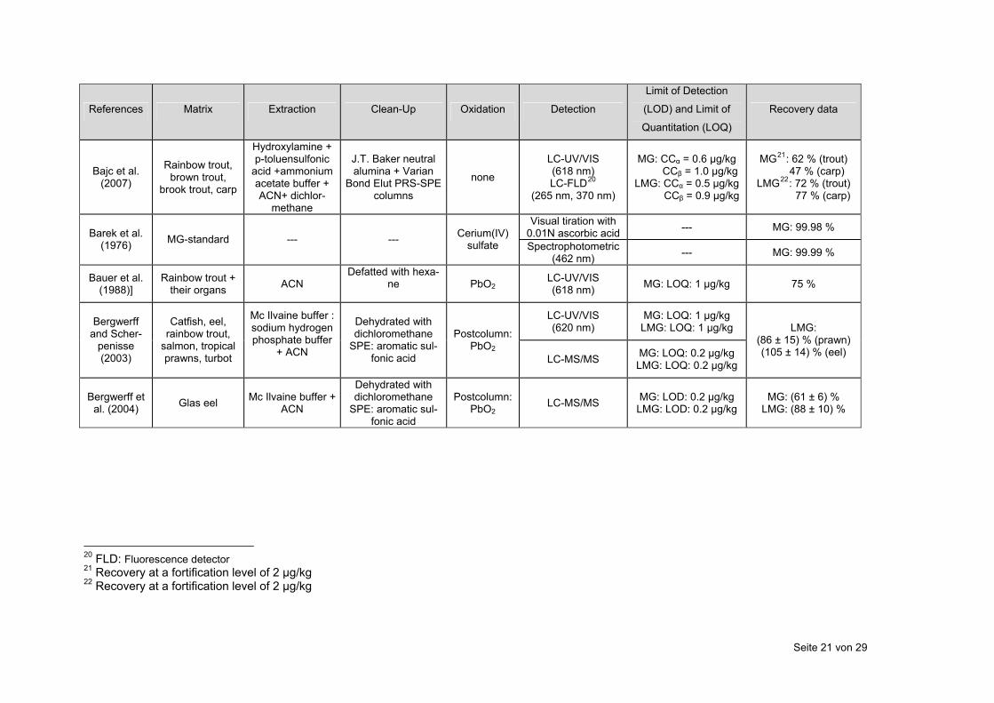
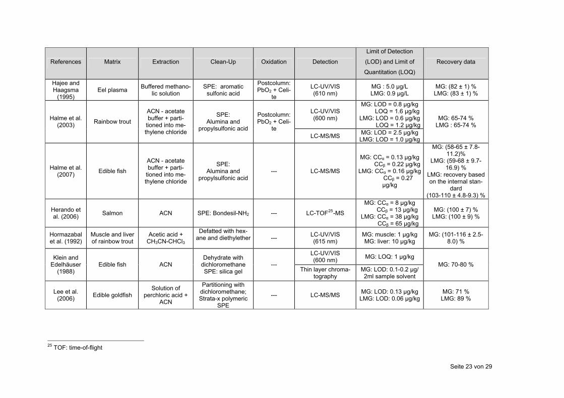
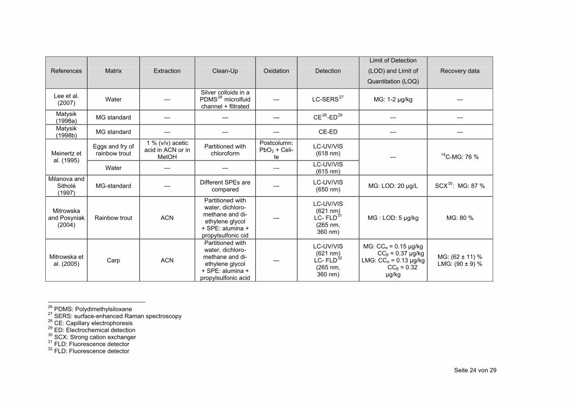
Annex III Overview of existing analytical methods for the determination of Malachite Green (MG) and Leucomalachite Green (LMG)
References Matrix Extraction Clean-Up Oxidation Detection
Limit of Detection
(LOD) and Limit of
Quantitation (LOQ)
Recovery data
Alexander (2006) MG standard --- --- --- flowing liquid-sheet
jet in CRDS15
MG: LOD: 71nM ---
Allen and Meinertz (1992)
MG oxalate stan-dard --- ---
Postcolumn: PbO2 + Ce-
lite
LC-UV/VIS (618 nm)
CMG16 and LMG: LOD: 0.12-0.28 ng ---
Allen et al. (1992) Water --- SPE: Diol column +
filtrated
Postcolumn: PbO2 + Celite
LC-UV/VIS (618 nm) MG: LOD 10 µg/L CMG: 95.4 %
LMG: 57.3 %
Allen et al. (1994)
rainbow trout: Eggs, fry, adult
muscle
1 % (v/v) acetic acid in ACN17 or
in MetOH18
Partitioned with chloroform
Postcolumn: PbO2 + Celi-
te
LC-UV/VIS (618 nm) ---
Eggs: MG: 85-98 % Fry: MG: 68 %
Muscle: MG: 66 %
Andersen et al.
(2005) Salmon ammonium aceta-
te buffer + ACN
P h artitioned witchloroform,
SPE: alumina + propylsulfonic acid
DDQ19 LC-UV/VIS (618 nm) MG: LOQ: 1 µg/kg
MG: ( 95.4 ± 11.1)%;(88.9 ± 2.6)%
LC-UV/VIS (618 nm) MG: LOQ: 1 µg/kg
Andersen et al.
(2006)
Channel catfish, rainbow trout, tilapia, basa,
atlantic salmon, tiger shrimps
ammonium aceta-te buffer + ACN
P h artitioned witchloroform,
SPE: alumina + propylsulfonic acid
DDQ
LC-MS MG: LOQ: 0.25 µg/kg
MG: Catfish: (88.7 ± 4.5)%Trout: (87.9 ± 3.4)%
Til % apia: (95.0 ± 4.1)Shrimps: (91.4 ±
4.2)%
Applied Bio-systems (2006)
MG- and LMG- standards --- --- --- LC-MS/MS
MG: LOD: 0.005 µg/kg LMG: LOD: 0.005
µg/kg ---
15 CRDS: cavity ring-down spectroscopy 16 CMG: chromatic malachite green 17 ACN: Acetonitril 18 MetOH: Methanol 19 DDQ: 2,3-dichloro-5,6-dicyano-1,4-benzoqinone
Seite 20 von 29
References Matrix Extraction Clean-Up Oxidation
Limit of Detection
Detection (LOD) and Limit of Recovery data
Quantitation (LOQ)
Bajc et al. (2007)
Rainbow trout, brown trout,
brook trout, carp
Hydroxylamine + p-toluensulfonic
acid +ammonium acetate buffer + ACN+ dichlor-
methane
J.T. Baker neutral alumina + Varian
Bond Elut PRS-SPE columns
none
LC-UV/VIS (618 nm) LC-FLD20
(265 nm, 370 nm)
MG: CCα = 0.6 µg/kg CCβ = 1.0 µg/kg LMG: CCα = 0.5 µg/kg CCβ = 0.9 µg/kg
MG21: 62 % (trout) 47 % (carp)
LMG22: 72 % (trout) 77 % (carp)
Visual tiration with 0.01N ascorbic acid --- MG: 99.98 % Barek et al.
(1976) MG-standard --- --- Cerium(IV) sulfate Spectrophotometric
(462 nm) --- MG: 99.99 %
Bauer et al. (1988)]
Rainbow trout + their organs ACN
Defatted with hexa-ne
PbO2 LC-UV/VIS (618 nm) MG: LOQ: 1 µg/kg 75 %
LC-UV/VIS (620 nm)
MG: LOQ: 1 µg/kg LMG: LOQ: 1 µg/kg Bergwerff
and Scher-penisse (2003)
Catfish, eel, rainbow trout,
salmon, tropical prawns, turbot
Mc Ilvaine buffer : sodium hydrogen phosphate buffer
+ ACN
Dehydrated with dichloromethane
SPE: aromatic sul-fonic acid
Postcolumn: PbO2
LC-MS/MS MG: LOQ: 0.2 µg/kg LMG: LOQ: 0.2 µg/kg
LMG: (86 ± 15) % (prawn) (105 ± 14) % (eel)
Bergwerff et al. (2004) Glas eel Mc Ilvaine buffer +
ACN
Dehydrated with dichloromethane
SPE: aromatic sul-fonic acid
Postcolumn: PbO2
LC-MS/MS MG: LOD: 0.2 µg/kg LMG: LOD: 0.2 µg/kg
MG: (61 ± 6) % LMG: (88 ± 10) %
20 FLD: Fluorescence detector 21 Recovery at a fortification level of 2 µg/kg 22 Recovery at a fortification level of 2 µg/kg
Seite 21 von 29

References Matrix Extraction Clean-Up Oxidation
Limit of Detection
Detection (LOD) and Limit of Recovery data
Quantitation (LOQ)
Ding et al. (2007) Roasted eel meat
Hydroxylamine hydrochloride + p-toluensulfonic acid + ammoniumace-tate buffer + CAN + dichlormethane
SPE: Oasis MCX car-
tridge --- LC-MS/MS MG: LOD: 0.004 µg/kg
LMG: LOD: 0.02 µg/kgMG23: (101 ± 3.7) % LMG24: (94 ± 11) %
Doerge et al. (1998b) Catfish, trout
ammonium acetate containing hydrxylamine HCl
+ p-toluenesulfonic
acid + ACN
Liquid-liquid parti-tioning + SPE --- LC-MS MG: LOD: 0.02 µg/kg
LMG: LOD: 0.5 µg/kg MG: 49.5 % LMG: 76.7 %
Dowling et al. (2007) Salmon Mc Ilvaine buffer +
ACN SPE: aromatic sul-
fonic acid --- LC-MS/MS
MG: CCα = 0.17 µg/kg CCβ = 0.30 µg/kgLMG: CCα = 0.15 µg/kg
CCβ = 0.35 µg/kg
MG: (103.7 ± 6.6) % LMG: (95.7 ± 4.7) %
thin layer chroma-tography No quantification Edelhäuser
and Klein (1986)
Edible fish ACN Defatted with hex-ane --- LC-UV/VIS
(600 nm) MG: LOD : 1 µg/kg
Effkemann (2007) Carp, trout ACN Dehydrated with
dichlormethane --- LC-MS/MS
MG: CCα = 1.2 µg/kg CCβ = 1.4 µg/kg LMG: CCα = 1.4 µg/kg CCβ = 1.7 µg/kg
---
Fink and Auch (1993) Trout ACN +
dichlormethane
Defatted with hex-ane
PbO2
LC-UV/VIS (618 nm)
MG : LOD = 3.6 µg/kg LOQ = 7.6 µg/kgLMG : LOD = 3.9 µg/kg
LOQ = 7.3 µg/kg
MG: 74 % LMG : 71%
Fornier de Violet et al.
(1995) Rainbow trout --- --- ---
Reflectance spectro-fluorimetry (640 nm)
MG: LOD: 5 µg/kg MG: 40 %
23 Recovery at a fortification level of 2 µg/kg 24 Recovery at a fortification level of 2 µg/kg
Seite 22 von 29
References Matrix Extraction Clean-Up Oxidation
Limit of Detection
Detection (LOD) and Limit of Recovery data
Quantitation (LOQ)
Hajee and Haagsma
(1995) Eel plasma Buffered methano-
lic solution SPE: aromatic
sulfonic acid
Postcolumn: PbO2 + Celi-
te
LC-UV/VIS (610 nm)
MG : 5.0 µg/L LMG: 0.9 µg/L
MG: (82 ± 1) % LMG: (83 ± 1) %
LC-UV/VIS (600 nm)
MG: LOD = 0.8 µg/kg LOQ = 1.6 µg/kg LMG: LOD = 0.6 µg/kg LOQ = 1.2 µg/kg
Halme et al. (2003) Rainbow trout
ACN - acetate buffer + parti-
tioned into me-thylene chloride
SPE: Alumina and
propylsulfonic acid
Postcolumn: PbO2 + Celi-
te LC-MS/MS MG: LOD = 2.5 µg/kg
LMG: LOD = 1.0 µg/kg
MG: 65-74 % LMG : 65-74 %
Halme et al. (2007) Edible fish
ACN - acetate buffer + parti-
tioned into me-thylene chloride
SPE: Alumina and
propylsulfonic acid --- LC-MS/MS
MG: CCα = 0.13 µg/kg CCβ = 0.22 µg/kgLMG: CCα = 0.16 µg/kg
CCβ = 0.27 µg/kg
MG: (58-65 ± 7.8-11.2)%
LMG: (59-68 ± 9.7-16.9) %
LMG: recovery based on the internal stan-
dard (103-110 ± 4.8-9.3) %
Herando et al. (2006) Salmon ACN SPE: Bondesil-NH2 --- LC-TOF25-MS
MG: CCα = 8 µg/kg CCβ = 13 µg/kg LMG: CCα = 38 µg/kg CCβ = 65 µg/kg
MG: (100 ± 7) % LMG: (100 ± 9) %
Hormazabal et al. (1992)
Muscle and liver of rainbow trout
Acetic acid + CH3CN-CHCl3
Defatted with hex-ane and diethylether
--- LC-UV/VIS
(615 nm) MG: muscle: 1 µg/kg MG: liver: 10 µg/kg
MG: (101-116 ± 2.5-8.0) %
LC-UV/VIS (600 nm) MG: LOQ: 1 µg/kg Klein and
Edelhäuser (1988)
Edible fish ACN Dehydrate with
dichloromethane SPE: silica gel
--- Thin layer chroma-
tography MG: LOD: 0.1-0.2 µg/ 2ml sample solvent
MG: 70-80 %
Lee et al. (2006) Edible goldfish
Solution of perchloric acid +
ACN
Partitioning with dichloromethane; Strata-x polymeric
SPE
--- LC-MS/MS MG: LOD: 0.13 µg/kg LMG: LOD: 0.06 µg/kg
MG: 71 % LMG: 89 %
25 TOF: time-of-flight
Seite 23 von 29
References Matrix Extraction Clean-Up Oxidation Detection
Limit of Detection
(LOD) and Limit of
Quantitation (LOQ)
Recovery data
Lee et al. (2007) Water ---
Silver colloids in a PDMS26 microlfuid channel + filtrated
--- LC-SERS27 MG: 1-2 µg/kg ---
Matysik (1998a) MG standard --- --- --- CE28-ED29 --- ---
Matysik (1998b) MG standard --- --- --- CE-ED --- ---
Eggs and fry of rainbow trout
1 % (v/v) acetic acid in ACN or in
MetOH
Partitioned with chloroform
Postcolumn: PbO2 + Celi-
te
LC-UV/VIS (618 nm) Meinertz et
al. (1995) Water --- --- --- LC-UV/VIS
(615 nm)
--- 14C-MG: 76 %
Milanova and Sitholé (1997)
MG-standard --- Different SPEs are compared --- LC-UV/VIS
(650 nm) MG: LOD: 20 µg/L SCX30: MG: 87 %
Mitrowska and Posyniak
(2004) Rainbow trout ACN
Partitioned with water, dichloro-methane and di-ethylene glycol
+ SPE: alumina + propylsulfonic cid
---
LC-UV/VIS (621 nm)
LC- FLD31 (265 nm, 360 nm)
MG : LOD: 5 µg/kg MG: 80 %
Mitrowska et al. (2005) Carp ACN
Partitioned with water, dichloro-methane and di-ethylene glycol
+ SPE: alumina + propylsulfonic acid
---
LC-UV/VIS (621 nm)
LC- FLD32 (265 nm, 360 nm)
MG: CCα = 0.15 µg/kg CCβ = 0.37 µg/kgLMG: CCα = 0.13 µg/kg
CCβ = 0.32 µg/kg
MG: (62 ± 11) % LMG: (90 ± 9) %
26 PDMS: Polydimethylsiloxane 27 SERS: surface-enhanced Raman spectroscopy 28 CE: Capillary electrophoresis 29 ED: Electrochemical detection 30 SCX: Strong cation exchanger 31 FLD: Fluorescence detector 32 FLD: Fluorescence detector
Seite 24 von 29

References Matrix Extraction Clean-Up Oxidation
Limit of Detection
Detection (LOD) and Limit of Recovery data
Quantitation (LOQ)
Möller (2007) Carp, rainbow trout
Mc Ilvaine buffer + ACN
Defatted with hex-ane,
SPE: aromatic sul-fonic acid
--- LC-MS/MS
MG: CCα = 0.65 µg/kg CCβ = 0.89 µg/kg LMG: CCα = 0.65 µg/kg
CCβ = 0.93 µg/kg
---
Ngamukot et al. (2006)
MG and LMG standards --- ---
BDD 33elec-trodes
(electrochem. oxidation)
FIA 34with ampho-teric detection
MG and LMG: LOD: 50 nM ---
Pourreza and Elhami (2007)
Fish farming and river water
Micelles of non-ionic surfactant
Triton-X-100 --- ---
LC-UV/VIS (618 nm)
MG: LOD: 1.2 µg/L MG: 99.4 %
Fish plasma ACN --- MG: LOD: 10 µg/kg LMG: LOD: 10 µg/kg
MG: (93 ± 6)% LMG: (87 ± 5) %
Plakas et al. (1995) Muscle of chan-
nel catfish
ACN-acetate buffer, reextracted with ACN, parti-tioned into me-thylene chloride
SPE: alumina + propylsulfonicacid
Postcolumn: PbO2
LC-UV/VIS (618 nm)
MG: LOD: 2 µg/kg LMG: LOD: 2 µg/kg
MG: (85 ± 4)% LMG: (95 ± 3)%
Roybal et al. (1995) Catfish
ACN buffer, partitioned into methylene chlo-
ride
SPE: Alumina and
propylsulfonic acid
Postcolumn: PbO2
LC-UV/VIS (618 nm)
MG: LOD = 0.1 µg/kg LOQ = 1.2 µg/kg LMG: LOD = 0.1 µg/kg LOQ = 1.5 µg/kg
MG: (72.7 ± 5.2)% LMG: (86.0 ± 6.8)%
Rushing and Thompson
(1997a) Catfish, trout
ACN buffer, partitioned into methylene chlo-
ride
SPE: Alumina and
propylsulfonic acid
Postcolumn: PbO2
LC-UV/VIS (588 nm)
Catfish: MG and LMG LOD = 0.45 µg/kg LOQ = 0.75 µg/kg
Trout : MG and LMG LOD = 0.83 µg/kg LOQ = 1.4 µg/kg
Catfish: MG and LMG(74.3 ± 3.3)%
Trout: MG and LMG: (71.8 ± 2.7)%
Rushing and Hansen (1997b)
Catfish
ACN buffer, partitioned into methylene chlo-
ride
SPE: Alumina and
propylsulfonic acid
Postcolumn: PbO2
LC-EC LC-UV/VIS
LC-FD
--- ---
33 BDD: Boron-doped diamond thin-film 34 FIA: Flow injection analysis
Seite 25 von 29

References Matrix Extraction Clean-Up Oxidation
Limit of Detection
Detection (LOD) and Limit of Recovery data
Quantitation (LOQ)
Safarik and Safarikova
(2002) Water --- Magnetic SPE --- LC-UV/VIS
(620 nm) MG: LOD: 0.5-1 µg/L MG: 22 %
Sagar and Smyth (1994) Water --- Cyano SPE ---
LC-amperometric detection at a carbon
fibre electrode MG: LOD: 0.28 µg/L CMG: 41-82 %
Scherpenisse and
Bergwerff (2005)
Finfish, pan-gasius, salmon,
tilapia, trout, victoria perch,
catfish, eel, tropi-cal prawns, tur-
bot
Mc Ilvaine buffer + ACN
Dehydrate with dichloromethane
SPE: aromatic sul-fonic acid
Postcolumn: PbO2
LC-MS/MS
MG and LMG for salmon:
CCα = 0.11 µg/kg CCβ = 0.18 µg/kg
LMG: From (86 ± 15)%;
prawn to (105 ± 14)%; eel
Stoev and Stoyanov
(2007) Edible fishs
Mc Ilvaine buffer + ACN, partitioned into methylene
chloride
Defatted with hex-ane,
SPE: SCX35
Postcolumn: PbO2
LC-DAD36, LC-MS/MS ---
MG: (42.7-51.6 ± 5.6-
21.9)% LMG:
(23.7-37.5 ± 10.8-20.4)%
Swarbick et al. (1997) Rainbow trout
Solvent of di-chlormethane,
ACN, perchloric acid
C18 SPE Postcolumn: PbO2
LV-UV/VIS (610 nm)
MG : LOD: 6 µg/kg LMG: 3 µg/kg
MG: 73-87% LMG: 89-98%
LC-UV/VIS (screening, 618 nm)
No LOD was estab-lished!
MG: (69.1-71.3 ± 4.4-
7.0)% LMG:
(91.2-97.3 ± 2.3-14.3)% Tarbin et al.
(1998) Trout Citrate buffer + ACN
Dichloromethane, SPE: SCX PbO2
LC-MS (confirma-tion)
MG: LOD: 0.4 µg/kg LMG: LOD: 0.5 µg/kg
MG: (61.0-64.2 ± 9.3-
11.7)% LMG:
(74.4-75.3 ± 5.0-14.0)%
35 SCX: Strong Cation Exchanging Agent 36 DAD: Diode array detection
Seite 26 von 29

References Matrix Extraction Clean-Up Oxidation
Limit of Detection
Detection (LOD) and Limit of Recovery data
Quantitation (LOQ)
Turnipseed et al. (1995a) Catfish
ACN buffer, partitioned into methylene chlo-
ride
SPE: Alumina and propyl-
sulfonic acid ---
Partical beam- LC-MS
MG and LMG: LOD: 20 µg/kg ---
Turnipseed et al. (1995b) Catfish
ACN buffer, partitioned into methylene chlo-
ride
SPE: Alumina and
propylsulfonic acid, cyano SPE
--- GC-MS LMG: LOQ: 5.0 µg/kg LMG: (95.9 ± 11.1)%
Turnipseed et al. (2005) Salmon
ACN buffer, partitioned into methylene chlo-
ride
SPE: Alumina and
propylsulfonic acid, cyano SPE
DDQ LC-MS MG: LOQ: 0.15 µg/kg MG and LMG: (86-109 ± 6.4-13)%
Turnipseed et al. (2006) Salmon
ACN buffer, partitioned into methylene chlo-
ride
SPE: Alumina and
propylsulfonic acid, cyano SPE
DDQ Comparison of:
LC-ND-APCI37-MS LC-ESI38-MS
LOD by: ND-APCI: 0.5 pg
ESI: 5 pg APCI: 500 pg
MG and LMG: (86-109 ± 6.4-13)%
Valle et al. (2005) Salmon Mc Ilvaine buffer +
ACN
Dichloromethane, SPE: Alumina and propylsulfonic acid
Precolumn: PbO2
LC-MS LOD: 0.15 µg/kg MG: (70 ± 3.1)% LMG: (85 ± 1.3)%
Van de Riet et al. (2005) Salmon Perchloric acid +
ACN SPE: octadecyl C18 --- LC-MS/MS
MG: LOD = 0.1 µg/kg LOQ = 0.3µpg/kg LMG: LOD = 0.1 µg/kg LOQ = 0.3 µg/kg
MG: 98 % LMG: 81 %
37 ND-APCI: No-discharge atmospheric pressure chemical ionization 38 ESI: Electospray ionization
Seite 27 von 29

References Matrix Extraction Clean-Up Oxidation
Limit of Detection
Detection (LOD) and Limit of Recovery data
Quantitation (LOQ)
Wu et al. (2007)
Grass carp, eel, salmon, shrimp,
shellfish
Mc Ilvaine buffer + p-toluensulfonic acid + TMPD39 +
ACN
SPE: OASIC MCX SPE
columns --- LC-MS/MS
MG:[ µg/kg] CCα CCβ Eel 0.06 0.09 Salmon 0.07 0.12 Shrimp 0.05 0.08 Shellfish 0.08 0.13 Grass carp 0.04 0.04 LMG:[ µg/kg] CCα CCβ Eel 0.04 0.06 Salmon 0.04 0.07 Shrimp 0.04 0.07 Shellfish. 0.02 0.04 Grass carp 0.05 0.09
MG [ %]40: Eel 101.1 Salmon 85.9 Shrimp 97.9 Shellfish 100.2 Grass carp 102.0 LMG [%]41: Eel 96.6 Salmon 105.2 Shrimp 99.6 Shellfish. 102.1 Grass carp 95.4
Xue and Yeung (1993) MG standard --- --- ---
On-column double –beam laser absorp-tion detection for CE
MG: LOD: 20 nM ---
Yang et al., (2007) Edible fish
McIlvaine buffer + ACN + dichlor-
methane
Anion exchanger AG 1 resin - ELISA MG and LMG:
LOD: 0.05 µg/L MG: 71-108 %
LMG: 62-105 %
39 TMPD: N,N,N´,N´-tetramethyl-1,4-phenylenedi-amine dihydrochloride 40 Recovery of MG [%] at the spiking level of 2 µg/kg 41 Recovery of LMG [%] at the spiking level of 2 µg/kg
Seite 28 von 29

Seite 29 von 29
References Matrix Extraction Clean-Up Oxidation Detection
Limit of Detection
(LOD) and Limit of
Quantitation (LOQ)
Recovery data
Zhu et al. (2007) Edible fish
ACN + ammonium acetate buffer,
partitioned against methylene chlo-
ride
SPE: Alumina and
propylsulfonic acid
Postcolumn: PbO2
LC-MS/MS MG and LMG: LOD: 0.5 µg/kg
MG and LMG: LOD: (77.6-98.1 ±
8.2)%