cmc review session 6k - critical care nursing · session 6: renal abnormalities ... 1 amp dextrose...
TRANSCRIPT
CMC Certification Review Course: Handout
Session #: 6
Renal Abnormalities, Endocrine Abnormalities, Hematologic Abnormalities, Multisystem Abnormalities
Presented by:
Leanna R. Miller RN, MN, CCRN-CSC, PCCN-CMC, CEN, CNRN, CMSRN, NP
An AACN Critical Care Publication 101 Columbia Aliso Viejo, CA 92656-1491 ©2013 American Association of Critical-Care Nurses All rights reserved. AACN grants permission for a single individual to print one copy of this electronic publication. No additional copies are permitted. No part of this electronic publication may be reproduced, uploaded, stored in a retrieval system, or transmitted, in any form or by any means (electronic, photocopying, recording or otherwise) without the prior written permission of AACN.
3/28/2013
1
EXIT Closes themodule
BACK Displays
previous slide
PAUSE/PLAY Pauses or
resumes audio
NEXT Displays next slide
PROGRESS BAR Purple shows your progress within the module
AUDIOMutes
audio for entire
module
TABLE of SLIDESNavigate to any slide by clicking on its tab.
CURRENT SLIDE
COMPLETED SLIDE
BOOKMARK
COMPLETED/TOTAL TIME
How to use this module:
CMC Review CourseSession 6:
Renal AbnormalitiesEndocrine Abnormalities
Hematologic AbnormalitiesMultisystem Abnormalities
Leanna R. Miller
RN, MN, CCRN-CSC, PCCN-CMC, CEN, CNRN, CMSRN, NP

3/28/2013
2
� Normal value
� 136–145 mEq/L
� Critical value
� < 120 mEq/L
� > 160 mEq/L
Sodium
Renal Abnormalities
� Decreased water intake
� Hypertonic IV fluids or tube feedings
� Fluid losses
� Osmotic diuresis
� Hyperosmolar hyperglycemia state (HHS)
� Diabetes insipidus
Hypernatremia: Etiology
Renal Abnormalities
� Extreme thirst
� Tachycardia
� Low-grade fever
� Disorientation
� Lethargy progressing to coma
� Seizures
Hypernatremia: Symptoms
Renal Abnormalities

3/28/2013
3
� Prolonged diuretic therapy
� Diaphoresis
� GI losses
� Hypotonic solutions
� Syndrome of inappropriate antidiuretic hormone
(SIADH)
Hyponatremia: Etiology
Renal Abnormalities
� Headache
� Lightheadedness
� Confusion
� Muscle cramps
� Convulsions
� Coma
Hyponatremia: Symptoms
Renal Abnormalities
� Normal value
� 3.5–5.5 mEq/L
� Critical values
� < 2.5 mEq/L
� > 6.5 mEq/L
Potassium
Renal Abnormalities

3/28/2013
4
� Increased intake
� Trauma
� Acidosis
� Kidney failure
Hyperkalemia: Etiology
Renal Abnormalities
� Central nervous system � Hyperactive reflexes� Paresthesia� Paralysis
� Cardiovascular � Tall, peaked T waves� Bradycardia� Escape beats� Prolonged PR interval� Asystole� Ventricular fibrillation
Hyperkalemia: Symptoms
� GI
� Abdominal cramps
� Diarrhea
� Intestinal ileus
� Neuromuscular
� Weakness
� Cramps
� Twitching
Renal Abnormalities
� Decreased intake
� Intracellular shift
� Increased GI loss
� Increased urinary loss
� Aldosterone excess
Hypokalemia: Etiology
Renal Abnormalities

3/28/2013
5
� CNS
� Hypoactive reflexes
� Paresthesia
� CV
� Flattened T waves
� Prominent U waves
� Peaked P waves
� Prolonged PR interval
� Ventricular asystole or
fibrillation
� Hypotension
Hypokalemia: Symptoms
� GI
� Abdominal distention
� Ileus
� Anorexia
� Nausea and vomiting
� Neuromuscular
� Weakness
� Fatigue
� Cramps
� Other
� Respiratory arrest
� Digitalis toxicity
Renal Abnormalities
� Normal values
� 8.5–10.5 mg/dL
� Ionized: 4.5–5.6 mg/dL
� Critical value
� < 7.0 mg/dL
� > 14.0 mg/dL
Calcium
Renal Abnormalities
� Hyperparathyroidism
� Bone release
� Immobilization
� Multiple fractures
� Acidosis
� Albumin
� Excessive vitamin D
� Decreased renal excretion
Hypercalcemia: Etiology
Renal Abnormalities

3/28/2013
6
� CNS
� Lethargy
� Coma
� CV
� Shortened QT interval
� Bradycardia
� Heart blocks
� GI
� Constipation
� Nausea
� Vomiting
Hypercalcemia: Symptoms
� Neuromuscular� Hypoactive deep tendon
reflexes
� Weakness
� Renal� Polyuria
� Renal calculi
� Flank pain
� Thirst
� Dehydration
� Skeletal� Deep bone pain
� Pathological fractures
Renal Abnormalities
� Hypoparathyroidism
� Chronic renal failure
� Decreased intestinal absorption
� Increased binding
� Alkalosis
Hypocalcemia: Etiology
Renal Abnormalities
� CV� Prolonged QT interval
� GI� Diarrhea
� Nausea and vomiting
� Hematologic� Bleeding
� Other� Bronchospasm
� Seizures
Hypocalcemia: Symptoms
� Neuromuscular
� Irritability
� Convulsions
� Hyperactive deep
tendon reflexes
� Tetany
� + Chvostek
� + Trousseau
Renal Abnormalities
3/28/2013
7
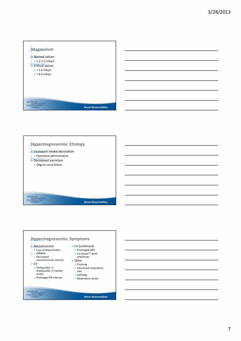
� Normal values
� 1.2–2.5 mEq/L
� Critical values
� < 1.0 mEq/L
� > 8.0 mEq/L
Magnesium
Renal Abnormalities
� Increased intake/absorption
� Parenteral administration
� Decreased excretion
� Oliguric renal failure
Hypermagnesemia: Etiology
Renal Abnormalities
� Neuromuscular
� Loss of deep tendon reflexes
� Decreased neuromuscular activity
� CV
� Tachycardia → bradycardia → cardiac arrest
� Prolonged PR interval
Hypermagnesemia: Symptoms
� CV (continued)
� Prolonged QRS
� Increased T wave amplitude
� Other
� Flushing
� Decreased respiratory rate
� Lethargy
� Respiratory arrest
Renal Abnormalities

3/28/2013
8
� Decreased intake/absorption
� Increased excretion
� Alcoholism
� Hyperaldosteronism
� Hyperparathyroidism
Hypomagnesemia: Etiology
Renal Abnormalities
� CNS� Confusion
� Coma
� Dizziness
� Neuromuscular� Twitches
� Muscle cramps
� Tetany
� Muscle weakness
Hypomagnesemia: Symptoms
� Other
� Anorexia
� Nausea
Renal Abnormalities
� Sudden decline in renal function
� Increase in BUN and creatinine
� Oliguria (< 400 mL/24 hours)
� Hyperkalemia and sodium retention
Acute Renal Failure: Definition
Renal Abnormalities

3/28/2013
9
RIFLE
ClassDefinition Frequency Need for
RRT
90-day
Mortality
Risk ↑ Scr x 1.5 or
↓ glomerular filtraJon rate
(GFR) > 25%
20-25% 1 % 8 %
Injury ↑ Scr x 2 or
↓ GFR > 50%
5-7 % 7% 21%
Failure ↑ Scr x 3 or
↓ GFR > 75%
1-3% 55% 33%
Loss (ARF) with complete loss of
kidney function > 4 weeks
1% 100% 40-50%
ESKD ESKD > 3 months 1% 100% >50%
RIFLE Criteria
Renal Abnormalities
� Prerenal� Reduced circulating
volume
� Diminished pump function
� Vasoconstrictor use
� Intrarenal� Intrarenal ischemia
� Nephrotoxins
� Immunologic processes
� Rhabdomyolysis
Acute Renal Failure: Pathophysiology
� Postrenal
� Renal calculi
� Urinary tract infection
� Enlarged prostate
� Trauma to plumbing
Renal Abnormalities
� Prerenal
� Decreased skin turgor
� Dry mucus membranes
� Weight loss
� Oliguria
� Hypotension
� Flat neck veins
� Tachycardia
Acute Renal Failure: Clinical Presentation
� Intrarenal
� Usually edema
� Postrenal
� Often anuria
Renal Abnormalities

3/28/2013
10
� Prerenal
� Urinalysis
� No abnormal casts
� Occasional hyaline casts
� BUN/Creatinine ratio
� > 20:1
� Fractional excretion of sodium (FENa),
renal failure index (RFI)
� < 1%
Acute Renal Failure: Diagnostics
Renal Abnormalities
� Intrarenal
� Urinalysis
� Abnormal casts
� BUN/Creatinine ratio
� 10–15:1
� FENa, RFI
� > 1%
Acute Renal Failure: Diagnostics
Renal Abnormalities
� Preventive
� Patients at risk
� Adequate hydration
� Avoid nephrotoxins
Acute Renal Failure: Management
Renal Abnormalities

3/28/2013
11
� Contrast-induced nephropathy
� Risk factors
� SBP < 80
� Heart failure
� NYHA III/IV
� IABP
� Diabetes
� Age > 75
� Preexisting renal disease
� Serum creatinine (SCr) > 1.5 mg/dL
� Creatinine clearance (CrC) < 60 mL/min
� Concomitant use of nephrotoxins
Acute Renal Failure: Management
Renal Abnormalities
� Contrast-induced nephropathy
� Low risk (no risk factors)
� No additional interventions required
� Moderate risk (1 risk factor)
� Check for decompensated heart failure, pulmonary edema,
or hyponatremia
� High risk (> 2 risk factors or SCr > 2.0 or CrC < 40)
� Check for decompensated heart failure, pulmonary edema,
or hyponatremia
Acute Renal Failure: Management
Renal Abnormalities
� Preventive
� Prevent and treat shock
� Monitor suspected patients
� Avoid infections
� Corrective and supportive
� Correct reversible causes
� Correct fluid excess or deficit
� Monitor for electrolyte imbalance
� Restore/maintain BP
� Maintain nutrition
� Assist with renal replacement therapy
Acute Renal Failure: Management
Renal Abnormalities

3/28/2013
12
� Pharmacological Strategies
� Loop diuretics
� Dopamine
� Fenoldopam
� N-Acetylcysteine
� Natriuretic peptides
Acute Renal Failure: Management
Renal Abnormalities
� Infection
� Dysrhythmias
� GI bleed
� Multiple organ failure
� Electrolyte abnormalities
Acute Renal Failure: Complications
Renal Abnormalities
Practice Exam Questions
Renal Abnormalities
3/28/2013
13
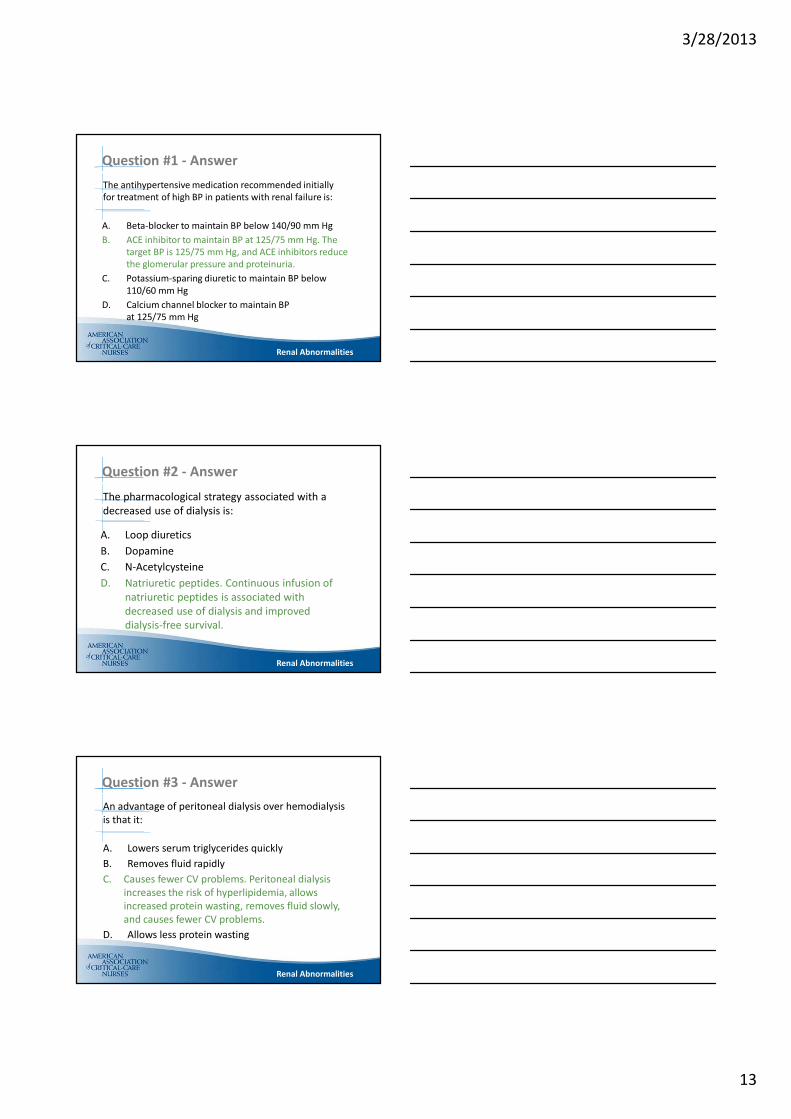
The antihypertensive medication recommended initially
for treatment of high BP in patients with renal failure is:
A. Beta-blocker to maintain BP below 140/90 mm Hg
B. ACE inhibitor to maintain BP at 125/75 mm Hg. The
target BP is 125/75 mm Hg, and ACE inhibitors reduce
the glomerular pressure and proteinuria.
C. Potassium-sparing diuretic to maintain BP below
110/60 mm Hg
D. Calcium channel blocker to maintain BP
at 125/75 mm Hg
Question #1 - Answer
Renal Abnormalities
The pharmacological strategy associated with a
decreased use of dialysis is:
A. Loop diuretics
B. Dopamine
C. N-Acetylcysteine
D. Natriuretic peptides. Continuous infusion of
natriuretic peptides is associated with
decreased use of dialysis and improved
dialysis-free survival.
Question #2 - Answer
Renal Abnormalities
An advantage of peritoneal dialysis over hemodialysis
is that it:
A. Lowers serum triglycerides quickly
B. Removes fluid rapidly
C. Causes fewer CV problems. Peritoneal dialysis
increases the risk of hyperlipidemia, allows
increased protein wasting, removes fluid slowly,
and causes fewer CV problems.
D. Allows less protein wasting
Question #3 - Answer
Renal Abnormalities
3/28/2013
14
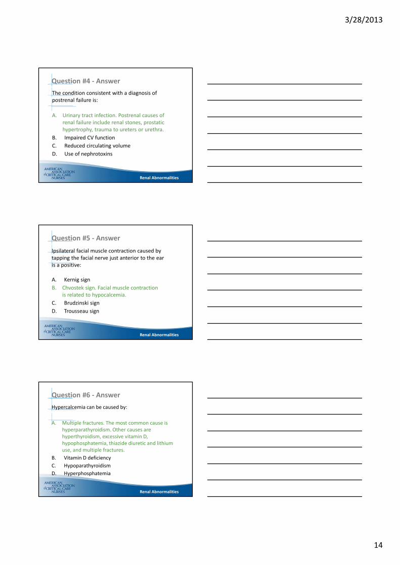
The condition consistent with a diagnosis of
postrenal failure is:
A. Urinary tract infection. Postrenal causes of
renal failure include renal stones, prostatic
hypertrophy, trauma to ureters or urethra.
B. Impaired CV function
C. Reduced circulating volume
D. Use of nephrotoxins
Question #4 - Answer
Renal Abnormalities
Ipsilateral facial muscle contraction caused by
tapping the facial nerve just anterior to the ear
is a positive:
A. Kernig sign
B. Chvostek sign. Facial muscle contraction
is related to hypocalcemia.
C. Brudzinski sign
D. Trousseau sign
Question #5 - Answer
Renal Abnormalities
Hypercalcemia can be caused by:
A. Multiple fractures. The most common cause is
hyperparathyroidism. Other causes are
hyperthyroidism, excessive vitamin D,
hypophosphatemia, thiazide diuretic and lithium
use, and multiple fractures.
B. Vitamin D deficiency
C. Hypoparathyroidism
D. Hyperphosphatemia
Question #6 - Answer
Renal Abnormalities
3/28/2013
15
Endocrine Abnormalities
� Metabolic disorder
� Hyperglycemia
� Defective insulin
� Production, secretion, or utilization
Diabetes Mellitus: Definition
Endocrine Abnormalities
� Type I
� Absolute or relative lack of insulin
� Type II
� Defect at cell level
� Impaired response to insulin
Diabetes Mellitus: Pathophysiology
Endocrine Abnormalities
3/28/2013
16
� Type I
� Viral
� Autoimmune
� Type II
� Heredity
� Obesity
Diabetes Mellitus: Etiology
Endocrine Abnormalities
� Hyperglycemia
� Weight loss
� Fatigue
� Polyuria, polydipsia, polyphagia
� Blurred vision
� Altered tissue response
� Delayed wound healing
� Recurrent infections (skin)
Diabetes Mellitus: Clinical Presentation
Endocrine Abnormalities
� Fasting blood glucose > 126 mg/dL
� Random blood glucose > 200 mg/dL
with classic symptoms
� Hgb A1c levels
Diabetes Mellitus: Diagnostics
Endocrine Abnormalities

3/28/2013
17
� Dietary control
� Exercise
� Medications
Diabetes Mellitus: Management
Endocrine Abnormalities
� Hypoglycemia
� Diabetic ketoacidosis (DKA)
� HHS
Diabetes Mellitus: Complications
Endocrine Abnormalities
� Serum glucose < 50 mg/dL
� Most common endocrine emergency
� < 35 mg/dL, brain is unable to extract O2
adequately
Hypoglycemia: Definition
Endocrine Abnormalities

3/28/2013
18
� Very old or very young
� Alcohol ingestion
� Diabetes
� Adrenal insufficiency
� Liver disease
� Drugs
� Propanolol
� Salicylates
� Sedatives
Hypoglycemia: Etiology
Endocrine Abnormalities
� Cool, diaphoretic skin
� Pale appearance
� Dilated pupils
� Confusion
� Combative behavior or coma
� Seizures
Hypoglycemia: Clinical Presentation
Endocrine Abnormalities
� Supplemental O2
� Monitor respiratory rate, breath sounds,
and signs of adequate oxygenation
� Determine blood glucose level
� 10–15 grams of fast-acting carbohydrate
� 1 amp dextrose 50% in water (D50W)
� Glucagon
� 0.5–2 mg IV
� Not effective in patients who have alcoholism
Hypoglycemia: Management
Endocrine Abnormalities

3/28/2013
19
� Peak insulin levels
� Regular: 3–4 hours
� NPH/lente: 4–18 hours
� Protamine zinc/ultralente: 18–30 hours
Hypoglycemia: Treatment
Endocrine Abnormalities
� Uncontrolled hyperglycemia
� Profound dehydration
� Electrolyte disturbances
� Acid-base abnormalities
DKA: Defining Characteristics
Endocrine Abnormalities
� Coma
� Abdominal pain
� Polydipsia, polyuria
� Kussmaul respirations
� Fruity breath
DKA: Signs and Symptoms
Endocrine Abnormalities
� Nausea and vomiting
� Weakness
� Weight loss
� Hypotension
� Ketonuria
� Tachycardia
3/28/2013
20
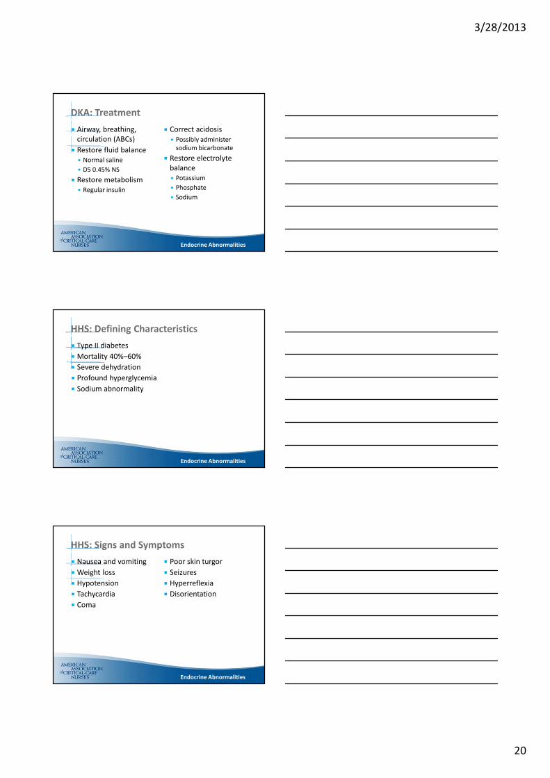
� Airway, breathing,
circulation (ABCs)
� Restore fluid balance
� Normal saline
� D5 0.45% NS
� Restore metabolism
� Regular insulin
DKA: Treatment
Endocrine Abnormalities
� Correct acidosis
� Possibly administer
sodium bicarbonate
� Restore electrolyte
balance
� Potassium
� Phosphate
� Sodium
� Type II diabetes
� Mortality 40%–60%
� Severe dehydration
� Profound hyperglycemia
� Sodium abnormality
HHS: Defining Characteristics
Endocrine Abnormalities
� Nausea and vomiting
� Weight loss
� Hypotension
� Tachycardia
� Coma
HHS: Signs and Symptoms
Endocrine Abnormalities
� Poor skin turgor
� Seizures
� Hyperreflexia
� Disorientation

3/28/2013
21
� ABCs
� Restore fluid balance
� Normal saline
� Restore metabolism
� Insulin
� Treat cause
HHS: Treatment
Endocrine Abnormalities
� Restore electrolytes
� Potassium
� Phosphate
� Monitor for cerebral
edema
� Thyroid gland does not make enough thyroid
hormone
� Iodine deficiency is most common cause
� 0.3% of the general American population have
overt hypothyroidism
� 4.3% have subclinical hypothyroidism
Hypothyroidism: Introduction
Endocrine Abnormalities
Type Origin Description
Primary Thyroid • Hashimoto’s thyroiditis
• Radioiodine therapy for hyperthyroidism
Secondary Pituitary • Does not create enough Thyroid Stimulating
Hormone (TSH)
• Damage to the pituitary gland such as
tumor, radiation, or surgery
Tertiary Hypothalamus • Fails to produce enough TSH
• Accounts for < 5% of cases
Hypothyroidism: Types
Endocrine Abnormalities

3/28/2013
22
� Early
� Cold intolerance
� Constipation
� Weight gain and water
retention
� Bradycardia
� Decreased sweating
Hypothyroidism: Signs and Symptoms
Endocrine Abnormalities
� Muscle cramps and
joint pain
� Dry, itchy skin
� Thin, brittle fingernails
� Poor muscle tone
� Depression
� Elevated serum
cholesterol
� Late
� Goiter
� Slow speech and a hoarse, breaking voice
� Dry puffy skin, especially face
� Low basal body temperature
� Thyroid-related depression
Hypothyroidism: Signs and Symptoms
Endocrine Abnormalities
� Primary
� TSH
� Free thyroxine (T4)
� Secondary and tertiary
� Free triiodothyronine (T3)
� Free T4
� Total T3
� Total T4
Hypothyroidism: Diagnosis
Endocrine Abnormalities

3/28/2013
23
� Free T3 from 24-hour urine
� Antithyroid antibiotics
� Serum cholesterol
� Prolactin
� Anemia testing, including ferritin
Hypothyroidism: Additional Diagnostics
Endocrine Abnormalities
� Levothyroxine (Synthroid)
Hypothyroidism: Treatment
Endocrine Abnormalities
� Overactive thyroid
� Produces too much T4
� Accelerates the body’s metabolism
� Can mimic other health problems,
making diagnosis difficult
Hyperthyroidism: Introduction
Endocrine Abnormalities
3/28/2013
24
� Conditions causing too much T4
� Graves’ disease
� Hyperfunctioning thyroid nodules
� Thyroiditis
Hyperthyroidism: Causes
Endocrine Abnormalities
� Sudden weight loss
� Tachycardia
� Increased appetite
� Nervousness, anxiety,
irritability
� Tremor
Hyperthyroidism: Signs and Symptoms
Endocrine Abnormalities
� Sweating
� Sensitivity to heat
� Change in bowel
patterns
� Fatigue, muscle
weakness
� Difficulty sleeping
� Enlarged thyroid
(goiter)
� Tissue and muscles behind the eyes swell
� Causes eyes to protrude
� Protruding eyeballs
� Red or swollen eyes
� Excessive tearing or discomfort
� Light sensitivity, blurry or double vision
� Inflammation or reduced eye movements
Hyperthyroidism: Graves’ Opthalmopathy
Endocrine Abnormalities

3/28/2013
25
� TSH levels
� T4 levels
� Radioactive iodine uptake test
� Thyroid scan
Hyperthyroidism: Diagnosis
Endocrine Abnormalities
� Radioactive iodine
� Antithyroid medications
� Βeta-blockers
� Thyroidectomy
Hyperthyroidism: Treatment
Endocrine Abnormalities
� Heart problems
� Brittle bones
� Graves’ opthalmopathy
� Graves’ dermopathy
� Thyrotoxic crisis
Hyperthyroidism: Complications
Endocrine Abnormalities
3/28/2013
26
� Combination of medical disorders that increase
risk of developing cardiovascular disease and
diabetes
� 25% of population
� Prevalence increases with age
Metabolic Syndrome: Introduction
Endocrine Abnormalities
� International Diabetes Foundation
� Raised triglycerides: > 150 mg/dL or treatment for lipid
abnormality
� Reduced high-density lipoprotein (HDL): < 40 mg/dL (male)
< 50 mg/dL (female) or treatment for lipid abnormality
� Elevated blood pressure (BP): systolic BP (SBP) > 130 mm Hg,
or diastolic BP (DBP) > 85 mm Hg, or treatment of BP
� Elevated fasting glucose: > 100 mg/dL or previously diagnosed
with type 2 diabetes
� Central obesity: waist: hip ratio > 0.9 (male);
> 0.85 (female) or body mass index > 30 kg/m2
Metabolic Syndrome: Definition
Endocrine Abnormalities
� Obesity
� Older age
� Sedentary lifestyle
� Degree of insulin resistance
� Stress
Metabolic Syndrome: Etiology
Endocrine Abnormalities

3/28/2013
27
� Signs and Symptoms
� Fasting hyperglycemia
� Hypertension
� Central obesity
� Decreased HDL
� Elevated triglycerides
Metabolic Syndrome
Endocrine Abnormalities
� Treatment
� Lifestyle changes
� Manage underlying
causes
� Manage cardiovascular
risk
Practice Exam Questions
Endocrine Abnormalities
The cornerstone of therapy in the management of DKA
is administering:
A. Sodium bicarbonate to correct acid-base imbalance
B. Insulin to correct metabolic abnormality. The priority
therapy is insulin replacement. If the pH is 7 or less, 1
amp of sodium bicarbonate is suggested. When the
potassium level reaches 4, potassium and phosphate
should be started. Fluid will be replaced hourly in
response to loss.
C. Normal saline to correct dehydration
D. Potassium phosphate to correct electrolyte imbalance
Question #1 - Answer
Endocrine Abnormalities

3/28/2013
28
The priority in the management of HHS is administering:
A. Sodium bicarbonate to correct acid-base imbalance
B. Insulin to correct metabolic abnormality
C. Normal saline to correct dehydration. Dehydration is
the priority problem in HHS. Since the patient has an
insulin resistance, insulin is used but only to slowly
lower the glucose. These patients are generally not
profoundly acidotic and usually stabilize with fluid
administration.
D. Potassium phosphate to correct electrolyte imbalance
Question #2 - Answer
Endocrine Abnormalities
The key criteria for identification of metabolic
syndrome is:
A. Elevated HDL
B. Hypoglycemia
C. Normal blood pressure
D. Central obesity. The criteria for metabolic
syndrome is low HDL, elevated fasting glucose,
hypertension, elevated triglycerides,
and central obesity.
Question #3 - Answer
Endocrine Abnormalities
The most common cause of hypothyroidism is:
A. Iodine deficiency. Iodine deficiency causes
primary hypothyroidism. This can also be
caused by acute stress and lithium use. Other
etiologies of hypothyroidism include pituitary
and hypothalamus abnormalities.
B. A pituitary tumor
C. Acute stress
D. Lithium use
Question #4 - Answer
Endocrine Abnormalities

3/28/2013
29
The most significant complication of antithyroid
medications is:
A. Osteoporosis
B. Congestive heart disease
C. Severe liver disease. Liver dysfunction is a limiting
factor in administration of antithyroid drugs (tapazole
and propylthiouracil). Osteoporosis and congestive
heart failure are complications of hyperthyroidism.
Renal dysfunction is not seen with these drugs.
D. Nephropathy
Question #5 - Answer
Endocrine Abnormalities
Hematologic Abnormalities
� Platelet (PLT) count < 100,000 per microliter (µL)
� Most common cause of bleeding disorders
Thrombocytopenia: Definition
Hematologic Abnormalities

3/28/2013
30
� Usually asymptomatic
� PLTs < 20,000/µL
� Petechiae
� Ecchymosis
� GI/GU bleed
� CNS bleed
Thrombocytopenia:Clinical Presentation
Hematologic Abnormalities
� Decreased hemoglobin, hematocrit,
and platelets
� Prolonged bleeding time, PT,
activated partial aPTT
Thrombocytopenia: Diagnosis
Hematologic Abnormalities
� Identify defects in hemostasis that can be
corrected
� Guide the management of hemostatic defects
that cannot be corrected
� Help manage the bleeding that cannot be
prevented
Thrombocytopenia: Admission Screening
Hematologic Abnormalities
3/28/2013
31
� Personal or family history of bleeding
� Abnormal bleeding associated with:
� Dental extractions
� Previous surgery
� Routine childhood trauma
Thrombocytopenia: History and Physical
Hematologic Abnormalities
� CBC
� Decreased hemoglobin and hematocrit
� Decreased PLTs
� PT/aPTT
� Bleeding time
Thrombocytopenia: Admission Screening
Hematologic Abnormalities
� CBC with coagulation studies
� Check for and correct hypothermia
� Review the history
� Review medications
Thrombocytopenia: Admission Screening
Hematologic Abnormalities
3/28/2013
32
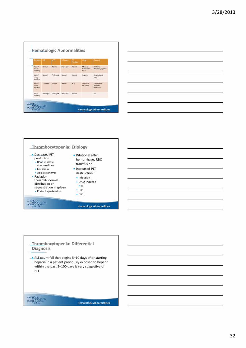
Symptom INR aPTT PLT Count PLT
Function
History Diagnosis
Major/
minor
bleeding
Normal Normal Decreased Normal Massive
transfusion;
fluids
Dilutional
thrombocytopenia
Major/
minor
bleeding
Normal Prolonged Normal Normal Negative Drug-induced
heparin
Major/
minor
bleeding
Increased Normal Normal N/A Vitamin K
deficiency
Liver disease,
warfarin,
antibiotics
Major
bleeding
Prolonged Prolonged Decreased Normal DIC
Hematologic Abnormalities
Hematologic Abnormalities
� Decreased PLT production
� Bone marrow abnormalities
� Leukemia
� Aplastic anemia
� Radiation therapyAbnormal distribution or sequestration in spleen
� Portal hypertension
Thrombocytopenia: Etiology
� Dilutional after
hemorrhage, RBC
transfusion
� Increased PLT
destruction
� Infection
� Drug-induced
� HIT
� ITP
� DIC
Hematologic Abnormalities
� PLT count fall that begins 5–10 days after starting
heparin in a patient previously exposed to heparin
within the past 5–100 days is very suggestive of
HIT
Thrombocytopenia: Differential Diagnosis
Hematologic Abnormalities

3/28/2013
33
� Diagnosis
� Thrombocytopenia
� > 50% fall in PLTs
� Thrombosis
� 50% VTE
� 25% PE
� ELISA assays
� Measure presence of antibodies to heparin-PF4 complex
� Heparin-induced platelet activation
� Serotonin release assay
Thrombocytopenia: Heparin-Induced Thrombocytopenia and Thrombosis (HITT)
Hematologic Abnormalities
Category 2 points 1 point 0 point
Thrombocytopenia > 50% fall 30%–50% fall < 30% fall
Timing of decrease
in PLT count
Days 5–10 or
< day 1 with
recent
heparin
> Day 10 or timing
unclear, or < day 1
if exposure 30–100
days
< Day 4
No recent
heparin
Thrombosis or
other sequelae
Proven
thrombosis,
skin
necrosis, or
acute
reaction
after heparin
Progressive,
recurrent, or silent
thrombosis
None
Other causes of
thrombocytopenia
None
evident
Possible Definite
Thrombocytopenia: 4 T System
Hematologic Abnormalities
� Discontinue the drug
� Administer direct thrombin inhibitor
� Hirudin
� Lepirudin
� Argatroban
HITT: Management
Hematologic Abnormalities

3/28/2013
34
� Serious bleeding disorder
� Thrombosis then hemorrhage
DIC: Definition
Hematologic Abnormalities
� Intrinsic Clotting
Cascade
� Endothelial injury
� Assessed by aPTT
DIC: Pathophysiology
� Extrinsic Clotting
Cascade
� Tissue thromboplastin
� Assessed by PT
Hematologic Abnormalities
� Obstetric
� Abruptio placentae
� Amniotic fluid embolus
� Eclampsia
� Hemolytic/ Immunologic
� Anaphylaxis
� Hemolytic blood reaction
� Massive blood transfusion
DIC: Etiology
� Infectious
� Bacterial
� Fungal
� Viral
� Rickettsial
� Vascular
� Shock
� Dissecting aneurysm
� Miscellaneous
� Emboli (fat)
� Aspirin poisoning
� GI disturbances� Pancreatitis
Hematologic Abnormalities

3/28/2013
35
� Decreased PLTs
� Decreased fibrinogen
� Increased PT and/or aPTT
� Increased d-dimer or fibrin split products (FSPs)
� Decreased ATIII
DIC: Laboratory Findings
Hematologic Abnormalities
� Treat underlying cause
� Surgery
� Antimicrobials
� Antineoplastics
� Stop thrombosis
� IV heparin
� ATIII
� Plasmapheresis
DIC: Management
� Administer blood
products
� PRBCs
� PLTs
� FFP
� Cryoprecipitate
Hematologic Abnormalities
� Hypovolemic shock
� Acute renal failure
� Infection
� ARDS
DIC: Complications
Hematologic Abnormalities

3/28/2013
36
� Treat underlying cause
� Administer PLTs
Thrombocytopenia: Treatment
Hematologic Abnormalities
� Hyperfibrinolysis
� Desmopressin (DDAVP®)
� Anifibrinolytics
� Aminocaproic acid (Amicar®)
Postoperative Bleeding
Hematologic Abnormalities
� Life-threatening hemorrhage
Thrombocytopenia: Complications
Hematologic Abnormalities

3/28/2013
37
� Definition
� Reduction in RBC concentration
� Causes
� Iron deficiency
� Thalassemia
� Anemia of chronic disease
Anemia
Hematologic Abnormalities
� Defects in production
� Increased destruction
� Increased loss of erythrocytes
Anemia: Etiology
Hematologic Abnormalities
� Mean corpuscular volume (MCV)
< 80 femtoliter (fL)
� Mean corpuscular hemoglobin
concentration (MCHC) < 32 gm/dL
� Examples
� Iron deficiency anemia
� Thalassemia
� Chronic lead poisoning
Anemia: Microcytic
Hematologic Abnormalities
3/28/2013
38
� MCV
� 80–100 fL
� MCHC
� 32–36 gm/dL
� Examples
� Acute blood loss
� Chronic disease
� Hemolytic
Anemia: Normocytic
Hematologic Abnormalities
� MCV > 130 fL and MCHC > 36 gm/dL
� Vitamin B12 deficiency
� Folate deficiency
� MCV 101–120 fL and MCHC > 36 gm/dL
� Liver disease
Anemia: Macrocytic
Hematologic Abnormalities
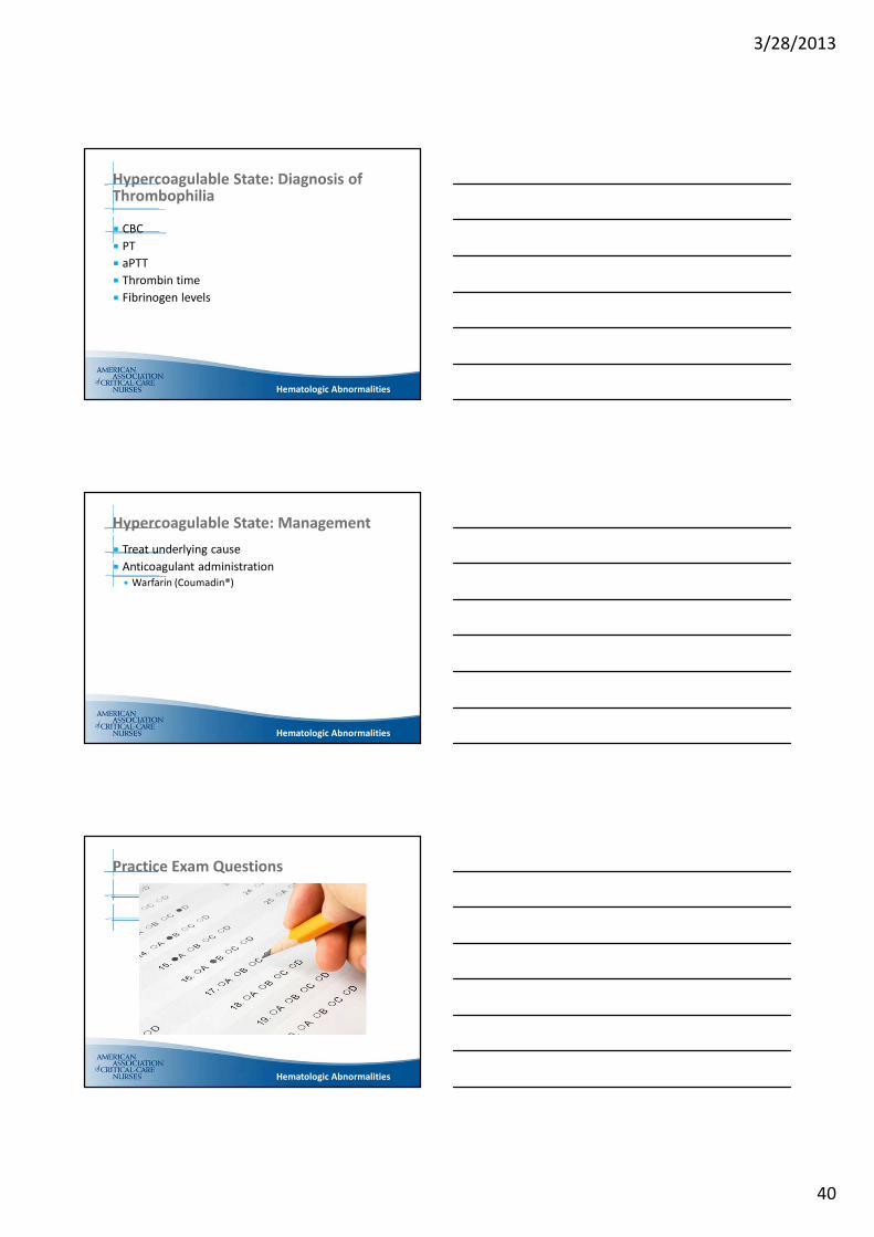
Test Iron Deficiency Chronic Disease Folic Acid Deficiency
Reticulocyte (ret)
count
Variable ↔ ↓
MCV ↓ ↔ or ↓ ↑
Iron ↓ ↓ ↑
Total iron-binding
capacity (TIBC)
↑ ↓ ↔
Ferritin ↓ ↔ or sl ↑ ↑
B12 ↔ ↔ ↔
Folate ↔ ↔ ↓
Transferrin ↑ ↔ or sl ↑ sl ↑
Anemia
Hematologic Abnormalities
3/28/2013
39
� Search for treatable disorders
� Deficiencies of iron, B12, folic acid
� Underlying infection, renal disease, inflammation,
or malignancy
� Other: bleeding disorder, alcohol abuse, hypothyroidism
� Symptomatic anemia
� RBC transfusion
� Erythropoiesis-stimulating agent
Anemia: Treatment
Hematologic Abnormalities
� Abnormality of blood coagulation
� Increases risk of thrombosis
� Found in 50% of people who have:
� DVT
� PE
Hypercoagulable State
Hematologic Abnormalities
� Congenital
� Type I defects
� ATIII deficiency
� Protein C deficiency
� Protein S deficiency
� Type II defects
� V Leiden
� Prothrombin G20210A
Hypercoagulable State: Etiology
� Acquired
� Autoimmune disease
� HIT
� Sickle cell anemia
� Metastatic cancer
� Nephrotic syndrome
� Pregnancy
� Obesity
Hematologic Abnormalities
3/28/2013
40
� CBC
� PT
� aPTT
� Thrombin time
� Fibrinogen levels
Hypercoagulable State: Diagnosis of Thrombophilia
Hematologic Abnormalities
� Treat underlying cause
� Anticoagulant administration
� Warfarin (Coumadin®)
Hypercoagulable State: Management
Hematologic Abnormalities
Practice Exam Questions
Hematologic Abnormalities
3/28/2013
41
The most common type of anemia in the United
States is:
A. Anemia of chronic disease
B. Folic-acid-deficiency anemia
C. Iron-deficiency anemia. Iron-deficiency anemia
is most often found in children and
nonpregnant women and is the most common
anemia worldwide.
D. Pernicious anemia
Question #1 - Answer
Hematologic Abnormalities
Hematologic Abnormalities
In iron-deficiency anemia, lab parameters should reveal:
A. Decreased MCV and increased TIBC. The lab values
associated with iron deficiency anemia are
decreased MCV, decreased iron, increased TIBC,
decreased ferritin, and increased transferrin.
B. Increased MCV and decreased ferritin
C. Increased transferrin and decreased serum iron
D. Decreased MCV and increased ferritin level
Question #2 - Answer
Hematologic Abnormalities
The lab data consistent with a diagnosis of DIC is:
A. Platelet count < 110,000
B. Fibrinogen < 100. The DIC panel shows a platelet
count < 80,000, D-dimer > 2.5, prolonged PT
and/or aPTT, and fibrinogen < 100.
C. D-dimer < 2
D. Normal PT and aPTT
Question #3 - Answer

3/28/2013
42
Hematologic Abnormalities
HIT may occur as rapidly as within 24 hours of heparin
therapy in patients:
A. Previously treated with heparin. HIT usually occurs
5–14 days after initiation of heparin therapy. If the
patient previously received heparin, HIT can occur
as rapidly as within 24 hours.
B. With DIC
C. With platelet dilution after resuscitation
D. Receiving continuous renal replacement therapy
Question #4 - Answer
Hematologic Abnormalities
The clotting that occurs with HIT begins when heparin binds
to:
A. Fragment crystallizable receptors on activated platelets
B. Immunoglobulin G (IgG) antibodies
C. Activated platelets
D. Platelet factor 4. Heparin binds to platelet factor 4,
which is released from an activated platelet. IgG
antibodies recognize and bind to this complex. This
complex binds to the Fc receptor on other platelets,
leading to platelet activation.
Question #5 - Answer
Hematologic Abnormalities
A risk for antiphospholipid syndrome is:
A. Heparin therapy
B. Smoking
C. Systemic lupus erythematosus. Antibodies are
developed against the cell membrane. It is found
in patients with autoimmune diseases, and risk
increases during pregnancy.
D. Obesity
Question #6 - Answer

3/28/2013
43
Hematologic Abnormalities
The lab finding consistent with DIC is:
A. Low fibrinogen level. In DIC the platelet count will be < 80,000, the ATIII level will be decreased, the FSP between 10–100, and the fibrinogen level will be < 100.
B. Elevated platelet level
C. Normal ATIII level
D. FSP > 100
Question #7 - Answer
Multisystem Abnormalities
� Progressive, cumulative organ dysfunction
� Secondary to inadequate substrate/O2
� Altered host homeostasis
Multiple Organ Dysfunction Syndrome (MODS): Definition
Multisystem Abnormalities
3/28/2013
44
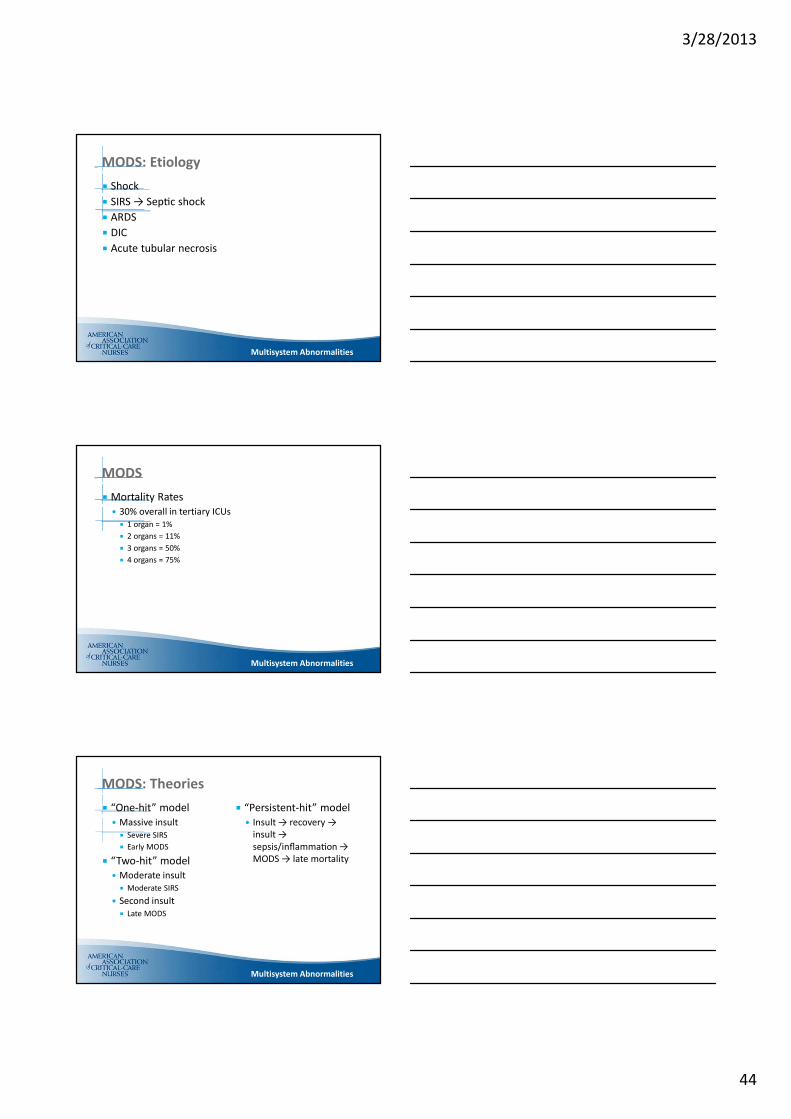
� Shock
� SIRS → SepJc shock
� ARDS
� DIC
� Acute tubular necrosis
MODS: Etiology
Multisystem Abnormalities
� Mortality Rates
� 30% overall in tertiary ICUs
� 1 organ = 1%
� 2 organs = 11%
� 3 organs = 50%
� 4 organs = 75%
MODS
Multisystem Abnormalities
� “One-hit” model
� Massive insult
� Severe SIRS
� Early MODS
� “Two-hit” model
� Moderate insult
� Moderate SIRS
� Second insult
� Late MODS
MODS: Theories
Multisystem Abnormalities
� “Persistent-hit” model
� Insult → recovery →
insult →
sepsis/inflammaJon →
MODS → late mortality

3/28/2013
45
� Cardiovascular
(1 or more)
� SBP < 60 mm Hg
� HR < 40 or > 200 bpm
� MAP < 50 mm Hg
� Cardiac arrest
� Continuous IV infusion
of inotropic agent to
maintain BP or CI
MODS: Assessing System Failure
Multisystem Abnormalities
� Pulmonary
(1 or more)
� RR > 70
� Mechanical ventilation
� pH < 7.2 with normal
PaCO2
� PaO2 < 40; PaCO2 > 65
� P/F ratio < 200 mm Hg
� Neurological
� GCS < 5 with no
sedation
� Fixed, dilated pupils
� ICP > 20 mm Hg for > 20
minutes
MODS: Assessing System Failure
Multisystem Abnormalities
� Renal (1 or more)
� Urine output < 0.5
mL/kg/h x 4 hours or
300 mL/24 h
� BUN > 100 mg/dL
� Serum creatinine > 1.2
� Requires renal
replacement therapy
� Hepatic
(presence of 3 or more)
� PT
� AST > 100 µ/L
� Serum albumin < 30 g/L
� Conjugated bilirubin > 10
mmol/L
MODS: Assessing System Failure
Multisystem Abnormalities
� Hematologic
(2 or more)
� Hgb < 5 g/dL
� WBCs < 3,000 cell/mm3
� PLTs < 20,000 mm3
� DIC
� Prolonged PT or aPTT
� FSPs > 10
� D-dimer > 2
� Fibrinogen < 100

3/28/2013
46
� SIRS
� Infection
� Trauma
� Ischemic or reperfusion
injury
� Single lactate > 4 or
more at initial
presentation
MODS: Identification of High-Risk Patient
Multisystem Abnormalities
� Failure to clear lactate levels during first 6hours associated with increased morbidity and mortality
� C-reactive protein (CRP)
� Brain natriuretic peptide (BNP)
� Procalcitonin
� Interleukin-6
� Protein C
� Altered mental status
� Band count > 5%
� Infection of lower respiratory tract
� Residence in nursing home
MODS: Identification of High-Risk Patient
Multisystem Abnormalities
� Ninth leading cause of death
� 50% develop due to hospital-associated infection
� Activation of immune/inflammatory response
Sepsis
Multisystem Abnormalities
3/28/2013
47
� Immune cellular interactions
� Endotoxin
� Arachidonic acid metabolites
� Tumor necrosis factor
� Complement
� Early systemic effects
� Alteration in myocardia function
� Maldistribution of blood flow
� Alteration in O2 delivery and utilization
SIRS
Multisystem Abnormalities
� Temperature� > 100.4°F or < 96.8°F (> 38°C or < 36°C)
� HR� > 90 bpm
� RR� > 20
� PaCO2 < 32 mm Hg
� WBC� > 12,000 or < 4,000
� > 10% bands
SIRS: Criteria
Multisystem Abnormalities
� Onset = minutes to hours
� Clinical signs of infection
� Improved CI
� Decreased afterload and increased HR
� Peripheral vasodilation
� Systemic edema
� Relative hypovolemia
Early Sepsis: Septicemia
Multisystem Abnormalities

3/28/2013
48
� Tachycardia
� Widened pulse pressure
� Tachypnea
� Decreased level of consciousness
� Thrombocytopenia
Early Sepsis: Signs and Symptoms
Multisystem Abnormalities
� Occurs most often during first 24 hours
of hospitalization
� Increase in mortality of 20%–46%
� Decreased O2 delivery and CV insufficiency
accompanies transition
� Usually not detected by vital signs or SIRS criteria
Transition From Early Sepsis to Severe Sepsis
Multisystem Abnormalities
� Global tissue hypoxia resulting from innate immune response Hypoxia stimulates further inflammation
� O2 delivery is insufficient to meet demands at cellular level
� Results in increased lactate levels
� SvO2 < 65% ScvO2 < 70% � Results in increased lactate
� Suggests presence of global tissue hypoxia� Greater extraction by tissues
O2 Transport and Utilization
Multisystem Abnormalities
3/28/2013
49
� Normal SvO2, ScvO2, and lactate levels suggest O2 supply
meets demands
� High SvO2, ScvO2 and lactate levels indicate that despite
adequate global systemic O2 delivery, tissues are unable
to extract O2
� Tissue hypoxia further activates endothelial mediators
� Loss of vascular integrity
� Inflammatory cytokines
� Procoagulants
� Reduced fibrinolysis
O2 Transport and Utilization
Multisystem Abnormalities
� Hypotension
� Metabolic acidosis
� Increased capillary permeability
→ systemic and pulmonary edema
� Hypoxemia
Early Septic Shock
Multisystem Abnormalities
� Severe LV dysfuncJon → decreased CI
� Increased peripheral vasoconstriction
� Cold extremities
� Hypotension
� Severe acidosis
Late: Cardiogenic Septic Shock
Multisystem Abnormalities

3/28/2013
50
� Support airway and ventilation
� Support CV function
� Prevent infection
� Provide nutritional support
Septic Shock: Treatment
Multisystem Abnormalities
� Resuscitation
� Cultures
� Antibiotics
� Early goal-directed therapy
� Delays in management of sepsis result in
high mortality rates and increased utilization
of hospital resources
First 6 Hours
Multisystem Abnormalities
� CVP 8–12 mm Hg
� MAP > 65 mm Hg
� Urine output > 0.5 mL/kg/hour
� ScvO2 > 70% or SvO2 > 65%
Early Goal-Directed Therapy
Multisystem Abnormalities

3/28/2013
51
Sepsis: Resuscitation Algorithm
Multisystem Abnormalities
Sepsis: Resuscitation Algorithm
Multisystem Abnormalities
� Initial Identification
� Increased serum lactate
� Identifies tissue hypoperfusion in patients who are not hypotensive
� Early antimicrobial therapy
� Empiric antibiotics within 4–8 hours of hospital presentation
� Surviving Sepsis Campaign recommends antibiotics within 1 hour
Management of IIR
Multisystem Abnormalities
� Source of infection and
local hospital sensitivity
and resistance patterns
� Surgical consultation
� Resistant organisms when
patients live in nursing
homes or are IV drug
users
3/28/2013
52
� Repletion of intravascular volume
� Rapid repeated 500 mL boluses of either
crystalloid or colloid (20 mL/kg)
� CVP 8–12 mm Hg
� 5% albumin or normal saline
� Found no significant difference in mortality
between the groups
Volume Therapy
Multisystem Abnormalities
� Norepinephrine 2–20 mcg/min
� Vasopressin 0.01–0.04 units/min
� Vasopressin and Septic Shock Trial (VASST) study
� Phenylephrine 40–300 mcg/min
� Dopamine 5–20 mcg/kg/min
Vasoactive Agents
Multisystem Abnormalities
� Adverse
consequences
� Splanchnic
hypoperfusion
� Excess tachycardia
� Coronary ischemia
� RBC replacement
� If ScvO2 remains < 70%
after optimization of
preload, afterload,
O2 sat
� Increase Hct to 30%
� Optimal erythrocyte
transfusion
� Fresh vs. stored blood
IIR Management
Multisystem Abnormalities
� Inotropic therapy
� Sepsis may be
accompanied by
myocardial suppression
in 10%–15% of patients
� Dobutamine titrated at
2.5 mcg/kg/min every
20–30 min to ScvO2 of
70%
� Milrinone
� Long half-life
� Accumulates in renal
failure
3/28/2013
53
� Decreasing O2 Consumption
� Intubation
� Sedation
� Analgesia
� Control fever
IIR Management
Multisystem Abnormalities
� Theoretical benefits
� Possible inhibition of
massive inflammatory
cascade
� Inflammatory cascade
leads to:
� Inadequate release or
response to
adrenocorticotropic
hormone (ACTH)
� Peripheral steroid
resistance at receptor level
Steroid Therapy
Multisystem Abnormalities
� On vasopressors
� Draw random cortisol
� < 25 mcg/mL give
corticosteroids
� Not on vasopressors
� Draw baseline random
cortisol level
� Cortisol stimulating test
� Levels at 30 and 60 min
� If difference is < 9 give
corticosteroids
� Low doses of hydrocortisone decreased
requirement of vasopressors and lowered
mortality
� Hydrocortisone 50 mg IV every 6 hours
� Dexamethasone 4 mg IV every 8 hours
� Fludrocortisone 100 mcg (PFT) daily
Steroid Therapy
Multisystem Abnormalities

3/28/2013
54
� IABP
� VAD
� ECMO
Mechanical Support of Heart
Multisystem Abnormalities
Practice Exam Questions
Multisystem Abnormalities
Multisystem Abnormalities
The parameter required to meet the SIRS criteria is:
A. HR 110 bpm. SIRS criteria: HR > 90;
WBC < 4,000 or > 12,000; PaCO2 < 32;
temperature < 36°C or > 38°C.
B. WBC 4,500
C. PaCO2 35 mm Hg
D. Temperature 37.4°C
Question #1 - Answer

3/28/2013
55
Multisystem Abnormalities
A sign or symptom consistent with alcohol withdrawal is:
The earliest definitive indicator of the development of
distributive shock is:
A. Increased HR
B. Widening pulse pressure. Distributive shock is
characterized by vasodilation, which results in
dropped diastolic pressure and widened pulse
pressure. Increased HR occurs in most shock (except
neurogenic). Altered LOC is a sign of all shocks as
perfusion to the brain decreases.
C. Decreased systolic pressure
D. Altered level of consciousness
Question #2 - Answer
Multisystem Abnormalities
The mortality rate associated with 3 organ failures
is:
A. 1%
B. 11%
C. 50%. One organ failure is 1% mortality,
2 organs failing is 11% mortality, 3 is 50%,
and 4 failing organs results in 75% mortality.
D. 75%
Question #3 - Answer
Multisystem Abnormalities
A sign or symptom consistent with alcohol withdrawal is:
The parameter consistent with CV failure is:
A. HR 180 bpm
B. MAP < 65 mm Hg
C. SBP < 90 mm Hg
D. Continuous use of dobutamine.
SBP < 60 mm Hg; HR < 40 or > 200;
MAP < 50 mm Hg; cardiac arrest or continuing
use of inotropic drugs to maintain CI or BP.
Question #4 - Answer
3/28/2013
56
Multisystem Abnormalities
A sign or symptom consistent with alcohol withdrawal is:
A patient with suspected septicemia had cultures obtained
and antibiotics started. The next intervention would be to
administer:
A. Normal saline to increase CVP to 8 mm Hg. The
priority is giving fluids. Next would be a vasopressor.
Later in management consider RBCs and steroids.
B. Packed red blood cells to boost O2-carrying capacity
C. Norepinephrine to improve BP
D. Corticosteroids to treat adrenal insufficiency
Question #5 - Answer
Multisystem Abnormalities
A sign or symptom consistent with alcohol withdrawal is:
The pathophysiology of distributive shock is:
A. The intravascular compartment fills beyond capacity
allowing fluid to leak out, compressing vital organs
B. The circulating volume causes excessive constriction of the
vessels, causing blood pooling
C. Widely fluctuating BPs stimulate vascular collapse, causing
severe alterations in peripheral perfusion
D. Although the circulating volume is intact, excessive vascular
dilation causes drastic drops in BP. The pathophysiology of
distributive shock is massive vascular dilation and possible
capillary leak leading to a drop in BP.
Question #6 - Answer