cloning and functional characterization of chondrichthyes ... · rhincodon typus estrogen receptors...
TRANSCRIPT

Instructions for use
Title Cloning and functional characterization of Chondrichthyes, cloudy catshark, Scyliorhinus torazame and whale shark,Rhincodon typus estrogen receptors
Author(s) Katsu, Yoshinao; Kohno, Satomi; Narita, Haruka; Urushitani, Hiroshi; Yamane, Koudai; Hara, Akihiko; Clauss, TonyaM.; Walsh, Michael T.; Miyagawa, Shinichi; Guillette, Louis J.; Iguchi, Taisen
Citation General and Comparative Endocrinology, 168(3): 496-504
Issue Date 2010-09-15
Doc URL http://hdl.handle.net/2115/47112
Type article (author version)
File Information GCE168-3_496-504.pdf
Hokkaido University Collection of Scholarly and Academic Papers : HUSCAP
1
Cloning and functional characterization of Chondrichthyes, cloudy
catshark, Scyliorhinus torazame and whale shark, Rhincodon typus
estrogen receptors
Yoshinao Katsu1,2,3, Satomi Kohno4, Haruka Narita3, Hiroshi Urushitani1, Koudai
Yamane5, Akihiko Hara5, Tonya M. Clauss6, Michael T. Walsh7, Shinichi Miyagawa1,2,
Louis J. Guillette Jr.4 and Taisen Iguchi1,2,*
1Okazaki Institute for Integrative Bioscience, National Institute for Basic Biology,
National Institutes of Natural Sciences, 5-1 Higashiyama, Myodaiji, Okazaki 444-8787,
Japan 2Department of Basic Biology, School of Life Science, Graduate University for
Advanced Studies, Okazaki 444-8787, Japan 3Department of Biological Sciences, Hokkaido University, Sapporo 060-0810, Japan 4Department of Biology, University of Florida, Gainesville, Florida 32611, USA 5Faculty of Fisheries Science, Hokkaido University, Hakodate, 041-8611, Japan 6Correll Center for Aquatic Animal Health, Georgia Aquarium, 225 Baker Street,
Atlanta, Georgia 30301, USA 7Aquatic Animal Health Program, College of Veterinary Medicine, University of Florida,
Gainesville, Florida 32611, USA
Short title: Molecular cloning and characterization of shark estrogen receptor
*To whom correspondence should be addressed:
Okazaki Institutes for Integrative Bioscience, National Institute for Basic Biology,
National Institutes of Natural Sciences, 5-1 Higashiyama, Myodaiji, Okazaki 444-8787,
Japan
TEL: +81-564-59-5235
FAX: +81-564-59-5236
e-mail: [email protected]

2
Abstract
Sex-steroid hormones are essential for normal reproductive activity in both sexes in all
vertebrates. Estrogens are required for ovarian differentiation during a critical
developmental stage and promote the growth and differentiation of the female
reproductive system following puberty. Recent studies have shown that environmental
estrogens influence the developing reproductive system as well as gametogenesis,
especially in males. To understand the molecular mechanisms of estrogen actions and to
evaluate estrogen receptor ligand interactions in Elasmobranchii, we cloned a single
estrogen receptor (ESR) from two shark species, the cloudy catshark (Scyliorhinus
torazame) and whale shark (Rhincodon typus) and used an ERE-luciferase reporter assay
system to characterize the interaction of these receptors with steroidal and other
environmental estrogens. In the transient transfection ERE-luciferase reporter assay
system, both shark ESR proteins displayed estrogen-dependent activation of transcription,
and shark ESRs were more sensitive to 17-estradiol compared with other natural and
synthetic estrogens. Further, the environmental chemicals, bisphenol A, nonylphenol,
octylphenol and DDT could activate both shark ESRs. The assay system provides a tool
for future studies examining the receptor-ligand interactions and estrogen disrupting
mechanisms in Elasmobranchii.
Keywords: shark; estrogen receptor; transactivation; evolution; environmental
chemicals
1. Introduction
Estrogens play important roles in the reproductive biology of all vertebrates
studied to date, including fish. Currently, the majority of the known actions of
estrogens are mediated by specific receptors that are localized in the nucleus of target
cells. These nuclear estrogen receptors (ESRs) belong to a superfamily of nuclear
transcription factors that include all other steroid hormone receptors including those for
progestogens, androgens, glucocorticoids, mineralocorticoids, the vitamin D receptor,
and the retinoic acid receptor (Blumberg and Evans, 1998). To date, in most
vertebrates, two distinct forms of ESR, ESR1 (ER) and ESR2 (ER) have been
isolated whereas in teleost fish, one ESR1 and two ESR2 (ESR2a and ESR2b) have
been identifiedThe ESR2b-form appears to be closely related to ESR2a suggesting a
3
gene duplication event that has occurred within the teleosts (Katsu et al., 2008). Thus,
the ancestral condition for the jawed vertebrates (Gnathostomata) is considered to have
had two forms of ESR, corresponding to ESR1 and ESR2 (Hawkins et al., 2000).
Chondrichthyes are jawed fish with skeletons consisting of cartilage rather than bone.
They are divided into two subclasses; Elasmobranchii including sharks, rays and skates,
and Holocephali (chimaera). Early study on steroids in Elasmobranchii ovaries
identified 17-estradiol, estrone and estriol (Wotiz et al., 1960; Chieffi and Lupo di
Prisco, 1963; Simpson et al., 1964). Further, Resse and Callard (1991) reported the
presence of estradiol-binding protein and characterized its ligand-specificity. However,
we were able to identify only one full length of ESR sequence of Chondrichthyes,
Squalus acanthias, in GenBank.
In vertebrates, 17-estradiol is the principle estrogen in circulation and
appears essential for normal ovarian development and function (Wallace, 1985).
Embryonic exposure to inhibitors of aromatase, the enzyme responsible for the
conversion of testosterone to 17-estradiol, causes genetic females to become
phenotypic males in chicken and at least one species of turtle (Elbrecht and Smith,
1992; Dorizzi et al., 1994). Likewise, embryonic exposure of various fishes,
amphibians or reptiles to 17-estradiol or estrogenic chemicals, pharmaceutical agents
or environmental contaminants can induce skewed sex ratios toward females (for
reviews, see Crews, 1996; Guillette et al., 1996; Tyler et al., 1998; Iguchi et al., 2001).
A number of studies strongly suggest that endogenous 17-estradiol acts as a natural
inducer of ovarian differentiation in non-mammalian vertebrates, including
Elasmobranchii (Devlin and Nagahama, 2002; Sinclair et al., 2002; Koob and Callard,
1999). However, the mechanisms of estrogen action on ovarian differentiation have
not been determined.
Disruption of the endocrine system has been shown to occur in wild fish
populations across the globe (Vos et al., 2000). Although the evidence for endocrine
disruption in wild fish is extensive, there are few verifications of a causal relation
between the presence of specific endocrine disrupting chemicals (ECDs) and a
functional effect (disruption). Several studies have now shown that wild freshwater
fish living in rivers heavily contaminated with treated wastewater treatment works
effluent have altered reproductive development and function (Hecker et al., 2002;
Jobling et al., 2002), and other investigations have shown that estrogens and estrogenic
4
chemicals are present in those effluents (Desbrow et al., 1998; Snyder et al., 2001) and
likely involved in these disruptions (Jobling et al., 2006; Katsu et al., 2007b). In
marine ecosystem, there are some reports of endocrine disruption in fish (Scott et al.,
2006, 2007), and the accumulations of various environmental chemicals with suspected
endocrine activity have been reported in sharks (Gelsleichter et al., 2005, 2006; Storelli
et al., 2006; Silva et al., 2007; Haraguchi et al., 2009). The mechanisms by which
estrogenic chemicals have an effect on sexual development and function in
Elasmobranchii, such as shark, however, are still to be determined.
In this study, we isolated cDNA clones encoding shark orthologs of ESR. The
transactivation function of shark ESR was subsequently determined by expressing these
cDNAs in transiently transfected HEK293 cells which were then used to determine
ligand-specificity of shark ESR with natural, synthetic and environmental estrogens.
The extensive global distribution of sharks in tropical to polar aquatic ecosystems and
their capability to tolerate highly contaminated environments make these animals an
interesting biological model for assessing endocrine disruptors in a wide range of
aquatic environments.
2. Methods
2.1. Animals
Cloudy catshark, Scyliorhinus torazame, were purchased from a local supplier. Whale
shark (Rhincodon typus) tissues were obtained for research purposes from the Georgia
Aquarium’s Correll Center for Aquatic Animal Health. All experiments in this study
involving sharks were carried out under the guidelines specified by the Institutional
Animal Care and Use Committee at the Gergia Aquarium’s Correll Center and
Hokkaido University.
2.2. Chemical reagents
17-Estradiol (E2), estrone (E1), estriol (E3), ethinylestradiol (EE2) and
diethylstilbestrol (DES) were purchased from Sigma-Aldrich Corp. (St. Louis, MO).
Bisphenol A (BPA) was purchased from Nacalai Tesque (Kyoto, Japan). The ER�
selective agonist, propyl pyrazole triol (PPT) and ER-selective agonist,
diarylpropionitrile (DPN) were obtained from Tocris Bioscience (Ellisville, MO).
4-nonylphenol (NP) and 4-tert-octylphenol (OP) were purchased from Tokyo Kasei

5
(Tokyo, Japan). Purity of BPA, NP and OP were over 99%. DDT and its metabolites
were purchased from Chem Service (West Chester, PA). Purity of p,p’-DDD,
p,p’-DDE, o,p’-DDE and p,p’-DDT were 99%, and o,p’-DDD and o,p’-DDT were
99.5%. All chemicals were dissolved in dimethylsulfoxide (DMSO). The
concentration of DMSO in the culture medium did not exceed 0.1%.
2.3. Molecular cloning of estrogen receptors
Two conserved amino acid regions in the DNA binding domain
(GYHYGVW) and the ligand binding domain (NKGM/IEH) of vertebrate ESRs were
selected for the design of degenerate oligonucleotides. The second PCR using the first
PCR amplicon, and nested primers that were selected in the DNA binding domain
(CEGCKAF) and the ligand binding domain (NKGM/IEH). As a template for PCR,
the first-strand cDNA was synthesized using total RNA isolated from liver. The
amplified DNA fragments were subcloned with TA-cloning plasmids pCR2.1-TOPO
(Invitrogen, Carlsbad, CA). The 5’ and 3’ ends of ER cDNAs were amplified by rapid
amplification of the cDNA end (RACE) using a SMART RACE cDNA Amplification
kit (BD Biosciences Clontech., Palo Alto, CA). Sequencing was performed using a
BigDye Terminator Cycle Sequencing kit (Applied Biosystems, Foster City, CA), and
analyzed on the ABI PRISM 377 automatic sequencer (Applied Biosystems).
2.4. Database and sequence analysis
All sequences generated were searched for similarity using Blastn and Blastp
at web servers of the National Center of Biotechnology Information (NCBI).
Phylogenetic analysis was demonstrated with the amino acid sequences for estrogen
receptor (ESR) from selected vertebrates and invertebrates as shown in Table S1. The
regions from immediately outside of the DNA binding domain through the ligand
binding domain of the sequences were trimmed according to the Conserved Domains
Database in NCBI. The trimmed sequences were aligned using the PRANK program,
a probabilistic multiple alignment program, which is good at insertions and deletions
(Löytynoja and Goldman, 2008). The aligned sequences were used for the estimation
of phylogenetic trees by the maximum likelihood analysis with the PhyML 3.0 program
with JTT substitution model, NNI (Nearest Neighbor Interchange) and SH-like aLRT
(approximately Likelihood-Ratio Test) branch supports (Guindon and Gascuel, 2003).

6
The estimated tree was edited on MEGA4 (Tamura et al., 2007). The aligned
sequences were also used for the rate shift analysis (Knudsen et al., 2003).
2.5. Construction of plasmid vectors
The full-coding region of shark ESR was amplified by PCR with KOD DNA
polymerase (TOYOBO Biochemicals, Osaka, Japan). The PCR product was
gel-purified and ligated into the pcDNA3.1 vector (Invitrogen). An estrogen-regulated
reporter vector containing four estrogen-responsive elements (4xERE), named
pGL3-4xEREtkLuc was constructed as described previously (Katsu et al., 2006).
2.6. Transactivation assays
To examine ligand (e.g., environmental estrogen) interactions with the cloned
estrogen receptors, HEK293 cells were seeded in 24-well plates at 5x10⁴ cells/well in
phenol-red free Dulbecco’s modified Eagle’s medium (Sigma-Aldrich) supplemented
with 10% charcoal/dextran treated fetal bovine serum (Hyclone, South Logan, UT).
After 24 h, the cells were transfected with 400 ng of pGL3-4xEREtkLuc, 100 ng of
pRL-TK (as an internal control to normalize for variation in transfection efficiency;
contains the Renilla reniformis luciferase gene with the herpes simplex virus thymidine
kinase promoter; Promega, Madison, WI), and 200 ng of pcDNA3.1-ESR using Fugene
6 transfection reagent (Roche Diagnostics, Basel, Switzerland) according to the
manufacturer’s instructions. After 4 h of incubation, various steroid hormones were
applied to the medium. After 44 h, the cells were collected, and the luciferase activity
of the cells was measured by a chemiluminescence assay with Dual-Luciferase Reporter
Assay System (Promega). Luminescence was measured using a Turner Designs
Luminometer TD-20/20 (Promega). Promoter activity was calculated as firefly
(Photinus pyralis)-luciferase activity / sea pansy (Renilla reniformis)-luciferase activity.
All transfections were performed at least three times, employing triplicate sample points
in each experiment. The values shown are mean ± SEM from three separate
experiments, and dose-response data and EC50 were analyzed using GraphPad Prism
(Graph Pad Software, Inc., San Diego, CA).
3. Results
3.1. Cloning of shark ER cDNAs

7
Using standard PCR techniques described above, partial DNA fragments were
amplified from catshark and whale shark liver RNA. For each species, only one type
of DNA fragment was obtained, and sequence analysis showed that these fragments had
similarity to spiny dogfish ESR2. Using the RACE technique, we were able to clone
full-length catshark and whale shark ESR cDNAs (GenBank Accession numbers,
catshark ESR: AB551715, and whale shark ESR: AB551716). The cDNA for catshark
ESR (Scyliorhinus torazame ESR, StESR) predicted 536 amino acids protein with a
calculated molecular mass of 60.5 kDa, whereas the whale shark ESR (Rhincodon typus
ESR, RtESR) is also predicted to be 536 amino acids but with a calculated molecular
mass of 60.8 kDa (Fig. 1). Using the nomenclature of Krust et al. (1986), the shark
ESR sequence can be divided into five domains based on its sequence identity to other
vertebrate ESRs (Fig. 2). When our catshark sequence (RtESR) is compared with
ESR1 from five other species (human, chicken, Xenopus, zebrafish, lungfish), catshark
ESR1 shared 29-28, 95-92, 33-27, 65-63, 19-13% identities to the A/B, C, D, E, and F
domains, respectively (Fig. 2A). In contrast, when RtESR is compared with the ESR2
of seven other vertebrate species, RtESR shared 45-32, 95-93, 38-29, 74-67, 28-16%
identities to the A/B, C, D, E, and F domains, respectively (Fig. 2B). Sequence
homology indicates that shark ESRs are more similar to the vertebrate ESR2 form than
the vertebrate ESR1 form. Using phylogenetic analysis, based on the protein
sequences of the C-, D- and E-domain, we were able to determine the relationship of
shark ESRs relative to other vertebrates (Fig. 3; Fig. S1). All three shark ESRs were
in the vertebrate ESR2 clade, and formed a group separate from bony vertebrates. No
ESR1 type receptor has been identified in cartilaginous fishes in the NCBI database nor
could we isolate one during this study. Lancelet ESR appears to be an ancient ESR
and ancestral to both forms (ESR1 and ESR2) of vertebrate ESRs (Fig. 3; Fig. S1).
Multiple sequence alignment and rate shift analysis revealed that most of the
functionally important sites were identical among the vertebrate ESRs except some of
the sites for the dimer interface (Fig. 4). Three of five non-conserved sites at 388, 416
and 442 for the dimer interface among vertebrates were positive at the rate shift analysis,
which indicates the potential sites to make the functional characteristics of each
subfamily, ESR1 and ESR2. Interestingly, sharks and hagfish (Myxine) ESR2 have the
tetrapod type residue (S) at 388 instead of the bony fish type (N) (Fig. 4).

8
3.2. Transcriptional activities of shark estrogen receptors by estrogens
A transactivation assay was used to determine whether shark ESRs display
estrogen-dependent transcriptional activity. ESRs activate transcription in an
estrogen-dependent manner through a variety of response sites, the most typical of
which is composed of two head-to-head AGGTCA half sites separated by three
nucleotides, an estrogen-response element, ERE (Zilliacus et al., 1995; Gruber et al.,
2004). To analyze the transcriptional activities of shark ESRs, we co-transfected a
reporter plasmid containing four EREs in front of a minimal tk promoter driving the
luciferase reporter gene (pGL3-4xEREtkLuc), together with a shark ESR encoding
expression plasmid and pRL-TK control plasmid (Kohno et al., 2008). We examined
the abilities of natural and synthetic estrogens to induce ESR-dependent transcriptional
activity. Both shark ESRs activated the expression of the luciferase reporter gene in a
dose-dependent manner for all estrogens examined (Fig. 5A and 5B). As shown in
Figure 5A and 5B, both shark ESRs exhibited significant transcriptional activity at a
concentration of 10-11 M E2. Concentration-response relationships were examined for
two other natural estrogens, E1 and E3 using the same assay system (Fig. 5A and 5B).
Both E1 and E3 activated shark ESR transcription, but were less effective (less potent)
compared with E2. Both synthetic estrogens, EE2 and DES, also stimulated luciferase
activity through the whale shark and catshark ESRs in a concentration-dependent
manner (Fig. 5). Based on EC50 values, both shark ESRs were more sensitive to E2
than to E1, E3, EE2 or DES (Table 1). Propyl pyrazole triol (PPT) and
diarylpropionitrile (DPN) are ESR1- and ESR2-selective agonists in mammals,
respectively (Stauffer et al., 2000; Meyers et al., 2001). These subtype selective ESR
agonists were used to examine and characterize shark ESRs. We found that both shark
ESRs are activated by DPN, the ESR2-agonist, but not PPT, the ESR1-agonist (Fig. 5C
and 5D). These results suggest the ligand-sensitivity of the shark ESRs isolated in this
study is similar to mammalian ESR2, but not ESR1.
3.3. Transcriptional activities of shark estrogen receptors by estrogenic chemicals
Considering the global contamination of aquatic ecosystems with various
persistent chemicals, such as organochlorines (e.g. DDT and its metabolites such as
DDE) and phenolic compounds (4-nonylphenol, 4-tert-octylphenol, and bisphenol A),
and the fact that a number of these chemicals have been shown to bind to the ESRs of

9
various vertebrates, we tested whether a similar phenomenon could be documented for
the shark ESRs identified here. First, we compared the ability of bisphenol A (BPA),
4-nonylphenol (NP), and 4-tert-octylphenol (OP) to induce shark ESR-dependent
reporter gene expression. The ESRs from both shark species activated the
transcription of the reporter gene when exposed to these three estrogenic chemicals;
shark ESRs were slightly more sensitive to NP than BPA and OP (Fig. 6). Next, we
examined whether the highly persistent and bioaccumulated/biomagnified
environmental contaminant DDT and its metabolites (p,p’-DDD, o,p’-DDD, p,p’-DDE,
p,p’-DDE, o,p’-DDT, and p,p’-DDT) could induce shark ESR-dependent reporter gene
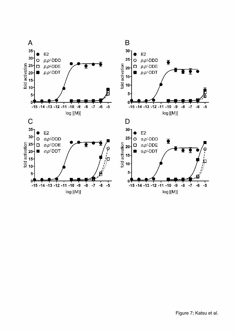
expression. Both shark ESRs were more sensitive to o,p’-forms than p,p’-forms (Figs.
7). Furthermore, o,p’-DDT was a more potent activator of shark ESRs than o,p’-DDD
and o,p’-DDE. These results are consistent with other vertebrate where ESR-induced
transcription by DDT and its metabolites has been reported. In order to truly
understand the implications of these assay results, the effects of estrogenic
environmental chemicals need to be studies using in vivo exposures on a model shark
species so that in vivo sensitivity can be fully assessed.
4. Discussion
Estrogens are implicated in a wide array of reproductive activities in
vertebrates, including gonadal differentiation, maturation of female reproductive tracts,
and reproductive behaviors (Iguchi et al., 2001; McLachlan, 2001; Moore et al., 2005).
In vertebrates, estrogens appear to induce both genomic actions through the nuclear ERs
and non-genomic cellular actions via ERs localized at the cell membrane that activate
G-protein coupled (Bjornstrom and Sjoberg, 2005). Reese and Callard (1991) reported
specific estrogen-interacted protein, perhaps estrogen receptor, from the little skate,
Raja erinacea. In 1997, a partial sequence for the nuclear estrogen receptor was
reported from Chondrichthyes, the smaller spotted catshark, Scyliorhinus canicula
(Escriva et al., 1997). Since then, only one additional sequence has been registered for
the spiny dogfish (GenBank accession No. AF147746). We report here the sequences
of two beta type ERs (ESR2) from the Elasmobranchii, one from the cloudy catshark
and the other from the whale shark. Although a growing literature exists on sequence
and evolutionary phylogeny for various vertebrate estrogen receptors (Bury and Strum,
2007; Howarth et al., 2008; Hu and Funder, 2006), few studies have examined the
10
estrogen receptor from Chondrichthyes, Elasmobranchii and Holocephali.
Shark ESRs isolated in this study belong to the vertebrate ESR2 family based on
sequence similarity, phylogenetic analysis, and characterization using specific agonists.
To date, there are no reports of an alpha-type ER (ESR1) nucleic and amino acid
sequences from Chondrichthyes. Do Chondrichthyes have alpha-type ER? Currently,
we have no answer of this question. Recently the elephant shark (Callorhinchus milii)
genome sequence project was initiated (Venkatesh et al., 2006; Venkatesh et al., 2007).
The elephant shark, also known as the elephant fish or ghost shark, is of the Holocephali
belonging to the Order Chimaeriformes and Family Callorhynchidae. When we
searched for ESR-like sequences in this database, we detected two amino acid
sequences, CAVCNDFASGY and CAVCSDYASGY corresponding to the DNA-binding
domain of the two vertebrate ESRs. The ESRs from three species of shark (spiny
dogfish, catshark, and whale shark) have the CAVCNDFASGY sequence. The
CAVCSDYASGY sequence, belonging to the ESR1 form, has two different amino acid
residues, S instead of N, and Y instead of F, that the elephant shark may express both
forms of ESR. Therefore, analysis of the Holocephali ESR will be useful for further
understanding estrogen receptor evolution in the Chondrichthyes. Moreover, two
types of ESRs in vertebrate were appeared by the gene duplication after the blanching of
lancelet ESR in our study, but it isn’t seen in Chondrichthyes. Since lamprey and
hagfish are very primitive vertebrates, we expected they could reveal how two types of
ESRs were appeared. However, lamprey and hagfish ESR belonged to ESR1 and
ESR2, respectively, although the support value was not high. To search both type of
ESR in these species or primitive vertebrates might be the key species to understand the
occurrences of ESR1 and ESR2 in vertebrates.
We examined the ability of newly identified shark ERs to transactivate and
ERE-dependent reporter gene. Both shark ESRs responded similarly to E2.
Relatively high concentrations of E1 and E3 were necessary for transactivation of the
shark ESRs compared to the induction by E2. This is similar to what we found other
vertebrates (Katsu et al., 2007a; 2007b; Naidoo et al., 2008; Katsu et al., 2010). We
found that E2 is the most potent transcriptional activator of shark ESR compared with
other natural and synthetic estrogens (Fig. 4A and 4B, Table 1). We found that DPN,
an ESR2-agonist, but not an ESR1-agonist, activated transcription of both shark ESRs,
suggesting that the shark ESRs are similar to mammalian ESR2. The receptor

11
dependent reporter gene assay system that we have used is suitable for the analysis of
ligand-induced ESR transactivation from a wide array of species. Additional studies
that focus on ontogenic and sexually dimorphic responses in several Elasmobranchii
species could provide insight into the function of these steroids during the life history of
Elasmobranchii species (Koob and Callard, 1999; Hamlett and Koob, 1999).
A significant number of contaminants have been shown to interact with the
ESRs of mammals and other vertebrates (Guillette et al., 2006). Furthermore, many of
these contaminants are persistent globally and potentially affect the developmental and
reproductive biology of various animal species from numerous ecosystems.
Concentrations of DDT and its metabolites were readily detectable in shark species
(Storelli et al., 2005, 2006; Silva et al., 2007). We examined possible interactions
between this pesticide and its metabolites (p,p’-DDD, o,p’-DDD, p,p’-DDE, o,p’-DDE,
p,p’-DDT, o,p’-DDT) and the ESRs from shark using our transactivation system. We
observed that o,p’-DDT could activate ESRs from both species of shark, consistent with
our findings with ESRs from other vertebrate species (Katsu et al., 2008). Further
studies should examine the molecular interactions between a species’ steroid receptors
and endogenous ligands as well as common contaminants in that species’ environment.
Our studies provide important tools with which to study the activation of ESRs and the
potential effects of environmental chemicals. For example, estrogens are not only
important for adult reproductive function but also for sex determination in many species,
thus a better understanding of the potential role of various contaminants as
environmental estrogens is critical for the ecotoxicology and conservation biology of
wildlife species.
In summary, we cloned and sequenced an ESR from two Elasmobranchii species
and then examined their ability to be activated by endogenous and xenobiotic estrogens.
This is the first report to characterize the ligand-dependent activation of cloned ESR
from Elasmobranchii. The Reproductive biology and endocrinology of
Elasmobranchii were reviewed in order to derive a working hypothesis that explaines
the complex nature of endocrine patterns observed in species utilizing disparate
reproductive modes (Koob and Callard, 1999, Pierantoni et al., 2002). Our data
provide useful tools for future studies examining the basic endocrinology of
Elasmobtranchii steroid hormone receptors. We have demonstrated that
transactivation assays, using ESRs from non-traditional model species, such as these
12
Elasmobranchii, provide important initial insights into potential risks from
environmental xenoestrogens and confirm that such an approach could provide
important data to aid in species conservation and ecological risk assessments. Our
study has provided basic molecular data useful in examining the role of ESRs in future
studies, such as those examining gonadal development, reproductive biology and
evolutionary endocrinology.
Acknowledgments
This work was supported by Grants-in-Aid for Scientific Research 20570064 (Y.K.)
and 19370027 (T.I.) from the Ministry of Education, Culture, Sports, Science and
Technology of Japan, and a grant for the United Kingdom and Japan Research
Collaboration from the Ministry of Environment, Japan (T.I.), and the Howard Hughes
Medical Institute Professors Program (L.J.G.).
References
Bjornstrom, L., Sjoberg, M., 2005. Mechanisms of estrogen receptor signaling:
convergence of genomic and nongenomic actions on target genes. Mol. Endocrinol. 19,
833-842.
Blumberg, B., Evans, R.M., 1998. Orphan nuclear receptors - new ligands and new
possibilities. Genes Dev. 12, 3149-3155.
Bury, N.R., Strum, A., 2007. Evolution of the corticosteroid receptor signalling pathway
in fish. Gen. Comp. Endocrinol. 153, 47-56.
Chieffi, G., Lupo di Prisco, C., 1963. Identification of sex hormones in the ovarian
extract of Torpedo marmorata and Bufo vulgaris. Gen. Comp. Endocrinol. 3, 149-152.
Crews, D., 1996. Temperature-dependent sex determination: the interplay of steroid
hormones and temperature. Zool. Sci. 13, 1-13.
Desbrow, C., Routledge, E.J., Brighty, G.C., Sumpter, J.P., Waldock, M., 1998.
Identification of estrogenic chemicals in STW effluent. 1. Chemical fractionation and in

13
vitro biological screening. Environ. Sci. Technol. 32, 1549-1558.
Devlin, R.H., Nagahama, Y., 2002. Sex determination and sex differentiation in fish: an
overview of genetic, physiological and environmental influences. Aquaculture 208,
191-364.
Dorizzi, M., Richard-Mercier, N., Desvages, G., Girondot, M., Pieau, C., 1994.
Masculization of gonads by aromatase inhibitors in a turtle with temperature-dependent
sex determination. Differentiation 58, 1-8.
Elbrecht, A., Smith, R.G., 1992. Aromatase enzyme activity and sex determination in
chickens. Science 255, 469-470.
Escriva, H., Safi, R., Hanni, C., Langlois, M-C., Saumitou-Laprade P., Stehelin D.,
Capron, A., Pierce, R., Laudet, V., 1997. Ligand binding was acquired during evolution
of nuclear receptors. Proc. Natl. Acad. Sci. USA 94, 6803-6808.
Gelsleichter, J., Manire, C.A., Szabo, N.J., Cortes, E., Carlson, J., Lombardi-Carlson, L.,
2005. Organochlorine concentrations in bonnethead sharks (Sphyrna tiburo) from Four
Florida Estuaries. Arch. Environ. Contam. Toxicol. 48, 474-483.
Gelsleichter, J.,Walsh, C.J., Szabo, N.J., Rasmussen, L.E., 2006. Organochlorine
concentrations, reproductive physiology, and immune function in unique populations of
freshwater Atlantic stingrays (Dasyatis sabina) from Florida’s St. Johns River.
Chemosphere 63, 1506-1522.
Gruber, C.J., Gruber, D.M., Gruber, I.M., Wieser, F., Huber, J.C., 2004. Anatomy of the
estrogen response element. Trends Endocrinol. Metab. 15, 73-78.
Guillette, L.J., Arnold, S.F, McLachlan, J.A., 1996. Ecoestrogens and embryos - is there
a scientific basis for concern? Anim. Reprod. Sci. 42, 13-24.
Guillette, L.J., Kools, S., Gunderson, M.P., Bermudez, D.S., 2006. DDT and its

14
analogues: new insights into their endocrine disruptive effects on wildlife. In: Norris,
DO, Carr JA (Eds.), Endocrine Disruption: Biological Basis for Health Effects in
Wildlife and humans. Oxford University Press, NY, pp. 332-355
Guindon, S., Gascual, O., 2003. A simple, fast, and accurate algorithm to estimate large
phylogenies by maximum likelihood. System. Biol. 52, 696-704.
Hamlett, W.C., Koob, T.J., 1999. Female reproductive system. In Hamlett, W.C., editor.
Sharks, skates and rays: the biology of elasmobranchi fishes. Baltimore, MD: Johns
Hopkins University Press. P398-443.
Haraguchi, K., Hisamichi, Y., Kotaki, Y., Kato, Y., Endo, T., 2009. Halogenated
bipyrroles and methoxylated tetrabromodiphenyl ethers in Tiger shark (Galeocerdo
cuvier) from the southern coast of Japan. Environ. Sci. Technol. 43, 2288-2294.
Hawkins, M.B., Thornton, J.W., Crews, D., Skipper, J.K., Dotte, A., Thomas, P., 2000.
Identification of a third estrogen receptor and reclassification of estrogen receptors in
teleosts. Proc. Natl. Acad. Sci. USA 97, 10751-10756.
Hecker, M., Tyler, C.R., Hoffmann, M., Maddix, S., Karbe, L., 2002. Plasma
biomarkers in fish provide evidence for endocrine modulation in the Elbe River,
Germany. Environ. Sci. Technol. 36, 2311-2321.
Howarth, D.L., Law, S.H.W., Barnes, B., Hall. J.M., Hinton, D.E., Moore, L., Maglich,
J.M., Moore, J.T., Kullman, S.W., 2008. Paralogous VDR’s in teleosts: transition of
nuclear receptor function. Endocrinology 149, 2411-2422.
Hu, X., Funder, J.W., 2006, The evolution of mineralocorticoid receptors. Mol.
Endocrinol. 20, 1471-1478.
Iguchi, T., Watanabe, H., Katsu, Y., 2001. Developmental effects of estrogenic agents on
mice, fish and frogs: a mini-review. Horm. Behav. 40, 248-251.

15
Jobling, S., Beresford, N., Nolan, M., Rodgers-Gray, T., Brighty, G.C., Sumpter, J.P.,
Tyler, C.R., 2002. Altered sexual maturation and gamete production in wild roach
(Rutilus rutilus) living in rivers that receive treated sewage effluents. Biol. Reprod. 66,
272-281.
Jobling, S., Williams, R., Johnson, A., Taylor, A., Gross-Sorokin, M., Nolan, M., Tyler,
C.R., van Aerle, R., Santos, E., Brighty, G., 2006. Predicted exposures to steroid
estrogens in U.K. rivers correlate with widespread sexual disruption in wild fish
populations. Environ. Health Perspect. 114, 32-39.
Katsu, Y., Kohno, S., Oka, T., Mitsui, N., Tooi, O., Santo, N., Urushitani, H., Fukumoto,
Y., Kuwabara, K., Ashikaga, K., Minami, S., Kato, S., Ohta, Y., Guillette, L.J., Iguchi, T.,
2006. Molecular cloning of estrogen receptor alpha (ER; ESR1) of the Japanese giant
salamander, Andrias japonicus. Mol. Cell. Endocrinol. 257-258, 84-94.
Katsu, Y., Hinago, M., Sone, K., Urushitani, H., Guillette, L.J., Iguchi, T., 2007a. In
vitro assessment of transcriptional activation of the estrogen and androgen receptors of
mosquitofish, Gambusia affinis affinis. Mol. Cell. Endocrinol. 276, 10-17.
Katsu, Y., Lange, A., Urushitani, H., Ichikawa, R., Paul, G.C., Cahill, L.L., Jobling, S.,
Tyler, C.R., Iguchi, T., 2007b. Functional associations between two estrogen receptors,
environmental estrogens, and sexual disruption in the roach (Rutilus rutilus). Environ.
Sci. Technol. 41, 3368-3374.
Katsu, Y., Kohno, S., hyodo, S., Ijiri, S., Adachi, S., Hara, A., Guillette, L.J., Iguchi, T.,
2008. Molecular cloning, characterization, and evolutionary analysis of estrogen
receptors form phylogenetically ancient fish. Endocrinology 149, 6300-6310.
Katsu, Y., Taniguchi, E., Urushitani, H., Miyagawa, S., Takase, M., Kubokawa, K., Tooi,
O., Oka, T., Santo, N., Myburgh, J., Matsuno, A., Iguchi, T., 2010. Molecular cloning
and characterization of ligand- and species-specificity of amphibian estrogen receptors.
Gen. Comp. Endocrinol. In press

16
Knudsen, B., Miyamoto, M.M., Laipis, P.J., and Silverman, D.N., 2003. Using
evolutionary rates to investigate protein functional divergence and conservation: A case
study of the carbonic anhydrases. Genetics 164, 1261-1269.
Koob, T.J., Callard, I.P., 1999. Reproductive endocrinology of female elasmobranchs:
lessons from the little skate (Raja erinacea) and spiny dogfish (Squalus acanthias). J.
Exp. Zool. 284, 557-574.
Kohno. S., Katsu, Y., Iguchi, T., Guillette, L.J., 2008. Novel approaches for the study of
vertebrate steroid hormone receptors. Integ. Comp. Biol. 48, 527-534.
Krust, A., Green, S., Argos, P., Bumar, V., Walter, J.M.B., Chambon, P., 1986. The
chicken oestrogen receptor sequence: homology with v-erb-A and the human oestrogen
and glucocorticoid receptor. EMBO J. 5, 891-897.
Löytynoja, A., Goldman, N., 2008. Phylogeny-aware gap placement prevents errors in
sequence alignment and evolutionary analysis. Science 320, 1632-1635.
McLachlan, J.A., 2001. Environmental signaling: what embryos and evolution teach us
about endocrine disrupting chemicals. Endocr. Rev. 22, 319–341.
Meyers, M.J., Sun, J., Carlson, K.E., Marriner, G.A., Katzenellenbogen, B.S.,
Katzenellenbogen, J.A., 2001. Estrogen receptor-beta potency-selective ligands:
structure-activity relationship studies of diarylpropionitriles and their acetylene and
polar analogues. J. Med. Chem. 44, 4230-4251
Moore, F.L., Boyd, S.K., Kelley, D.B., 2005. Historical perspective: hormonal
regulation of behaviors in amphibians. Horm. Behav. 48, 373-383.
Naidoo, V., Katsu, Y., Iguchi, T., 2008. The influence of non-toxic concentrations of
DDT and DDE on the old world vulture estrogen receptor alpha. Gen. Comp.
Endocrinol. 159, 188-195.
17
Pierantoni, R., Cobellis, G., Meccariello, R., Fasano, S., 2002. Evolutionary aspects of
cellular communication in the vertebrate hypothalamo-hypophysio-gonadal axis. Int.
Rev. Cytol. 218, 69-141.
Reese, J.C., Callard, I.P., 1991. Characterization of a specific estrogen receptor in the
oviduct of the little skate, Raja erinacea. Gen. Comp. Endocrinol. 84, 170-181.
Scott, A.P., Katsiadaki, I., Witthames, P.R., Hylland, K., Davies, I.M., McIntosh, A.D.,
Thain, J., 2006. Vitellogenin in the blood plasma of male cod (Gadus morhua): a sign of
oestrogenic endocrine disruption in the open sea? Mar. Environ. Res. 61, 149-170.
Scott, A.P., Sanders, M., Stentiford, G.D., Reese, R.A., Katsiadaki, I., 2007. Evidence
for estrogenic endocrine disruption in an offshore flatfish, the dab (Limanda limanda L.).
Mar. Environ. Res. 64,128-148.
Silva, C.E.A., Azeredo, A., Lailson-Brito, J., Torres, J.P.M., Malm, O., 2007.
Polychlorinated biphenyls and DDT in swordfish (Xiphias gladius) and blue shark
(Prionace glauca) from Brazilian coast. Chemosphere 67, S48-S53.
Simpson, T.H., Wright, R.S., Hunt, S.V., 1964. Sex hormones in fish: II. The oestrogens
of Scyliorhinus canicula. J. Endocrinol. 26, 499-507.
Sinclair, A.H., Smith, C., Western, P., McClive, P., 2002. A comparative analysis of
vertebrate sex determination. Genet. Biol. Sex Deter. Novartis Found. Symp. 244,
102-114.
Snyder, S.A., Villeneuve, D.L., Snyder, E.M., Giesy, J.P., 2001. Identification and
quantification of estrogen receptor agonists in wastewater effluents. Environ. Sci.
Technol. 35, 3620-3625.
Stauffer, S.R., Coletta, C.J., Tedesco, R., Nishiguchi, G., Carlso, K., Katzenellenbogen,
B.S., Katzenellenbogen, J.A., 2000. Pyrozole ligands: structure-affinity/activity
relationships and estrogen receptor-alpha-selective agonists. J. Med. Chem. 43,
18
4934-4947
Storelli, M.M., Storelli, A., Marcotrigiano, G.O., 2005. Concentrations and hazard
assessment of polychlorinated biphenyls and organochlorine pesticides in shark liver
from the Mediterranean Sea. Mar. Poll. Bull. 50, 850-855.
Storelli, M.M., Barone, G., Santamaria, N., Marcotrigiano, G.O., 2006. Residue levels of
DDTs and toxic evaluation of polychlorinated biphenyls (PCBs) in Scyliorhinus
canicula liver from the Mediterranean Sea (Italy). Mar. Poll. Bull. 52, 696-718.
Tamura, K., Dudley, J., Nei, M., Kumar, S., 2007. MEGA4: Molecular evolutionary
genetics analysis (MEGA) software version 4. Mol. Biol. Evol. 24, 1596-1599.
Tyler, C.R., Jobling, S., Sumpter, J.P., 1998. Endocrine disruption in wildlife: a critical
review of the evidence. Crit. Rev. Toxicol. 28, 319-361.
Venkatesh, B., Kirkness, E.F., Loh, Y.H., Halpern, A.L., Lee, A.P., Johnson, J., Dandona,
N., Viswanathan, L.D., Tay, A., Venter, J.C., Strausberg, R.L., Brenner, S., 2006.
Ancient noncoding elements conserved in the human genome. Science 314, 1892.
Venkatesh, B., Kirkness, E.F., Loh, Y.H., Halpern, A.L., Lee, A.P., Johnson, J., Dandona,
N., Viswanathan, L.D., Tay, A., Venter, J.C., Strausberg, R.L., Brenner, S., 2007. Survey
sequencing and comparative analysis of the elephant shark (Callorhinchus milii)
genome. PLoS Biol. 5, e101.
Vos, J.G., Dybing, E., Greim, H.A., Ladefoged, O., Lambre, C., Tarazona, J.V., Brandt,
I., Vethaak, A.D., 2000. Health effects of endocrine-disrupting chemicals on wildlife
with special reference to the European situation. Crit. Rev. Toxicol. 30, 71-133.
Wallace, R.A., 1985. Vitellogenesis and oocyte growth in non-mammalian vertebtares.
In: Browder, L.W. (Ed.), Development Biology. Plenum Press, New York. 127-177.
Wotiz, H.H., Boticelli, C., Hisaw, F.L., Olsen, A.G., 1960. Estradiol-17, estrone and

19
progesterone in the ovaries of dogfish (Squalus suckleyi). Proc. Natl. Acad. Sci. USA 46,
580-585.
Zilliacus, J., Wright, A.P., Carlstedt-Duke, J., Gustafsson, J.A., 1995. Structural
determinants of DNA-binding specificity by steroid receptors. Mol. Endocrinol. 9,
389-400.

20
Figure Legends
Figure 1. Amino acid sequence comparison of ESR proteins from shark. Full amino
acid sequences of spiny dogfish (Squalus acanthias, SaESR; Genbank accession No.
AF147746), catshark (Scyliorhinus torazame, StESR; Genbank accession No.
AB551715), and whale shark (Rhincodon typus, RtESR; Genbank accession No.
AB551716) were aligned with Clustal W software. DNA-binding domain, DBD and
ligand-binding domain, LBD are enclosed within a box. BOXSHADE
(http://www.ch.embnet.org/software/BOX_form.html) was used to make this figure.
Figure 2. Domain structure of whale shark ESR (RtESR), and homology with ESR
from other species. (A) Domain structure of ESR in whale shark (RtESR), and
identity with catshark ESR (StESR), human ESR1 (HsESR1), chicken ESR1 (GgESR),
western clawed frog ESR1 (XlESR1), zebrafish ESR1 (DrESR1), and lungfish ESR1
(PdESR1). (B) Domain structure of ESR in whale shark (RtESR), and identity with
human ESR2 (HsESR2), chicken ESR2 (Gg ESR2), western clawed frog ESR2 (Xl
ESR2), zebrafish ESR2a (DrESR2a), zebrafish ESR2b (DrESR2b), and lungfish ESR2
(Pd ESR2). The functional A/B to F domains are schematically represented with the
numbers of amino acid residues indicated. The numbers within each box indicated the
percent identity of the domain compared to whale shark ESR
Figure 3. Phylogenetic tree of vertebrate and invertebrate ESRs using predicted amino
acid sequences. Two of our new ESRs belonged to ESR2 group. The phylogenetic
tree was constructed by using the maximum likelihood analysis on PhyML 3.0 with JTT
substitution model, NNI (Nearest Neighbor Interchange) and SH-like aLRT
(approximate Likelihood-Ratio Test) for branches blanch supports indicating but the
numbers on the blanches. Two of our new ESRs were highlighted. The scale bar
indicates 0.2 expected amino acid substitutions per site. For complete phylogeny and
accessions, see supplemental Fig. S1 and supplemental Table S1.
Figure 4. Multiple sequence alignments of amino acid for two types of ESRs among
vertebrates. The results suggest the sites of dimer interface at 388, 416 and 442 in this
alignment would be important to become the different type of ESR functionally. Shark
ESR2 has tetrapod type of amino acid at 338 in this alignment. The sequences were

21
aligned by PRANK program, a probabilistic multiple alignment program. Highlighted
sites are the possible critical residues for functional similarities and difference estimated
by rate shift analysis: Magenta, type-I faster; blue, type-I slower; red, type-I & II faster;
cyan, type-I & II slower; green, type-II. The functionally important sites from NCBI
Conserved Domains Database were indicated above the alignment with inversed letters
and bold letters in alignment: Z, zinc binding site; D, DNA binding site; 2, dimer
interface; L, ligand binding site; C, coactivator recognition site. Symbols under the
alignment indicate the degree of conservation at each column: *, identical; :, conserved
substitutions ; ., semi-conserved substitutions.
Figure 5. Transcriptional activities of shark ESR exposed to estrogens.
Concentration-response profile for whale shark ESR (A and C), and catshark ESR (B
and D) for natural and synthetic estrogens (A and B), and ER agonists (C and D).
Natural estrogens, E1 (estrone), E2 (17estradiol), E3 (estriol), synthetic estrogens,
EE2 (ethinylestradiol), DES (diethylstilbestrol), and ER agonist, PPT (propyl pyrazole
triol, ESR1-selective agonist), and DPN (diarylpropionitrile, ESR2-selective agonist)
are examined. Each point represents the mean of triplicate determinations, and vertical
bars represent the mean±SEM.
Figure 6. Transcriptional activities of shark ESR exposed to bisphenol A (BPA),
nonylphenol (NP), and 4-tert-octylphenol (OP). Concentration-response profile for
whale shark ESR (A) and catshark ESR (B) for BPA, NO, and OP. Each point
represents the mean of triplicate determinations, and vertical bars represent the mean±
SEM.
Figure 7. Transcriptional activities of shark ESR exposed to DDT and its metabolites.
Concentration-response profile for whale shark ESR (A and C) and catshark ESR (B
and D) for p,p’-DDD, p,p’-DDE, or p,p’-DDT (A and B), and o,p’-DDD, o,p’-DDE, or
o,p’-DDT (C and D). Each point represents the mean of triplicate determinations, and
vertical bars represent the mean±SEM.

1 MATSPRKESSQMPELQELRPGTVENHIKNSPTGLTTQPQ--YSSTLPGLSEHGPVCIPSS 1 MATSPMKESS-MSELQELRAGPIENQIKNSPLAGPNQPQ--YGSSVPSLSEHGPICIPS- 1 MTTSSMKEPSQMPELQELRTGPIEQHVKTTSPVGPLHPQPQYGNSLPSLTEHTPVCIPS-
59 YVENRHEFPTLAFYSPSILGYSMPSDASGPDGTIVRQSLSPSMYWSSTGHVSPITLHCQQ 57 ----PNEYPTLAFYNPSILGYSMSSDAGGPDGTIVKQCLSPSMYWSSPGHASPITLHCQQ 60 ----PNEYPTLAFYSPSILGYSVSGDTSGPDATIVRQSLSPSMYWSSAGHVSPISLHCQQ
119 PIMYAEPPKSPWDDLRSGDQHLLNRENLKKKPAPPGSSVNGVCSRRDAHFCAVCNDFASG113 PIMYAEPSKSPWDDLRSGDQHLLARENLRKKPTAPGSSVNSVCSRRDAHFCAVCNDFASG116 PIMYAEPASSPWDDLRPGEQHLLNRENLRKKPVPPNSSMNSVCSRRDAHFCAVCNDFASG
179 YHYGVWSCEGCKAFFKRSIQGHNAYICPATNQCTIDKNRRKSCQACRLRKCYEVGMMKSE173 YHYGVWSCEGCKAFFKRSIQGHNAYICPATNQCTIDKNRRKSCQACRLRKCYEVGMMKSD176 YHYGVWSCEGCKAFFKRSIQGHNAYICPATNQCTIDKNRRKSCQACRLRKCYEVGMMKSD
239 TRRDRCSYRLTRQNRLSSAQAQHWSRGKRSGESDTTNISDTCLNELAPEKLLSSLLEAEP233 TRRDRCSYRLTRQNRLSSPHAQHGNRGKRIMESELTNISDTCLSQLAPERLLSSLLEAEP236 TRRDRSSYRFSRQSRLSSPQAQHGSRSKRTVQNDLNNSTQPCLGQLDPEKLLSSLLEAEP
299 PNVYSLNHPNKPYTEVSMMMSLTNLADRELVHMIAWAKKVPGFVELDLHDQVQLLECCWL293 PNVYSLKHPNKPYTEASMMMSLTNLADRELVHMIAWAKKVPGFLELDLHEQVQLLECCWL296 PNVYSLKQPNKPYTEASMMMSLTNLADRELVHMIAWAKKVPGFLELDLHDQVQLLECCWL
359 EVLMVGLMWRSIEYPGKLLFAPDLILDRDEGQCVEGILEIFDMLLAATSRFRDLKLQHEE353 EVLMVGLMWRSIEYPGKLLFAPDLILDRDEGQCVEGILEIFDMLLAATSRFRELKLQHEE356 EVLMVGLMWRSIEYPGKLLFAPDLILDRDEGQCVEGILEIFDMLLAATSRFRELKLQHEE
419 YLCLKAMVLLNSSMFPRSGVTEEHENREKLHKILDTITDTLIWCMSKSGIPPQQQATRLA413 YLCLKAMVLLNSSMFPRSAVSEEHESREKLHKILDTITDTLIWCMSKSGIPPPQQATRLA416 YLCLKAMVLLNSSMFPRSGVTEEHENREKLHKILDTITDTLIWCMSKSGVPPQQQATRLA
479 HLLMLLSHIRHASNKGMEHLYSMKCKNVVPFYDLLLEMLDAHVIYSRTKPSDDHDCGQSK473 HLLMLLSHIRHASNKGMEHLYSMKCKNVVPFYDLLLEMLDAHVIYSRKKPSDDHDNNQSK476 HLLMLLSHIRHASNKGMEHLYSMKCKNVVPFYDLLLEMLDAHVIYSHRKPSDDHVLQPVK
539 GDNL533 ADKL536 I---
SaESR StESR RtESR
SaESR StESR RtESR
SaESR StESR RtESR
SaESR StESR RtESR
SaESR StESR RtESR
SaESR StESR RtESR
SaESR StESR RtESR
SaESR StESR RtESR
SaESR StESR RtESR
SaESR StESR RtESR
DBD
LBD
Figure 1; Katsu et al.

1 166 232 286 520 536!
1 163 229 283 517 536!
1 185 251 311 550 595!
1 179 245 306 544 589!
1 179 245 303 541 585!
1 152 218 276 514 565!
1 186 252 309 547 591!
A/B C D E F!
75 100 56 95 50!
28 95 28 65 18!
29 95 30 65 19!
29 93 28 64 16!
28 92 33 63 13!
29 95 27 63 19!
1 166 232 286 520 536!
1 149 215 265 501 533!
1 174 240 287 521 541!
1 167 233 283 520 548!
1 165 231 297 535 592!
1 159 225 282 520 553!
1 173 239 290 526 561!
A/B C D E F!
36 95 33 70 18!
43 95 29 74 28!
38 93 35 70 21!
39 93 34 67 22!
32 93 29 67 16!
45 95 38 70 25!
RtESR!
StESR!
HsESR1!
GgESR1!
XlESR1!
DrESR1!
PdES1R!
RtESR!
HsESR2!
GgESR2!
XlESR2!
DrESR2a!
DrESR2b!
PdESR2!
A!
B!
Figure 2; Katsu et al.!

Pro
tost
omes
E
SR
Mollusks Annelid worms Lancelets
Lampreys Ray-finned fishes Lung fishes Tetrapods E
SR
1 Hagfishes
Squalus acanthias (spiny dogfish) Scyliorhinus torazame (cloudy catshark) Rhincodon typus (whale shark)
Car
tilag
inou
s fis
hes
Ray-finned fishes Lung fishes Tetrapods
ES
R2 Ve
rteb
rate
s D
eute
rost
omes
E
SR
99
9992
87
86
99
95
69
99
99 92
95
71
97
98 99
97
99
0.2
Figure 3; Katsu et al.
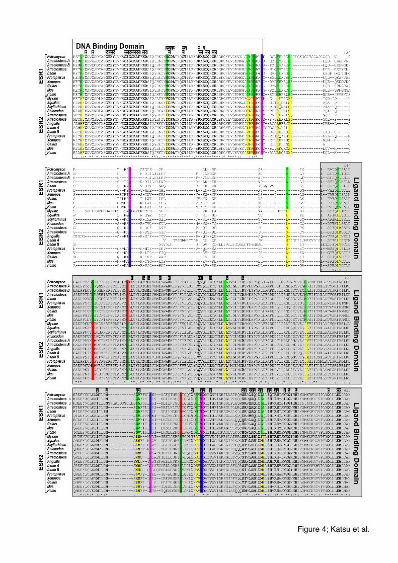
Figure 4; Katsu et al.!

B!A!
Figure 5; Katsu et al.!
C! D!

A!
B!
Figure 6; Katsu et al.!
A! B!
C! D!
Figure 7; Katsu et al.!

Table 1. Gene Transcriptional Activities of Estrogens Mediated by shark ESR RtESR EC50 (M) 95% CIa(M) RPb (%)
E1 8.636 x 10⁻¹⁰ 6.851 x 10⁻¹⁰ to 1.089 x 10⁻⁹ 1.1 E2 1.561 x 10⁻¹¹ 1.114 x 10⁻¹¹ to 2.186 x 10⁻¹¹ 100 E3 2.151 x 10⁻¹⁰ 1.539 x 10⁻¹⁰ to 3.006 x 10⁻¹⁰ 4.5 EE2 2.364 x 10⁻¹¹ 1.674 x 10⁻¹¹ to 3.338 x 10⁻¹¹ 39.6 DES 2.239 x 10⁻¹¹ 1.623 x 10⁻¹¹ to 3.087 x 10⁻¹¹ 48.2
StESR EC50 (M) 95% CI (M) RP (%)
E1 4.976 x 10⁻¹⁰ 3.135 x 10⁻¹⁰ to 7.898 x 10⁻¹⁰ 1.8 E2 5.659 x 10⁻¹² 3.418 x 10⁻¹² to 9.374 x 10⁻¹² 100 E3 1.254 x 10⁻¹⁰ 7.429 x 10⁻¹¹ to 2.118 x 10⁻¹⁰ 7.3 EE2 1.430 x 10⁻¹¹ 9.175 x 10⁻¹² to 2.228 x 10⁻¹¹ 66.0 DES 1.173 x 10⁻¹¹ 6.777 x 10⁻¹² to 2.031 x 10⁻¹¹ 69.7
a95% CI: 95% confidence intervals of EC50. bRP: relative potency = (EC50 E2/ EC50 chemical X) x 100. RtESR: whale shark ESR, StESR: catshark ESR
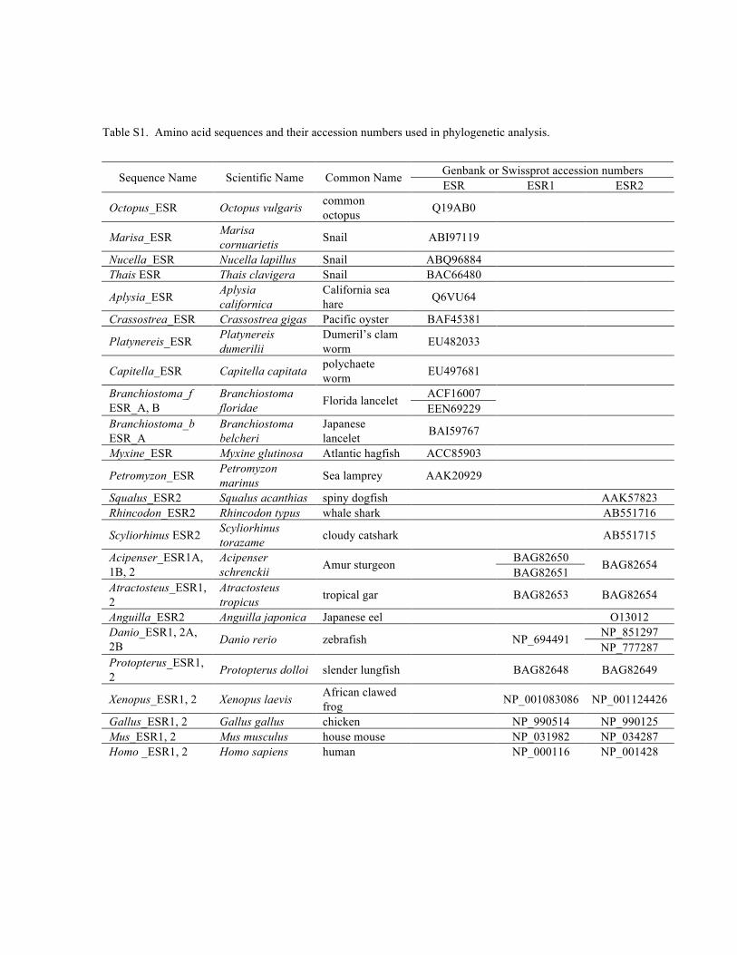
Table S1. Amino acid sequences and their accession numbers used in phylogenetic analysis.
Genbank or Swissprot accession numbers Sequence Name Scientific Name Common Name ESR ESR1 ESR2
Octopus_ESR Octopus vulgaris common octopus Q19AB0
Marisa_ESR Marisa cornuarietis Snail ABI97119
Nucella_ESR Nucella lapillus Snail ABQ96884 Thais ESR Thais clavigera Snail BAC66480
Aplysia_ESR Aplysia californica
California sea hare Q6VU64
Crassostrea_ESR Crassostrea gigas Pacific oyster BAF45381
Platynereis_ESR Platynereis dumerilii
Dumeril’s clam worm EU482033
Capitella_ESR Capitella capitata polychaete worm EU497681
ACF16007 Branchiostoma_f ESR_A, B
Branchiostoma floridae Florida lancelet
EEN69229
Branchiostoma_b ESR_A
Branchiostoma belcheri
Japanese lancelet BAI59767
Myxine_ESR Myxine glutinosa Atlantic hagfish ACC85903
Petromyzon_ESR Petromyzon marinus Sea lamprey AAK20929
Squalus_ESR2 Squalus acanthias spiny dogfish AAK57823 Rhincodon_ESR2 Rhincodon typus whale shark AB551716
Scyliorhinus ESR2 Scyliorhinus torazame cloudy catshark AB551715
BAG82650 Acipenser_ESR1A, 1B, 2
Acipenser schrenckii Amur sturgeon
BAG82651 BAG82654
Atractosteus_ESR1, 2
Atractosteus tropicus tropical gar BAG82653 BAG82654
Anguilla_ESR2 Anguilla japonica Japanese eel O13012 NP_851297 Danio_ESR1, 2A,
2B Danio rerio zebrafish NP_694491 NP_777287
Protopterus_ESR1, 2 Protopterus dolloi slender lungfish BAG82648 BAG82649
Xenopus_ESR1, 2 Xenopus laevis African clawed frog NP_001083086 NP_001124426
Gallus_ESR1, 2 Gallus gallus chicken NP_990514 NP_990125 Mus_ESR1, 2 Mus musculus house mouse NP_031982 NP_034287 Homo _ESR1, 2 Homo sapiens human NP_000116 NP_001428