clinical studydownloads.hindawi.com/journals/trt/2012/240132.pdffrom the dr. saiful anwar hospital,...
TRANSCRIPT
Hindawi Publishing CorporationTuberculosis Research and TreatmentVolume 2012, Article ID 240132, 5 pagesdoi:10.1155/2012/240132
Clinical Study
Soluble Urokinase Plasminogen Activator Receptor Levels inTuberculosis Patients at High Risk for Multidrug Resistance
Tri Yudani Mardining Raras,1 Triwahju Astuti,2 and Iin Noor Chozin2
1 Laboratory of Biochemistry and Molecular Biology, Faculty of Medicine, Brawijaya University, Malang, Indonesia2 Department of Pulmonology, Faculty of Medicine, Brawijaya University, Dr. Saiful Anwar Hospital, Malang, Indonesia
Correspondence should be addressed to Tri Yudani Mardining Raras, [email protected]
Received 9 October 2012; Accepted 19 November 2012
Academic Editor: T. Ottenhoff
Copyright © 2012 Tri Yudani Mardining Raras et al. This is an open access article distributed under the Creative CommonsAttribution License, which permits unrestricted use, distribution, and reproduction in any medium, provided the original work isproperly cited.
The soluble urokinase plasminogen activator receptor (suPAR) has been shown to be a strong prognostic biomarker fortuberculosis (TB). In the present study, the profiles of plasma suPAR levels in pulmonary TB patients at high risk for multidrugresistance were analyzed and compared with those in multidrug resistant (MDR)-TB patients. Forty patients were prospectivelyincluded, consisting of 10 MDR-TB patients and 30 TB patients at high risk for MDR, underwent clinical assesment. PlasmasuPAR levels were measured using ELISA (SUPARnostic, Denmark) and bacterial cultures were performed in addition to drugsusceptibility tests. All patients of suspected MDR-TB group demonstrated significantly higher suPAR levels compared with thehealthy TB-negative group (1.79 ng/mL). Among the three groups at high risk for MDR-TB, only the relapse group (7.87 ng/mL)demonstrated suPAR levels comparable with those of MDR-TB patients (7.67 ng/mL). suPAR levels in the two-month negativeacid-fast bacilli conversion group (9.29 ng/mL) were higher than positive control, whereas levels in the group consisting of therapyfailure patients (5.32 ng/mL) were lower. Our results strongly suggest that suPAR levels enable rapid screening of suspected MDR-TB patients, but cannot differentiate between groups.
1. Background
For decades, tuberculosis (TB) has been one of the leadinginfectious diseases causing death in Indonesia. Presently, thesituation is worsening due to the appearance of multidrug-resistant TB (MDR-TB), which accounts for 4% to 5% ofTB cases. MDR-TB is a condition in which Mycobacteriumtuberculosis (Mtb) becomes resistant to the most powerfulfirst-line anti-TB drugs (ie. isoniazid [INH] and rifampicin)[1]. Incomplete and improper standard intensive-phasetreatments are considered to be factors that lead to MDR-TB.There are several scenarios in which TB patients are likelyto become at high risk for multidrug resistance includingnegative acid-fast bacilli (AFB) conversion after two months,therapy failure and relapse.
To date, the most common test recommended by theWHO to diagnose multidrug resistance is the drug suscep-tibility test (DST), which takes two months to complete
[2]. The use of surrogate immunological prognostic markersin TB patients at high risk for multidrug resistance ableto predict multidrug resistance is expected to be beneficialin reducing the duration of drug susceptibility testing[3]. Similarly, biomarkers that accurately predict high riskmultidrug resistance in specific individuals may promptearlier initiation of the DST. A potential role for biomarkersin routine clinical care has recently been proposed [3].One such marker is soluble urokinase plasminogen acti-vator receptor (suPAR), a receptor for the serine proteaseurokinase plasminogen activator (uPA). This molecule playsa role in both innate and adaptive immunity through thefibrinolysis pathway. Bacterial endotoxins and cytokines ofthe innate immune system stimulate the secretion of uPA inseveral cell types including monocytes and neutrophils [4].Plasma levels of suPAR may reflect health status includingnormal biological processes, infection, or therapy progress.suPAR levels rise in TB patients and decrease as they progress

2 Tuberculosis Research and Treatment
through treatment [5]. Recent research on the prognosticpower of suPAR has also been demonstrated in assumed TBnegative patients [6].
Our previous study showed that suPAR could be usedas a biomarker to monitor the progress of therapy in TB-AFB(+) patients [7]. The purpose of the present study wasto observe the profile of suPAR levels in pulmonary TBpatients at high risk for multidrug resistance including thosewith negative AFB conversion at two months, therapy failureand relapse, and compare it with the incidence of multidrugresistance. Patients in this category are likely to be resistantto rifampicin and INH. Therefore, patients were also testedfor their sensitivity to antibiotics.
2. Materials and Methods
2.1. Subjects. The sample consisted of 10 MDR-TB patientsand 30 TB patients at high risk for multidrug resistance. Tenhealthy patients serving as negative controls were recruitedfrom the Dr. Saiful Anwar Hospital, Malang, Indonesia. Thepatients were categorized into suspected MDR-TB, includingnegative AFB conversion after two months of intensive-phasetherapy, relapse and therapy failure groups. All patients,except the healthy control group, fulfilled the followinginclusion criteria: having pulmonary TB, regularly takingmedication, male or female between 15 and 50 years of age,having a body mass index (BMI) > 16 kg/m2, agreeing to bea subject of the research, and providing informed consent.TB patients with other diseases, such as severe bacterialpneumonia, HIV-AIDS, heart disease, diabetes mellitus,heart and kidney problems, or extrapulmonary TB, andpregnant patients and patients with psychiatric problemswere excluded from the study.
All patients, except TB-AFB(−) conversion, receiveddirectly observed, six-month (26-week), short-course anti-TB therapy as recommended by the Indonesian NationalTuberculosis Program, which is based on WHO TB guide-lines [8].
2.2. TB Treatment. For patients in the AFB conversionafter two months and therapy failure groups, the drugregimen consisted of a fixed, weight-dependent combinationof INH (320–400 mg/day), rifampicin (480–600 mg/day),ethambutol (800–1200 mg/day), and pyrazinamide (1000–1250 mg/day) for the two-month intensive-phase treatment,followed by rifampicin and INH in the four-month continu-ation phase. In the case of therapy failure groups, the patientsshowed to be AFB positive at the end of five and six monthsof therapy.
2.3. Routine Examination. Several routine clinical and lab-oratory investigations were performed in accordance withstandard procedures conducted in Indonesian hospitals [9].These included patient complaints (ie., history), X-ray, BMI,lesion width, erythrocyte sedimentation rate (ESR), bacterialculture, and DSTs. Chest X-rays were taken before therapybegan and included standard posteroanterior and lateral
views, the results of which were reviewed by a pulmonologist.Lesion width was measured according to chest X-ray gradingof disease (i: minimal lesion ie, lesion width less than arearestricted to median line, apex and front costae, solitarylesion located anywhere and no cavity found; ii: moderateadvanced ie, width of cavity is less than one lobe, if a cavity ispresent, should be in no more than one lobe; iii: far advancedcorresponding to a lesion width greater than minimal andmoderate lesions and, if with cavity, should not exceed awidth of 4 cm).
2.4. Microbiological Research Method. To observe the viabil-ity of Mtb and to determine resistance to basic anti-TB drugs,sputum samples were collected from patients in each group.Sputa were washed in 0.9% NaCl solution and concentrated,and the sediment was subsequently cultured on Lowenstein-Jensen medium. The DST was performed with at least twoanti-TB drugs, rifampicin and INH, using the bacteriologicalabsolute concentration method [9].
2.5. Sample Handling. A 3 mL blood sample was obtainedfrom new patients by venipuncture before they initiatedsecond-line anti-TB drug treatment as guided by the WHO[8]. Sera were centrifuged (6000×g) at 4◦C for 10 min,divided into aliquots of 0.5 mL, and subsequently stored at−80◦C.
2.6. Enzyme-Linked Immunosorbent Assay (ELISA). Serumlevels of suPAR were measured in duplicate using commer-cially available ELISA kits according to the manufacture’sprotocol (suPARnostic, ViroGates A/S, Copenhagen, Den-mark) [10]. The optical density of the samples at 450 nmwere read using a Biotech microplate reader, with thewavelength correction set at 650 nm. Results of the respectivemeasurements were analyzed using SPSS version 3.4 (IBMCorporation, USA).
2.7. Statistical Analysis. All statistical analyses were per-formed using SPSS software. Multivariate analyses wereconducted using SPSS version 16 (IBM Corporation, USA).Differences between the groups and controls were analyzedusing one-way ANOVA.
2.8. Ethics. The present study was approved by the EthicsCommittee of Syaiful Anwar Public Hospital, BrawijayaUniversity, Indonesia. All patients provided written informedconsent.
3. Results
3.1. Patient Characteristics. The study initially included 41patients consisting of 31 pulmonary TB patients at highrisk for multidrug resistance and 10 MDR-TB patients.During the course of the study, however, one patientacquired pneumonia and was subsequently excluded. Table 1summarizes the clinical characteristics of patients based onthe criteria for the MDR-TB group as well as groups at highrisk for multidrug resistance. One-half (50%) of the patients
Tuberculosis Research and Treatment 3
Table 1: Characteristics of tuberculosis (TB) patients at high risk for multidrug resistance (MDR) and multidrug resistant TB.
Patient characteristicMDR
(n = 10)n (%)
NC(n = 10)n (%)
TF(n = 10)n (%)
R(n = 10)n (%)
Total(n = 40)n (%)
BMI (kg/m2)<18 5 (50) 7 (70) 4 (40) 6 (60) 32 (80)
18–22 3 (30) 3 (30) 4 (40) 4 (40) 18 (45)
>22 2 (20) 0 2 (20) 0 4 (10)
Lesion width
Normal 0 (0) 0 1 (10) 0 1 (2.5)
Minimal 0 (0) 1 (10) 1 (10) 1 (10) 3 (7.5)
Moderate 0 (0) 2 (20) 3 (30) 3 (30) 9 (22.5)
Far advanced 10 (100) 7 (70) 5 (50) 6 (60) 38 (95)
Milier 0 (0) 0 (0) 0 (0) 0 (0) 3 (7.5)
ESR (mm/h) 65.1 63.5 24.6 62.1
Culture and DST
(+) MDR 10 (100) 2 (20) 1 (10) 2 (20) 15 (37.5)
(+) nMDR 0 (0) 1 (10) 4 (40) 5 (50) 10 (40)
Culture (−) 0 (0) 7 (70) 4 (40) 2 (20) 13 (52.5)
MOTT 0 (0) 0 (0) 1 (10) 1 (10) 2 (5)
BMI: Body mass index; DST: drug susceptibility test; eSR: erythrocyte sedimentation rate; MOTT: mycobacterium other than tuberculosis; NC: no conversionof AFB (+) into AFB (−) after two-month therapy; TF: therapy failure; R: relapse.
in the present study had a low BMI or were underweight(BMI < 18 kg/m2), while only a few (8%) maintained theirbody weight (BMI > 22 kg/m2). Underweight patients wereevenly distributed among the groups, with no significantdifferences in BMI between the MDR-TB group and thegroup at high risk for multidrug resistance (P = 0.647).
Examination of chest X-rays showed that lesion areas cor-responding to the far advanced category most often occurredin patients in the MDR-TB group (100%). In contrast,the high risk for multidrug resistance group demonstratedvariations in lesion width: highly advanced lesions (60%to 70%), moderately advanced lesions (20% to 30%), andminimal lesions (10%).
ESR is often elevated in patients with active TB, althougha normal ESR does not rule out TB [2]. Surprisingly, ESRsamong MDR-TB patients were not significantly differentfrom those in the relapse group and the two-month AFBconversion group (P = 0.555). In fact, the therapy failuregroup demonstrated the lowest ESRs.
3.2. suPAR Measurement. TB patients at high risk for mul-tidrug resistance had significantly higher suPAR levels thanthose in the healthy purified protein derivative-negative (ie.,control) group (1.78 ng/mL) (P < 0.0001). However, whensuPAR levels were compared between the MDR-TB group(7.63 ng/mL) and those in the suspected TB group, onlythe relapse groups showed comparable results (7.85 ng/mL).Patients in the two-month negative AFB conversion grouphad the highest suPAR levels (9.24 ng/mL), while patients inthe therapy failure group demonstrated significantly lowersuPAR concentrations (5.04 ng/mL) (P = 0.028).
16
12
8
4
7.85 8
5.54
1.76∗
MD
R-T
B
Posi
tive
cu
ltu
re
but
not
MD
R
Neg
ativ
e cu
ltu
re
Con
trol
gro
up
Culture growth and drug sensitivity test
Pla
sma
suPA
R le
vel (
ng/
mL)
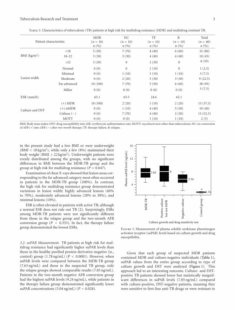
Figure 1: Measurement of plasma soluble urokinase plasminogenactivator receptor (suPAR) levels based on culture growth and drugsusceptibility.
Given that each group of suspected MDR patientscontained MDR and culture-negative individuals (Table 1),suPAR values from the entire group according to type ofculture growth and DST were analyzed (Figure 1). Thisapproach led to an interesting outcome. Culture- and DST-positive TB patients showed lower but statistically insignif-icant differences in suPAR levels (7.85 ng/mL) comparedwith culture-positive, DST-negative patients, meaning theywere sensitive to first-line anti-TB drugs or were resistant to
4 Tuberculosis Research and Treatment
one of rifampicin or INH (non-MDR) (8.03 ng/mL) (P =0.890). However, the mean concentration of plasma suPARin culture-negative patients (5.54 ng/mL) was significantlylower than that of Mtb-positive culture MDR (7.85 ng/mL)as well as Mtb-positive culture non-MDR (8.03 ng/mL)patients (P = 0.027). Finally, patients with cultures ofMycobacterium other than TB (MOTT) showed no signifi-cant differences in suPAR levels (6.48 ng/mL) compared withculture-positive and culture-negative groups (P = 0.55).
4. Discussion
In the present study, all TB patients at high risk for multidrugresistance shared characteristics that were also found inMDR-TB patients, including low BMI and high ESR. The factthat BMI values were below normal in all TB groups suggeststhat BMI is not a characteristic of MDR-TB patients becauseit was also prevalent in patients in the TB at high risk formultidrug resistance as well as those who were TB-AFB(+)[9]. The lack of adequate nutritional status or malnutritioncan be a risk factor contributing to TB infection as a resultof decreases in T helper cell 1 responses [11, 12]. Anotherreason for low BMI might be that TB patients demonstrateanorexia with decreased appetite, which is consistent withgeneral conditions present in chronic and advanced disease.The ESR values of the relapse patients were, surprisingly,comparable with those of MDR-TB patients. Although ESRis not considered to be a specific marker for TB, it has beenshown to be an effective inflammatory marker in active TB[13].
The measurement of suPAR levels has been shown toreflect pathological status in pulmonary TB patients, makingapplication in clinical settings relevant to disease progress.We report the profile of suPAR levels from three groups of TBpatients at high risk for MDR-TB (ie., two-month negativeAFB conversion, therapy failure and relapse). Among thethree groups at high risk for MDR-TB, only the relapsegroup demonstrated suPAR levels comparable with MDR-TB patients (7.67 ng/mL). The other groups (negative AFBconversion after two months and therapy failure) showedeither higher or lower levels subsequently (9.29 ng/mL and5.32 ng/mL, resp.). Given that patients from the relapsegroup were completely cured of TB for a considerable lengthof time but then reactivated, results of the present studydemonstrate that suPAR levels are quite stably maintainedin the plasma of relapse patients. One possible explanationis that a small proportion of Mtb in the macrophages ofTB relapse patients survives first-line anti-TB treatment andpersists for an uncertain length of time and subsequentlyreemerges upon host immunosuppression [14]. This corre-lates best with the culture test, in which as much as 50%of the bacteria are Mtb positive, although monoresistant toanti-TB drugs. These microorganisms belong to minoritybacillary subpopulations that persist in the biofilm [15].Such subpopulations of bacteria can develop multidrugresistance [16, 17]. It is not surprising that the DST indicatedthat 20% of patients from the relapse group demonstratedMDR-positive cultures. Moreover, because suPAR levels
between culture-positive MDR and non-MDR groups werenot statistically different, these made up 70% of the finalvalue of suPAR from the relapse group. The reason(s) whyMtb resistant to anti-TB drugs elicited comparable suPARlevels compared with nativeMtb is unknown. Kiran et al.[18] found a slight reduction in the number of lymphocytesin MDR-TB patients. Nevertheless the level of CD19(+) Blymphocytes responsible for antibody production and theiractivation receptor CD23 were nonsignificantly reduced inMDR-TB groups, thereby influencing humoral as well ascellular immunity, and this effect is more profound in MDR-TB groups than in TB-AFB(+) groups. A study by Eugen-Olsen et al. [5] demonstrated that blood suPAR levels areassociated with the number of bacteria in the sputum andwithin the bronchi [5].
The mean suPAR level in patients in the two-monthAFB negative conversion group (9.29 ng/mL) was slightlybut significantly higher than in patients in the MDR-TBgroup, indicating that pathological processes continued toproceed, possibly due to the survival of a subpopulation ofbacteria. These bacteria persist within latent foci containedin granulomas and in the sputum of large cavities [15].As a general rule, Mtb can be eradicated by anti-TB drugregimens in two months [17]. This was also shown in thepresent study, in which 70% of the culture growth wasnegative. Surprisingly, suPAR levels remained high, and 20%of the cultures were MDR positive, which may have occurreddue to bacteria that could not be identified using the acid-fast test [19]. “Invisible” mycobacteria were also confirmedin a study by Al-Moamary et al. [20], who demonstratednegative cultures in 63% to 73% of AFB(+) patients. Thesestudies suggest that, in addition to clinical improvementand X-rays, another accurate prognostic marker is requiredto make a decision whether therapy should be terminatedor continued. Although culture tests were negative after 2months of therapy (ie., bacteria did not grow), we suggestthat because suPAR levels remained high, continuation oftreatment with anti-TB regimens is warranted.
The therapy failure group showed the lowest suPARlevels, but these levels were still comparable with those ofthe cured TB patients from the previous study (5.32 ng/mLversus 5.09 ng/mL) [7]. Despite 40% of lesion widths beingin the far-advanced category, only 10% of patients hadMDR-positive cultures, with 40% culture positive althoughmonoresistant or sensitive to an anti-TB drug. This indicatesthat the response to therapy did not improve with antitu-berculosis first line; therefore, the damage in the lungs ofMDR-TB patients continued to progress and became morewidespread. After six months, the lesion widths remained thesame.
To date, culture growth and the DST have been the onlymethods able to discriminate multidrug resistance from sus-pected multidrug resistance. Our novel finding that suPARlevels could reflect the immune status of different groupsof TB patients at high risk for multidrug resistance adds tothe literature. Although suPAR level could not differentiateMDR-TB patients from suspected MDR patients, TB patientswith anti-TB drug sensitivity after therapy demonstrated thatsuPAR levels > 5 ng/mL warrant performance of DSTs.

Tuberculosis Research and Treatment 5
5. Conclusion
The results of our study suggest that measurement ofsuPAR levels could be used to screen TB patients at highrisk for multidrug resistance after therapy. However, thismarker cannot differentiate between suspected multidrugresistance and MDR-TB patients. Larger studies are expectedto confirm the prognostic value for MDR from TB patientsat high risk of MDR.
Acknowledgment
The authors thank the suPARNOSTIC Inc. that gave specialprice for the kit that enabled them to conduct this paper.This study was funded by the Faculty of Medicine, BrawijayaUniversity. A thank you for Dian, M.D., Rahadi, MD thatprovided samples and Fitri for technical assistant.
References
[1] Anonim, Standard Internasional Penanganan Tuberculosis,Ditjen PP & PL Departemen Kesehatan RI, 2006.
[2] WHO, TB/HIV a Clinical Manual, Stop TB Department Deptof HIV/AIDS, Dept Child and Adolescent Health and Devel-opment, World Health Organization, Geneva, Switzerland,2004.
[3] R. S. Wallis, T. M. Doherty, P. Onyebujoh et al., “Biomarkersfor tuberculosis disease activity, cure, and relapse,” The LancetInfectious Diseases, vol. 9, no. 3, pp. 162–172, 2009.
[4] M. Del Rosso, F. Margheri, S. Serrati, A. Chilla, A. Laurenzana,and G. Fibbi, “The urokinase reptor system, a key regulatorat the intersection between imflammation, immunity andcoagulation,” Current Pharmaceutical Design, vol. 17, pp.1924–1943, 2011.
[5] J. Eugen-Olsen, P. Gustafson, N. Sidenius et al., “The serumlevel of soluble urokinase receptor is elevated in tuberculosispatients and predicts mortality during treatment: a com-munity study from Guinea-Bissau,” International Journal ofTuberculosis and Lung Disease, vol. 6, no. 8, pp. 686–692, 2002.
[6] P. Rabna, A. Andersen, C. Wejse et al., “High mortality riskamong individuals assumed to be TB-negative can be pre-dicted using a simple test,” Tropical Medicine and InternationalHealth, vol. 14, no. 9, pp. 986–994, 2009.
[7] T. Mardining Raras and I. Noor Chozin, “The solubleplasminogen Activator receptor as a biomarker on monitoringthe therapy progress of pulmonary TB-AFB(+) patients,”Tuberculosis Research and Treatment, vol. 2010, Article ID406346, 6 pages, 2010.
[8] WHO, Policy Guidence on Drug-Susceptibility Test of SecondLine Antituberculosis Drug, The Stop TB Department, WorldHealth Organization, Geneva, Switzerland, 2008.
[9] Perhimpunan Dokter Paru Indonesia, Pedoman Diagnosisdan Penatalaksanaan di Indonesia, Jakarta, Indah Ofset CitraGrafika, hal 1-64, 2006.
[10] V. Gates, suPARnostic ELISA Kit: Enzyme-Linked Immunosor-bent Assay for Quantitative Determination of Soluble UrokinasePlasminogen Activator Receptor in Human Plasma, Copen-hagen, Denmark, 2007.
[11] G. A. W. Rook and T. M. Doherty, “Host susceptibilityand resistance to Mycobacterium tuberculosis,” in Genetic,Neuroendocrine and Acquired Factors, H. S. Schaaf and A. I.
Zumla, Eds., pp. 96–106, Saunders Elsevier, Haryana, India,2009, Hrsg.Tuberculosis. A Comprehensive Clinical Reference.
[12] J. S. Mukherjee and S. Schaaf, “Pediatric MDR-TB,” inTuberculosis: A Comprehensive Clinical Treatise, A. Zumla andS. Schaaf, Eds., 2008.
[13] K. Furuhashi, T. Shirai, T. Suda, and K. Chida, “Inflammatorymarkers in active pulmonary tuberculosis: association withTh1/Th2 and Tc1/Tc2 balance,” Kekkaku, vol. 87, no. 1, pp.1–7, 2012.
[14] L. G. Wayne and C. D. Sohaskey, “Nonreplicating persistenceof Mycobacterium tuberculosis,” Annual Review of Microbiol-ogy, vol. 55, pp. 139–163, 2001.
[15] A. K. Ojha, A. D. Baughn, D. Sambandan et al., “Growth ofMycobacterium tuberculosis biofilms containing free mycolicacids and harbouring drug-tolerant bacteria,” MolecularMicrobiology, vol. 69, no. 1, pp. 164–174, 2008.
[16] N. Dhar and J. D. McKinney, “Microbial phenotypic het-erogeneity and antibiotic tolerance,” Current Opinion inMicrobiology, vol. 10, no. 1, pp. 30–38, 2007.
[17] K. Lewis, “Persister cells, dormancy and infectious disease,”Nature Reviews Microbiology, vol. 5, no. 1, pp. 48–56, 2007.
[18] B. Kiran, T. Cagatay, P. Clark et al., “Can immune parametersbe used as predictors to distinguish between pulmonarymultidrug-resistant and drug-sensitive tuberculosis?” Archivesof Medical Science, vol. 6, no. 1, pp. 77–82, 2010.
[19] C. C. Shu, J. T. Wang, C. H. Lee, J. Y. Wang, L. N. Lee,and C. J. Yu, “Predicting results of mycobacterial culture onsputum smear reversion after anti-tuberculous treatment: acase control study,” BMC Infectious Diseases, vol. 10, article 48,2010.
[20] M. S. Al-Moamary, W. Black, E. Bessuille, R. K. Elwood, and S.Vedal, “The significance of the persistent presence of acid-fastbacilli in sputum smears in pulmonary tuberculosis,” Chest,vol. 116, no. 3, pp. 726–731, 1999.

Submit your manuscripts athttp://www.hindawi.com
Stem CellsInternational
Hindawi Publishing Corporationhttp://www.hindawi.com Volume 2014
Hindawi Publishing Corporationhttp://www.hindawi.com Volume 2014
MEDIATORSINFLAMMATION
of
Hindawi Publishing Corporationhttp://www.hindawi.com Volume 2014
Behavioural Neurology
EndocrinologyInternational Journal of
Hindawi Publishing Corporationhttp://www.hindawi.com Volume 2014
Hindawi Publishing Corporationhttp://www.hindawi.com Volume 2014
Disease Markers
Hindawi Publishing Corporationhttp://www.hindawi.com Volume 2014
BioMed Research International
OncologyJournal of
Hindawi Publishing Corporationhttp://www.hindawi.com Volume 2014
Hindawi Publishing Corporationhttp://www.hindawi.com Volume 2014
Oxidative Medicine and Cellular Longevity
Hindawi Publishing Corporationhttp://www.hindawi.com Volume 2014
PPAR Research
The Scientific World JournalHindawi Publishing Corporation http://www.hindawi.com Volume 2014
Immunology ResearchHindawi Publishing Corporationhttp://www.hindawi.com Volume 2014
Journal of
ObesityJournal of
Hindawi Publishing Corporationhttp://www.hindawi.com Volume 2014
Hindawi Publishing Corporationhttp://www.hindawi.com Volume 2014
Computational and Mathematical Methods in Medicine
OphthalmologyJournal of
Hindawi Publishing Corporationhttp://www.hindawi.com Volume 2014
Diabetes ResearchJournal of
Hindawi Publishing Corporationhttp://www.hindawi.com Volume 2014
Hindawi Publishing Corporationhttp://www.hindawi.com Volume 2014
Research and TreatmentAIDS
Hindawi Publishing Corporationhttp://www.hindawi.com Volume 2014
Gastroenterology Research and Practice
Hindawi Publishing Corporationhttp://www.hindawi.com Volume 2014
Parkinson’s Disease
Evidence-Based Complementary and Alternative Medicine
Volume 2014Hindawi Publishing Corporationhttp://www.hindawi.com