clinical study fascia wrapping technique: a modified
TRANSCRIPT
Clinical StudyFascia Wrapping Technique: A Modified Method forthe Treatment of Cubital Tunnel Syndrome
Hyun Ho Han, Hae Won Kang, Jun Yong Lee, and Sung-No Jung
Department of Plastic and Reconstructive Surgery, Uijeongbu St.Mary’s Hospital, College ofMedicine,TheCatholic University of Korea,222 Banpo-daero, Seocho-gu, Seoul 137-701, Republic of Korea
Correspondence should be addressed to Sung-No Jung; [email protected]
Received 2 July 2014; Accepted 26 August 2014; Published 15 October 2014
Academic Editor: Anastasios V. Korompilias
Copyright © 2014 Hyun Ho Han et al. This is an open access article distributed under the Creative Commons Attribution License,which permits unrestricted use, distribution, and reproduction in any medium, provided the original work is properly cited.
Variations of the anterior transposition of the ulnar nerve for cubital tunnel syndrome include subcutaneous, submuscular,intramuscular, and subfascial methods. We introduce a modification of subfascial transposition, which is designed to facilitatenerve gliding by wrapping the nerve with fascia. Twenty patients with wrapping surgery following the diagnosis of cubital tunnelsyndrome were reviewed retrospectively. Preoperative electrodiagnostic studies were performed in all patients and all of themwererechecked postoperatively. The preoperative mean value of motor conduction velocity (MCV) was 37.1 ± 6.7m/s within the elbowsegment and this result showed a decrease compared to the result of MCVwith 53.9±6.9m/s in the below the elbow-wrist segmentwith statistical significance (𝑃 < 0.05). Postoperative mean values of MCV were improved in all of 20 patients to 47.6 ± 5.5m/s(𝑃 < 0.05). 19 patients of 20 (95%) reported good or excellent clinical outcomes according to amodified Bishop scoring system.Thesurgical treatment methods for cubital tunnel syndrome have their own advantages and disadvantages, and the preferred methoddiffers depending on the surgeon.The wrapping method of anterior transposition is a newly designed alternative method modifiedfrom subfascial transposition. This method could be an alternative option to treat cubital tunnel syndrome.
1. Introductions
Ulnar nerve compression at the elbow region, which is namedcubital tunnel syndrome, is the second most common com-pressive neuropathy of the upper limb after carpal tunnel syn-drome [1].Multiple surgical options have been recommendedin the literature and reflect the controversy surrounding thesurgical treatment of cubital tunnel syndrome. The surgicalmanagement is broadly divided into three types of proce-dures [2]: simple decompression [3, 4], medial epicondylec-tomy [5, 6], and anterior transposition of the ulnar nerve.Also variations of anterior transposition of the ulnar nervehave been proposed; these include subcutaneous [7–9],submuscular [10–12], intramuscular [13–15], and subfascial[2, 16] methods. A subcutaneous transposition is a simpleand reliable procedure that facilitates an early postoperativemobilization. However, it is more vulnerable to trauma andhypersensitivity. A submuscular or intramuscular transpo-sition is well protected as it lies deeply under a substantial
amount of soft tissue. However, it has the disadvantages ofprolonged postoperative elbow immobilization and potentialsubsequent contracture. A subfascial transposition protectsthe transposed nerve and avoids problems like scarring,recurrence, and elbow contracture [2, 16].
Themethodwe are introducing in this study is amodifiedmethod of subfascial transposition.The method of subfascialtransposition hasmerits asmentioned above, but whether thenerve would not adhere between the fascia and the muscleand gliding would be facilitated was doubted, and there was adifficulty in fixing the fascial flap to the muscle after elevationsince the muscular tissue was friable [17, 18]. In addition, ifthere is a defect on a region like dorsum of hand for whichtendon gliding is necessary, to cover it with temporoparietalfascia free flap to facilitate tendon gliding after coverageand conduct split thickness skin graft is a widely knownmethod [19–21] based onwhichwe thought that wrapping thenerve with fascia would cause less adhesion and be helpfulfor gliding. Thus this new method is designed to facilitate
Hindawi Publishing Corporatione Scientific World JournalVolume 2014, Article ID 482702, 6 pageshttp://dx.doi.org/10.1155/2014/482702
2 The Scientific World Journal
(a) (b)
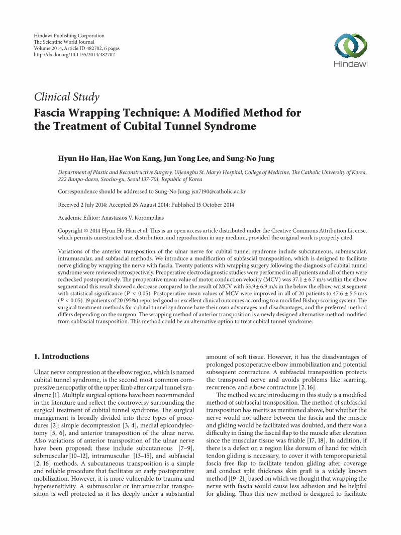
Figure 1: Nerve release and anterior transposition. (a) Feeding artery vessel (arrow) of nerve must be saved. (b) Anterior transposition of theulnar nerve was conducted followed by dissection to achieve sufficient release without compression of the ulnar nerve.
nerve gliding by wrapping the nerve with fascia. Here, wesummarize and report the surgerymethod aswell as the resultof surgery.
2. Patients and Method
Twenty patients who had surgery with the wrapping methoddue to the diagnosis of cubital tunnel syndrome werereviewed retrospectively. The study patient pool consists ofpatients who had a surgical operation at a single centre inUijeongbu St. Mary’s Hospital from January 2008 throughJanuary 2012. All operations have been conducted by thecorresponding author, Sung-No Jung. Diagnosis of cubitaltunnel syndrome was made on a typical history of pain. Sen-sory deficit according to the distribution of ulnar nerve wasmeasured by static and dynamic 2-point discrimination tests.And the loss of intrinsic bulk and weakness of grip strengthwere measured as well using a hand grip dynamometer andcompared with that of the normal part on the opposite side.Preoperatively, the condition of the ulnar nerve was gradedaccording to severity, based on Dellon’s classification [22].Preoperative electromyography of the flexor carpi ulnaris,abductor digiti minimi, and first interosseous muscle wasdone in all patients. Also we evaluated the preoperativemotor conduction velocity (MCV) of the ulnar nerve inthe segments of below the elbow-wrist, above the elbow-below the elbow, and axilla-above the elbow in all patients.A section survey was simultaneously applied between 4 cmdistal and 6 cm proximal to the medical epicondyle in allpatients to determine the exact location of the compressionsite. Patients were selected for the operation by using thefollowing 2 criteria: (1) an absolute MCV from above theelbow to below the elbow of less than 50m/s or (2) slowingof greater than 10m/s in the above the elbow to below theelbow segment compared with the below the elbow to wristsegment [23]. Also differential diagnosis was conducted for
polyneuropathy. All patients were preoperatively examinedwith standard radiographs of the elbow.
The fascia wrapping method was used for the surgeryin all patients. The postoperative MCV test and outcomeassessment for the patients were based on the modifiedBishop scoring system [14]. It was examined in all 20 patientsabout 1 year later.
TheMCV results between preoperative and postoperativedata were statistically analyzed by independent 𝑡-test andthe results between preoperative below the elbow-wrist andwithin the elbow segment were analyzed by one sample 𝑡-test.SPSS version 13.0 software (SPSS Inc., Chicago, IL, USA) wasused.
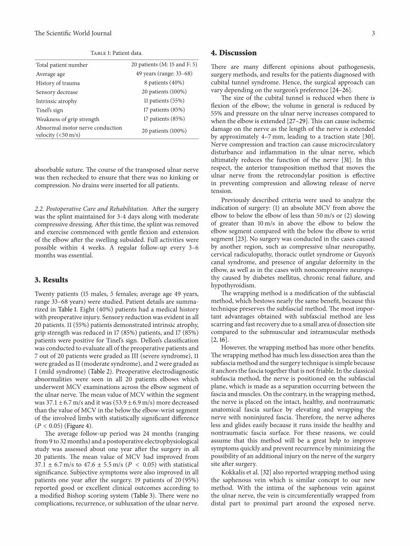
2.1. Operative Procedure. Skin and underlying subcutaneoustissues were curvilinear incised midway between the ole-cranon and medial epicondyle under general anesthesiaand tourniquet control. The fascia was divided between themedial epicondyle and olecranon, passing proximally fromthemedial intermuscular septum to the postcondylar groove,releasing Osborne’s band, which is an aponeurosis locatedbetween the two heads of flexor carpi ulnaris muscle. Thefeeding artery vessel of the nerve should be saved (Figure 1).An anterior transposition of the ulnar nerve was conductedfollowed by dissection to achieve a sufficient release withoutcompression of the nerve (Figure 1). Superficial fascia belong-ing to the flexor pronator muscle group was elevated as abroad fascia flap with a width exceeding approximately 3 cmand a position of 1-2 cm apart from the medical epicondyleorigin. Unlike the existing subfascial transposition, which islocated in the nerve betweenmuscle and fascia after elevatingfascia flap, we conducted the wrapping procedure by locatingthe ulnar nerve over the fascia and very loosely rolling theulnar nerve with the elevated fascia flap (Figures 2 and 3).The elevated fascia flap was firmly anchored onto the fascialocated to the side ofmedial epicondyle through a continuous
The Scientific World Journal 3
Table 1: Patient data.
Total patient number 20 patients (M: 15 and F: 5)Average age 49 years (range: 33–68)History of trauma 8 patients (40%)Sensory decrease 20 patients (100%)Intrinsic atrophy 11 patients (55%)Tinel’s sign 17 patients (85%)Weakness of grip strength 17 patients (85%)Abnormal motor nerve conductionvelocity (<50m/s) 20 patients (100%)
absorbable suture. The course of the transposed ulnar nervewas then rechecked to ensure that there was no kinking orcompression. No drains were inserted for all patients.
2.2. Postoperative Care and Rehabilitation. After the surgerywas the splint maintained for 3-4 days along with moderatecompressive dressing. After this time, the splint was removedand exercise commenced with gentle flexion and extensionof the elbow after the swelling subsided. Full activities werepossible within 4 weeks. A regular follow-up every 3–6months was essential.
3. Results
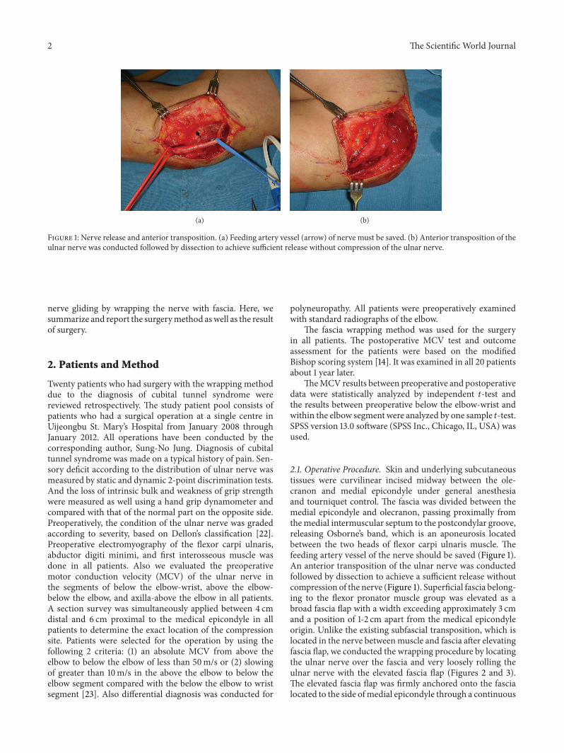
Twenty patients (15 males, 5 females; average age 49 years,range 33–68 years) were studied. Patient details are summa-rized in Table 1. Eight (40%) patients had a medical historywith preoperative injury. Sensory reduction was evident in all20 patients. 11 (55%) patients demonstrated intrinsic atrophy,grip strength was reduced in 17 (85%) patients, and 17 (85%)patients were positive for Tinel’s sign. Dellon’s classificationwas conducted to evaluate all of the preoperative patients and7 out of 20 patients were graded as III (severe syndrome), 11were graded as II (moderate syndrome), and 2 were graded asI (mild syndrome) (Table 2). Preoperative electrodiagnosticabnormalities were seen in all 20 patients elbows whichunderwent MCV examinations across the elbow segment ofthe ulnar nerve. The mean value of MCV within the segmentwas 37.1 ± 6.7m/s and it was (53.9 ± 6.9m/s) more decreasedthan the value of MCV in the below the elbow-wrist segmentof the involved limbs with statistically significant difference(𝑃 < 0.05) (Figure 4).
The average follow-up period was 24 months (rangingfrom9 to 32months) and a postoperative electrophysiologicalstudy was assessed about one year after the surgery in all20 patients. The mean value of MCV had improved from37.1 ± 6.7m/s to 47.6 ± 5.5m/s (𝑃 < 0.05) with statisticalsignificance. Subjective symptoms were also improved in allpatients one year after the surgery. 19 patients of 20 (95%)reported good or excellent clinical outcomes according toa modified Bishop scoring system (Table 3). There were nocomplications, recurrence, or subluxation of the ulnar nerve.
4. Discussion
There are many different opinions about pathogenesis,surgery methods, and results for the patients diagnosed withcubital tunnel syndrome. Hence, the surgical approach canvary depending on the surgeon’s preference [24–26].
The size of the cubital tunnel is reduced when there isflexion of the elbow; the volume in general is reduced by55% and pressure on the ulnar nerve increases compared towhen the elbow is extended [27–29].This can cause ischemicdamage on the nerve as the length of the nerve is extendedby approximately 4–7mm, leading to a traction state [30].Nerve compression and traction can cause microcirculatorydisturbance and inflammation in the ulnar nerve, whichultimately reduces the function of the nerve [31]. In thisrespect, the anterior transposition method that moves theulnar nerve from the retrocondylar position is effectivein preventing compression and allowing release of nervetension.
Previously described criteria were used to analyze theindication of surgery: (1) an absolute MCV from above theelbow to below the elbow of less than 50m/s or (2) slowingof greater than 10m/s in above the elbow to below theelbow segment compared with the below the elbow to wristsegment [23]. No surgery was conducted in the cases causedby another region, such as compressive ulnar neuropathy,cervical radiculopathy, thoracic outlet syndrome or Guyon’scanal syndrome, and presence of angular deformity in theelbow, as well as in the cases with noncompressive neuropa-thy caused by diabetes mellitus, chronic renal failure, andhypothyroidism.
The wrapping method is a modification of the subfascialmethod, which bestows nearly the same benefit, because thistechnique preserves the subfascial method. The most impor-tant advantages obtained with subfascial method are lessscarring and fast recovery due to a small area of dissection sitecompared to the submuscular and intramuscular methods[2, 16].
However, the wrapping method has more other benefits.The wrapping method has much less dissection area than thesubfasciamethod and the surgery technique is simple becauseit anchors the fascia together that is not friable. In the classicalsubfascia method, the nerve is positioned on the subfascialplane, which is made as a separation occurring between thefascia andmuscles. On the contrary, in the wrappingmethod,the nerve is placed on the intact, healthy, and nontraumaticanatomical fascia surface by elevating and wrapping thenerve with noninjured fascia. Therefore, the nerve adheresless and glides easily because it runs inside the healthy andnontraumatic fascia surface. For these reasons, we couldassume that this method will be a great help to improvesymptoms quickly and prevent recurrence byminimizing thepossibility of an additional injury on the nerve of the surgerysite after surgery.
Kokkalis et al. [32] also reported wrapping method usingthe saphenous vein which is similar concept to our newmethod. With the intima of the saphenous vein againstthe ulnar nerve, the vein is circumferentially wrapped fromdistal part to proximal part around the exposed nerve.

4 The Scientific World Journal
Figure 2: Wrapping procedure. Wrapping procedure was conducted by locating the ulnar nerve over the fascia and very loosely rolling theulnar nerve with the elevated fascia flap. Closure could be tightly made together with the fascia.
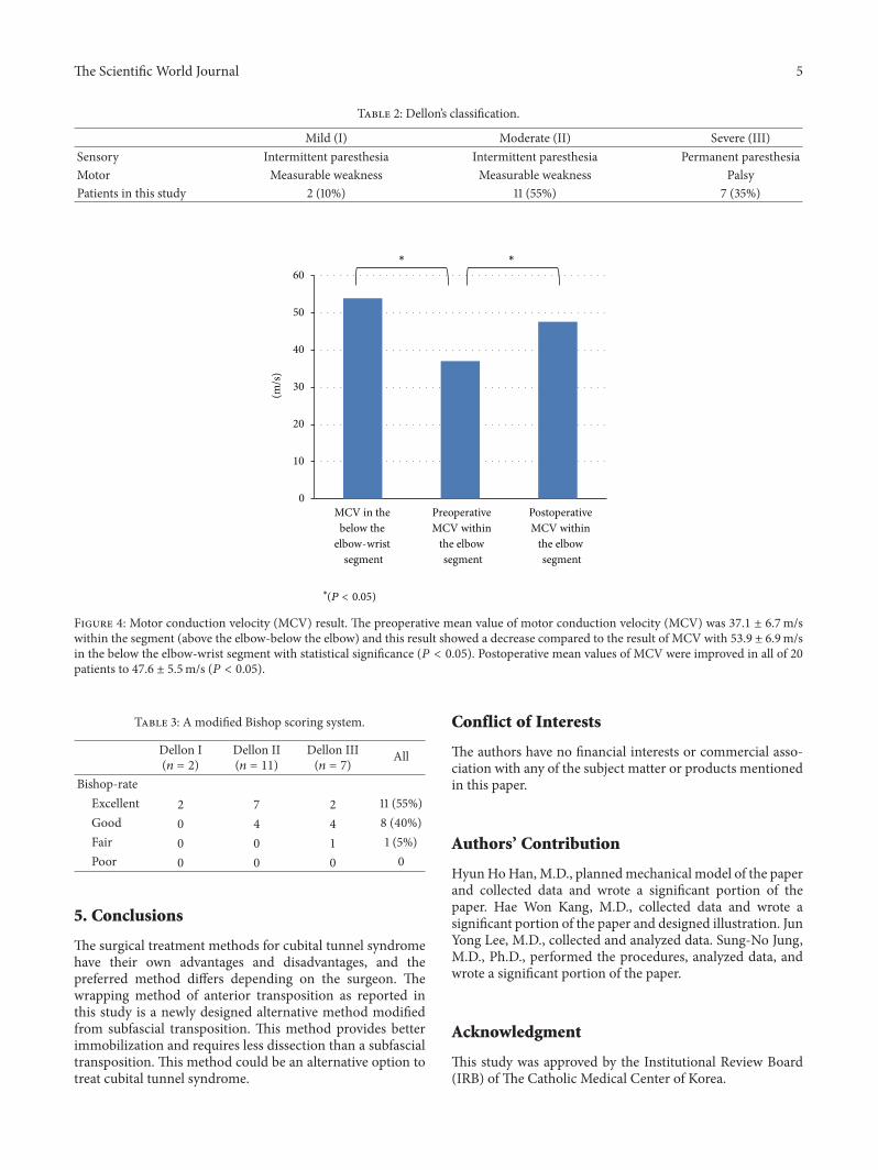
Fascia flap
Proximal side
Distal side
Ulnarnerve
Medial epicondyle
Distal side
Proximal sideFascia
wrapping
Figure 3: Schematic illustration of the wrapping procedures.
They assumed that autologous vein graft with its smoothinner surface should improve the gliding function of thenerve and reduce scar formation around the nerve.
A further advantage for this surgical method is that thenerve can be placed in a more superficial position than thefascia. It is less likely that therewill be problems of nerve kink-ing or iatrogenic compression on the new surgery site becausethe nerve plane is placed on the same plane before surgery,above the fascia. In the classical subfascial transpositionmethod, there is the potential that compression on a certainregion between the two heads of the FCU, which is mainlythe distal part, can developed, or compression can worsenbecause the plane is changed from suprafascia to subfascia.For this reason, the confirmation and release of six anatomiccompression sites of ulnar nerve should be completed whenan anterior transposition is conducted [33]. By our wrappingmethod, the nerve is pre- and postoperatively moved on thesame plane so the compression potential can be reduced.
Finally, the last benefit of this surgical method is thatit is simple to fix the fascia. Fascia Z-plasty or step ladderincision that sutures the fascia together is used for conductionof subfascia nerve transposition due to the high potential ofloosening when fascia is fixed onto muscle that is friable afternerve transposition [17, 18]. In this situation, a dissection hasto be donemore on the radial side. On the contrary, the wrap-ping method can provide simple and firm immobilizationby elevating the fascia only, placing the nerve on the fascia,turning over the elevated fascia, and finally suturing the fasciatogether.
The limitation of our study is that we were not ableto objectively analyze the surgery results of the wrappingmethod compared with the classical subfascia method by acontrol group. This study has left much to be desired and,therefore, we are looking forward to a comparative analysisbetween the wrapping method and subfascia method toachieve more interesting and significant results.
The Scientific World Journal 5
Table 2: Dellon’s classification.
Mild (I) Moderate (II) Severe (III)Sensory Intermittent paresthesia Intermittent paresthesia Permanent paresthesiaMotor Measurable weakness Measurable weakness PalsyPatients in this study 2 (10%) 11 (55%) 7 (35%)
0
10
20
30
40
50
60∗ ∗
MCV in the below the
elbow-wrist segment
Preoperative MCV within
the elbow segment
Postoperative MCV within
the elbow segment
(m/s
)
∗
(P < 0.05)
Figure 4: Motor conduction velocity (MCV) result. The preoperative mean value of motor conduction velocity (MCV) was 37.1 ± 6.7m/swithin the segment (above the elbow-below the elbow) and this result showed a decrease compared to the result of MCV with 53.9 ± 6.9m/sin the below the elbow-wrist segment with statistical significance (𝑃 < 0.05). Postoperative mean values of MCV were improved in all of 20patients to 47.6 ± 5.5m/s (𝑃 < 0.05).
Table 3: A modified Bishop scoring system.
Dellon I(𝑛 = 2)
Dellon II(𝑛 = 11)
Dellon III(𝑛 = 7) All
Bishop-rateExcellent 2 7 2 11 (55%)Good 0 4 4 8 (40%)Fair 0 0 1 1 (5%)Poor 0 0 0 0
5. Conclusions
The surgical treatment methods for cubital tunnel syndromehave their own advantages and disadvantages, and thepreferred method differs depending on the surgeon. Thewrapping method of anterior transposition as reported inthis study is a newly designed alternative method modifiedfrom subfascial transposition. This method provides betterimmobilization and requires less dissection than a subfascialtransposition. This method could be an alternative option totreat cubital tunnel syndrome.
Conflict of Interests
The authors have no financial interests or commercial asso-ciation with any of the subject matter or products mentionedin this paper.
Authors’ Contribution
HyunHoHan,M.D., plannedmechanical model of the paperand collected data and wrote a significant portion of thepaper. Hae Won Kang, M.D., collected data and wrote asignificant portion of the paper and designed illustration. JunYong Lee, M.D., collected and analyzed data. Sung-No Jung,M.D., Ph.D., performed the procedures, analyzed data, andwrote a significant portion of the paper.
Acknowledgment
This study was approved by the Institutional Review Board(IRB) of The Catholic Medical Center of Korea.

6 The Scientific World Journal
References
[1] C. Folberg, A. P. Weiss, and E. Akelman, “Cubital tunnelsyndrome,”Orthopaedic Review, vol. 23, no. 3, pp. 233–241, 1994.
[2] D. C. Chuang and M. A. Treciak, “Subfascial anterior transpo-sition: a modified method for the treatment of cubital tunnelsyndrome (CuTS),” Techniques in Hand & Upper ExtremitySurgery, vol. 2, no. 3, pp. 178–183, 1998.
[3] D. H. Wilson and R. Krout, “Surgery of ulnar neuropathy at theelbow: 16 cases treated by decompressionwithout transposition:Technical note,” Journal of Neurosurgery, vol. 38, no. 6, pp. 780–785, 1973.
[4] M. H. Lavyne and W. O. Bell, “Simple decompression andoccasional microsurgical epineurolysis under local anesthesiaas treatment for ulnar neuropathy at the elbow,” Neurosurgery,vol. 11, no. 1, pp. 6–11, 1982.
[5] S. Heithoff and L. H. Millender, “Medial epicondylectomy,” inOperative Nerve Repair and Reconstruction, R. H. Gelberman,Ed., pp. 1087–1096, Lippincott Williams & Wilkins, Philadel-phia, Pa, USA, 1991.
[6] B. J. Goldberg, T. R. Light, and S. J. Blair, “Ulnar neuropathy atthe elbow: results of medial epicondylectomy,” Journal of HandSurgery, vol. 14, no. 2, part 1, pp. 182–188, 1989.
[7] R. G. Eaton, J. F. Crowe, and J. C. Parkes III, “Anterior transpo-sition of the ulnar nerve using a non-compressing fasciodermalsling,” Journal of Bone and Joint Surgery A, vol. 62, no. 5, pp.820–825, 1980.
[8] M. J. Harrison and S. Nurick, “Results of anterior transpositionof the ulnar nerve for ulnar neuritis,” British Medical Journal,vol. 1, no. 687, pp. 27–29, 1970.
[9] J. C. Richmond and W. W. Southmayd, “Superficial anteriortransposition of the ulnar nerve at the elbow for ulnar neuritis,”Clinical Orthopaedics and Related Research, vol. 164, pp. 42–44,1982.
[10] R. D. Leffert, “Anterior submuscular transposition of the ulnarnerves by the Learmonth technique,” Journal of Hand Surgery,vol. 7, no. 2, pp. 147–155, 1982.
[11] P. C. Amadio and G. T. Gabel, “Treatment and complicationsof failed decompression of the ulnar nerve at the elbow,” inOperat Nerve Repair and Reconstruction, R. H. Gelberman, Ed.,pp. 1107–1119, J. B. Lippincott, Philadelphia, Pa, USA, 1991.
[12] N. Zemel, F.W. Jobe, and L. A. Yocum, “Submuscular transposi-tion/ulnar nerve decompression in athletes,” inOperative NerveRepair and Reconstruction, R. H. Gelberman, Ed., pp. 1097–1104,Lippincott Williams &Wilkins, Philadelphia, Pa, USA, 1991.
[13] A. W. Adson, “The surgical treatment of progressive ulnarparalysis,”Minnesota Medical, vol. 1, pp. 455–460, 1918.
[14] W. B. Kleinman and A. T. Bishop, “Anterior intramusculartransposition of the ulnar nerve,” Journal of Hand Surgery, vol.14, no. 6, pp. 972–979, 1989.
[15] W. B. Kleinman, “Anterior intramuscular transposition,” inOperative Nerve Repair and Reconstruction, R. H. Gelberman,Ed., pp. 1069–1076, J.B. Lippincott, Philadelphia, Pa, USA, 1991.
[16] L.-C. Teoh, F. C. Yong, S. H. Tan, and H. A. Chin, “Anteriorsubfascial transposition of the ulnar nerve,” Journal of HandSurgery, vol. 28, no. 1, pp. 73–76, 2003.
[17] J. B. Lowe III, C. B. Novak, and S. E. Mackinnon, “Currentapproach to cubital tunnel syndrome,” Neurosurgery Clinics ofNorth America, vol. 12, no. 2, pp. 267–284, 2001.
[18] S. E. Mackinnon and C. B. Novak, “Compressive neuropathies,”in Operative Hand Surgery, D. Green, Ed., pp. 1001–1002,Churchill Livingstone, New York, NY, USA, 6th edition, 2011.
[19] G. Biswas, I. Lohani, and P. S. Chari, “The sandwich tem-poroparietal free fascial flap for tendon gliding,” Plastic andReconstructive Surgery, vol. 108, no. 6, pp. 1639–1640, 2001.
[20] D. N. Hing, H. J. Buncke, and B. S. Alpert, “Use of thetemporoparietal free fascial flap in the upper extremity,” Plasticand Reconstructive Surgery, vol. 19, pp. 485–498, 1987.
[21] J. Upton, C. Rogers, G. Durham-Smith, and W. M. Swartz,“Clinical applications of free temporoparietal flaps in handreconstruction,” Journal of Hand Surgery, vol. 11, no. 4, pp. 475–483, 1986.
[22] A. L. Dellon, “Technique for successful management of ulnarnerve entrapment at the elbow,” Neurosurgery Clinics of NorthAmerica, vol. 2, no. 1, pp. 57–73, 1991.
[23] R. K.Olney andR.G.Miller, “Conduction block in compressionneuropathy: recognition and quantification,”Muscle and Nerve,vol. 7, no. 8, pp. 869–871, 1984.
[24] A. L. Dellon, W. Hament, and A. Gittelshon, “Nonoperativemanagement of cubital tunnel syndrome: an 8-year prospectivestudy,” Neurology, vol. 43, no. 9, pp. 1673–1677, 1993.
[25] M. M. Tomaino, P. J. Brach, and D. P. Vansickle, “The rationalefor and efficacy of surgical intervention for electrodiagnostic-negative cubital tunnel syndrome,” Journal of Hand Surgery, vol.26, no. 6, pp. 1077–1081, 2001.
[26] G. H. Baek, B. C. Kwon, and M. S. Chung, “Comparativestudy between minimal medial epicondylectomy and anteriorsubcutaneous transposition of the ulnar nerve for cubital tunnelsyndrome,” Journal of Shoulder and Elbow Surgery, vol. 15, no. 5,pp. 609–613, 2006.
[27] M. F.Macnicol, “Extraneural pressures affecting the ulnar nerveat the elbow,” Hand, vol. 14, no. 1, pp. 5–11, 1982.
[28] J. Pechan and I. Julis, “The pressure measurement in the ulnarnerve. A contribution to the pathophysiology of the cubitaltunnel syndrome,” Journal of Biomechanics, vol. 8, no. 1, pp. 75–79, 1975.
[29] C.-O.Werner, P. Ohlin, and D. Elmqvist, “Pressures recorded inulnar neuropathy,” Acta Orthopaedica Scandinavica, vol. 56, no.5, pp. 404–406, 1985.
[30] D. W. Vanderpool, J. Chalmers, D. W. Lamb, and T. B. Whiston,“Peripheral compression lesions of the ulnar nerve,” Journal ofBone and Joint Surgery, vol. 50, no. 4, pp. 792–803, 1968.
[31] G. Lundborg, “Structure and function of the intraneuralmicrovessels as related to trauma, edema formation, and nervefunction,”The Journal of Bone & Joint Surgery, vol. 57, no. 7, pp.938–948, 1975.
[32] Z. T. Kokkalis, S. Jain, and D. G. Sotereanos, “Vein wrappingat cubital tunnel for ulnar nerve problems,” Journal of Shoulderand Elbow Surgery, vol. 19, no. 2, pp. 91–97, 2010.
[33] W. B. Kleinman, “Cubital tunnel syndrome: anterior transpo-sition as a logical approach to complete nerve decompression,”Journal of Hand Surgery, vol. 24, no. 5, pp. 886–897, 1999.

Submit your manuscripts athttp://www.hindawi.com
Stem CellsInternational
Hindawi Publishing Corporationhttp://www.hindawi.com Volume 2014
Hindawi Publishing Corporationhttp://www.hindawi.com Volume 2014
MEDIATORSINFLAMMATION
of
Hindawi Publishing Corporationhttp://www.hindawi.com Volume 2014
Behavioural Neurology
EndocrinologyInternational Journal of
Hindawi Publishing Corporationhttp://www.hindawi.com Volume 2014
Hindawi Publishing Corporationhttp://www.hindawi.com Volume 2014
Disease Markers
Hindawi Publishing Corporationhttp://www.hindawi.com Volume 2014
BioMed Research International
OncologyJournal of
Hindawi Publishing Corporationhttp://www.hindawi.com Volume 2014
Hindawi Publishing Corporationhttp://www.hindawi.com Volume 2014
Oxidative Medicine and Cellular Longevity
Hindawi Publishing Corporationhttp://www.hindawi.com Volume 2014
PPAR Research
The Scientific World JournalHindawi Publishing Corporation http://www.hindawi.com Volume 2014
Immunology ResearchHindawi Publishing Corporationhttp://www.hindawi.com Volume 2014
Journal of
ObesityJournal of
Hindawi Publishing Corporationhttp://www.hindawi.com Volume 2014
Hindawi Publishing Corporationhttp://www.hindawi.com Volume 2014
Computational and Mathematical Methods in Medicine
OphthalmologyJournal of
Hindawi Publishing Corporationhttp://www.hindawi.com Volume 2014
Diabetes ResearchJournal of
Hindawi Publishing Corporationhttp://www.hindawi.com Volume 2014
Hindawi Publishing Corporationhttp://www.hindawi.com Volume 2014
Research and TreatmentAIDS
Hindawi Publishing Corporationhttp://www.hindawi.com Volume 2014
Gastroenterology Research and Practice
Hindawi Publishing Corporationhttp://www.hindawi.com Volume 2014
Parkinson’s Disease
Evidence-Based Complementary and Alternative Medicine
Volume 2014Hindawi Publishing Corporationhttp://www.hindawi.com