clinical characteristics and prognosis of peripartum
TRANSCRIPT

Clinical characteristics and prognosis of Peripartum Cardiomyopathy Kamilu Musa Karaye
Department of Public Health and Clinical Medicine, Division of Medicine Umeå University, Umeå, Sweden, 2016

Responsible publisher under swedish law: the Dean of the Medical Faculty This work is protected by the Swedish Copyright Legislation (Act 1960:729) ISBN: 978-91-7601-441-7 ISSN: 0346-6612 New series number: 1791 Electronic version available at: http://umu.diva-portal.org/ Printed by: Print and Media, Umea University
Umeå, Sweden, 2016

iii
“She is heavy but toils and tries
Her heart pounding but slowly
Lungs never full, deep breaths running
Who? An unlucky African mother.”
- KK
Dedicated to my parents, wife and children.

iv
Table of contents Table of contents iv
Abstract vi
List of papers ix
Abbreviations x
Introduction 1
Definition 1
Epidemiology 3
PPCM risk factors 4
Aetiology 5
Diagnosis and clinical presentation 8
Novel echocardiographic techniques in the study of PPCM 9
Prognosis 13
Treatment 14
Objectives 17
Materials and methods 19
Study 1 20
Study 2 22
Study 3 24
Study 4 26
Results 29
Study 1 29
Study 2 33
Study 3 42
Study 4 52

v
Discussion 60
Study 1 61
Study 2 64
Study 3 66
Study 4 67
Conclusion 70
Acknowledgement 72
References 73
vi
Abstract
Background:
Peripartum cardiomyopathy (PPCM) is an incompletely understood disease that causes significant morbidity and mortality in many parts of the world, including Northern Nigeria. This Thesis thus aimed to carry out 4 studies with the following objectives: [1] to determine if selenium deficiency, serum ceruloplasmin and traditional birth practices are risk factors for PPCM, in Kano, Nigeria; [2] to describe the one year survival and left ventricular reverse remodeling (LVRR) in a group of patients with PPCM from three referral hospitals in Kano, Nigeria; [3] to identify potential electrocardiographic (ECG) predictors of PPCM; and [4] to assess right ventricular systolic dysfunction (RVSD) and remodelling in a cohort of PPCM patients in Kano, Nigeria.
Materials and Methods:
The studies were carried out in 3 referral hospitals in Kano, Nigeria. PPCM was defined according to recommendations of the Heart Failure (HF) Association of the European Society of Cardiology (ESC) Working Group on PPCM.
Study 1: This was a case-control study. Critically low serum selenium concentration was defined as <70µg/L.
Study 2: This was a longitudinal study. LVRR was defined as absolute increase in LV ejection fraction (LVEF) by ≥10.0% and decrease in LV end-diastolic dimension indexed to body surface area (LVEDDi) ≤33.0 mm/m2, while recovered LV systolic function as LVEF ≥55%, at 12 months follow-up.
Study 3: This was a case-control study. Logistic regression models and a risk score were developed to determine ECG predictors of PPCM.
Study 4: This was a longitudinal study. Consecutive PPCM patients who had satisfied the inclusion criteria were recruited and followed up for 12 months. RVSD was defined as the presence of either tricuspid annular plane systolic excursion (TAPSE) <16mm or peak systolic wave (S‟) tissue Doppler velocity of RV free wall <10cm/s. For the purpose of this study, recovery of RV systolic function was defined as an improvement of reduced TAPSE to

vii
≥16mm or S‟ to ≥10cm/s, without falling to reduced levels again, during follow-up.
Results:
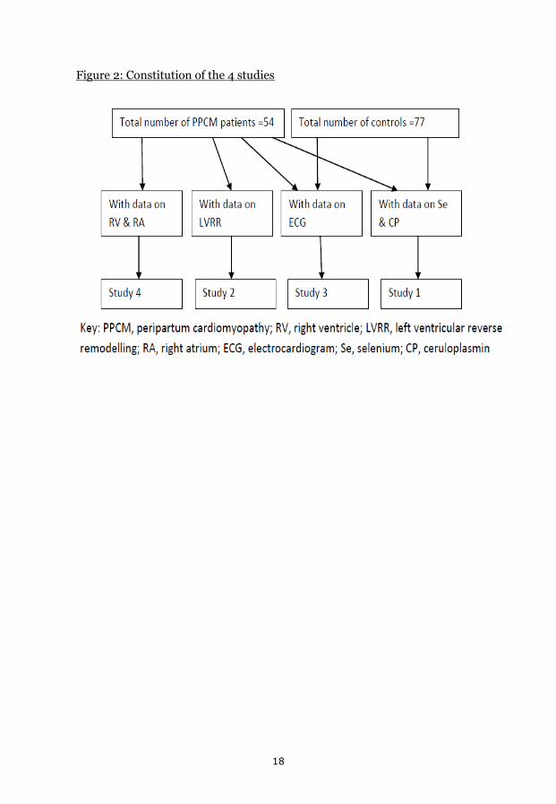
Study 1: Total of 39 PPCM patients and 50 controls were consecutively recruited after satisfying the inclusion criteria. Mean serum selenium in patients (61.7±14.9µg/L) was significantly lower than in controls (118.4±45.6µg/L) (p<0.001). The prevalence of serum selenium <70µg/L was significantly higher among patients (76.9%) than controls (22.0%) (p<0.001). The mean ceruloplasmin and prevalence of socio-economic indices, multiparity, pregnancy-induced hypertension, obesity and twin pregnancy were not different between the groups (p>0.05). Logistic regression showed that rural residency significantly increased the odds for serum selenium <70µg/L by 2.773 fold (p=0.037).
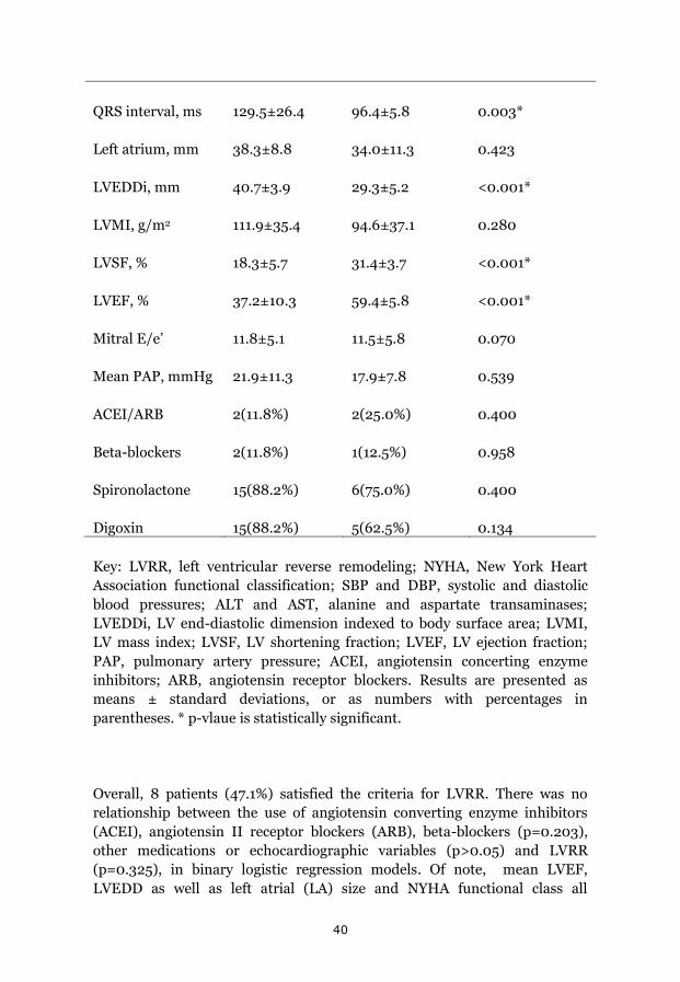
Study 2: A total of 54 newly diagnosed PPCM patients with mean age of 26.6±6.7 years, presented with classical features of predominantly left-sided HF and 33 of them qualified for follow-up. Of the 17 survivors at 12 months, 8 patients (47.1%) satisfied the criteria for LVRR, of whom 5 (29.4%) had recovered LV systolic function, but LVRR was not predicted by any variable in the regression models. The prevalence of normal LV diastolic function increased from 11.1% at baseline to 35.3% at twelve months (p=0.02). At one year follow-up, 41.4% of patients had died (two thirds of them within the first 6 months), but mortality wasn‟t predicted by any variable including LVRR.
Study 3: A total of 54 PPCM and 77 controls were consecutively recruited after satisfying the inclusion criteria. After controlling for confounding variables, a rise in heart rate by 1 beat/minute increased the odds of PPCM by 6.4% (p=0.001), while presence of ST-T-wave changes increased the odds of PPCM by 12.06 fold (p<0.001). In patients, QRS duration modestly correlated (r=0.4; p<0.003) with LV dimensions and end-systolic volume index (LVESVI), and was responsible for 19.9% of the variability of the latter (R2 = 0.199; p=0.003). A risk score of ≥2, developed by scoring 1 for each of the three ECG disturbances (tachycardia, ST-T-wave abnormalities and QRS duration), had a sensitivity of 85.2%, specificity of 64.9%, negative predictive value of 86.2% and area under the curve of 83.8% (p<0.0001) for potentially predicting PPCM.
Study 4: A total of 45 patients were recruited over 6 months with a mean age of 26.6±7.0 years. RV systolic function recovery occurred in a total of 8 patients (8/45; 17.8%), of whom 6 (75.0%) recovered in 6 months after
viii
diagnosis. The prevalence of RVSD fell from 71.1% at baseline to 36.4% at 6 months (p=0.007) and 18.8% at one year (p=0.0008 vs baseline; p=0.41 vs 6 month). Patients with RVSD had higher serum creatinine, and TAPSE accounted for 19.2% (p=0.008) of the variability of serum creatinine at 6 months. Although 83.3% of the deceased had RVSD, it didn‟t predict mortality in the regression models (p>0.05).
Conclusion:
These studies have shown that selenium deficiency seems to be a risk factor for PPCM in Kano, Nigeria, related to rural residency. However, serum ceruloplasmin, customary birth practices and some other characteristics were not associated with PPCM in the study area. They have also shown that PPCM patients had modest LVRR but high mortality at one year. Further studies should be carried out to identify reasons for the high mortality and how to curb it. In addition, using the risk score could help to streamline the diagnosis of PPCM with significant accuracy, prior to confirmatory investigations in postpartum women. Finally, RVSD and reverse remodelling were common in Nigerians with PPCM, in whom the first 6 months after diagnosis seem to be critical for RV recovery and survival.

ix
List of papers
1. Kamilu M Karaye, Isah A Yahaya, Krister Lindmark, Michael Y Henein. Serum Selenium and Ceruloplasmin in Nigerians with Peripartum Cardiomyopathy. Int J Mol Sci 2015; 16: 7644-7654.
2. Karaye KM, Lindmark K, Henein MY. One year survival in Nigerians with peripartum cardiomyopathy. Heart Views 2015, in press.
3. Karaye KM, Lindmark K, Henein MY. Electrocardiographic predictors of peripartum cardiomyopathy. Cardiovasc J Afr 2015; in press.
4. Karaye KM, Lindmark K, Henein MY. Right ventricular systolic dysfunction and remodelling in Nigerians with peripartum cardiomyopathy: a longitudinal study. BMC Cardiovasc Disord 2016; 16: 27. DOI 10.1186/s12872-016-0204-8
x
Abbreviations
ACEI Angiotensin converting enzyme inhibitors
AKTH Aminu Kano Teaching Hospital
ALT Alanine transaminase
ARB Angiotensin II receptor blocker
AST Aspartate transaminase
AUC Area under the curve
BNP B-type natriuretic peptide
BMI Body mass index
BP Blood pressure
CAD Coronary artery disease
CI Confidence interval
CMR Cardiac magnetic resonance imaging
CRT Cardiac resynchronisation therapy
DBP Diastolic BP
DCM Dilated cardiomyopathy
3DE 3-Dimensional echocardiography
DEFF Design effect
DT Deceleration time
ECG Electrocardiogram
ESC European Society of Cardiology

xi
Hb Haemoglobin
HF Heart failure
HIV Human immunodeficiency virus
HR Heart rate
ICD Implantable cardioverter defibrillator
IPAC Investigation in Pregnancy Associated Cardiomyopathy
IQR Inter quartile range
IU International units
IV Intravenous
LA Left atrium
LGE Late Gadolinium enhancement
LV Left ventricle
LVDD LV diastolic dysfunction
LVEDD LV end-diastolic dimension
LVEDDi LVEDD index
LVEF LV ejection fraction
LVESD LV end-systolic dimension
LVESVi LV end-systolic volume index
LVMI LV mass index
LVRR LV reverse remodelling
mPAP Mean pulmonary artery pressure

xii
MMSH Murtala Mohammed Specialist Hospital
MRI Magnetic resonance imaging
N Total number of subjects
NHLBI National Heart, Lung and Blood Institute
NIH National Institute of Health
NPV Negative predictive value
NYHA New York Heart Association
OR Odds ratio
P Prevalence
PA Pulmonary artery
PASP PA systolic pressure
PHT Pulmonary hypertension
PI Principal investigator
PPCF Peripartum cardiac failure
PPCM Peripartum cardiomyopathy
PPV Positive predictive value
RA Right atrium
RAA RA area
RAL RA length
RAP RA pressure
RIMP Right ventricular index of myocardial performance

xiii
ROC Receiver-operating characteristics curve
RV Right ventricle
RVb RV basal dimension
RVSD RV systolic dysfunction
SBP Systolic BP
SPSS Statistical package for social sciences
STAT3 Signal transducer and activator of transcription 3
TAPSE Tricuspid annular plane systolic excursion
TDI Tissue Doppler imaging
TNF Tissue necrosis factor
TV Tricuspid valve
USA United States of America

1
1.0: Introduction
The first description of Peripartum Cardiomyopathy (PPCM) dates back to 1880, when Virchow and Porak in two separate studies established an association between cardiac failure and the puerperium.[1,2] In 1937, Gouley et al described the clinical and pathological features of a severe and fatal type of heart failure (HF) in seven pregnant women; establishing the syndrome as a distinct clinical entity.[3] These women had dilated hearts in the last months of pregnancy, which persisted after delivery. An autopsy on four out of the seven patients who died demonstrated enlarged hearts with distinct widespread severe focal areas of necrosis and fibrosis.[3] Hull and Hidden then described 80 patients with this condition in New Orleans in 1938, and called it „Postpartal HF‟.[4] Over the years, research on PPCM had dragged on at a relatively slow pace.
1.1: Definition
Demakis et al were the first to define and describe the diagnostic criteria of PPCM in 1971.[5,6] They defined PPCM as the development of HF within the last month of pregnancy or first five months postpartum, in the absence of any identifiable cause for HF, and any recognizable heart disease before the last month of pregnancy.[5,6] Since then, the definition of PPCM has undergone several modifications.
In April 1997, the National Heart, Lung, and Blood Institute (NHLBI) and the Office of Rare Diseases of the National Institutes of Health (NIH) of the United States convened a Workshop on PPCM, to foster a systematic review of information and to develop recommendations for research and education. The agreed definition of PPCM, was based on the definition by Demakis et al of 1971, but including left ventricular (LV) systolic dysfunction demonstrated by classic echocardiographic criteria, such as depressed LV shortening fraction (LVSF) (<30%) or ejection fraction (LVEF) (<45%).[7]
In 2007, the European Society of Cardiology (ESC) working group on myocardial and pericardial diseases redefined cardiomyopathies including
2
PPCM, which they defined as a form of dilated cardiomyopathy (DCM) that presents with signs of cardiac failure during the last month of pregnancy or within 5 months of delivery.[8] DCM itself was defined by the presence of LV dilatation and systolic dysfunction in the absence of abnormal loading conditions (hypertension, valve disease) or coronary artery disease (CAD) sufficient to cause global systolic impairment. Right ventricular (RV) dilatation and dysfunction may be present but is not a diagnostic criterion.[8] In this classification, the focus was mainly on the concept of morphology and function of the heart, and away from that of diagnosis by exclusion. The aim was to promote a greater appreciation of the broad spectrum of diseases that can cause cardiomyopathies in everyday clinical practice.[8]
The most recent definition of PPCM was by the HF Association of ESC Working Group on PPCM, who believed that the time frame along with the echocardiographic cut-offs in the definition by NHLBI group are arbitrary and may lead to under-diagnosis of PPCM.[7,9] They therefore proposed the following simplified definition: “PPCM is an idiopathic cardiomyopathy presenting with HF secondary to LV systolic dysfunction towards the end of pregnancy or in the months following delivery, where no other cause of HF is found. It is a diagnosis of exclusion. The LV may not be dilated but the LVEF is nearly always reduced below 45%”.[9] The decision by this group to expand the definition to include women who present earlier in pregnancy was informed by the results of studies which have confirmed that such presentation was not uncommon, although most PPCM patients present in the last month of the pregnancy and puerperium.[10] Elkayam et al have demonstrated that the clinical presentation and outcome of patients with pregnancy-associated cardiomyopathy, diagnosed between the 17th – 36th weeks of gestation, were indistinguishable from those of patients meeting classic criteria for PPCM, and therefore concluded that the pregnancy-associated cardiomyopathy and PPCM represent a continuum of the same disease.[10] HF could be defined as a syndrome in which the patients should have the following features: symptoms of HF, typically shortness of breath at rest or during exertion, and/or fatigue; signs of fluid retention such as pulmonary congestion or ankle swelling; and objective evidence of an abnormality of the structure or function of the heart at rest.[11]
In general, experts now concur that PPCM is currently considered to be a distinct disease, and not a clinically silent idiopathic DCM unmasked by the
3
stresses of pregnancy.[7,9] This is mainly because the reported incidence is higher than the incidence of idiopathic DCM, its prognosis is better than that of idiopathic DCM, and because the high frequency of myocarditis would not be expected in a population presenting with decompensation of pre-existing heart disease due to hemodynamic stress.[7]
1.2: Epidemiology
The true incidence or prevalence of PPCM is unknown. This is largely because there have been only very few population-based studies on PPCM worldwide. PPCM is rare in some parts of the world and more common in others. For example, PPCM is very rare in Europe, but common in West Africa.[12,13] Recent studies suggest an estimated incidence of 1 case per 299 live births in Haiti, one case per 1000 live births in South Africa, and 1 case per 2289 - 4000 live births in the USA.[7,14,15,16] The reasons for this variation in incidence between and within countries remain unknown, but probably reflect an overestimation of the disorder in earlier studies that relied upon clinical criteria alone for the diagnosis.
The Hausa tribe of northern Nigeria appears to have the highest known incidence in the world of a HF within the time frame of PPCM, peripartum cardiac failure (PPCF); the incidence is reported to be as high as 1:100.[17,18] This was probably related to some local Hausa-Fulani customs, such as ingestion of a form of dry lake salt in the immediate postpartum period, a practice that can produce significant volume overload.[17,18] The other identified customary birth practices include frequent hot baths by breastfeeding mothers during the puerperal period, together with regular ingestion of a thick drink made from millet and rich in dry lake salt, „Kunun Kanwa‟ (in Hausa Language), and lying on heated mud beds.[17,18]
PPCM was recently described as the most prevalent type of cardiomyopathy in Kano, north-western Nigeria, found in 55 out of 1296 patients (4.2%) referred for echocardiography over a period of 7 months, representing 52.4% of all cardiomyopathies.[19] The prevalence of PPCM is still very high, despite the fact that the cultural practices identified decades ago to be important in the aetiology of PPCF in Zaria, a city about 120 km from Kano, are no longer popular.[17,18] However, some of the customs are still being
4
practised by the Hausa and Fulani women across northern Nigeria, although with lesser frequency, for shorter duration of time within the puerperal period, or even abandoned (as in the case of lying on heated mud beds) by most women in the present day northern Nigeria.[ 20,21] The disease is less common among other ethnic groups in the Country.
1.3: PPCM risk factors
PPCM has been associated with several risk factors over the years, but there is significant inconsistency between studies of their association with the disease. The risk factors include increased age, gravidity or parity, African origin, toxaemia or hypertension of pregnancy, use of tocolytics, twin pregnancy, obesity and low socioeconomic status.[22,23,24]
Although PPCM is thought to be more prevalent in the upper and lower extremes of childbearing age, and in older women of high parity, it is important to note that 24–37% of cases may occur in young primigravid patients.[14,15,25] Several case series reports from Nigeria, Haiti and South Africa did not show a disproportionate role for older age, multiparity, and long-term use of tocolytic agents in the development of PPCM.[14,19,25,26] In addition, Elkayam et al clearly showed that PPCM in the United States is not limited to black women, and their study did not support a strong association between multiparity and development of PPCM because almost 40% of the cases occurred in association with the first pregnancy and >50% with the first 2 pregnancies.[10] However, there was a significantly higher incidence in African American women as compared with other races.[27] Gentry et al conducted a case-control study in Augusta, Georgia, and Memphis Tennessee, and found almost a 16-fold higher incidence of PPCM in African American compared with non–African American women.[27]
What is the relationship between hypertension and PPCM? The answers to this question are debatable. The Zaria syndrome of PPCF was significantly found to be related to raised blood pressure (BP) during the acute phase of the HF, which was secondary to volume expansion following intake of large quantities of „kanwa‟, but in which 22% of the patients developed sustained hypertension during 2-5 years of follow up.[17,20] However, it is important to note that this Zaria syndrome was actually defined by its authors to be “a

5
high-output HF with well preserved ventricular function”.[17] For this reason therefore, PPCF is an entity different from what we know today as PPCM, going by the current definitions.[7-9]
A recent review by ElKayam described „hypertension‟ as a strong „associated condition‟, and not an aetiologic factor.[28] In societies where both PPCM and sustained hypertension are common in women, differentiating PPCM from hypertensive heart disease could be difficult if high BP is considered a clinical feature of PPCM. In support of this point, we found eccentric left ventricular hypertrophy, irrespective of gender, to be the most common type of abnormal LV geometry in hypertensive subjects in Kano, Nigeria.[29] These patients tend to present in HF with similar clinical and echocardiographic features to PPCM, except for the high BP or history of hypertension.[29] Some PPCM registries have shown that high BP in PPCM is rare, as reported by Sliwa et al from South Africa (seen 2%) and Fett et al from Haiti (4%).[14,25] In contrast, a retrospective study in Japan has reported the incidence of hypertension in PPCM as 41%, which is substantially higher than in the overall pregnant population.[ 30,31] Moreover, Kamiya et al revealed that hypertensive disorder is independently associated with a shorter hospital stay and higher LVEF.[ 30,31] However, hypertensive patients with and without PPCM had the same LV size and systolic dysfunction at diagnosis and discharge. In addition, parameters such as LV systolic diameter, fractional shortening, and LVEF showed greater improvement in the hypertensive patients. Thus, they concluded that the hypertensive state is not causative in the development of PPCM and might be a subset of PPCM that is characterized by relatively swift recovery, except in fatal cases.[30]
1.4: Aetiology
The aetiology and pathogenesis of PPCM is unknown, but several hypotheses have been proposed over the years.
Myocarditis hypothesis: For a long time, this hypothesis had enjoyed greater evidence and acceptance as a cause of PPCM than any other proposed aetiological factor. It is believed that the absent or muted immune response during pregnancy allows for unchecked viral replication and thus a greater
6
likelihood of myocarditis in the setting of a viral infection.[7] However, the prevalence of myocarditis in PPCM is highly variable between the different studies, ranging from none to 100%.[32] The reasons for this variability include: (a) the difficulty in defining PPCM clinically; (b) the inclusion of patients outside the accepted time frame of PPCM; (c) the difficulty in establishing the diagnosis by endomyocardial biopsy; (d) the variability in the inclusion of patients with borderline myocarditis together with those with histologic myocarditis as defined by the Dallas histological criteria; (e) the potential geographic variability of patient populations affected; and (f) the variable interval between presentation and the performance of endomyocardial biopsy.[7]
Prolactin, 16 kDa prolactin and Cathepsin D hypothesis: Experimental work suggested a novel and specific pathogenic mechanism by demonstrating the development of PPCM in female mice with a cardiomyocyte-specific deletion of the transcription factor, signal transducer and activator of transcription 3 (STAT3) protein.[25] Absence of cardiomyocyte STAT3 in the postpartum heart blunts the induction of the antioxidant enzyme manganese superoxide dismutase, resulting in increased oxidative stress. This then leads to increased expression and proteolytic activity of cardiac cathepsin D, which causes cleavage of the nursing hormone prolactin into an antiangiogenic and proapoptotic 16-kDa form of the prolactin. The latter has a detrimental effect on the myocardial microvasculature resulting in myocardial hypoxemia and apoptosis, and the development of PPCM.[25] Preliminary studies in a limited number of patients with PPCM have shown a favourable effect of bromocriptine, a pharmacological inhibitor of prolactin, in support of this hypothesis of PPCM.[33,34]
Autoimmune hypothesis: In women with PPCM, high titres of autoantibodies against some cardiac tissue proteins (adenine nucleotide translocator, branched chain α-keto acid dehydrogenase) and increased levels of tumor necrosis factor-alpha (TNF), interleukin-6, and soluble Fas receptors (an apoptosis signalling receptor) have been reported, suggesting a possible role of abnormal immunologic activities and inflammatory cytokines in pathogenesis of this disease.[35]

7
Malnutrition hypothesis: Malnutrition was thought to play an important role in the aetiopathogenesis of PPCM, but the occurrence of the disease in well-nourished patients has put this theory to doubt.[18] Furthermore, the investigation of micronutrients such as Vitamin A, Vitamin B12, Vitamin C, and beta carotene has not proved fruitful.[36] Nevertheless, selenium deficiency has been reported in 35 women with PPCM from the Sahel region of Africa.[37] However, a similar study among 18 women in Haiti didn‟t corroborate the findings, and an analysis by Fett et al of the data from the study by Cenac et al revealed a wide range of selenium values in PPCM patients, which probably represented a non-causal overlap in the Sahel of two common, but unrelated conditions.[36,37] It is likely that these geographic differences in serum levels of selenium in PPCM patients may be related to the deficiency of selenium in the soil.[32] Still, in the northern Nigerian city of Kano, we observed that PPCM almost always occurs in poor women; seen in 7.3% of the 164 women with low income referred for echocardiography, and absent among 113 women who had “higher income”.[24] The most plausible explanation for this observation could be poor nutrition predisposing the poor women to the disease, although the confounding effect of other risk/aetiological factors associated with poverty, such as increased tendency to infections, cannot be dismissed.
Epidemiologic studies showed that low selenium levels in the soil and in local foodstuffs correlated with low selenium levels in whole-blood and hair samples, and treatment with sodium selenite prevented Keshan disease and mitigated the clinical manifestations in patients with the disease.[38,39] Selenium is a critical component of central antioxidant enzymes, including glutathione peroxidases.[40] Owing to the importance of many selenoproteins in attenuating oxidant stress in a variety of cellular compartments, one can readily conclude that selenium deficiency promotes oxidant stress and injury, which may also potentiate the oxidant injury of other contributing pathogenic factors, including viral and other infections. [40,41] Therefore in spite of the observations by Fett et al, it is still conceivable that selenium deficiency is related to PPCM as illustrated in Figure 1, at least in some parts of the world.[36]
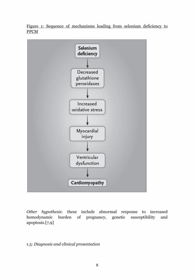
8
Figure 1: Sequence of mechanisms leading from selenium deficiency to PPCM
Other hypothesis: these include abnormal response to increased hemodynamic burden of pregnancy, genetic susceptibility and apoptosis.[7,9]
1.5: Diagnosis and clinical presentation
9
The diagnosis of PPCM rests on the development of HF and echocardiographic identification of new LV systolic dysfunction during a limited period surrounding parturition. This presents a challenge because many women experience dyspnoea, fatigue, and pedal oedema towards the end of a normal pregnancy; symptoms identical to early congestive HF. PPCM may therefore go unrecognized, leading to underestimation of its incidence or prevalence.[7,9] Strong consideration should be given to screening family members of PPCM patients because there are reports that suggested genetic predisposition to the cardiomyopathy.[7,9]
Symptoms and signs that should raise the suspicion of HF include paroxysmal nocturnal dyspnoea, chest pain, cough, raised jugular venous pressure, new murmurs consistent with mitral or tricuspid valve (TV) regurgitation, and pulmonary basal crackles. There are no specific criteria for differentiating subtle symptoms of HF from normal late pregnancy, so it is important that a high index of suspicion be maintained to identify the rare case of PPCM. Several studies have shown that formation of intracardiac thrombus and thromboembolic complications are not uncommon in PPCM patients.[7,9,19,26] In a cross-sectional study on patients admitted with HF, 6 out of 11 patients with PPCM were found to have mural thrombi, and 4 of them presented with cardioembolic stroke.[26]
The diagnosis of PPCM requires excluding other causes of cardiomyopathy and is confirmed by standard echocardiographic assessment of LV systolic dysfunction, including depressed LVSF and LVEF. As mentioned earlier, excluding cases of hypertensive heart disease could prove to be very challenging in populations where both PPCM and hypertension are common. An electrocardiogram (ECG) is recommended for all PPCM patients, and a recent study found 96% of PPCM patients to have „abnormal‟ ECGs at presentation, and suggested its potential usefulness in screening and prognosticating patients at risk, in resource-poor settings.[7,9,42]
1.51: Novel echocardiographic techniques in the study of PPCM
The advent of novel echocardiographic techniques provides the opportunity to study PPCM further. These techniques include those for studying ventricular long-axis function, right ventricular (RV) function, tissue
10
Doppler imaging (TDI) techniques including strain and strain rate echocardiography, and speckle tracking echocardiography. Unfortunately, these techniques have not been widely utilised to study PPCM. Although LV systolic dysfunction defines PPCM, information on both LV and RV diastolic dysfunction would provide important clinical information that could further characterise the disease. This hypothesis is supported by evidence in DCM patients, in whom parameters of LV diastolic dysfunction were found to be powerful and important predictors of major cardiac events such as heart transplantation and non-sudden death.[43] In this study, invasive and echocardiographic parameters of diastolic function revealed comparable information for the estimation of prognosis of patients with DCM.[43]
Ventricular long-axis function: It has been recognised for ages that the fall in cavity volume with LV systole involves longitudinal as well as circumferential shortening, although the latter plays a dominant role. Though longitudinally directed fibres situated mainly in the subepicardial and subendocardial regions of the LV and RV free walls and the papillary muscles comprise only a small proportion of the total ventricular myocardial mass, they play a major role in the maintenance of normal EF and in determining atrioventricular interactions.[44,45] Not surprisingly therefore, loss of longitudinal fibre function leads to characteristic disturbances.[44,45] Longitudinal function is always reduced when ventricular cavity size is increased, but in addition, EF is also reduced.[46] This relation is consistent enough for long axis amplitude, „the amplitude of atrioventricular ring motion‟, to be used as an index of EF.[47] It applies not only to the LV, where it can be shown to relate to prognosis but also to the RV, where it provides a simple method of assessing RV function.[48]
Tricuspid annular plane systolic excursion (TAPSE) has been recently used to study RV systolic function in PPCM patients in comparison to patients with DCM.[21] In this cross-sectional study, reduced TAPSE, signifying RV systolic dysfunction, was defined as value of ≤14 mm.[21] A total of 90 patients were recruited; 55 of them had PPCM while the remaining 35 had DCM. Mean TAPSE was significantly less in PPCM (12.58±4.27mm) as compared with DCM patients (14.46±3.21mm) (P<0.028), while TAPSE ≤14 mm was found in 54.6% of PPCM patients and in 37.1% of DCM patients (P= 0.05).[21] The study showed perhaps for the first time, that RV systolic function in PPCM patients was worse than that of patients with idiopathic DCM.

11
In addition, long-axis measurements have provided the opportunity for detailed analysis of ventricular segmental and global function in patients with ischemic cardiomyopathy. An increase in long-axis amplitude with stress (a normal response) has been found to be a reliable sign of myocardial viability irrespective of the appearance of additional markers of ischemic dysfunction such as incoordination/dyssynchrony.[49,50] It remains to be explored whether this concept can be extended to the prediction of recovery of LVEF in patients with PPCM.
Myocardial Strain and Strain Rate Imaging: Myocardial velocities measured with TDI may be overestimated or underestimated by translational motion or tethering of the myocardium, respectively. This limitation can be overcome by measuring the actual extent of myocardial deformation (stretching or contraction) by strain and strain rate imaging.[51] Strain rate is the rate of change in length calculated as the difference between two velocities normalised to the distance between them; it is expressed as seconds-1. By convention, shortening is expressed by negative values and lengthening by positive values for both strain and strain rate.[51] To the best of our knowledge, strain imaging has not been used to study PPCM.
Speckle tracking echocardiography: This technique is a method for quantifying myocardial motion in various planes using 2D images. Reflection, scattering, and interference of the ultrasound beam in the myocardial tissue produce a speckle formation.[51] Myocardial regions with unique speckle patterns in the grey scale 2D image can be tracked from frame to frame throughout the cardiac cycle. This allows assessment of LV rotational motion, often referred to as torsion or twist. Speckle tracking is an alternative method for quantification of LV systolic, and potentially diastolic function. It is also another method for studying strain using 2D images instead of the TDI methods. Speckle tracking does not have the limitation of angle dependence that TDI-derived strain measurements have.[51]
Speckle tracking imaging has been used to gain greater understanding into the pathophysiology of cardiac ischemia and infarction, primary diseases of

12
the myocardium, and the effects of valvular disease on myocardial function.[52] Speckle tracking has also been used to quantify abnormalities in the timing of mechanical activation for HF patients undergoing cardiac resynchronization pacing therapy.[52] Further advances, such as 3-dimensional speckle tracking strain imaging, have emerged to provide even greater insight. Strain imaging has become established as a robust research tool and has great potential to play many roles in routine clinical practice to advance the care of the cardiovascular patient.[52] Again, it appears that this technique has not been used to study PPCM.
Cardiac magnetic resonance imaging (CMR): This technique has been used in a limited number of PPCM patients for the assessment of cardiac function and the detection of mural thrombi or myocardial fibrosis.[28] In a group of 8 women with PPCM who were studied with CMR, none exhibited abnormal myocardial late enhancement (i.e. no detectable myocardial fibrosis), and no difference was found in the MRI patterns in 4 patients who recovered normal LV function compared with those who did not.[53] The authors concluded that patients with PPCM do not exhibit a specific cardiac CMR pattern, and do not exhibit myocardial late enhancement. They suggested that myocardial fibrosis does not play a major role in the limitation of cardiac function recovery after PPCM.[53] This assertion goes against the earlier findings by Gouley et al, reported in 1937, who found widespread areas of focal necrosis and fibrosis in the hearts of PPCM patients.[3] A more recent retrospective cohort study by Arora et al found late gadolinium enhancement (LGE) in 4 out of 10 PPCM patients on follow up, who had multiple readmissions because of HF exacerbations and persistently low LVEF on subsequent echocardiograms.[54] The authors thus concluded that LGE seems to be associated with a poor prognosis in the setting of PPCM.[54]
The role of CMR in PPCM is being further explored in an arm of the Investigation in Pregnancy Associated Cardiomyopathy (IPAC) study in the United States.[28] One of its objectives is to investigate the frequency of myocardial injury or inflammation on cardiac MRI and the ability of tissue characteristics to predict subsequent recovery of LVEF.[28]
13
CMR is now considered the reference method of non-invasive cardiac imaging. The accuracy of echo-Doppler modalities and measurements does not refer to autopsy data anymore, but to CMR. In interventional trials, CMR allows to reduce the sample size of the study population because of the smaller variability of its measurements. New echo technologies, mainly three-dimensional echocardiography (3DE) and speckle-tracking echocardiography, have become available and are competitive with CMR in accuracy while being less expensive and more widely available.[55]
1.6: Prognosis
The prognosis in PPCM varies geographically. In the Unites States, reported mortality rates associated with PPCM have varied widely between 0% and 19%, while rates of cardiac transplantation have ranged from 6% to 11%.[28] Substantial differences in the reports are probably due to variations in patient populations, diagnostic criteria, and treatments, as well as reporting bias.[28] Goland et al provided detailed information regarding mortality in 13 patients, most of whom died either suddenly (38%) or of progressive HF (45%) between the day of delivery and 8 years postpartum.[56] Mortality was found to be higher in women with baseline LVEF ≤25% as well as in women in whom the diagnosis of PPCM was delayed.[56] In addition, Whitehead et al reported that mortality increased with maternal age, in women with parity of more than 4, and in black women, who were 6.4 times more likely to die compared with whites.[57] Eighteen percent of deaths occurred within 1 week and 87% within 6 months of diagnosis, and mortality was due either to progressive HF or to sudden cardiac death.[57] In South Africa, case series have demonstrated that mortality rates have slowly improved over time but 6 months and 2-year mortality rates remain at 10% and 28% respectively.[9] Single centre studies in Brazil and Haiti reported mortality rates of 14-16% within 6 months.[9] Other factors found to independently predict mortality, although inconsistently, are New York Heart Association (NYHA) functional classes, LVEF, QRS duration on ECG and late onset of symptoms.[7,9]
LV systolic function is believed to return to normal in about 23-41% of PPCM patients over time.[9] In the recent IPAC study carried out in 30 North American centres however, 13% of well-treated PPCM patients had experienced major events or had persistent severe cardiomyopathy with an LVEF <35% and 72% achieved an LVEF ≥50%.[58] An initial LVEF <30%,

14
an LVEDD ≥6.0 cm, black race, and presentation after 6 weeks post-partum were associated with a lower LVEF at 12 months. No subjects with both a baseline LVEF <30% and an LVEDD ≥6.0 cm recovered by 1 year post-partum, whereas 91% with both a baseline LVEF≥30% and an LVEDD <6.0cm recovered.[58]
Subsequent pregnancies in women with history of PPCM are associated with a risk for recurrent and persistent cardiac dysfunction and even mortality. The risk is substantially higher in patients with persistent LV dysfunction before subsequent pregnancy. At the same time, however, recovery of LV systolic function does not guarantee an uncomplicated subsequent pregnancy. Although mortality in such patients is rare, marked decreases in LV function have been reported in approximately 20% of patients, with persisting dysfunction after pregnancy in about 50%.[59]
1.7: Treatment:
1.71: Initial management of acute heart failure
The principles of managing acute HF due to PPCM are no different than those applying to acute HF arising from any other cause and are summarized in recent ESC guidelines.[11] In summary, rapid treatment is essential, especially when the patient has pulmonary oedema and/or hypoxaemia. Oxygen should be administered in order to achieve an arterial oxygen saturation of ≥95%, using, where necessary, non-invasive ventilation with a positive end-expiratory pressure of 5–7.5 cm of water. Intravenous (IV) diuretics should be given when there is congestion and volume overload, with an initial bolus of frusemide 20–40mg. IV nitrate is recommended (e.g. nitroglycerine starting at 10–20 up to 200 mg/min) in patients with a systolic blood pressure (SBP) >110 mmHg and may be used with caution in patients with SBP between 90 and 110 mmHg. Inotropic agents should be considered in patients with a low output state, indicated by signs of hypoperfusion.[9,11] Further care in the form of ventricular mechanical support or cardiac transplantation should be offered to patients when needed according to the recommendations in standard guidelines.[9,11]
1.72: Management of chronic heart failure in PPCM

15
I: Management during pregnancy: this should follow standard guidelines with some exceptions.[9,11] These exceptions include avoiding angiotensin converting enzyme inhibitors (ACEI) and angiotensin receptor blockers (ARB) because they are contraindicated in pregnancy, in view of renal and other foetal toxicities.[9,11] Hydrallazine and long-acting nitrates could be used in combination as substitute. Spironolactone is thought to have antiandrogenic effects in the 1st trimester.[9,11] Because the effects of eplerenone on the human foetus are uncertain, it should also be avoided during pregnancy. Beta blockers and diuretics can be used but cautiously, given the latter‟s effect on placental blood flow. Digoxin has been used in pregnancy without causing foetal harm. It is excreted into breast milk, but no adverse effects have been reported, and the drug is compatible with breast-feeding.[60]
II: Management of HF after delivery: this should follow standard guidelines:[9,11]
1.73: Bromocriptine in the treatment of PPCM
Bromocriptine, a dopamine 2D agonist which blocks prolactin, may be a novel disease-specific treatment for PPCM. Several case reports have suggested that the addition of bromocriptine to standard therapy for HF may be beneficial in patients with acute onset of PPCM.[30,61] Because the drug appears to increase the risk of thromboembolic phenomena including myocardial infarction, anticoagulation therapy is strongly encouraged in PPCM patients with a low LVEF in general and in those taking bromocriptine in particular. Before this treatment can be recommended as a routine strategy, there is a need for a larger randomized trial, although some physicians currently add bromocriptine to conventional therapy on an individual basis.[9]
1.74: Anticoagulation in the treatment of PPCM
In PPCM, anticoagulation is advisable from the time of the diagnosis until LV function recovers (LVEF >35%), or for the treatment of atrial fibrillation, because of the high incidence of thromboembolism associated with the disease.[7,9,19,26] Anticoagulation is particularly important during pregnancy and the first 6 to 8 weeks postpartum because of persistent hypercoagulable state.[62] In contrast to warfarin which causes foeto-

16
toxicity, both unfractionated heparin and low-molecular-weight heparin do not cross the placenta and are safe during pregnancy.[63] The use of unfractionated heparin is preferred during pregnancy because of its shorter half-life and reversible effect, in the event of premature labour and a possible need for urgent delivery. Both warfarin and heparin are not secreted into breast milk, and are therefore safe during breast-feeding.[63]
1.75: Cardiac resynchronisation therapy (CRT) (with or without Implantable Cardioverter Defibrillator (ICD))
ICD and CRT have become standard of care in modern treatment for HF. The recent focused update of the ESC guidelines on device therapy in HF recommended that CRT with pacemaker or defibrillator function should be offered to patients in HF with NYHA class III or IV despite optimal medical therapy, LVEF ≤35%, QRS duration ≥120 ms, and in sinus rhythm.[64] However, patients in NYHA class IV should be ambulatory.[64] In this group of patients, CRT has been shown to confer sustained and significant alleviation of symptoms and to increase exercise capacity.[65,66] CRT also reduces all-cause mortality as well as rehospitalisation rate for HF, and to reverse remodelling particularly in non-ischemic heart diseases.[66,67] In addition, the guidelines recommend CRT preferably with defibrillator function, to reduce morbidity or to prevent disease progression, for patients with mild symptoms (NYHA class II) despite optimal medical therapy, with LVEF ≤35%, QRS duration ≥150 ms, and in sinus rhythm.[64]
Decisions about both the necessity and the timing of CRT or ICD implantation in PPCM patients are extremely difficult and require careful consideration of the advantages and otherwise, in the context of the natural history of PPCM. The major issues are the cost and potential complications of implanting a device in a patient who may not need it for a long time because of subsequent recovery of ventricular function.[9] However, CRT could be considered in a patient with PPCM who has persistently severe LV dysfunction 6 months following presentation, despite optimal medical therapy.[9] Six months after ICD-CRT treatment, 2 PPCM patients who didn‟t improve on medical treatment were observed to have a significant improvement in LVEF from 25% to 45% and 28% to 50%, respectively, and positive remodelling with reduction of left ventricular end-diastolic volume from 216 to 144ml and from 354 to 105ml, which represents a 34% and a 70% reduction, respectively.[68]

17
1.8: Objectives
The general objective of the thesis was to study the clinical characteristics and prognosis of peripartum cardiomyopathy in Kano, Nigeria.
Study 1
To assess the significance of serum levels of selenium and ceruloplasmin as well as the previously described traditional birth practices, as PPCM risk factors, in Kano, Nigeria.
Study 2
To describe the one year survival and LV reverse remodelling (LVRR) in a cohort of PPCM patients from three referral hospitals in Kano, Nigeria.
Study 3
To determine potential ECG variables that predicts the diagnosis of PPCM.
Study 4
To assess RV systolic dysfunction (RVSD) and reverse remodelling over 1 year in a cohort of PPCM patients in Kano, Nigeria.
18
Figure 2: Constitution of the 4 studies
19
2.0: Materials and methods
2.1: Study centres
The study was carried out in Aminu Kano Teaching Hospital (AKTH), Murtala Mohammed Specialist Hospital (MMSH), and a private cardiac clinic in Kano, Nigeria. Kano is a densely populated city in north-western region of Nigeria.
2.2: Sample size estimation
Studies 1 and 3 had a case-control design, while studies 2 and 4 were prospective and longitudinal in design. Minimum sample size for patients for all the studies was estimated at 45 subjects, calculated using a validated formula (see below), using an estimated prevalence of PPCM patients of 4.2% among patients referred for cardiologic evaluation, at a confidence level of 90%.[24]
Sample size (n) = [DEFF*Np(1-p)]/ [(d2/Z21-α/2*(N-1)+p*(1-p)]
Where:
Population size (N) = 100,000
Hypothesized frequency of outcome factor in the population (p) = 4.2%
Confidence limits (d) = 10%
Design effect (for cluster surveys-DEFF) = 1
The sample size for the controls was estimated at 1.5 times the total number of patients (±5 patients).
2.3: Ethics

20
Informed consent was obtained from all recruited subjects and a consent form signed or thumb-printed (if illiterate). Ethical approval for the study was sought from the Research Ethics Committees of AKTH and Kano State Hospitals Management Board before the commencement of the study, and the research conformed to the ethical guidelines of the Declaration of Helsinki; on the principles for medical research involving human subjects.[69]
2.4: Study 1 (Serum selenium and ceruloplasmin in Nigerians with peripartum cardiomyopathy)
This was a case-control study but the patients were followed up for 6 months. Inclusion criteria for the patients were: (i) new diagnosis of PPCM before commencement of medical treatment; (ii) presenting within 5 months postpartum; (iii) at least 18 years of age; and (iv) giving written informed consent. To be included, the controls had to satisfy the following criteria: (i) being apparently healthy; (ii) no past history of any cardiac disease or systemic hypertension (except if pregnancy-induced during the last pregnancy); (iii) having normal ECG; (iv) presenting to the study centers within 5 months postpartum for routine immunizations for their children; and (v) giving written informed consent. Subjects were excluded if their results for serum selenium and ceruloplasmin were not available. PPCM was defined according to the recommendations of the HF Association of the ESC Working Group on PPCM [9]. At the study sites, physicians and obstetricians were approached and requested to refer all patients with suspected PPCM to the principal investigator (PI) for further evaluation at no cost. Patients were then interviewed, clinically evaluated and recruited consecutively. Hospital in-patients with PPCM were clinically evaluated and underwent investigations within the first 48 h of admission. Demographic data, relevant aspects of history and physical signs, results of investigations, medications, co-morbid conditions, and complications were included in a detailed questionnaire. For each subject, a 12-lead ECG at rest and trans-thoracic echocardiogram (for PPCM patients only) were carried out by the PI at the study centers according to standard recommendations [9]. The echocardiographic examination was carried out using Sonoscape S8 Doppler Ultrasound Machine (Shenzhen, China, 2010) and the ECG using Mindray DECG-03A digital electrocardiograph (Shenzhen, China, 2008) according to
21
standad ecommendations [70,71,72]. In view of the lack of facilities in Nigeria, serum samples for selenium and ceruloplasmin assays were frozen and sent to Metropolis Diagnostic Centre, Metropolis Healthcare Ltd, New Delhi, India. Serum selenium was measured using the Inductive Coupled Plasma Mass Spectrometry Method, while ceruloplasmin was measured using the Nephelometry Method with BN Prospec Machine (Made by Siemens). Patients were followed up by the physicians in the respective study centres for six months, and the PI re-evaluated the patients at the completion of the six months including ECG and echocardiogram examinations, but blood tests were not repeated. A high attrition rate of at least 40% was anticipated given that many of the patients did not have contact telephone or might not have money to pay for transportation or because of the frequent terrorist attacks on Kano City, including the main study centre (MMSH). The reference range of serum selenium and ceruloplasmin at the Metropolis Laboratory were 74.0–90.0 µg/L and 20.0–60.0 mg/dL, respectively. In this study, low serum selenium was defined as values <70 µg/L, which seems to be the minimal selenium level that is critical for expression of key antioxidant enzymes such as glutathione peroxidases and the thioredoxin reductases [40,73]. Increased ceruloplasmin level was defined as >60 mg/dL. Anemia was defined as hemoglobin <12 g/dl, according to the recommendation of World Health Organization in non-pregnant women [74].
Statistical Analysis
Continuous variables were explored for the presence of skewness, which was corrected with logarithmic (log10) transformation. Patients‟ baseline characteristics were described using frequencies and mean, while time period between patients‟ delivery and recruitment as well
22
as serum levels of selenium and ceruloplasmin were described using the median and inter-quartile range (IQR). Chi-square, Fisher‟s exact probability and Student t- tests were used to compare categorical and continuous variables as appropriate. Binary logistic regression models were used to assess for association between low selenium and variables of interest, and values were expressed as Odds Ratios (OR) and 95% Confidence Intervals (CI). The statistical analysis was carried out using SPSS version 16.0 software (Chicago, IL, USA). A p-value < 0.05 was considered as minimum level of statistical significance.
2.5: Study 2 (One year survival in Nigerians with peripartum cardiomyopathy)
This was a longitudinal study carried out in three referral hospitals in Kano, Nigeria. Patients‟ inclusion criteria were: (i) new diagnosis of PPCM before commencement of medical treatment; (ii) onset of HF symptoms between last few months of pregnancy and first five months postpartum, (iii) at least 18 years of age; (iv) contact telephone number, except patients who gave reassurance that they were willing to attend the follow-up visits, and (v) giving written informed consent. PPCM was defined according to the recommendations of the HF Association of the European Society of Cardiology Working Group on PPCM.[9] At the study sites, physicians and obstetricians were approached and requested to refer all patients with suspected PPCM to the principal investigator (PI) for further evaluation at no cost. Patients were then interviewed, clinically evaluated and recruited consecutively. Hospital in-patients with PPCM were clinically evaluated and underwent investigations within the first 48 hours of admission. Demographic data, relevant aspects of history and physical signs, results of investigations, medications, co-morbid conditions, and complications were included in a detailed questionnaire. For patients who didn‟t have contact telephone number but who agreed to participate, attending the first month follow up with the supervising physician was used as evidence of commitment and for inclusion. All patients were given appointment cards as a reminder, and were also telephoned to be informed of the six and twelve month follow-up
23
visits. At the recruitment visit, each subject underwent a 12-lead ECG at rest and trans-thoracic echocardiogram at the study centres according to standard recommendations.[70] The echocardiographic examination was carried out using Sonoscape S8 Doppler Ultrasound system (Shenzhen, China, 2010). Other baseline investigations recommended for the management of patients with PPCM were carried out at the laboratories of the study centre, including plasma hemoglobin; serum urea, electrolytes and creatinine; blood counts; liver enzymes and serum albumin.[9] The PI re-evaluated the patients at six and twelve month follow-up, using the same protocol as at recruitment including ECG and echocardiogram examinations, but blood tests were not repeated.
Systemic hypertension was defined using standard cut-off values for systolic BP (SBP) ≥140mmHg and or diastolic BP (DBP) ≥90mmHg. Anaemia was defined as haemoglobin <12g/dl, according to the recommendation of the World Health Organisation in non-pregnant women, renal impairment as serum creatinine >150μmol/l (>2 mg/dl), hyponatraemia as serum sodium <135mmol/l and hypoalbuminaemia as serum albumin <30g/L.[11,74]
Cardiac function assessment: Of the standard echocardiographic recordings obtained, LVRR was defined as the presence of both absolute increase in LVEF ≥10.0% and decrease in LV end-diastolic dimension indexed to body surface area (LVEDDi) ≤33.0 mm/m2, while recovered LV systolic function as LVEF ≥55%, at 12 months follow-up.[75]
LV diastolic function (LVDD) was defined and graded using trans-mitral flow and LV myocardial TDI velocities at the mitral annular level as follows:[70,76]
Normal LV diastolic function: E:A ratio 1-2, deceleration time (DT) 160-230 milliseconds(ms) and E/e‟ <8.
Grade I LVDD (impaired myocardial relaxation): E:A <1.0 and DT >240ms.

24
Grade II LVDD (pseudonormalised pattern): E:A 1-1.5, DT 160-230ms, e‟ <7cm/s and E/e‟ >15.0.
Grade III LVDD (restrictive filling): E:A >2.0, DT <160ms, e‟ <7cm/s and E/e‟ >15.0.
Statistical analysis
Continuous variables were explored for the presence of skewness. Patients‟ baseline characteristics were described using frequencies and mean, while survival time from diagnosis to the patients‟ demise and serum aspartate transaminase (AST) and alanine transaminase (ALT) were described using the median and IQR. Chi-square, Fisher‟s exact probability and Student t- tests were used to compare categorical and continuous variables as appropriate. Spearman correlation coefficient and logistic regression models were used to assess potential associations between LVRR or survival and variables of interest. The statistical analysis was carried out using SPSS version 16.0 software. Two-sided p-value <0.05 was considered as minimum level of statistical significance.
2.6: Study 3 (ECG predictors of PPCM)
This was a case-control study carried out in MMSH, AKTH and a private cardiology clinic in Kano, Nigeria. The inclusion criteria for the patients were: (i) confirmed diagnosis of PPCM; (ii) onset of symptoms towards the end of pregnancy or within the puerperium, and presentation to hospital within nine months postpartum; (iii) age of at least 18 years; and (iv) written informed consent. Patients were excluded if: (i) their symptoms could be explained by diagnoses other than PPCM; (ii) their symptoms started in early pregnancy or after the first 5 months postpartum; (iii) they were younger than 18 years or older than 45 years; (iv) they denied consent to participate. To be included, the controls had to satisfy the following criteria: (i) apparently healthy; (ii) no past history of any cardiac disease or systemic hypertension (except pregnancy-induced hypertension) (iii) normal ECG (except for flat T-waves in leads III or aVF, and inverted T-waves in aVR, V1 or V2, which are considered as non-specific);[77] (iv) presenting to the study centres within 9 months postpartum for routine immunisations for their children; and (v) written informed consent. In addition, subjects taking drugs known to affect ECG intervals were excluded from the study.[78] T-

25
wave inversion with or without ST segment depression were considered abnormal in all leads except aVR, V1 and V2.[77] In addition, flat T-waves in leads III or aVF were also considered as non-specific.[77] Controls were excluded if: (i) they presented their children for immunisation after 5 months postpartum; (ii) they presented to the hospital as patients; (iii) they were younger than 18 years or older than 45 years; (iv) they were known or found clinically to have any cardiac disease; (v) they denied consent. PPCM was defined according to the recommendations of the HF Association of the ESC Working Group on PPCM.[9]
At the study centres, physicians in Internal Medicine and Obstetrics and Gynaecology Departments were approached and requested to refer all patients with suspected PPCM to the PI for further evaluation. Patients were then interviewed, clinically evaluated and recruited consecutively. Hospital in-patients with PPCM were clinically assessed and underwent investigations within the first 48 hours of admission. Demographic data, relevant aspects of history and physical signs, results of investigations, co-morbid conditions, and complications were included in a detailed questionnaire. Baseline serum urea, electrolytes and creatinine were carried out in the laboratories of AKTH, while 12-lead ECG at rest and trans-thoracic echocardiogram (for PPCM patients only) were all carried out by the PI at the study sites according to the standard recommendations.[70] The echocardiographic examination was performed using Sonoscape S8 Doppler Ultrasound Machine (Shenzhen, China, 2010) and the ECG was recorded using Mindray DECG-03A digital electrocardiograph (Shenzhen, China, 2008).[70,71] All ECG recordings were studied and interpreted by the investigators in the standard fashion, and ECG intervals/durations were measured using manual callipers.[ 72,79] The controls were evaluated using the same protocol as patients including the ECG, but echocardiogram was not performed.
Statistical analysis
Frequencies and mean (with standard deviation) were used to describe patients‟ characteristics. Chi-square, Fisher‟s exact probability, Student‟s t- and Mann-Whitney U tests were used to compare categorical and continuous variables as appropriate. Binary logistic regression models were used to determine predictors of PPCM among ECG variables, and values were expressed as OR and 95%CI. Spearman‟s correlation coefficient and linear regression models were used to further assess relationships between
26
variables of interest. A simple score assigning 1 to each identified independent ECG predictor was composed and its accuracy in predicting PPCM was determined using the area under the receiver-operating characteristics (ROC) curve (AUC), and AUC >0.75 was considered satisfactory. P-value <0.05 was considered statistically significant. The statistical analysis was carried out using SPSS version 16.0 software.
2.7: Study 4 (RVSD and remodelling in Nigerians with PPCM)
This is a longitudinal study carried out in MMSH, AKTH and a private cardiology clinic in Kano, Nigeria. Inclusion criteria were: (i) new diagnosis of PPCM before commencement of medical treatment; (ii) onset of HF symptoms between last few months of pregnancy and first five months postpartum, (iii) at least 18 years of age; (iv) contact telephone number, except patients who gave reassurance that they were willing to attend the follow-up, and (v) giving written informed consent. We excluded PPCM patients who were on HF treatment, as well as those who presented more than 5 months since delivery. PPCM was defined according to the recommendations of the HF Association of the Working Group on PPCM.[9]
At the study sites, physicians and obstetricians were invited to refer all patients with suspected PPCM to the PI for further evaluation. Patients were then interviewed, clinically evaluated and recruited consecutively. For each subject, a 12-lead ECG at rest and trans-thoracic echocardiogram were carried out by the PI at the study centres according to standard recommendations [70]. The echocardiographic examination was carried out using Sonoscape S8 Doppler Ultrasound System (Shenzhen, China, 2010). Plasma haemoglobin and serum urea, electrolytes and creatinine were measured at the laboratories of AKTH according to standard protocols. The PI re-evaluated the patients at 6 and 12 months follow-up, using the same protocol as at recruitment including ECG and echocardiographic examinations, but blood tests were not repeated.
Cardiac function assessment: Echocardiography was performed according to standard recommendations.[70,80] RV basal diameter (RVb), Right atrial

27
longitudinal dimension (RAL) and RA end-systolic area (RAA) were measured in each patient. TAPSE was recorded from the apical four-chamber view with the M-mode cursor positioned at the free wall angle of the TV annulus [81]. RV long axis amplitude of motion (i.e. TAPSE) was measured from end-systolic to end-diastolic points [81], and its peak systolic velocity (S‟) was measured from myocardial TDI. All recordings of TAPSE and S‟ were obtained during held end-expiration. Care was taken to align M-mode or TDI beam along the direction of tricuspid annulus motion, with the minimum angle in between. TDI sample volume was positioned at 10 mm from the insertion site of the tricuspid leaflets or 10 mm away within RV lateral wall and adjusted to cover the longitudinal excursion of the tricuspid annulus both in systole and diastole.[70] RVSD was defined as the presence of either TAPSE <16mm or S‟ of RV lateral tricuspid annulus <10cm/s.[80] For the purpose of this study, recovery of RV systolic function was defined as an improvement of reduced TAPSE to ≥16mm or S‟ to ≥10cm/s, without falling to reduced levels again, during follow-up.
Pulmonary artery systolic pressure (PASP) was estimated using continuous wave Doppler of the maximum velocity of the tricuspid regurgitant jet (V), from which the retrograde pressure drop was calculated using the modified Bernoulli equation (4V2), and adding to it the estimated right atrial pressure (RAP).[82] RAP was estimated using the diameter and collapse of the inferior vena cava during spontaneous respiration, as previously described.[83] Pulmonary hypertension (PHT) was defined as mean pulmonary arterial pressure (mPAP) of ≥25mmHg at rest.[84] mPAP was estimated from PASP using the Chemla formula as: mPAP = (0.61 X PASP) +2 (mmHg).[85]
Statistical analysis Continuous variables were explored for the presence of skewness, which was corrected with log10 transformation. Patients‟ baseline characteristics were described using frequencies and mean, while time period between patients‟ delivery and recruitment was described using the median and IQR. Chi-square, Fisher‟s exact probability and Student t- tests were used to compare categorical and continuous variables as appropriate. Spearman correlation coefficient (ρs) was used to assess the association between TAPSE and mPAP and serum creatinine, while logistic regression models were used to assess the

28
associations between RVSD or mortality and variables of interest. Linear regression was also used to assess the relationship between TAPSE and mPAP. Estimates for regression analyses were expressed as OR and 95%CI. The regression results were tested with Hosmer and Lemeshow‟s goodness of fit test, and a p-value ≥0.05 implied that the model‟s estimates fit the data at an acceptable level. The statistical analysis was carried out using SPSS version 16.0 software. Two-sided p-value <0.05 was considered as minimum level of statistical significance.
29
3.0: Results 3.1: Study 1 (Serum selenium and ceruloplasmin in Nigerians with PPCM) A total of 54 PPCM patients and 77 controls were consecutively screened but only 39 patients and 50 controls were recruited after satisfying all the inclusion criteria, including availability of serum selenium and ceruloplasmin results. Their baseline characteristics are presented in Table 1. The mean age, systolic and diastolic blood pressures, body mass index, monthly family income, and years of education were not different (p>0.05) between the two groups, but PPCM patients had significantly higher heart rates (p <0.001). Screening for human immunodeficiency virus (HIV) was not carried out, and none of the subjects gave such history. All the subjects were in sinus rhythm, but premature ventricular or atrial extrasystoles were seen in 3 (7.7%) patients and 2 (4.0%) controls (p=0.453; not significant). The mean LVEF among patients was 33.4±10.1%, and 2 (5.1%) patients had LVEF of 45 to 49%. The median time from childbirth to presentation was 7.0 weeks among patients and 6.0 weeks among controls (p=0.589). However, HF was evident in all patients during the puerperium. All blood samples were taken at the time of evaluation and recruitment.

30
Table 1: Baseline characteristics of PPCM patients and controls
PPCM patients
N=39
Controls
N=50
p-value
Age, years 25.9±6.5 26.4±6.2 0.674
Body mass index, Kg/m2 21.2±3.9 21.9±4.2 0.377
Systolic blood pressure, mmHg 119±26 126±18 0.124
Diastolic blood pressure, mmHg 86±19 83±14 0.469
Hypotension (SBP<100mmHg) 13(33.3%) 2(4%) <0.001*
Heart rate, beats/min 108±17 94±16 <0.001*
Family income/month, ₦ 26180±20 28290±34155 0.701
Education, years 4.4±5.7 7.4±4.9 0.009*
NYHA class:
I
II
III
IV
0
14(35.9%)
18(46.2%)
7(18.0%)
50(100%)
0
0
0
<0.001*
Haemoglobin, g/dL 12.3±1.8 13.4±2.8 0.058
Serum sodium, mmol/L 136.0±5.5 140.4±4.1 <0.001*
Serum creatinine, µmol/L 79.8±21.9 73.3±18.0 0.149
Anaemia 16(41.0%) 11(22.0%) 0.053
Serum albumin, g/dL 32.4±4.9 35.8±7.3 0.061
ALT, IU/L 25.3±30.7 23.0±18.3 0.664
AST, IU/L 30.3±31.6 30.6±16.9 0.950
ACEI or ARB 18(46.2%) 0 -
Digoxin 37(94.9%) 0 -
Beta-blockers 3(7.7%) 0 -
Spironolactone 37(94.9%) 0 -
Nifedipine 0 7(14.0%) -
α-Methyl Dopa 5(12.8%) 14(28.0% 0.083
Thiazide diuretic 10(25.6%) 8(16.0%) 0.261
31
Key: ₦, Nigerian Naira. *, p-value statistically significant. SBP, systolic blood pressure; NYHA, New York Heart Association; ALT, alanine transaminase; AST, aspartate transaminase; ACEI, Angiotensin Converting Enzyme Inhibitors; ARB, Angiotensin Receptor Blocker. Results are presented as means ± standard deviations, or as numbers with percentages in parentheses.
Risk factors for PPCM are compared in Table 2. The mean serum selenium for patients (61.7±14.9µg/L) was significantly lower than for controls (118.4±45.6µg/L) (p<0.001). The prevalence of serum selenium levels of <70µg/L and <45µg/L were also significantly higher among the patients than controls (p<0.001 for both comparisons). The mean ceruloplasmin and prevalence of high ceruloplasmin, multiparity, pregnancy-induced hypertension, obesity and twin pregnancy were not significantly different between the two groups (p>0.05). The prevalence of the traditional birth practices (traditional hot baths and use of “kunun kanwa”) were remarkably higher among the controls than PPCM patients (p<0.001), while rural residency was significantly higher among the patients than controls (p=0.001). None of the subjects ever used or seen the clay beds.
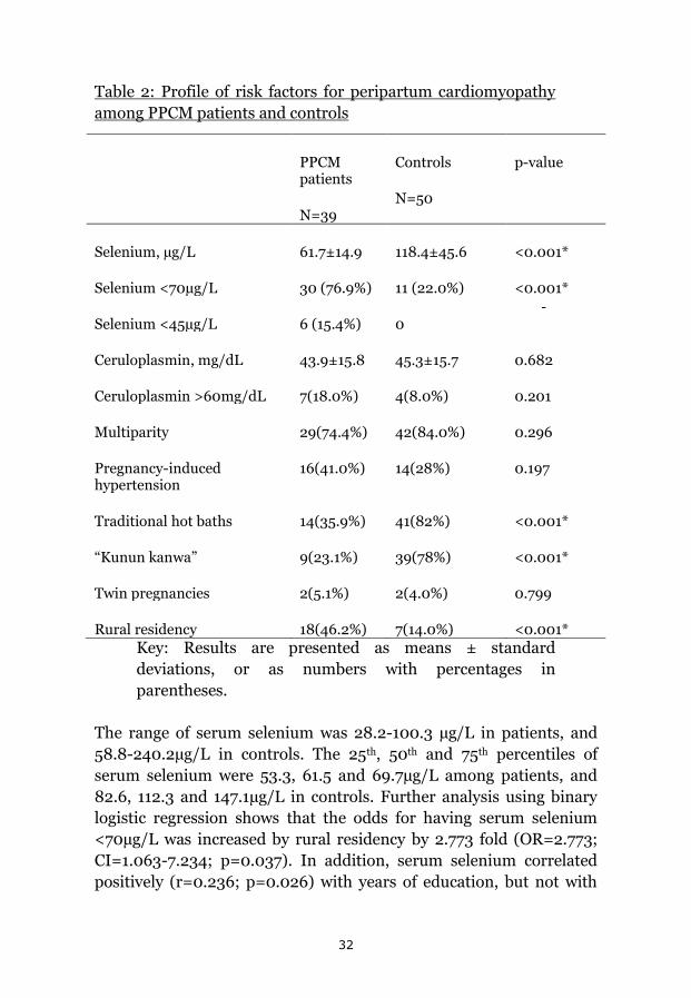
32
Table 2: Profile of risk factors for peripartum cardiomyopathy among PPCM patients and controls
PPCM patients
N=39
Controls
N=50
p-value
Selenium, µg/L 61.7±14.9 118.4±45.6 <0.001*
Selenium <70µg/L 30 (76.9%) 11 (22.0%) <0.001*
Selenium <45µg/L 6 (15.4%) 0 -
Ceruloplasmin, mg/dL 43.9±15.8 45.3±15.7 0.682
Ceruloplasmin >60mg/dL 7(18.0%) 4(8.0%) 0.201
Multiparity 29(74.4%) 42(84.0%) 0.296
Pregnancy-induced hypertension
16(41.0%) 14(28%) 0.197
Traditional hot baths 14(35.9%) 41(82%) <0.001*
“Kunun kanwa” 9(23.1%) 39(78%) <0.001*
Twin pregnancies 2(5.1%) 2(4.0%) 0.799
Rural residency 18(46.2%) 7(14.0%) <0.001* Key: Results are presented as means ± standard deviations, or as numbers with percentages in parentheses.
The range of serum selenium was 28.2-100.3 µg/L in patients, and 58.8-240.2µg/L in controls. The 25th, 50th and 75th percentiles of serum selenium were 53.3, 61.5 and 69.7µg/L among patients, and 82.6, 112.3 and 147.1µg/L in controls. Further analysis using binary logistic regression shows that the odds for having serum selenium <70µg/L was increased by rural residency by 2.773 fold (OR=2.773; CI=1.063-7.234; p=0.037). In addition, serum selenium correlated positively (r=0.236; p=0.026) with years of education, but not with

33
haemoglobin, age, LVEF, family income, parity, blood pressures, BMI or serum creatinine of the subjects (p>0.05). 3.2: Study 2 (One year survival in Nigerians with peripartum cardiomyopathy)
A total of 72 patients were referred to the investigators with a diagnosis of PPCM based on clinical features and findings on chest radiograph. After further evaluation including echocardiography, 18 subjects (25.0%) were excluded and the remaining 54 (75.0%) were confirmed to have PPCM, and all were of Hausa-Fulani ethnic group (Figure 3).
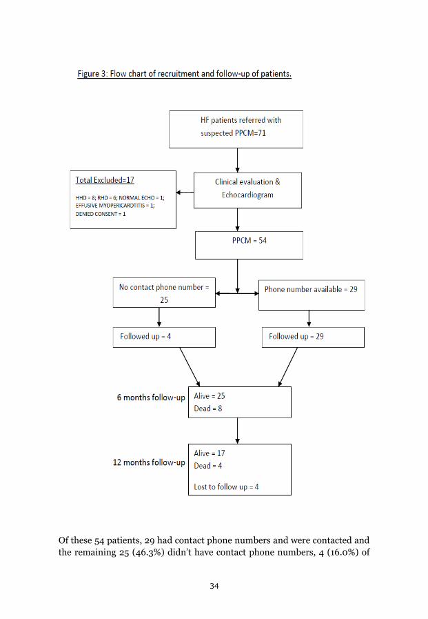
34
Of these 54 patients, 29 had contact phone numbers and were contacted and the remaining 25 (46.3%) didn‟t have contact phone numbers, 4 (16.0%) of
35
whom qualified for follow-up. Overall therefore, 33 patients were followed up as shown in Figure 3. When the 21 patients who didn‟t have contact phone numbers and didn‟t attend follow-up were compared with the 33 followed-up patients, their baseline characteristics were similar (p>0.05) except for the lower mean haemoglobin in the former group (11.5±2.0g/dL) compared with the latter group (12.8±1.6g/dL) (p=0.026).
Patients’ baseline demographics and clinical characteristics: The age of the patients ranged between 18 and 45 years with a mean of 26.6±6.7 years, and 19 of them (35.2%) were between 18 and 20 years, 24 (44.5%) between 20 and 30 years, and the remaining 11 (20.4%) were older than 30 years. No patient had history of smoking, diabetes mellitus, alcohol drinking, stroke, or morbid arrhythmias. One patient had LV thrombus and developed lower limb gangrene needing bilateral below knee amputations. Screening for human immunodeficiency virus was not carried out and none of the recruited patients was known to have the disease.
Patients‟ body mass index (BMI) <18.5Kg/m2 (under-weight) was found in 14 (25.9%), 18.5-24.9 Kg/m2 (normal body weight) in 29 (53.7%), 25.0-29.9 Kg/m2 in 8 (14.8%) and ≥30.05 Kg/m2 in only 2 (3.7%) patients. Nine (16.7%) patients had hypotension (systolic BP <100mmHg) and 25 (46.3%) had pregnancy-induced hypertension at presentation. Two patients became pregnant again before the six month follow-up, and both survived follow-up; their LVEF increased from 43.5± 5.0% at baseline to 52.5±5.0% and 54.5±3.5% at six and twelve month follow-up, respectively.
Mortality: Of the 33 patients followed-up, 12 died (36.4%), 8 (66.7%) within the first six months and the remaining 4 (33.3%) before the twelve month follow-up, 4 patients were lost to follow-up (12.1%) and 17 (51.5%) completed the follow-up (Figure 3). The baseline characteristics of the 17 survivors (58.6%) were similar to the deceased (41.4%) except for lower serum albumin and LV mass index (LVMI), and higher plasma haemoglobin (p<0.05) (Table 3). The mean NYHA class for survivors reduced from 2.53±0.80 at baseline to 1.47±0.62 at one year follow-up (p<0.001). None of these or other variables including LVRR predicted mortality in the regression models (>0.05). The median survival time from diagnosis for the deceased patients was 20.0 (IQR=9-26) weeks.

36
Table 3: Baseline characteristics of survivors and deceased patients.
Characteristics Alive
(N=17, 58.6%)
Deceased
(N=12, 41.4%)
p-value
Age, years 26.4±7.1 28.5±7.2 0.432
Rural residency 3(17.7%) 6(50.0%) 0.064
Parity 4±3 6±4 0.179
NYHA:
II
III
IV
9(52.9%)
6(35.3%)
2(11.8%)
5(41.7%)
4(33.3%)
3(25.0%)
0.776
Pneumonia 4(23.5%) 4(33.3%) 0.683
Pedal oedema 2(11.8%) 3(25.0%) 0.622
Hepatomegaly 6(35.3%) 6(50.0%) 0.471
Pregnancy-induced hypertension
7(41.2%) 7(58.3%) 0.462
SBP, mmHg 111.8±19.7 123.7±23.5 0.152
DBP, mmHg 80.8±16.1 90.2±16.5 0.139
Heart rate, beats/min
105.5±18.0 116.6±15.0 0.102
BMI, Kg/m2 20.2±4.5 22.5±5.4 0.242
Sodium, mmol/L 136.2±5.5 135.3±8.3 0.732
Median serum Creatinine, µmol/L
82.0(IQR=70-104) 77.0(=60.5-103.5)
0.671
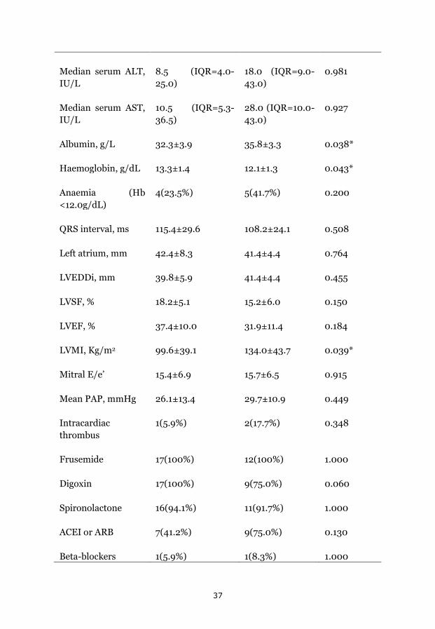
37
Median serum ALT, IU/L
8.5 (IQR=4.0-25.0)
18.0 (IQR=9.0-43.0)
0.981
Median serum AST, IU/L
10.5 (IQR=5.3-36.5)
28.0 (IQR=10.0-43.0)
0.927
Albumin, g/L 32.3±3.9 35.8±3.3 0.038*
Haemoglobin, g/dL 13.3±1.4 12.1±1.3 0.043*
Anaemia (Hb <12.0g/dL)
4(23.5%) 5(41.7%) 0.200
QRS interval, ms 115.4±29.6 108.2±24.1 0.508
Left atrium, mm 42.4±8.3 41.4±4.4 0.764
LVEDDi, mm 39.8±5.9 41.4±4.4 0.455
LVSF, % 18.2±5.1 15.2±6.0 0.150
LVEF, % 37.4±10.0 31.9±11.4 0.184
LVMI, Kg/m2 99.6±39.1 134.0±43.7 0.039*
Mitral E/e‟ 15.4±6.9 15.7±6.5 0.915
Mean PAP, mmHg 26.1±13.4 29.7±10.9 0.449
Intracardiac thrombus
1(5.9%) 2(17.7%) 0.348
Frusemide 17(100%) 12(100%) 1.000
Digoxin 17(100%) 9(75.0%) 0.060
Spironolactone 16(94.1%) 11(91.7%) 1.000
ACEI or ARB 7(41.2%) 9(75.0%) 0.130
Beta-blockers 1(5.9%) 1(8.3%) 1.000
38
Key: NYHA, New York Heart Association functional classification; SBP and DBP, systolic and diastolic blood pressures; BMI, body mass index; ALT and AST, alanine and aspartate transaminases; LVEDDi, LV end-diastolic dimension indexed to body surface area; LVMI, LV mass index; LVSF, LV shortening fraction; LVEF, LV ejection fraction; PAP, pulmonary artery pressure; ACEI, angiotensin concerting enzyme inhibitors; ARB, angiotensin receptor blockers. Results are presented as means ± standard deviations, or as numbers with percentages in parentheses. * p-vlaue is statistically significant.
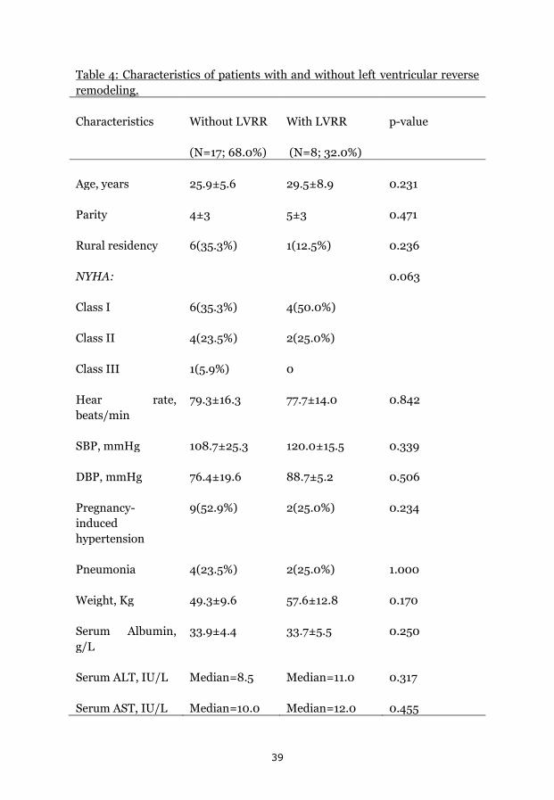
LVRR: Of the 17 survivors at 12 months follow-up, five (29.4%) had recovered LV systolic function (LVEF≥55%), ten (58.8%) had increased LVEF of at least 10%, and eight (47.1%) had reduced LVEDDi ≤33.0mm/m2 (Table 4).
39
Table 4: Characteristics of patients with and without left ventricular reverse remodeling.
Characteristics Without LVRR
(N=17; 68.0%)
With LVRR
(N=8; 32.0%)
p-value
Age, years 25.9±5.6 29.5±8.9 0.231
Parity 4±3 5±3 0.471
Rural residency 6(35.3%) 1(12.5%) 0.236
NYHA:
Class I
Class II
Class III
6(35.3%)
4(23.5%)
1(5.9%)
4(50.0%)
2(25.0%)
0
0.063
Hear rate, beats/min
79.3±16.3 77.7±14.0 0.842
SBP, mmHg 108.7±25.3 120.0±15.5 0.339
DBP, mmHg 76.4±19.6 88.7±5.2 0.506
Pregnancy-induced hypertension
9(52.9%) 2(25.0%) 0.234
Pneumonia 4(23.5%) 2(25.0%) 1.000
Weight, Kg 49.3±9.6 57.6±12.8 0.170
Serum Albumin, g/L
33.9±4.4 33.7±5.5 0.250
Serum ALT, IU/L Median=8.5 Median=11.0 0.317
Serum AST, IU/L Median=10.0 Median=12.0 0.455
40
QRS interval, ms 129.5±26.4 96.4±5.8 0.003*
Left atrium, mm 38.3±8.8 34.0±11.3 0.423
LVEDDi, mm 40.7±3.9 29.3±5.2 <0.001*
LVMI, g/m2 111.9±35.4 94.6±37.1 0.280
LVSF, % 18.3±5.7 31.4±3.7 <0.001*
LVEF, % 37.2±10.3 59.4±5.8 <0.001*
Mitral E/e‟ 11.8±5.1 11.5±5.8 0.070
Mean PAP, mmHg 21.9±11.3 17.9±7.8 0.539
ACEI/ARB 2(11.8%) 2(25.0%) 0.400
Beta-blockers 2(11.8%) 1(12.5%) 0.958
Spironolactone 15(88.2%) 6(75.0%) 0.400
Digoxin 15(88.2%) 5(62.5%) 0.134
Key: LVRR, left ventricular reverse remodeling; NYHA, New York Heart Association functional classification; SBP and DBP, systolic and diastolic blood pressures; ALT and AST, alanine and aspartate transaminases; LVEDDi, LV end-diastolic dimension indexed to body surface area; LVMI, LV mass index; LVSF, LV shortening fraction; LVEF, LV ejection fraction; PAP, pulmonary artery pressure; ACEI, angiotensin concerting enzyme inhibitors; ARB, angiotensin receptor blockers. Results are presented as means ± standard deviations, or as numbers with percentages in parentheses. * p-vlaue is statistically significant.
Overall, 8 patients (47.1%) satisfied the criteria for LVRR. There was no relationship between the use of angiotensin converting enzyme inhibitors (ACEI), angiotensin II receptor blockers (ARB), beta-blockers (p=0.203), other medications or echocardiographic variables (p>0.05) and LVRR (p=0.325), in binary logistic regression models. Of note, mean LVEF, LVEDD as well as left atrial (LA) size and NYHA functional class all
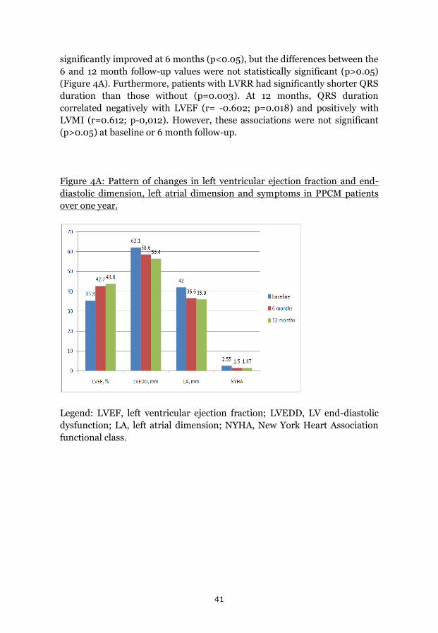
41
significantly improved at 6 months (p<0.05), but the differences between the 6 and 12 month follow-up values were not statistically significant (p>0.05) (Figure 4A). Furthermore, patients with LVRR had significantly shorter QRS duration than those without (p=0.003). At 12 months, QRS duration correlated negatively with LVEF (r= -0.602; p=0.018) and positively with LVMI (r=0.612; p-0,012). However, these associations were not significant (p>0.05) at baseline or 6 month follow-up.
Figure 4A: Pattern of changes in left ventricular ejection fraction and end-diastolic dimension, left atrial dimension and symptoms in PPCM patients over one year.
Legend: LVEF, left ventricular ejection fraction; LVEDD, LV end-diastolic dysfunction; LA, left atrial dimension; NYHA, New York Heart Association functional class.
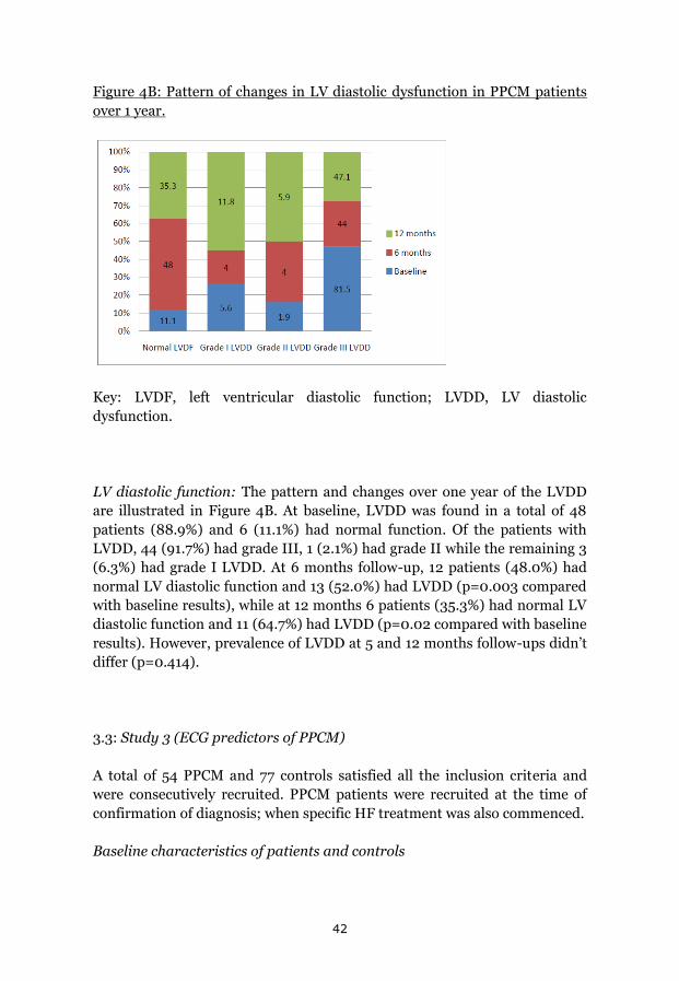
42
Figure 4B: Pattern of changes in LV diastolic dysfunction in PPCM patients over 1 year.
Key: LVDF, left ventricular diastolic function; LVDD, LV diastolic dysfunction.
LV diastolic function: The pattern and changes over one year of the LVDD are illustrated in Figure 4B. At baseline, LVDD was found in a total of 48 patients (88.9%) and 6 (11.1%) had normal function. Of the patients with LVDD, 44 (91.7%) had grade III, 1 (2.1%) had grade II while the remaining 3 (6.3%) had grade I LVDD. At 6 months follow-up, 12 patients (48.0%) had normal LV diastolic function and 13 (52.0%) had LVDD (p=0.003 compared with baseline results), while at 12 months 6 patients (35.3%) had normal LV diastolic function and 11 (64.7%) had LVDD (p=0.02 compared with baseline results). However, prevalence of LVDD at 5 and 12 months follow-ups didn‟t differ (p=0.414).
3.3: Study 3 (ECG predictors of PPCM)
A total of 54 PPCM and 77 controls satisfied all the inclusion criteria and were consecutively recruited. PPCM patients were recruited at the time of confirmation of diagnosis; when specific HF treatment was also commenced.
Baseline characteristics of patients and controls
43
The baseline characteristics of the subjects are presented in Table 5. The mean age, body mass index (BMI), systolic blood pressure (SBP) and diastolic BP (DBP) and prevalence of pregnancy-induced hypertension were not significantly different between the two groups (p>0.05). However, mean serum creatinine was higher (p=0.045), while mean serum sodium and potassium were significantly lower (p=0.009 and <0.001 respectively) in patients compared to controls.

44
Table 5: Baseline characteristics of PPCM patients and controls
PPCM patients (N=54)
Controls
(N=77)
p-value
Mean age (years) 26.6±6.7 25.7±5.7 0.450
Body Mass Index (Kg/m2)
21.6±4.3 21.8±4.3 0.836
Systolic BP(mmHg)
119±24 123±16 0.293
Diastolic BP(mmHg)
86±18 82±12 0.099
Pregnancy-induced hypertension
16(41.0%) 14(28%) 0.197
Serum creatinine (µmol/L)
93.2±67.1 74.7±19.3 0.045*
Serum sodium (mmol/L)
136.9±5.9 139.6±4.4 0.009*
Serum potassium (mmol/l)
3.9±0.8 4.6±0.7 <0.001*
Key: * p-value statistically significant; values are expressed as means ± standard deviations or as numbers with percentages in parentheses.
ECG findings in patients and controls
ECG findings are presented in Table 6. All subjects were in sinus rhythm, and ectopic beats and PR-interval were not significantly different (p>0.05) between the two groups. However, patients had significantly faster heart rate, broader QRS duration, prolonged QTc interval, and more frequent tachycardia and ST-T-wave abnormalities (T-wave inversion with or without

45
ST segment depression in all leads except aVR, V1 and V2), than the controls (p<0.004 for all comparisons).

46
Table 6: ECG features of PPCM patients and controls
PPCM patients (N=54)
Controls
(N=77)
p-value
Sinus rhythm 54(100%) 77(100%) -
Premature ventricular or atrial extrasystoles
5(9.3%) 4(5.2%) 0.365
Heart rate, beats/min
111±16 90±16 <0.001*
Tachycardia 36 (66.7%) 17(22.1%) <0.001*
QRS duration (ms)
109.9±23.6 98.6±12.8 0.004*
QRS duration ≥110ms
19(35.2%) 8(10.4%) 0.001*
QTc duration (ms)
445.0±34.2 421.2±18.9 <0.001*
QTc duration ≥460ms
12 (22.2%) 3(3.9%) 0.001*
PR interval (ms) 148.1±20.4 149.1±21.1 0.799
ST-T-wave abnormalities
37(68.5%) 13(16.9%) <0.001*
Key: * p-value statistically significant; values are expressed as means ± standard deviations or as numbers with percentages in parentheses.
ECG predictors of PPCM
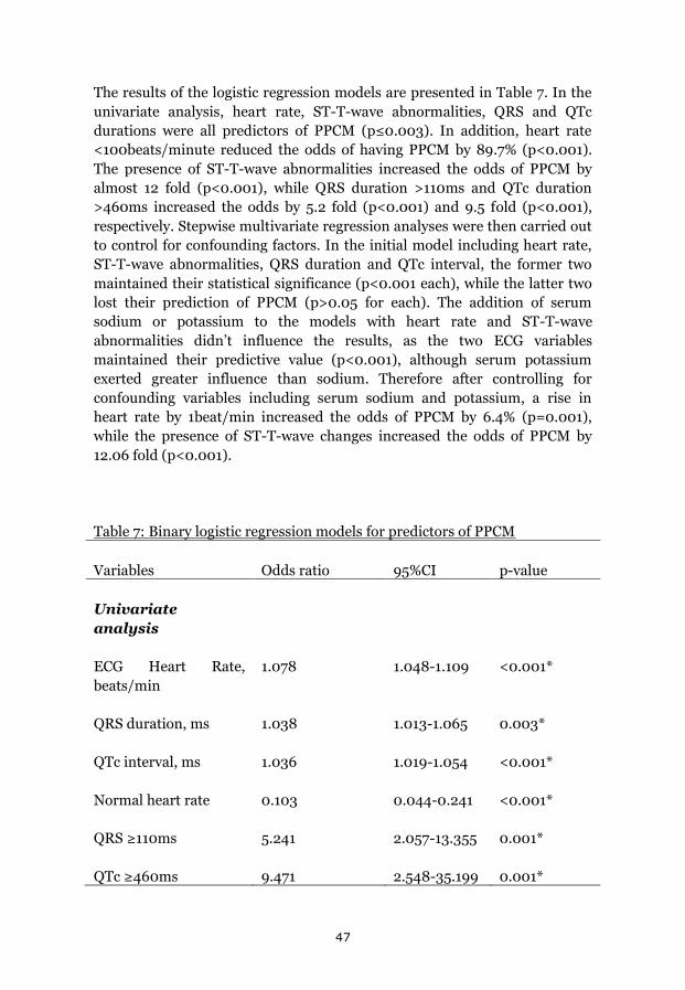
47
The results of the logistic regression models are presented in Table 7. In the univariate analysis, heart rate, ST-T-wave abnormalities, QRS and QTc durations were all predictors of PPCM (p≤0.003). In addition, heart rate <100beats/minute reduced the odds of having PPCM by 89.7% (p<0.001). The presence of ST-T-wave abnormalities increased the odds of PPCM by almost 12 fold (p<0.001), while QRS duration >110ms and QTc duration >460ms increased the odds by 5.2 fold (p<0.001) and 9.5 fold (p<0.001), respectively. Stepwise multivariate regression analyses were then carried out to control for confounding factors. In the initial model including heart rate, ST-T-wave abnormalities, QRS duration and QTc interval, the former two maintained their statistical significance (p<0.001 each), while the latter two lost their prediction of PPCM (p>0.05 for each). The addition of serum sodium or potassium to the models with heart rate and ST-T-wave abnormalities didn‟t influence the results, as the two ECG variables maintained their predictive value (p<0.001), although serum potassium exerted greater influence than sodium. Therefore after controlling for confounding variables including serum sodium and potassium, a rise in heart rate by 1beat/min increased the odds of PPCM by 6.4% (p=0.001), while the presence of ST-T-wave changes increased the odds of PPCM by 12.06 fold (p<0.001).
Table 7: Binary logistic regression models for predictors of PPCM
Variables Odds ratio 95%CI p-value
Univariate analysis
ECG Heart Rate, beats/min
1.078 1.048-1.109 <0.001*
QRS duration, ms 1.038 1.013-1.065 0.003*
QTc interval, ms 1.036 1.019-1.054 <0.001*
Normal heart rate 0.103 0.044-0.241 <0.001*
QRS ≥110ms 5.241 2.057-13.355 0.001*
QTc ≥460ms 9.471 2.548-35.199 0.001*

48
ST-T-wave abnormalities
11.970 5.160-22.770 <0.001*
Multivariate analyses
A: included variables: heart rate; QRS; QTc; ST-T-wave abnormalities
ECG Heart Rate, beats/min
1.073 1.036-1.112 <0.001*
ST-T-wave abnormalities
14.591 4.581-46.480 <0.001*
QRS duration, ms 1.028 0.994-1.062 0.105
QTc interval, ms 1.014 0.993-1.035 0.202
B: included variables: heart rate; ST-T-wave abnormalities; serum potassium
ECG Heart Rate, beats/min
1.066 1.029-1.104 <0.001*
ST-T-wave abnormalities
12.056 3.507-4.443 <0.001*
C: included variables: heart rate; ST-T-wave abnormalities; serum sodium:
ECG Heart Rate, beats/min
13.415 4.203-42.825 <0.001*

49
ST-T-wave abnormalities
1.064 1.029-1.101 <0.001*
Key: * p-value statistically significant; ECG, electrocardiogram.
Relationship between ECG and echocardiographic variables
Patients were evaluated with echocardiography. Mean end-diastolic and end-systolic dimensions (LVEDD, and LVESD, respectively), LV end-systolic volume index (LVESVi) and LVEF were 61.4±8.8mm, 51.0±9.1mm, 85.1±33.1ml/m2 and 34.4±9.9% respectively. The relationship between these echocardiographic and ECG variables are presented in Table 8, which shows that QRS duration was the only variable that modestly correlated with LV dimensions. Figure 5 shows the relationship between QRS duration and LVESD, which was responsible for 19.9% of its variability (R2 = 0.199; p=0.002).
Table 8: Correlation between ECG and echocardiographic variables.
ST-T-wave abnormalities
ECG-QRS
ECG-QT
ECG-HR
LVESD Spearman‟s Correlation
+0.095 +0.330 +0.111 +0.070
p-value 0.495 0.022* 0.454 0.637
LVEDD Spearman‟s Correlation
+0.070 +0.2870 +0.076 -0.040
p-value 0.613 0.048* 0.610 0.789
LVEF Spearman‟s Correlation
-0.100 -0.167 -0.040 -0.260
p-value 0.472 0.256 0.789 0.074
LVESVi Spearman‟s Correlation
+0.165 +0.004 +0.005 -0.230
p-value 0.268 0.979 0.976 0.144

50
Key: * p-value statistically significant; ECG, electrocardiogram; HR, heart rate; LVESD, left ventricular end systolic dimension; LVEDD, left ventricular end diastolic dimension; LVEF, left ventricular ejection fraction; LVESVi, left ventricular end systolic volume index.
Figure 5: Relationship between QRS duration and LV end-systolic dimension.
Legend: R2 = 0.199; B=0.571 (CI=0.200-0.943); p=0.002. LVESD, left ventricular end-systolic dimension.
The ECG risk score for PPCM
Three variables, namely tachycardia, ST-T-wave abnormalities and QRS duration, were included in the risk score, counting 1 for each if present and 0 if absent (Table 9). Tachycardia was defined as heart rate >100 beats/minute, ST-T-wave abnormalities as T-wave inversion with or without
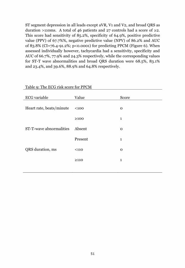
51
ST segment depression in all leads except aVR, V1 and V2, and broad QRS as duration >110ms. A total of 46 patients and 27 controls had a score of ≥2. This score had sensitivity of 85.2%, specificity of 64.9%, positive predictive value (PPV) of 67.7%%, negative predictive value (NPV) of 86.2% and AUC of 83.8% (CI=76.4-91.2%; p<0.0001) for predicting PPCM (Figure 6). When assessed individually however, tachycardia had a sensitivity, specificity and AUC of 66.7%, 77.9% and 24.3% respectively, while the corresponding values for ST-T wave abnormalities and broad QRS duration were 68.5%, 83.1% and 23.4%, and 39.6%, 88.9% and 64.8% respectively.
Table 9: The ECG risk score for PPCM
ECG variable Value Score
Heart rate, beats/minute <100
≥100
0
1
ST-T-wave abnormalities Absent
Present
0
1
QRS duration, ms <110
≥110
0
1

52
Figure 6: ROC curve of PPCM risk score
Legend: Area under the curve = 83.8% (CI=76.4%-91.18%); p<0.0001
3.4: Study 4 (Right ventricular systolic dysfunction and remodelling in Nigerians with peripartum cardiomyopathy)
The study flow is shown in Figure 3. A total of 71 patients suspected to have PPCM were referred to the PI for possible inclusion, but only 54 (76.1%) satisfied the diagnostic criteria, and all of them developed the disease postpartum. Of the 54 PPCM patients, 45 (83.3%) had complete data for RVSD assessment at baseline, and of these 22 (48.9%) were reviewed at 6 months, 8 (17.8%) had died, while the remaining 15 (33.3%) were lost to follow-up. At 1 year, 16/45 (35.6%) patients were alive, 12 (26.7%) had died and 17 (37.8%) were lost to follow-up. The median time from delivery to recruitment for the patients was 6.0 (IQR: 3-15) weeks, and they were recruited in July to December 2013.

53
Baseline clinical characteristics of patients with and without RVSD (Table 10): Of the 45 recruited patients, 32 (71.1%) had RVSD while the remaining 13 (28.9%) had normal RV systolic function. When these 2 groups were compared, the differences were not statistically significant except for higher serum creatinine among those with RVSD (p=0.02), who also tended to have lower diastolic blood pressure (DBP) (p=0.05).
Table 10: Baseline clinical characteristics of patients with and without RVSD
Variables Patients with RVSD N=32(71.1%)
Patients without RVSD N=13(28.9%)
p-value
Age (years) 26.1±7.5 27.7±5.4 0.500
NYHA class: 0.809
II 13(40.6%) 6(46.2%)
III 12(37.5%) 5(38.5%)
IV 7(21.9%) 1(7.7%)
Parity ≥2 25(78.9%) 10(76.9%) 0.758
Breastfeeding 30(93.8%) 13(100%) -
BMI(Kg/m2) 21.4±4.6 21.9±3.9 0.704
Systolic BP(mmHg)
116±23 128±26 0.172
Diastolic BP(mmHg)
83±18 94±16 0.051
Heart rate/minute 111±16 104±21 0.302

54
Pedal oedema 26(81.3%) 8(61.5%) 0.312
Hepatomegaly 19(59.4%) 6(46.2%) 0.515
Pregnancy associated hypertension
13(40.6%) 9(69.2%) 0.158
Haemoglobin (g/dL)
12.2±1.8 12.9±1.3 0.202
Log10 Creatinine 2.0±0.2 1.9±0.1 0.020*
Sodium (mmol/L) 136.3±6.3 135.5±5.2 0.655
Treatment:
ACEI/ ARB 15(46.9%) 7(53.9%) 0.749
Frusemide 32(100%) 13(100%) -
Spironolactone 31(96.9%) 11(84.6%) 0.196
Digoxin 28(87.57%) 12(92.3%) >0.999
Beta blockers 2(6.3%) 1(7.7%) >0.999
Warfarin 1(3.1%) 1(7.7%) 0.499
α-Methyl Dopa 4(12.5%) 2(15.4%) >0.999
Key: Key: NYHA, New York Heart Association functional classification; BMI, body mass index; ACEI, angiotensin concerting enzyme inhibitors; ARB, angiotensin receptor blockers. Results are presented as means ± standard deviations, or as numbers with percentages in parentheses. * p-vlaue is statistically significant.
RV remodelling: Baseline echocardiographic variables were compared between patients with and without RVSD in Table 11. Those with RVSD had significantly lower mean TAPSE and S‟ (p<0.001), but RV basal diameter,

55
RAA, RAL, mPAP, and prevalence of PHT was not significantly different (p>0.05) between the groups. Mean PAP was 29.3±12.1mmHg among all subjects respectively, out of whom a total of 30 (66.7%) had PHT.
Table 11: Baseline echocardiographic characteristics of patients with and without RVSD.
Variables Patients with RVSD (N=32)
Patients without RVSD (N=13)
p-value
Right atrial end-systolic area, cm2
19.7±7.6 15.8±8.1 0.149
Right atrial length, mm
42.9±10.2 42.5±11.1 0.916
Right ventricular basal diameter, mm
49.6±9.3 47.7±10.7 0.583
TAPSE, mm 13.1±2.6 20.3±2.2 <0.001*
Right ventricular S‟, cm/s
10.6±3.2 15.3±3.9 <0.001*
RIMP 1.09±0.60 1.76±1.30 0.032*
RV wall thickness, mm
3.6±1.2 3.5±0.8 0.952
Tricuspid Valve E:A ratio
1.21±0.44 0.90±0.35 0.019*
Tricuspid Valve E/e‟ 5.08±2.73 3.46±1.45 0.023*
Left atrial diameter, mm
43.2±6.4 41.9±6.2 0.548
LV end-diastolic diameter, mm
62.0±7.5 62.4±11.7 0.910

56
LV ejection fraction, %
32.4±9.3 36.6±7.7 0.127
LV stroke volume index, L/m2
39.9±12.3 51.4±17.9 0.017
Anti-log10 mitral valve E:A
1.50±0.62 1.31±0.25 0.154
Mitral valve E/e‟ 15.96±7.13 15.61±7.06 0.882
PA acceleration time, ms
61.8±22.4 78.9±28.0 0.037*
PA systolic pressure, mmHg
47.4±19.4 39.2±21.5 0.246
Mean PAP, mmHg 30.6±11.6 25.9±13.1 0.273
Pulmonary hypertension
23(71.9%) 7(53.9%) 0.304
Key: TAPSE, Tricuspid annular plane systolic excursion; LV, left ventricle; E:A, transvalvular filling velocities ratio; E/e‟, ratio of early filling to averaged early diastolic tissue Doppler velocities of mitral or tricuspid valves; PAP, pulmonary artery pressure; RIMP, right ventricular (RV) index of myocardial performance. Results are presented as means ± standard deviations, or as numbers with percentages in parentheses. * p-vlaue is statistically significant.
The pattern of RV reverse remodelling is presented in Figure 7, which shows increased TAPSE (p=0.049) at 6 months, but RAL (p=0.04) reduced at 12 months and mPAP fell at both 6 (p=0.008) and 12 months (p=0.020) follow up. The prevalence of RVSD reduced from 71.1% at baseline to 36.4% (8/22 patients) at 6 months (p=0.007) and to 18.8% (3/16 patients) at 12 months (p=0.001 vs baseline and p=0.41 vs 6 month). In addition, the prevalence of
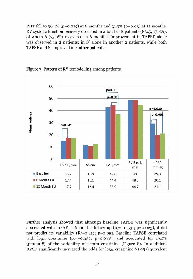
57
PHT fell to 36.4% (p=0.019) at 6 months and 31.3% (p=0.03) at 12 months. RV systolic function recovery occurred in a total of 8 patients (8/45; 17.8%), of whom 6 (75.0%) recovered in 6 months. Improvement in TAPSE alone was observed in 2 patients; in S‟ alone in another 2 patients, while both TAPSE and S‟ improved in 4 other patients.
Figure 7: Pattern of RV remodelling among patients
Further analysis showed that although baseline TAPSE was significantly associated with mPAP at 6 months follow-up (ρs= -0.531; p=0.023), it did not predict its variability (R2=0.217; p=0.051. Baseline TAPSE correlated with log10 creatinine (ρs=+0.332; p=0.048), and accounted for 19.2% (p=0.008) of the variability of serum creatinine (Figure 8). In addition, RVSD significantly increased the odds for log10 creatinine >1.95 (equivalent
TAPSE, mm S', cm RAL, mm RV Basal, mm
mPAP, mmHg
Baseline 15.2 11.9 42.8 49 29.3
6 Month FU 17.4 11.1 44.4 48.5 20.1
12 Month FU 17.2 12.4 36.9 44.7 21.1
0
10
20
30
40
50
60
Mea
n va
lues
p=0.049
p=0.013
p=0.0
p=0.008p=0.020
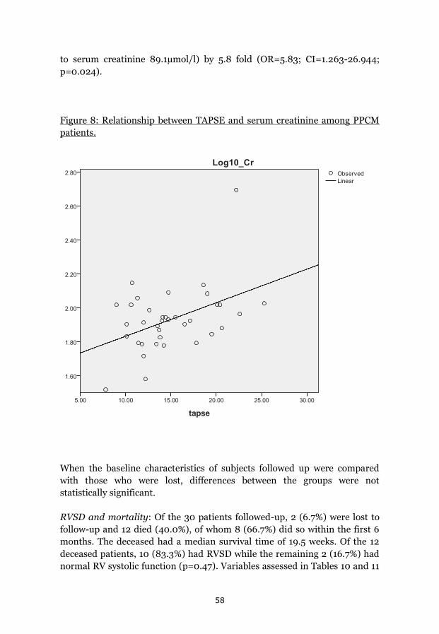
58
to serum creatinine 89.1µmol/l) by 5.8 fold (OR=5.83; CI=1.263-26.944; p=0.024).
Figure 8: Relationship between TAPSE and serum creatinine among PPCM patients.
When the baseline characteristics of subjects followed up were compared with those who were lost, differences between the groups were not statistically significant.
RVSD and mortality: Of the 30 patients followed-up, 2 (6.7%) were lost to follow-up and 12 died (40.0%), of whom 8 (66.7%) did so within the first 6 months. The deceased had a median survival time of 19.5 weeks. Of the 12 deceased patients, 10 (83.3%) had RVSD while the remaining 2 (16.7%) had normal RV systolic function (p=0.47). Variables assessed in Tables 10 and 11

59
were compared between the deceased (12 subjects) and the survivors (16 subjects) at 1 year follow up, and the only significant difference between the groups was a lower serum haemoglobin level in the former (12.1±1.3g/dl) as compared to the latter (13.5±1.4g/dl) (p=0.012). Step wise univariate regression analyses were then carried out in which the serum haemoglobin and the other variables in the Tables were assessed for possible association with one year mortality. However, the one year mortality wasn‟t predicted by any variable in the univariate regression models, including RVSD (p=0.284), serum creatinine (p=0.441) and haemoglobin (p=0.053) (Hosmer & Lemeshow test Χ2=9.69; p=0.288).
60
4.0: Discussion In this project, the clinical characteristics and prognosis of PPCM were studied in 3 referral hospitals in Kano, Nigeria. Firstly, the study has shown that low serum selenium and rural residency seemed to be significant risk factors for PPCM in Kano. In addition, rural residency increased the odds for having critically low serum selenium levels by up to 2.77 fold. However, ceruloplasmin, socio-economic indices, multiparity, pregnancy-induced hypertension, obesity and twin pregnancy didn‟t seem to be associated with PPCM in the study area. In addition, the traditional birth practices that were previously associated with PPCF in northern Nigeria were still being commonly practiced by most women recruited in the study but were not associated with PPCM among the participants.
The second study showed that of the 33 patients (80% <30 years old) followed-up for 1 year, 47.1% had LVRR and 29.4% recovered LV systolic function. Of the 33 patients, 4 (12.1%) were lost to follow-up, 17 (51.5%) (58.6% of followed up patients) survived and 12 (36.4%) (41.4% of followed up patients) died during the follow-up period, with a median survival time of 20 weeks.
The third study describes, perhaps for the first time, the use of a simple risk score using ECG variables to predict PPCM diagnosis among women at risk of the disease. PPCM patients and controls had similar age, systolic and diastolic blood pressures and body mass index.
The fourth study assessed, perhaps for the first time, RVSD and remodelling, and its potential recovery. The prevalence of RVSD, as defined by reduced TAPSE and RV free wall S‟ velocity, was 71.1% at baseline, which fell to 36.4% at 6 months and to 18.8% at 12 months follow-up. Likewise, PHT was found in 66.7% of patients at baseline, and persisted in 36.4% at 6 months, and to 31.3% at 12 months follow up. RV systolic function recovery occurred in a total of 8 patients (8/45; 17.8%), of whom 6 (75.0%) recovered in 6 months. Forty percent of the followed-up patients died within 1 year; two-thirds of them within the first 6 months after diagnosis.

61
4.1: Study 1 (Serum selenium and ceruloplasmin in Nigerians with PPCM)
4.1.1: Selenium deficiency as PPCM risk factor
Selenium is a naturally occurring element found in soil, rocks and water [86]. The selenium content in food principally depends on the concentration and physico-chemical forms existing in the soil [86]. Although food is the main source of selenium for man, the dietary selenium intake principally depends on the region of origin of the foodstuffs and the protein content [86,87]. Thus, animal products (meat, fish and especially shrimps) tend to be richer in selenium than plant materials [88]. It is also important to appreciate that levels of serum selenium are determined by many factors, including its availability in foods, absorption, cooking, lactation, alcohol, chronic illnesses, etc [86,89]. Cenac et al reported for the first time from Niger Republic, where PPCM is an endemic disease, that selenium deficiency may be an important problem in Sahelian African patients with PPCM, akin to what was described for Keshan disease [13,40,73,90-92]. In support of the PPCM selenium theory by Cenac et al, our results have shown critically low selenium levels among 76.9% of the studied PPCM patients [13,93]. North-western Nigeria shares a long border, geography and customs with Niger republic, hence the common food types and dietary habits, which are the sources of selenium. Our results have shown that PPCM patients had significantly lower serum selenium levels and significantly higher prevalence of rural residency than controls in spite of similar income and educational levels. In addition, rural residency significantly increased the odds of having critically low serum selenium levels. It is well known that in Nigeria, most rural residents are subsistence farmers who tend to consume the locally produced food and grown animals. Urban residents on the other hand are exposed to imported foods and animals, from regions where there is no selenium deficient soil and animals. Therefore, it is reasonable to hypothesize that most women in Kano (and the Sahel region) develop PPCM if they depend on locally produced foods and animals. The serum selenium levels among PPCM patients in Kano (61.7±14.9µg/L) and Niamey

62
(48.0±25µg/L) were similar, most likely because of their geographical and cultural similarities which explains the heavy burden of the disease in the region, in comparison with respective values in Haiti of 110µg/L (range 67–145µg/L) [13,14]. This difference could be explained on the basis of geographical and cultural factors. In spite of the foregoing, the question still remains: does selenium deficiency have a cause-and-effect relationship with PPCM? For a convincing answer, similar epidemiologic studies to those carried out in China are needed, where Keshan disease has been relegated to history [91]. Our finding of a significant difference in residency between patients and controls has not been previously described, and could be of epidemiologic importance in the planning of interventions, if selenium deficiency is eventually proven to be a cause of PPCM. Selenium is a key component of the antioxidant glutathione peroxidase. A decrease in the activity of this enzyme secondary to selenium deficiency may lead to an increase in the oxidative stress in myocardial cells, and this can make the cells more susceptible to infections, especially by viruses [94]. This mechanism was demonstrated in mice models by injecting viral genomes in mice deficient in selenium, and compared with mice with good nutritional support. The incidence of myocarditis in the first group was much higher [94].
4.1.2. PPCM and traditional birth practices peculiar to northern Nigeria
About four decades ago, some studies described an association between puerperal customary practices and PPCF in Zaria, a city about 120Km from Kano in north-western Nigeria [17,18,20]. Davidson et al reported that the customs of Hausa women in Zaria were important in the pathogenesis of PPCF, given that only 1% did not take postpartum baths, 3% did not lie on the hot beds, and 6% did not take “kanwa” [20]. However, it is important to note that the Zaria syndrome of PPCF was defined as mainly a “high-output HF with well preserved ventricular function”, making it different from what is
63
known today as PPCM [9,17]. In a cross-sectional study, Isezuo et al reported that up to 81.5% of PPCM patients in Sokoto practiced the hot baths for 30 days [95]. In our case-control study, the hot baths and intake of “kunun kanwa” were still commonly practiced by the great majority of healthy women postpartum. The prevalence of both practices was significantly higher among the healthy controls than among the PPCM patients, perhaps because many of the latter were hospitalised. Therefore, the notion that those traditional birth practices are PPCM risk factors should be downplayed.
4.1.3. Other PPCM risk factors
Unlike previous reports, the present study has shown that socio-economic indices, multiparity, pregnancy-induced hypertension, obesity and twin pregnancy were not associated with PPCM in the study area [95,97]. In addition, the study has shown for the first time that CP was not associated with PPCM, in contrast to a recent report that found it to be related to some biochemical changes and LV systolic dysfunction in heart failure [98]. The findings of the present study have therefore shed more light on the multi-factorial nature of PPCM, and have suggested that the risk factors might vary between individuals and geographic regions. Selenium levels in the soil and domestic animals in Kano, as well as in other geographical regions are relevant information but beyond the scope of the present study.
4.1.4. Limitations
Although relevant, blood levels of vitamins, C-reactive proteins, natriuretic peptides, thyroid hormones and prolactin were not assessed in the present study. Still the present study has shed more light on the selenium theory of PPCM and has discarded the theory of traditional birth practices as risk factors for PPCM. Future studies should ideally be community-based, prospective and comparative between various regions, and should involve assessment of selenium

64
in the environment (foods and soil) as well as blood levels of the above-mentioned biomarkers.
4.2: Study 2 (One year survival in Nigerians with PPCM)
4.2.1: Mortality in PPCM patients: Of the 29 patients with complete 12 months follow-up, the survivors (58.6%) had significantly lower serum albumin and LVMI, and higher plasma hemoglobin than the deceased (41.4%). However, none of these or other variables could predict mortality in the regression models. Two-thirds of the deaths occurred within 6 months of the diagnosis while the remaining one-third died before the 12-month follow-up visit. Much lower mortality rates of 13.0% over 6 months in South Africa, 15.3% over 2 years in Haiti and 28.0% over 5 years in USA, respectively, have been reported.[99-101] Although some researchers didn‟t identify any predictors of mortality in agreement with our findings, others reported inconsistent predictors of mortality including younger age at diagnosis, lower BMI and echocardiographic variables.[28,99,100] It is clearly difficult to compare mortality results between studies given the varying follow-up times and several other challenges including possible strong influence of socio-political crises on the mortality.
Similar to our findings, 53.3% of those who died in the Haiti study did so within 60 days after diagnosis.[100] Whitehead et al also reported that in a cohort of PPCM patients in the USA, 18% of deaths occurred within 1 week and 87% within 6 months of diagnosis.[57] Therefore, most of the mortality in PPCM, as well as recovery, seem to occur within 6 months of diagnosis. Although the reasons for these observations are not clear, they seem to give hope to patients of better outcomes once they survive the first 6 months of the disease. It is therefore imperative that patients are supported with optimal medical treatment to be able to survive the first critical 6 months of the disease.
4.2.2: LVRR: Of the 17 survivors at 12 months follow-up, 47.1% satisfied the criteria for LVRR while 29.4% recovered LV systolic function. There was no relationship between the use of heart failure medications or other variables and LVRR in the regression models. In addition, significant improvement in mean LVEF, LVEDD, LA size, LVDD and NYHA functional class had already manifested at six month, with no further significant improvement at 12

65
months follow-up. LVDD was similar; showing a maximum fall in its prevalence from 89% at baseline to 52% at six months follow-up. Therefore, LV recovery in our PPCM patients involved both systolic and diastolic functions, with maximum improvement achieved at 6 months follow-up in most patients, independent of ACEIs, ARBs and beta-blockers. However, almost all the patients were treated with furosemide, spironolactone and digoxin, as treatment for HF.
Our findings on LVRR seem to be similar to those previously reported from South Africa (21%) and Haiti (28%) but lower than what was reported from USA (54%) likely because of the differences in the level of health care between the three countries.[28,99,100] Furthermore, the achievement of maximum recovery within 6 months in our cohort of patients is also supported by Fett et al and Elkayam.[28,100] Finally, our inability to identify predictors of recovery of LV function or reverse remodelling, including HF medications, was similarly reported by Fett et al.[100]
However, Blauwet et al found older age and smaller LVESDto be significant predictors of LV recovery among South Africans.[100] In addition, our results show that patients with LVRR had significantly shorter QRS duration than those without, and QRS duration correlated with LVEF and with LVMI, but only at 1 year follow-up. Thus, aside from “mechanical remodelling”, our results also show that “electrical remodelling” does occur in PPCM, and is likely related to the progression or otherwise of the LV disease. Similar observations were previously made by Henein et al among patients with dilated and ischemic cardiomyopathies.[102] Progressive LV disease eventually results in increased myocardial stiffness and a rise in diastolic pressures. The resulting increase in LV filling pressures and consequent ischemia of the sub-endocardium predispose the latter to conduction “depolarization” delay, which if prolonged could result in sub-endocardial fibrosis and permanent conduction disturbance.[102] Thus, in dilated and dysfunctioning ventricles, progressive broadening of the QRS complex can be taken as a marker of worsening of ventricular disease.[102]
4.2.3: Study limitations: The present study has some limitations. Firstly, natriuretic peptides and other biomarkers of HF were not assessed because the tests were not readily available at the study centres. Secondly, 21 out of 54 patients could not be followed-up because they lacked phone numbers and so could not be contacted, and didn‟t turn up for follow-up in spite of adequate counselling. However, these patients had similar baseline characteristics as those followed up except for their lower mean haemoglobin levels.
66
4.3: Study 3 (ECG predictors of PPCM)
4.3.1: ECG and diagnosis of PPCM: Our findings show that the presence of heart rate less than 100 beats/minute reduced the risk of diagnosing PPCM by 89.7%, while the presence of ST-T-wave abnormalities, QRS duration more than 110ms and QTc duration longer than 460ms increased the odds of PPCM by 12.0, 5.2 and 9.5 folds, respectively. In the initial multiple regression model, heart rate and ST-T-wave abnormalities maintained their high statistical significance in predicting PPCM but not QRS duration and QTc interval. This finding was not influenced by serum sodium or potassium. None of our patients had malignant arrhythmia and ectopic beats were equally uncommon in both groups. The latter results were supported by previous reports in women with PPCM as well as in healthy pregnant women.[42,103] Serum sodium and potassium levels were significantly lower in patients than controls, possibly because of the water retention caused by the HF syndrome. [9] It should be noted that patients were all recruited at the time of confirmation of the diagnosis, when most HF treatments were commenced and none was on drugs that could affect ECG measurements.
We developed a simple scoring system using the 3 ECG variables that could potentially predict PPCM (HR, ST-T wave abnormalities and QRS duration) with an accuracy of 83.8%. A risk score of ≥2 had a sensitivity of 85.2%, specificity of 64.9% and NPV of 86.2%. The performance of the score was weak when each of the 3 variables was used separately rather than in combination. The score‟s satisfactory accuracy in predicting the diagnosis of PPCM makes it appealing for routine use, particularly in areas where the disease is prevalent and more expensive diagnostic facilities are limited.
4.3.2: ECG and cardiac function in PPCM: Our results have shown that in the patients, QRS duration modestly correlated with LVEDD and LVESD, explaining 19.9% of the variability of the latter and suggesting diffuse pathological changes. Furthermore, QRS duration of >110ms significantly increased the odds of PPCM by 5.2 fold, in spite of the faster heart rate in patients compared to controls. These results are supported by a previous study which showed an association between QRS duration and mortality among predominantly male (98%) elderly Caucasians with moderate-severe
67
LV systolic dysfunction caused by a variety of diseases (LVEF <40%).[104] Therefore in patients presenting with clinical features of HF, QRS duration >110ms could suggest significant LV dilatation and systolic dysfunction, as well as poor prognosis.
T-wave abnormalities were identified in 68.5% of our patients. Similar findings were reported by Ntusi et al in DCM patients, with lower prevalence in idiopathic (68.8%) compared to those with familial disease (87.5%), but there was no association between such electric disturbance and survival.[105] It seems therefore that T wave changes, which reflect disturbed repolarisation, might have less detrimental effect on patient‟s survival compared with QRS duration which, in most, mirrors the impact of systolic dysfunction. In our series, QRS duration was the only ECG variable that correlated with LV dimensions, while heart rate and ST-T-wave abnormalities had higher sensitivity in predicting PPCM. Longitudinal follow up studies should determine the prognostic impact of ECG variables in the setting of PPCM.
4.3.3: Limitations: The present study has some limitations. Firstly, serum levels of calcium and magnesium were neither assessed nor controlled for, but deficiencies of these electrolytes have not been reported to be common in PPCM patients.[9] Secondly, PPCM patients were not directly compared with patients with other conditions such as DCM but we relied on published evidence; thus the findings cannot be considered specific to PPCM. The sample size was small but our results are consistent and seem to provide evidence for using easily measured ECG variables to predict PPCM in women at risk of the disease. Finally, echocardiography was not performed in the controls. They were screened using clinical evaluation and ECG only, which can be used to identify healthy women after delivery.[42] Our findings should be considered as representative of the early stage of the disease, while the long term electrical abnormalities remain to be determined.
4.4: Study 4 (RVSD and remodelling in Nigerians with PPCM)
RVSD, its recovery and potential relationship with mortality are not well studied in PPCM. Based on reduced TAPSE, we have previously reported a prevalence of RVSD of 54.6% in PPCM patients.[21] Adding reduced S‟ raised the prevalence of patients with RVSD in this study to 71.1%, suggesting a more sensitive means for identifying such patients. The second
68
important observation in the current study is the significant recovery of RVSD along with its pressure afterload in the form of PHT. Indeed, 6 months from the time of presentation, the prevalence of RVSD and PHT fell by more than 50% despite poor adherence to heart failure conventional medications. Thus, the observed RV reverse remodelling seems to be related to the recovery of the pulmonary circulation status rather than to the effect of medications as has been previously observed in the LV.[28] This claim is supported by the modest relationship we found between TAPSE and mPAP at 6 months follow-up. Our finding might be explained by the significant RV function recovery in more than 50% of patients at 6 months, thus suggesting a different pathophysiologic mechanism to right ventricular-pulmonary circulation coupling in PPCM compared with DCM, which has a more chronic course of disturbances of ventricular function.[106] In addition, TAPSE was linearly related to serum creatinine, confirming the adverse relationship between RVSD and renal impairment.[107,108] Whether this reflects the aggressive nature of cardiac decompensation in PPCM, since all the patients developed LV systolic dysfunction with LVEF <45% within a few postpartum days, remains to be verified in a larger group of patients.
Thus, our findings show that the first 6 months of PPCM diagnosis seem to be crucial for RV function recovery as well as survival. These findings are supported by Whitehead et al who reported 87% of PPCM deaths occurring within the first 6 months of diagnosis, and Elkayam who observed that LV function normalises in approximately 50% of women with PPCM within the same period.[28, 57] These findings urge intensive medical treatment for such young patients within the first few days and weeks of their presentation. Finally, our mortality rate of 40% in the present study seems high compared to 13% over 6 months in South Africa [99]. This could be partly explained by the high maternal mortality in Nigeria in general, which was as high as 496.4 per 100,000 live births in 2013.[109]
4.4.2: Study limitations: Although important, B-type natriuretic peptide (BNP) or N-terminal pro-BNP and other biomarkers could not be assessed because the tests were not readily available at the study centres. Secondly, it is important to point out that 15 of the 45 patients could not be followed-up because they were not contactable in spite of adequate counselling. Their inability to come for follow-up wasn‟t unrelated to the significant security challenges that were faced by northern Nigeria including Kano, which made movements and keeping to appointments extremely difficult. Thirdly, cardiac magnetic resonance imaging could have shed more light on the

69
nature of the myocardial pathology in PPCM, but this facility was not available at the study centres.[110]
70
5.0: Conclusion The 1st study has shown that low serum selenium and rural residency seemed to be significant risk factors for PPCM in Kano, Nigeria. Rural residency increased the odds of having critically low serum selenium levels by up to 2.77 fold. In addition, ceruloplasmin, socio-economic indices, multiparity, pregnancy-induced hypertension, traditional birth practices, obesity and twin pregnancy were not associated with PPCM in the study area. Future community comparative studies are needed to further explore the relationship between selenium deficiency and PPCM, in view of its high prevalence in sub-Saharan Africa and associated morbidity and mortality.
In the 2nd study, we showed that LVRR occurred in almost half the patients at 6 months but was not predicted by any clinical or echocardiographic variable. Overtime, the prevalence of normal LV diastolic function tripled independent of heart failure medications. Furthermore, LVRR was associated with shorter QRS duration which correlated with LVEF and LV mass index at one year follow-up, thus signifying “electrical remodelling” in PPCM. Finally, none of the variables predicted the 41.4% mortality, of which two thirds occurred before the first 6 months follow-up.
The 3rd study seems to be the first to show that in women presenting within the first 9 months after delivery with symptoms of HF, heart rate and ST-T-wave abnormalities were potential predictors of the diagnosis of PPCM. In addition, QRS duration modestly correlated with LV dimensions. A simple ECG based score could potentially predict the diagnosis of PPCM, a finding that could help to streamline the diagnosis of PPCM prior to confirmatory investigations, particularly in limited resource settings, which is where the disease is common. A simple score of ≥2, counting 1 for each of three ECG abnormalities (tachycardia, ST-T-wave abnormalities and QRS duration >110ms), had 83.8% accuracy for predicting PPCM in women at risk.
The 4th study assessed perhaps for the first time RVSD, RV reverse remodelling and pulmonary circulation disturbances in a group of PPCM patients. These were prevalent in over two-thirds of the patients at

71
presentation and improved by more than 50% at 6 months despite the lack of adherence to medical therapy. However, the studied cohort suffered a relatively high mortality which was not predicted by RVSD or other variables. These findings urge intensive care for such young patients during the acute phase of the disease to support cardiac function recovery and reduce mortality.

72
Acknowledgements
My deepest gratitude is to my mentor, Professor Michael Henein, who has tremendously supported, taught and guided me throughout my Cardiology education. Michael, I am most grateful.
I am also deeply grateful to my Co-Supervisor, Dr Krister Lindmark, for his support, encouragement and useful suggestions throughout the Thesis.
I am very grateful to Kerstin Rosenqvist for her assistance and support, without which this Thesis would have been extremely difficult! Thank you very much. My sincere appreciation also goes to members of Department of Public Health and Clinical Medicine, Umea University, who have in any way taught or assisted me during the PhD program. I am grateful to the management and staff of Bayero University, Aminu Kano Teaching Hospital and Murtala Mohammed Specialist Hospital, Kano, Nigeria, for their support during the PhD program. I am most grateful to my parents and to my wife and children, for all their love, affection and patience throughout my PhD program. Finally, all gratitude is to the Almighty God for all his Blessings.
73
References 1. Virchow R. Sitzing der Berliner Geburtshilflisher Gersellskhalt, cited
by Porak, C. De l'influence réciproque de la grossesse et des maladies du Coeur, thesis, Paris, 1880.
2. Porak C. De l'influence réciproque de la grossesse et des maladies du Coeur, thesis, Paris, 1880.
3. Gouley BA, McMillan TM, Bellet S. Idiopathic myocardial degeneration associated with pregnancy and especially the puerperium. Am J Med Sci 1937; 19: 185–199
4. Hull E, Hidden E. Postpartal heart failure. South Med J 1938; 31: 265–270
5. Demakis JG, Rahimtoola SH. Peripartum cardiomyopathy. Circulation 1971; 44: 964-968.
6. Demakis JG, Rahimtoola SH, Sutton GC, Meadows WR, Szanto PB, Tobin JR, et al. Natural course of Peripartum cardiomyopathy. Circulation 1971; 44: 1053-1061.
7. Pearson GD, Veille JC, Rahimtoola S, Hsia J, Oakley CM, Hosenpud JD, et al. Peripartum cardiomyopathy: National Heart, Lung, and Blood Institute and Office of Rare Diseases (National Institutes of Health) workshop recommendations and review. JAMA 2000; 283: 1183–1188.
8. Elliott P, Andersson B, Arbustini E, Bilinska Z, Cecchi F, Charron P, et al. Classification of the cardiomyopathies: a position statement from the European Society of Cardiology Working Group on Myocardial and Pericardial Diseases. Eur Heart J 2008; 29: 270–276.
9. Sliwa K, Hilfiker-Kleiner D, Petrie MC, Mebazaa A, Pieske B, Buchmann E, et al. Heart Failure Association of the European Society of Cardiology Working Group on Peripartum Cardiomyopathy. Current state of knowledge on aetiology, diagnosis, management, and therapy of peripartum cardiomyopathy: a position statement from the Heart Failure Association of the European Society of Cardiology Working Group on peripartum cardiomyopathy. Eur J Heart Fail 2010; 12(8): 767-778
10. Elkayam U, Akhter MW, Singh H, Khan S, Bitar F, Hameed A, et al. Pregnancy-associated cardiomyopathy: clinical characteristics and a comparison between early and late presentation. Circulation 2005; 111: 2050–2055
11. Dickstein K, Cohen-Solal A, Filippatos G, McMurray JJ, Ponikowski P, Poole-Wilson PA, et al. ESC guidelines for the diagnosis and treatment of acute and chronic heart failure 2008: the Task Force for the diagnosis and treatment of acute and chronic heart failure

74
2008 of the European Society of Cardiology. Developed in collaboration with the Heart Failure Association of the ESC (HFA) and endorsed by the European Society of Intensive Care Medicine (ESICM). Eur J Heart Fail 2008; 10: 933–989
12. Ferriere M, Sacrez A, Bouhour JB, Cassagnes J, Geslin P, Dubourg O, et al. Cardiomyopathy in the peripartum period: current aspects. A multicenter study. 11 cases. Arch Mal Coeur Vaiss 1990; 83(10): 1563–1569.
13. Cénac A, Djibo A. Postpartum cardiac failure in Sudanese–Sahelian Africa: clinical prevalence in western Niger. Am J Trop Med Hyg 1998; 58: 319–323.
14. Fett JD, Christie LG, Carraway RD, Murphy JG. Five-year prospective study of the incidence and prognosis of peripartum cardiomyopathy at a single institution. Mayo Proceed 2005; 80: 1602–1606
15. Desai D, Moodley J, Naidoo D. Peripartum cardiomyopathy: experiences at King Edward VIII Hospital, Durban, South Africa and a review of the literature. Trop Doct 1995; 25: 118–123.
16. Mielniczuk LM, Williams K, Davis DR, Tang AS, Lemery R, Green MS, et al. Frequency of peripartum cardiomyopathy. Am J Cardiol 2006; 97: 1765-1768.
17. Sanderson JE, Adesanya CO, Anjorin FI, Parry EHO. Postpartum cardiac failure-heart failure due to volume overload? Am Heart J 1979; 97: 613-621.
18. Fillmore SJ, Parry EH. The evolution of peripartal heart failure in Zaria. Circulation 1977; 56: 1058-1061.
19. Karaye KM, Sa‟idu H, Habib AG. Peripartum and other cardiomyopathies in a Nigerian adult population. Int J Cardiol 2011; 147(2): 342-343.
20. Davidson NM, Parry EH. Peri-partum cardiac failure. Q J Med 1978; 47: 431–461.
21. Karaye KM. Right ventricular systolic function in peripartum and dilated cardiomyopathies. Eur J Echocardiogr 2011; 12(5): 372-374.
22. Sliwa K, Fett J, Elkayam U. Peripartum cardiomyopathy. Lancet 2006; 368: 687-693.
23. Abboud J, Murad Y, Chen-Scarabelli C, Saravolatz L, Scarabelli TM. Peripartum cardiomyopathy: a comprehensive review. Int J Cardiol 2007; 12; 118(3): 295-303.
75
24. Karaye KM, Sani MU. The Impact of Income on the Echocardiographic Pattern of Heart Diseases in Kano, Nigeria. Niger J Med 2008; 17(3): 350-355.
25. Sliwa K, Forster O, Libhaber E, Fett JD, Sundstrom JB, Hilfiker-Kleiner D, et al. Peripartum cardiomyopathy: inflammatory markers as predictors of outcome in 100 prospectively studied patients. Eur Heart J 2006; 27: 441–46.
26. Karaye KM, Sani MU. Factors associated with poor prognosis among patients admitted with heart failure in a Nigerian tertiary medical centre: a cross-sectional study. BMC Cardiovasc Disord 2008, 8(1): 16.
27. Gentry MB, Dias JK, Luis A, Petel R, Thornton J, Reed GL. African-American women have a higher risk for developing Peripartum cardiomyopathy. J Am Coll Cardiol 2010; 55: 654 –659.
28. Elkayam U. Clinical characteristics of peripartum cardiomyopathy in the United States: diagnosis, prognosis and management. J Am Coll Cardiol 2011; 58: 659–670
29. Karaye KM, Habib AG. Pattern of left ventricular geometry in hypertension: a study of a hypertensive population in Nigeria. Sahel Medical Journal 2009; 12(4): 148-154.
30. Kamiya CA, Kitakaze M, Ishibashi-Ueda H, Nakatani S, Murohara T, Tomoike H, et al. Different characteristics of peripartum cardiomyopathy between patients complicated with and without hypertensive disorders: Results from the Japanese nationwide survey of peripartum cardiomyopathy. Circ J 2011; 75: 1975 – 1981.
31. Okamoto H, Takenaka T, Saitoh Y. Is Hypertensive Disorder a Unique Risk Factor for Peripartum Cardiomyopathy and Pregnancy-Associated Cardiomyopathy? Circ J 2011; 75: 1827-1828.
32. Ntusi NBA, Mayosi BM. Aetiology and risk factors of peripartum cardiomyopathy: a systematic review. Int J cardiol 2009; 131: 168-179
33. Hilfiker-Kleiner D, Kaminski K, Podewski E, Bonda T, Schaefer A, et al. A cathepsin D-cleaved 16 kDa form of prolactin mediates postpartum cardiomyopathy. Cell 2007; 128: 589–600.
34. Sliwa K, Blauwet L, Tibazarwa K, Libhaber E, Smedema J-P, Becker A, et al. Evaluation of bromocriptine in the treatment of acute severe peripartum cardiomyopathy: a proof of concept pilot study. Circulation 2010; 121: 1465–1473.
35. Sliwa K, Skudicky D, Bergemann A, Candy G, Puren A, Sareli P. Peripartum cardiomyopathy: analysis of clinical outcome, left

76
ventricular function, plasma levels of cytokines and Fas/APO-1. J Am Coll Cardiol 2000; 35: 701–705
36. Fett JD, Ansari AA, Sundstrom JB, Combs GF. Peripartum cardiomyopathy: a selenium disconnection and an autoimmune connection. Int J Cardiol 2002; 86: 311–316.
37. Cenac A, Simonoff M, Moretto P, Djibo A. A low plasma selenium is a risk factor for peripartum cardiomyopathy. A comparative study in Sahelian Africa. Int J Cardiol 1992; 36: 57–59.
38. Wang, GY, Zhou R, Sun, S, Yin T, Ying G. The difference between whole blood and hair selenium levels of inhabitants in the endemic and nonendemic areas of Keshan disease (author‟s translation). Zhonghua Yu Fang Yi Xue Za Zhi 1979; 13: 204–206.
39. Keshan Disease Research Group of the Chinese Academy of Medical Sciences, Beijing. Epidemiologic studies on the etiologic relationship of selenium and Keshan disease. Chin Med J (Engl.) 1979; 92: 477–482.
40. Loscalzo J. Keshan Disease, Selenium Deficiency, and the Selenoproteome. N Engl J Med 2014; 370 (18): 1756-1760
41. Beck MA, Levander OA, Handy J. Selenium deficiency and viral infection. J Nutr 2003; 133: Suppl 1:1463S-1467S.
42. Tibazarwa K, Lee G, Mayosi B, Carrington M, Stewart S, Sliwa K. The 12-lead ECG in peripartum cardiomyopathy. Cardiovasc J Afr 2012; 23(6): 322-329.
43. Schannwell CM, Schoebel FC, Marx R, Plehn G, Leschke M, Strauer BE. Prognostic relevance of left ventricular diastolic function parameters in dilated cardiomyopathy. Z Kardiol 2001; 90(4): 269-79.
44. Henein MY, Gibson DG. Normal long axis function. Heart 1999; 81: 111–113
45. HeneinMY, Gibson DG. Long axis function in disease. Heart 1999; 81: 229–231
46. Alam M, Hoglund C, Philip A. Atrioventricular plane displacement in severe congestive heart failure following dilated cardiomyopathy or myocardial infarction. J Intern Med 1990; 228: 569–575
47. Pai RG, Bodenheimer MM, Pai SM, Koss JH, Adamick RD. Usefulness of systolic excursion of the mitral annulus as an index of left ventricular systolic function. Am J Cardiol 1991; 67: 222–224
77
48. Kaul S, Tei C, Hopkins JM, Shah PM. Assessment of right ventricular function using two-dimensional echocardiography. Am Heart J 1984; 107(3): 526-531.
49. Coronary artery disease. In: Henein MY, Sheppard M, Pepper J, Rigby M (Eds). Clinical Echocardiography. Springer-Verlag London Limited, London, 2004: 119-148
50. Duncan AM, Francis DP, Gibson DG, Henein MY. Differentiation of ischemic from nonischemic cardiomyopathy during dobutamine stress by left ventricular long-axis function: additional effect of left bundle-branch block. Circulation 2003; 108(10): 1214-1220
51. Powell BD, Espinosa RE, Yu CM, Oh JK. Tissue Doppler imaging, strain imaging and dyssynchrony assessment. In Oh JK, Seward JB, Tajik AJ, eds. The Echo Manual. 3rd edition. Philadelphia, Baltimore, New York, London, Buenos Aires, Hong Kong, Sydney, Tokyo: Lippincott Williams & Wilkins (a Wolters Kluwer business), 2006; 80-98
52. Gorcsan III J, Tanaka H. Echocardiographic assessment of myocardial strain. J Am Coll Cardiol 2011; 58: 1401-1413.
53. Mouquet F, Lions C, de Groote P, Bouabdallaoui N, Willoteaux S, Dagorn J, et al. Characterisation of peripartum cardiomyopathy by cardiac magnetic resonance imaging. Eur Radiol 2008; 18: 2765–2769.
54. Arora NP, Mohamad T, Mahajan N, Danrad R, Kottam A, Li T, et al. Cardiac magnetic resonance imaging in peripartum cardiomyopathy. Am J Med Sci 2014; 347(2): 112-117.
55. Galderisi M, Henein MY, D'hooge J, Sicari R, Badano LP, Zamorano JL, et al; European Association of Echocardiography. Recommendations of the European Association of Echocardiography: how to use echo-Doppler in clinical trials: different modalities for different purposes. Eur J Echocardiogr 2011; 12(5): 339-353
56. Goland S, Modi K, Bitar F, Janmohamed M, Mirocha JM, Czer LS, et al. Clinical profile and predictors of complications in peripartum cardiomyopathy. J Card Fail 2009; 15: 645–650
57. Whitehead SJ, Berg CJ, Chang J. Pregnancy-related mortality due to cardiomyopathy: United States, 1991–1997. Obstet Gynecol 2003; 102: 1326 –1331.

78
58. McNamara DM, Elkayam U, Alharethi R, Damp J, Hsich E, Ewald G, et al; IPAC Investigators. Clinical Outcomes for Peripartum Cardiomyopathy in North America: Results of the IPAC Study (Investigations of Pregnancy-Associated Cardiomyopathy). J Am Coll Cardiol 2015; 66(8): 905-914.
59. Elkayam U, Tummala PP, Rao K, Akhter MW, Karaalp IS, Wani OR, et al. Maternal and fetal outcomes of subsequent pregnancies in women with peripartum cardiomyopathy. N Engl J Med 2001; 334: 1567–1571
60. American Academy of Paediatrics, Committee on Drugs. The transfer of drugs and other chemicals into human milk. Paediatrics 2001; 108: 776–789
61. Habedank D, Kuhnle Y, Elgeti T, Dudenhausen JW, Haverkamp W, Dietz R. Recovery from peripartum cardiomyopathy after treatment with bromocriptine. Eur J Heart Fail 2008; 10: 1149–1151
62. Hellgren M. Hemostasis during normal pregnancy and puerperium. Semin Thromb Hemost 2003; 29: 125–130.
63. Briggs GG, Freeman RK, Yatte SJ. Drugs in Pregnancy and Lactation. 8th edition, Philadelphia, PA: Wolters Kluwer, 2008.
64. Dickstein K, Vardas PE, Auricchio A, Daubert JC, Linde C, McMurray J, et al; Committee for Practice Guidelines of the European Society of Cardiology. 2010 focused update of ESC Guidelines on device therapy in heart failure: an update of the 2008 ESC Guidelines for the diagnosis and treatment of acute and chronic heart failure and the 2007 ESC Guidelines for cardiac and resynchronization therapy. Developed with the special contribution of the Heart Failure Association and the European Heart Rhythm Association. Eur J Heart Fail 2010; 12(11): 1143-1153.
65. Cleland JG, Daubert JC, Erdmann E, Freemantle N, Gras D, Kappenberger L, et al. The effect of cardiac resynchronization on morbidity and mortality in heart failure. N Engl J Med 2005; 352: 1539–1549.
66. Cleland JG, Daubert JC, Erdmann E, Freemantle N, Gras D, Kappenberger L, et al. Longer-term effects of cardiac resynchronization therapy on mortality in heart failure [the CArdiac REsynchronization-Heart Failure (CARE-HF) trial extension phase]. Eur Heart J 2006; 27: 1928–1932.
67. Bristow MR, Saxon LA, Boehmer J, Krueger S, Kass DA, De Marco T, et al. Cardiac-resynchronization therapy with or

79
without an implantable defibrillator in advanced chronic heart failure. N Engl J Med 2004; 350: 2140–2150.
68. Mouquet F, Mostefa KM, Lamblin N, Coulon C, Langlois S, Marquie C, et al. Unexpected and rapid recovery of left ventricular function in patients with peripartum cardiomyopathy: impact of cardiac resynchronization therapy. Eur J Heart Fail 2012; 14(5): 526-529.
69. World Medical Association Declaration of Helsinki, Ethical Principles for Medical Research Involving Human Subjects. J Postgrad Med 2002; 48: 206-208.
70. Lang RM, Badano LP, Mor-Avi V, Afilalo J, Armstrong A, Ernande L, et al. Recommendations for cardiac chamber quantification by echocardiography in adults: an update from the American Society of Echocardiography and the European Association of Cardiovascular Imaging. J Am Soc Echocardiogr 2015; 28(1): 1-39.
71. Quiñones MA, Otto CM, Stoddard M, Waggoner A, Zoghbi WA.Recommendations for quantification of Doppler Echocardiography. A report from the Doppler Quantification Task Force of the Nomenclature and Standards Committee of the American Society of Echocardiography. J Am Soc Echocardiogr 2002; 15: 167–184.
72. Kadish AH, Buxton AE, Kennedy HL, Knight BP, Mason JW, Schuger CD, et al. ACC/AHA clinical competence statement on electrocardiography and ambulatory electrocardiography: a report of the American College of Cardiology/American Heart Association/ American College of Physicians − American Society of Internal Medicine Task Force on Clinical Competence (ACC/AHA Committee to Develop a Clinical Competence Statement on Electrocardiography and Ambulatory Electrocardiography). J Am Coll Cardiol 2001; 38: 2091−2100.
73. Yang GQ, Ge KY, Chen JS, Chen XS. Selenium-related endemic diseases and the daily selenium requirement of humans. World Rev Nutr Diet 1988; 55: 98-152.
74. World Health Organization. Nutritional anemia. Report of a WHO Scientific Group. WHO Technical Report Series, No 405. Geneva, Switzerland: World Health Organization; 1968.
75. Merlo M, Pyxaras SA, Pinamonti B, Barbati G, Di Lenarda A, Sinagra G. Prevalence and prognostic significance of left
80
ventricular reverse remodelling in dilated cardiomyopathy receiving tailored medical treatment. J Am Coll Cardiol 2011; 57(13): 1468-76.
76. Oh JK, Park SJ, Nagueh SF. Established and novel clinical applications of diastolic function assessment by echocardiography. Circ Cardiovasc Imaging 2011; 4; 444-455.
77. Goldberger AL, Goldberger ZD, Shvilkin A. Understanding the normal ECG. In Goldberger’s Clinical Electrocardiography, A Simplified Approach. Eighth edition. Edited by Goldberger AL, Goldberger ZD, Shvilkin A. Philadelphia: Elsevier Saunders; 2013: 26-34.
78. Woosley RL. Drugs that prolong the QT interval and/or induce Torsades de Pointes. http://www.azcert.org/medical-pros/drug-lists/browse-drug-list.cfm (accessed on 13th December 2014)
79. Buxton AE, Calkins H, Callans DJ, DiMarco JP, Fisher JD, Greene HL, et al. ACC/AHA/HRS 2006 key data elements and definitions for electrophysiological studies and procedures: a report of the American College of Cardiology/ American Heart Association task Force on Clinical Data Standards (ACC/AHA/HRS Writing Committee to Develop Data Standards on Electrophysiology). Circulation 2006; 114: 2523-2570.
80. Rudski LG, Lai WW, Afilalo J, Hua L, Handschumacher MD, Chandrasckaran K, et al. Guidelines for the echocardiographic assessment of the right heart in adults: a report from the American Society of echocardiography. Endorsed by the European Society of Cardiology, and the Canadian Society of echocardiography. J Am Soc Echocardiogr 2010; 23: 685-713.
81. Lindqvist P, Henein M, Kazzam E. Right ventricular outflow tract fractional shortening: an applicable measure of right ventricular systolic function. Eur J Echocardiogr 2003; 4:29–35.
82. Sciomer S, Magri D, Badagliacca R. Non-invasive assessment of pulmonary hypertension: Doppler echocardiography. Pulm Pharmacol Ther 2007; 20: 135-140.
83. Posteraro A, Salustri A, Trambaiolo P, Amici E, Gambelli G. Echocardiographic estimation of pulmonary pressures. J Cardiovasc Med 2006; 7: 545-554.
81
84. Galie` N, Hoeper MM, Humbert M, Torbicki A, Vachiery JL, Barbera JA, et al. Guidelines for the diagnosis and treatment of pulmonary hypertension. The Task Force for the Diagnosis and Treatment of Pulmonary Hypertension of the European Society of Cardiology (ESC) and the European Respiratory Society (ERS), endorsed by the International Society of Heart and Lung Transplantation (ISHLT). Eur Heart J 2009; 30: 2493-2537.
85. Chemla D, Castelain V, Humbert M, Hébert JL, Simonneau G, Lecarpentier Y, et al. New Formula for Predicting Mean Pulmonary Artery Pressure Using Systolic Pulmonary Artery Pressure. Chest 2004; 126: 1313–1317.
86. Tato Rocha RE, Cardenas Viedma E, Herrero Huerta E. Selenio: implicaciones fisiopatol´ogicas y cl´ınicas. Ann Med Intern 1994; 2: 457-463.
87. Diaz-Alarcon JP, Navarro-Alarcon M, Lopez-Ga de la Serrana H, Lopez-Martinez MC. Determination and chemical speciation of selenium in farmlands from southeastern Spain: relation to levels found in sugar cane. J Agric Food Chem 1996; 44: 2423-2427.
88. Diaz-Alarcon JP, Navarro-Alarcon M, Lopez-Ga de la Serrana H, Lopez-Martinez MC. Determination of selenium in meat products by hydride generation atomic absorption spectrometry } selenium levels in meats, organ meats, and sausages in Spain. J Agric Food Chem 1996; 44: 1494-1497.
89. Navarro-Alarcon M, Lopez-Martinez MC. Essentiality of selenium in the human body: relationship with different diseases. The Science of the Total Environment 2000; 249: 347-371.
90. Chen J. An original discovery: selenium deficiency and Keshan disease (an endemic heart disease). Asia Pac J Clin Nutr 2012; 21: 320-326.
91. Wang GY, Zhou R, Sun S, Yin T, Ying G. The difference between whole blood and hair selenium levels of inhabitants in the endemic and nonendemic areas of Keshan disease (author‟s translation). Zhonghua Yu Fang Yi Xue Za Zhi 1979; 13: 204-206.
82
92. Epidemiologic studies on the etiologic relationship of selenium and Keshan disease. Chin Med J (Engl) 1979; 92: 477-482.
93. Karaye KM, Saidu H, Bala MS, Yahaya IA. Prevalence, clinical characteristics and outcome of pulmonary hypertension among admitted heart failure patients. Ann Afr Med 2013; 12(4): 197-204.
94. Ne`ve J. Human selenium supplementation as assessed by changes in blood selenium concentration and glutathione peroxidase activity. J Trace Elem Med Biol 1995; 9: 65–73.
95. Isezuo SA, Abubakar SA. Epidemiologic profile of peripartum cardiomyopathy in a tertiary care hospital. Ethn Dis 2007; 17: 228-233.
96. Karaye KM, Henein MY. Peripartum cardiomyopathy: a review article. Int J Cardiol 2013; 164: 33-38.
97. Sliwa K, Mayosi BM. Recent advances in the epidemiology, pathogenesis and prognosis of acute heart failure and cardiomyopathy in Africa. Heart 2013; 99(18): 1317-1322.
98. Xu Y, Lin H, Zhou Y, Cheng G, Xu G. Ceruloplasmin and the extent of heart failure in ischemic and nonischemic cardiomyopathy patients. Mediators Inflamm 2013; 2013: 348145. doi:10.1155/2013/348145.
99. Blauwet LA, Libhaber E, Forster O, Tibazarwa K, Mebazaa A, Hilfiker-Kleiner D, et al. Predictors of outcome in 176 South African patients with peripartum cardiomyopathy. Heart 2013; 99(5): 308-313.
100. Fett JD, Christie LG, Carraway RD, Murphy JG. Five-year prospective study of the incidence and prognosis of peripartum cardiomyopathy at a single institution. Mayo Clin Proc 2005; 80(12): 1602-1606.
101. Witlin AG, Mabie WC, Sibai BM. Peripartum cardiomyopathy: an omnious diagnosis. Am J Obstet Gynecol 1997; 176: 182–188.
102. Shamim W, Yousufuddin M, Cicoria M, Gibson DG, Coats AJ, Henein MY. Incremental changes in QRS duration in serial ECGs over time identify high risk elderly patients with heart failure. Heart 2002; 88(1): 47-51.
83
103. Salisu AI, Karaye KM. Electrocardiographic indices in a rural pregnant Nigerian women population. Sahel Medical Journal 2010; 13(3): 147-152.
104. Silvet H, Amin J, Padmanabhan S, Pai RG. Prognostic implications of increased QRS duration in patients with moderate and severe left ventricular systolic dysfunction. Am J Cardiol 2001; 88(2): 182-185, A6.
105. Ntusi NB, Badri M, Gumedze F, Wonkam A, Mayosi BM. Clinical characteristics and outcomes of familial and idiopathic dilated cardiomyopathy in Cape Town: a comparative study of 120 cases followed up over 14 years. S Afr Med J 2011; 101(6): 399-404.
106. Di Mauro M, Calafiore AM, Penco M, Romano S, Di Giammarco G, Gallina S. Mitral valve repair for dilated cardiomyopathy: predictive role of right ventricular dysfunction. Eur Heart J 2007; 28(20): 2510-2516.
107. Mullens W, Abrahams Z, Francis GS, Sokos G, Taylor DO, Starling RC, et al. Importance of venous congestion for worsening of renal function in advanced decompensated heart failure. J Am Coll Cardiol 2009; 53(7): 589-596.
108. Guinot PG, Arab OA, Longrois D, Dupont H. Right ventricular systolic dysfunction and vena cava dilatation precede alteration of renal function in adult patients undergoing cardiac surgery: an observational study. Eur J Anaesthesiol 2014; 31: 1–8.
109. Kassebaum NJ, Bertozzi-Villa A, Coggeshall MS, Shackelford KA, Steiner C, Heuton KR, et al. Global, regional and national levels and causes of maternal mortality during 1990-2013: a systematic analysis for the Global Burden of Disease Study 2013. Lancet 2014; 384(9947): 980-1004.
110. Blecker GB, Steendijk P, Holman ER, Yu C-M, Breithardt OA, Kaandorp TAM, et al. Assessing right ventricular function: the role of echocardiography and complementary technologies. Heart 2006; 2(Suppl I): i19-i26.