cli global society at the amputation prevention symposium ... · gangrene of the Þrst great toe of...
TRANSCRIPT

LLC,TM
an HMP Communications Holdings Company September 2017
Leadership and members of the CLI Global Society were pres-ent at the 7th Annual AMPuta-tion Prevention Symposium held
August 9-12, 2017 in Chicago, Illinois, attended by over 800 attendees from 25 countries and 46 states.
The first annual Alan T. Hirsch Memorial Keynote Address “A Call to Action – The CLI Global Society” was presented by Dr. Barry T. Katzen, MD, FACC, FACR, FSIR. Dr. Katzen is the Founder and Chief Medical Executive of Miami Cardiac and Vascular Institute, a Founding Member and President of the Critical Limb Ischemia (CLI) Global Society and a pioneer in CLI therapy.
Dr. Katzen recognized the vast body of work of Dr. Hirsch who passed away earlier this year. “Dr. Hirsch was a con-summate clinician who provided data driven, evidence-based care long before it was fashionable. He joined as a Founding Board member of the fledging CLI Global Society with his usual delibera-tion but ultimate incredible enthusiasm! His contribution to science, and specifi-cally CLI, will be sorely missed.”
Dr. Katzen called attention to the first angioplasty procedure, performed in 1963 by Charles Dotter, MD, “the Father of Interventional Radiology,” which was performed on a patient with CLI with a non-healing ulcer of her foot. “Endovascular therapy today represents a field that includes multiple disciplines working collaboratively to advance this
important area of less invasive therapy. The field has matured over the past de-cades bringing rapid technological ad-vancements, increasing cost pressure on the healthcare system along with a need for improved clinical science, data and proof of benefit. The treatment of CLI is much more than endovascular therapy or surgery revascularization.”
The CLI Global Society was formed in 2016 to address the unmet need of CLI. In the United States, lower extremity PAD manifests as CLI in nearly 1 million Medicare patients per year, with an esti-mated annual cost of over 3 billion dol-lars.1 One in 190 Americans (1.6 million) living with loss of a limb. Unchecked, this number may more than double by 2050 to 3.6 million.2
The CLI Global Society’s mission is to improve quality of life by preventing amputations and death due to critical limb ischemia. Dr. Katzen underscored that mission by addressing this unmet need must start with a definition of CLI. CLI was first formally defined in 1982 by the Working Party of the International Vascular Symposium as a condition in pa-tients without diabetes with chronic isch-emia as the major threat to a limb with ankle pressure <40 mm Hg in patients with rest pain and <60 mm HG in those with tissue necrosis.
The Rutherford classification was first developed in 1986 and revised in 1997 with CLI falling under Rutherford cat-egories 4-6. The Inter-Society Consensus
for the Management of Peripheral Arterial Disease (TASC II) was published in 2008 and defined CLI as all patients with chronic ischemic rest pain, ulcers or gan-grene attributed to objectively proven ar-terial occlusion disease.3 The AHA/ACC adopted the TASC II definition in 2016.4 In 2014, the Society of Vascular Surgery (SVS) defined CLI as an objective classifi-cation of the threatened limb based on the degree of ischemia, wound extent, gan-grene and infection. SVS developed the SVS Lower Extremity Threatened Limb Classification System based on grading of these 3 factors (The WIfi Classification).5
So why is CLI a problem? The CLI Global Society recognizes the
following contributing factors to the challenge of CLI:1. Lack of consensus on definition; 2. Lack of awareness within healthcare
community and general public;3. CLI morbidity and mortality are
akin to the most aggressive cancer diagnoses;
4. Limited research; 5. Lack of consensus on best meth-
ods to prevent, diagnose, treat and rehabilitate;
6. Limited number of CLI specialists; 7. No diagnosis code for CLI;
Critical limb ischemia (CLI), the most advanced form of peripheral artery disease (PAD), is defined by all societal guidelines and consensus
statements as the presence of tissue loss or gangrene with documented evidence of hy-poperfusion.1,2 Hypoperfusion has been tra-ditionally assessed using the ankle brachial index (ABI) or ankle systolic pressure; how-ever, other tests such as toe pressure or toe brachial index (TBI), TcPO2, and skin perfu-sion pressure have also been used, though not widely. In 2014, in a single center study, we showed for the first time that approximately
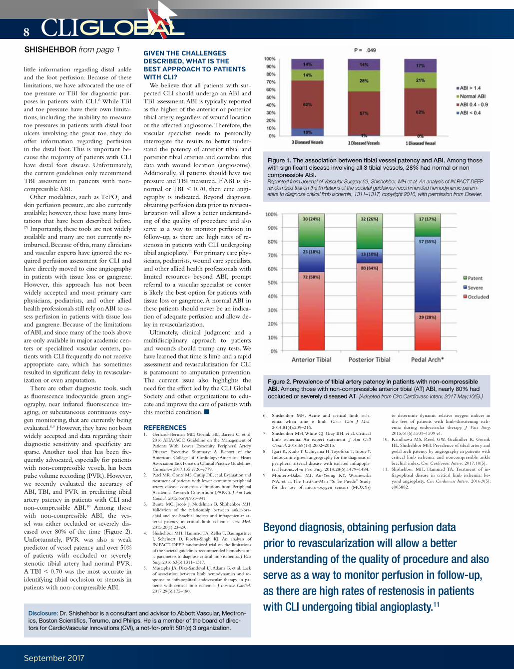
30% of patients with documented CLI have near normal, normal, or non-compress-ible ABI.3 Subsequently, using the data from IN.PACT DEEP Trial, we validated and ex-tended our previous findings (Figure 1), showing that approximately one-third of pa-tients with tibial disease had normal or non-compressible ABI.4 Since then, others have reproduced, validated, and extended our pre-vious work.5
While the exact reasons for normal or non-compressible ABI in patients with CLI is unknown, a few mechanisms have been postulated: (1) A significant proportion of
patients with CLI are diabetic or have end-stage renal disease (ESRD); hence, because of heavy medial calcinosis, these arteries are partially or fully non-compressible, resulting in elevated or non-interpretable pressures. (2) There is extensive collateral flow below the knee; therefore, while the main tibial arter-ies are occluded, the collateral flow provides adequate pressure. (3) A significant portion of patients with CLI have isolated below knee disease; it is possible that ABI reflects pres-sure to proximal and mid ankle but provides
CLI Global Society at the AMPutation Prevention Symposium 2017
Flaws in the Objective Diagnosis of Critical Limb IschemiaMehdi H. Shishehbor, DO, MPH, PhD, Professor of Medicine, Case Western Reserve University; Co-Chair, Harrington Heart & Vascular Institute; Director, Interventional Cardiovascular Center, Co-Director, Vascular Center, University Hospitals of Cleveland, Cleveland, OH
Mehdi H. Shishehbor, DO, MPH, PhD
CLI THE OFFICIAL PUBLICATION OF THE CRITICAL LIMB ISCHEMIA GLOBAL SOCIETY
GLOBAL
Dr. Barry T Katzen delivers the ignaugural Alan T. Hirsch Keynote Address at the AMPutation Prevention Symposium 2017 in Chicago, IL.
Continued on page 8
Continued on page 17

ORBITAL ATHERECTOMY
VESSEL PREPWORKS.
TO TREAT PAD, MODIFYING CALCIUM CHANGES COMPLIANCE...
...ALLOWING FOR LOW PRESSURE PTA INFLATION—TO MINIMIZE VESSEL DAMAGE
Indication: The CSI Orbital Atherectomy System is a percutaneous orbital atherectomy system indicated for use as therapy in patients
with occlusive atherosclerotic disease in peripheral arteries and stenotic material from artificial arteriovenous dialysis fistulae.
Contraindications for the system include use in coronary arteries, bypass grafts, stents, or where thrombus or dissections are present.
Although the incidence of adverse events is rare, potential events that can occur with atherectomy include: pain, hypotension, CVA/TIA,
death, dissection, perforation, distal embolization, thrombus formation, hematuria, abrupt or acute vessel closure, or arterial spasm.
Caution: Federal law (USA) restricts this device to sale by, or on the order of, a physician.
CSI and Diamondback 360 are registered trademarks of Cardiovascular Systems, Inc.
©2017 Cardiovascular Systems, Inc. EN-4389 0817
CSI360.com
Date: August 2017
Client: Cardiovascular Systems, Inc.
Job Number: CSI1713
Job Description: CSI Peripheral Full Page Ad
Headline: “Orbital atherectomy vessel prep works.”
Pub: (CLIGP) CLI Global Compendium
Keyline built to: 100%
Color: CMYK
BL: 10.75" x 14.25"
TR: 10.5" x 14"
LV: .5" from all trim edges
NOTE: MAGENTA = Bleed needed
Epson Stock: ❑ Oris Super White ❑ Oris Comm ❑ Oris News
• 692 Mendelssohn Ave N • Golden Valley, MN 55427 • 612.767.3455
Epson Color Profile: ❑ Gracol ❑ Swop3 ❑ News ❑ Supplied
Client:
Job No:
Epson SureColor P7000sleepingdogltd.com
78277
Lee Branding
✔
✔
WHEN YOU SEE CALCIUM, THINK
78277_CSI1713_Peripheral_Balloon_Ad_CLIgp_0816.indd 1 8/16/17 2:55 PMCSI_CLIG0917.indd 1 8/17/17 9:13 AM

3
September 2017
CLIGLOBAL
J.A. MUSTAPHA, MD, FACC, FSCAIClinical EditorMetro Health University of Michigan HealthWyoming, MIClinical Associate Professor of MedicineMichigan State University COM, East Lansing, MI
Jeff Martin, PublisherCarmen Heaney, Executive EditorLaurie Gustafson, Managing EditorVic Geanopulos, Creative DirectorElizabeth Vasil, Graphic Production Manager
EDITORIAL CORRESPONDENCE: Laurie Gustafson, Managing Editor HMP Communications 70 East Swedesford Road, Suite 100 [email protected]
GEORGE ADAMS, MDGarner, NC
VICKIE R. DRIVER, DPM, MSBoston, MA
LAWRENCE GARCIA, MDBoston, MA
PHILIP P. GOODNEY, MDLebanon, NH
MICHAEL R. JAFF, DONewton, MA
BARRY T. KATZEN, MDMiami, FL
ROBERT LOOKSTEIN, MDNew York, NY
D. CHRIS METZGER, MDKingsport, TN
RICHARD F. NEVILLE, MDFairfax, VA
CONSTANTINO S. PENA, MDMiami, FL
FADI A. SAAB, MDWyoming, MI
DIERK SCHEINERT, MDLeipzig, Germany
ANDREJ SCHMIDT, MDLeipzig, Germany
RAMON VARCOE, MBBS, MSSydney, Australia
FRANK J. VEITH, MDNew York, NY
RENU VIRMANI, MDGaithersburg, MD
CRAIG M. WALKER, MDHouma, LA
THOMAS ZELLER, MDBad Krozingen, Germany
Published in collaboration with
Editor’s note: Articles in this supplement to Cath Lab Digest did not undergo peer review.
EDITORIAL
SCIENTIFIC ADVISORY BOARD
LLCan HMP Communications Holdings Company
,™
An 80-year-old female patient presented to our hospital with gangrene of the first great toe of the right foot (Figure 1). She
was being followed by her primary phy-sician for multiple medical problems, in-cluding mild pain in the right calf and foot when walking, relieved at rest. The pain worsened during the previous sever-al months, progressing to rest pain and er-ythema of the foot, and finally, develop-ment of gangrene involving the great toe. Her other relevant medical problems in-cluded prior myocardial infarction, atri-al fibrillation, diabetes mellitus (Type 2), hypertension, hyperlipidemia, and chronic
kidney disease (Stage IV). Although the patient was taking warfarin and aspirin, she was never started on pentoxifylline or cilostazol for management of her ini-tial symptoms.
Given her renal insufficiency (Cr. 1.6, GFR 27), arterial duplex imaging was obtained of both lower extremities, which demonstrated occlusion of the femoropoliteal artery with trace reconsti-tution of the anterior tibial artery (Figure 2). No flow could be identified in the remaining infra-popliteal arteries. Based on spectral waveforms, no inflow disease was suspected. Additionally, no embolic disease was suspected clinically.
Continued on page 10
TABLE OF CONTENTS
CLI Global Society at the AMPutation Prevention Sym-posium 2017 ...................................................................................1
Flaws in the Objective Diagnosis of Critical Limb Ischemia ...............................................................................1
Case Study: An Endovascular First Approach to CLI .......3
Key Findings of the Registry of First-Line Treatments in Patients with Critical Limb Ischemia (CRITISCH) ..............4
The Effectiveness of a Team Approach in Treating Critical Limb Ischemia................................................................6
Selected Abstracts from the AMP Symposium ............... 18
Calendar of Future Educational Events ............................22
©2017, Critical Limb Ischemia Global, LLC (CLIG). All rights reserved. Repro-duction in whole or in part prohibited. Opinions expressed by authors, con-tributors, and advertisers are their own and not necessarily those of Critical Limb Ischemia Global or the editorial staff. Critical Limb Ischemia Global is not responsible for accuracy of dosag-es given in articles printed herein. The appearance of advertisements in this journal is not a warranty, endorsement or approval of the products or services advertised or of their effectiveness, quality or safety. Critical Limb Ischemia Global disclaims responsibility for any injury to persons or property resulting from any ideas or products referred to in the articles or advertisements.
Content may not be reproduced in any form without written permission. Contact [email protected] for rights and permission.
Case Study: An Endovascular First Approach to CLI Sabeen Dhand, MDPresbyterian Intercommunity Hospital, Whittier, CA
Dr. Sabeen Dhand
Figure 1. Photograph of the patient’s right foot demonstrating well-demarcated dry gangrene of the great toe. The patient also suffers from white discoloration of the forefoot, worst in the proximal first digit. All toes show nail and skin changes, as well as hair loss. No additional ulcers are identified. There is a mottled appearance of the foot, which is cool to touch. No infra-inguinal pulses are palpable on physical exam.
Disclosure: Dr. Dhand reports that he has been a consultant for Abbott.

4
September 2017
CLIGLOBAL
Critical limb ischemia (CLI) rep-resents the most severe form of lower extremity atherosclerosis and is associated with excessive
cardiovascular morbidity and mortali-ty and increased rates of limb loss. The severity of the disease and the inten-sive vascular care needed pose a signifi-cant socioeconomic burden for all west-ern societies. Although claudicants still represent the majority of peripheral ar-terial disease (PAD) patients, it remains interesting that the costs for CLI health-care accounted for 56% of all PAD reim-bursement costs in Germany.1 In a retro-spective Medicare study, the mean cost of inpatient care in the year before ma-jor PAD-related amputation amounted to $22,405.2
Despite some evidence that an intensive medical therapy and aggressive risk factor modification might change the natural history of CLI, revascularization of the affected limb still remains the corner-stone of CLI treatment.3,4 Unfortunately, many CLI patients will undergo major amputation as primary therapy, while in Germany, a retrospective insurance reg-istry revealed that 44% of patients who received a primary amputation had not received at least a diagnostic angiography in the year prior to the procedure.1
Surgery, especially in cases of autoge-nous vein conduits, is associated with en-couraging primary patency and increased amputation-free survival rates, despite the
prolonged recovery and rather invasive nature of the procedure.5 Endovascular revascularization offers an alternative minimally-invasive approach to surgery. The continuous development of the en-dovascular field over the past decades led to a paradigm shift in the treatment of pe-ripheral atherosclerosis, and endovascular therapy replaced surgery as the first-line treatment strategy. However, the impact of many novel endovascular modalities in the treatment of CLI remains unclear, since in many recent trials, the majority of the enrolled patients were claudicants.6
Unfortunately, the Bypass Versus Angioplasty for Severe Ischemia of the
Leg (BASIL) trial,7 which compared the outcomes of plain angioplasty and open repair in CLI patients, still remains the only published randomized controlled trial. Nonetheless, there is a paucity of comparative data between the new tech-nologies and bypass surgery.
Notwithstanding the scarce level 1 evidence about the different treatment options, previous studies did not include consecutive patients and did not evalu-ate endovascular revascularization or hybrid procedures as possible treatment options.7,8 For this reason, we conduct-ed a prospective, multicenter registry to
examine the performance of all first-line treatment strategies.
The First-Line Treatments in Patients with Critical Limb Ischemia (CRITISCH) study is a prospective, in-terdisciplinary, multicenter registry evalu-ating the current practice of all available treatment strategies in 1200 unselected CLI patients treated in 27 vascular centers in Germany.9 The single inclusion crite-rion was the presence of new onset CLI. CLI was defined as an ankle-brachial in-dex <0.40 or ischemic rest pain, or both, with or without tissue loss in the pres-ence of PAD (Rutherford classes 4–6). In cases with a non-calculable index, ankle pressure or transcutaneous oxygen pres-sure was measured. Only one limb per patient was included. Patients with acute limb ischemia, isolated aortic or iliac in-terventions, vascular trauma, and known clotting disorders or non-atherosclerotic occlusive disease were excluded from the registry. The study was validated via an external audit at participating cen-ters. The recruitment started in January 2013 and was completed in September 2014. Follow-up was planned at 6, 12, and 24 months after the patient’s enroll-ment. The applied treatment was left at the discretion of the treating physician following the principle of “best medi-cal practice” and there was no restriction concerning the selected treatment op-
tion. The various treatment options were categorized into five groups. Group I in-cluded patients undergoing endovascular treatment. Group II consisted of patients treated with bypass surgery, and group III included patients who were treated by common or deep femoral artery surgical revascularization, with or without con-comitant inflow or outflow endovascular intervention. Group IV included patients treated conservatively (i.e., prostaglan-din intravenous therapy, sympathicolysis) and group V was comprised of patients who underwent major amputation with-out any revascularization attempt. The
primary endpoint of the CRITISCH registry was amputation-free survival (AFS). Secondary endpoints were over-all survival (OS), amputation-free time (AF), and freedom of any re-intervention or above ankle amputation of the index limb (RAO).
Endovascular therapy was the pre-ferred treatment option in 642 patients (53.5%), while bypass surgery (group II), common/deep femoral artery open re-pair (group III), conservative treatment (group IV), and primary major ampu-tation (group V) were selected in 284 (23.7%), 126 (10.5%), 118 (9.8%), and 30 patients (2.5%), respectively. Because of the lack of randomization, a multi-variate multinomial logistic regression model provided estimates of the Odds Ratios (OR) for the selection criteria between bypass surgery and endovascular treatment depending on five binary co-variates: chronic kidney disease (CKD), TASC II class C/D, diabetes, a previous peripheral vascular intervention (PVI) at the index leg, and the absence of run-off vessels. Bypass was preferred over endo-vascular revascularization in patients with normal renal function (OR: 2.00, 95% CI: 1.47–2.73), patients presenting with TASC II C/D lesions (OR: 8.99, 95% CI: 5.44–14.87), after previous PVI (OR: 1.40, 95% CI: 1.03–1.89), or with at least one patent tibial vessel (OR: 4.18, 95% CI: 2.73–6.40).
The main risk factors for amputation or death during the hospital stay were coronary artery disease (odds ratio, OR: 2.96), acute coronary syndrome the last 6 months (OR: 3.67), end stage renal disease (OR: 3.31), stages 3 and 4 of CKD (OR: 6.34), and bypass surgery (OR: 3.34).
In the framework of a preplanned in-terim analysis, we used a prospective confirmatory analysis to compare am-putation-free survival using the endovas-cular approach to treatment using bypass surgery (Hazard ratio [HR] of Wald test <1.33). Our hypothesis was that endo-vascular therapy would be shown to be non-inferior to conventional bypass. The 12-month amputation-free survival after endovascular revascularization and bypass surgery was 75% and 72%, respectively. The non-inferiority of endovascular therapy versus bypass surgery was con-firmed (HR: 0.91). The Wald test (upper bound of 1-sided (1-0.0058) confidence interval [CI]: 1.29; P=.003) confirmed a statistically significant non-inferiority of endovascular therapy compared to bypass surgery. An effect of the selected treat-ment strategy on time until death (HR: 1.14; 95% CI: 0.80 to 1.63; P=.453), ma-jor amputation (HR: 0.86; 95% CI: 0.56 to 1.30; P=.463), and re-intervention and/or above-ankle amputation (HR: 0.89; 95% CI: 0.70 to 1.14; P=.348) was not observed.
In regard to secondary prevention, univariate and multivariable statistical
Key Findings of the Registry of First-Line Treatments in Patients with Critical Limb Ischemia (CRITISCH) Konstantinos Stavroulakis, MD and Theodosios Bisdas, MDDepartment of Vascular Surgery, St. Franziskus Hospital and Department of Vascular Surgery and Endovascular Surgery University Clinic of Muenster, Muenster, Germany
Continued on page 14
Konstantinos Stavroulakis, MD
Endovascular therapy was the preferred treatment option in 53.5%, while bypass surgery, common/deep femoral artery open repair, conservative treatment, and primary major amputation were selected in 23.7%, 10.5%, 9.8%, and 2.5%, respectively…The main risk factors for amputation or death during the hospital stay were coronary artery disease, acute coronary syndrome the last 6 months, end stage renal disease stages 3 and 4 of CKD, and bypass surgery.

ACHIEVE MORETOGETHER
Committed to helping you do moreLearn about our hands-on training and expert clinical support.
The perfect combinationfor crossing complex lesions
©2017 Terumo Medical Corporation. All rights reserved. All brand names are trademarks or registered trademarks of Terumo. TIS-285-04192017/TJP

6
September 2017
CLIGLOBAL
Peripheral arterial disease (PAD) is a highly prevalent, substantial-ly under-diagnosed global dis-ease that carries a significant risk
of morbidity and mortality.1 As of 2009, PAD was estimated to afflict 8 to 12 mil-lion people, but more recent numbers ap-proach 18 million in the United States.2 Mortality rates associated with this devas-tating disease rival or exceed those asso-ciated with most lethal cancers and cor-onary conditions, including breast cancer, colon cancer, and congestive heart fail-ure.3,4 Fowkes et al noted in Lancet that PAD afflicts > 202 million people world-wide, and as such, PAD has been stated to be both more prevalent and more le-thal than HIV.5 In regard to prognosis, one year following the diagnosis of PAD/CLI, 25% of these patients will be dead, while 30% will have undergone an am-putation. At year 5, over 60% of those di-agnosed with CLI will be dead. Addi-tionally, within 5 years of the diagnosis of PAD/CLI, 20% will have sustained a non-fatal myocardial infarction (MI) or cerebrovascular accident (CVA), while 30% will have a fatal MI or CVA.6
Unfortunately, many patients present-ing with PAD are not diagnosed until
they exhibit severe ischemic symptom-atology or advanced non-healing wounds of the lower extremities. During initial examination, it is vital that the PAD risk factors of each patient are identified. These include diabetics over the age of 50, diabetics under the age of 50 with co-morbidities of hypertension or hyperlip-idemia, renal disease patients, past or cur-rent smokers, patients with a past history of cardiovascular disease such as MI or CVA, patients over the age of 65, and all chronic wound patients.7 Complicating the diagnosis of this devastating disease is the fact that 50–60% of PAD patients present without symptomatology 8 As we are aware, diabetic patients with severe neuropathy can undergo pedal amputa-tions without the use of any anesthesia. With this profoundly impaired sense, is it realistic for us to believe that they will be able to recognize the symptoms of PAD, such as claudication?
The PARTNERS Study was con-ducted from June 1999 to October 1999 and reported in JAMA in 2001.1 This study accumulated data from 27 sites, located within 25 cities, and looked at nearly 7,000 patients of 350 primary care physicians. Patients were identified as
being at risk for having PAD, and a de-tailed history and ABI vascular test was performed. Those patents with an ABI of 0.9 or less, or those with a history of recent or prior interventions, were con-sidered to be positive for PAD. Peripheral arterial disease was found in 29% of this group, with only 11% having any symp-tomatology. Another concerning finding of the PARTNERS study was that while nearly 83% of patients with PAD were aware that they had PAD, less than 50% of their own treating primary care phy-sicians were aware that they did indeed have PAD.
Economic costs associated with PAD are astronomical, with estimates of $58 billion in annual hospital costs and as-sociated costs of all vascular events and interventions in 2004. Annual outpatient medication costs and in-patient inter-ventions were estimated to exceed $290 billion in 2010.9,10 To illustrate the enor-mous benefits of limb salvage, not only must we consider the untoward psycho-logical, personal, and financial effects of primary amputation, but the unfortunate fact that, typically, a patient undergoing a below-the-knee amputation (BKA) loses their contralateral limb in 2 years and dies within 5 years of this amputation. From an economic standpoint, the rehabilita-tive costs following major primary am-putation are $500,000–$600,000 during the first 5 years, not including the costs of home renovations such as ramps, shower and bath grab bars, etc.11 The following case history is an illustration of all the aforementioned data and statements.
A 75-year-old Caucasian male present-ed to my podiatry practice as an out-of-state referral from a revascularization spe-cialist. The patient had presented initially with severe bilateral ischemic peripheral arterial disease and severely calcified ves-sel disease below the knee. His primary complaint was a painful, non-healing, gangrenous second digit of the left foot. The patient was a nonsmoker, lived an active lifestyle with no history of diabetes mellitus, cardiac, or renal dysfunction.
Prior to presentation to the referring physician and following the development of gangrenous changes of his left second
toe, he had undergone an unsuccessful endovascular revascularization procedure and was referred to the referring revascu-larization specialist. Unfortunately, at that time, the patient sought a second opinion at a large academic hospital. Following an extensive work-up and additional unsuc-cessful endovascular work at that center, he was advised that his best option would be below-the-knee (BTK) amputation. In fact, he was given
psychological counseling to prepare for BTK amputation. At this point, he decided to travel to the referring revas-cularization specialist for a third opinion.
It was there that he was diagnosed with severe PAD and extensive infra-popliteal calcified vessel disease. The patient un-derwent CO2 angiography and drug eluting stent placement in the peroneal artery followed by crossing of a long cal-cified anterior tibial occlusion and treat-ment of the dorsalis pedis with percuta-neous transluminal angioplasty.
The Effectiveness of a Team Approach in Treating Critical Limb Ischemia Frank J. Tursi, DPM, FACFASOur Lady of Lourdes Medical Center, Voorhees, NJ
Craig Walker, MDInterventional Cardiologist, Founder, President, and Medical Di-rector, Cardiovascular Institute of the South, Houma, LAClinical Professor of Medicine, Tulane University School of Medicine, Louisiana State University School of Medicine,New Orleans, LA
Frank J Tursi, DPM, FACFAS Craig Walker, MD
Disclosure: Dr. Tursi reports that he is a Speaker/Consultant for Kerecis.Dr. Walker has no relevant disclosures.
Dr. Tursi graduated from Temple University Medical School, School of Podiatric Medicine and completed his surgical residency at Atlanta Hospital and Regional Dia-betes Center in Atlanta, Georgia. He has extensive experience in complex foot and ankle traumatic injuries and the management of diabetic wounds and foot care. He is board certified and is a fellow in American College of Foot and Ankle Surgeons. He is the Chief of Foot and Ankle Surgery at Our Lady of Lourdes Medical Center and is part of the podiatric staff at Virtua and Inspira Health Systems.
Dr. Tursi is Hyperbaric Medicine trained and also earned the position of Assistant Clini-cal Professor at the University of Medicine and Dentistry of New Jersey and the Univer-sity of Pennsylvania/Presbyterian Podiatric Residency Training Program. Dr. Tursi has served as a foot and ankle consultant to the Philadelphia Flyers for the last 25 years.
Dr. Walker is an Interventional Cardiologist and Founder, President, and Medical Director of the Cardiovascular Institute of the South in Houma, Louisiana. He is a Clinical Professor of Medicine at Tulane University School of Medicine, Louisiana State University School of Medicine in New Orleans, Louisiana.
Dr. Tursi and Dr. Walker share a case study that demonstrates the effectiveness of a team approach to treating critical limb ischemia (CLI).
Figure 1. Initial presentation to podiatrist.
Continued on page 16

The LUTONIX® 035 Drug Coated Balloon PTA catheter is indicated for percutaneous transluminal angioplasty, after appropriate ves-sel preparation, of de novo or restenotic lesions up to 150 mm in length in native superfi cial femoral or popliteal arteries with refer-ence vessel diameters of 4-7 mm. The LIFESTENT® Vascular Stent System is intended to improve luminal diameter in the treatment of symptomatic de novo or restenotic lesions up to 240 mm in length in the native superfi cial femoral artery (SFA) and popliteal artery with reference vessel diameters ranging from 4.0 - 6.5 mm. These products have not been clinically studied in com-bination.Please consult product labels and instructions for indi-cations, contraindications, hazards, warnings, and pre-cautions. Bard, Advancing Lives and the Delivery of Health Care, LifeStent, and Lutonix are trademarks and/or registered trademarks of C. R. Bard, Inc. or an affi litate. Copyright © 2016, C. R. Bard, Inc. All Rights Reserved. Illustrations by Mike Austin. Copyright © 2016. All Rights Reserved. Bard Peripheral Vascular, Inc. | 1 800 321 4254 | www.bardpv.com | 1625 W. 3rd Street | Tempe, AZ 85281 BPV/BPAL/1016/0052
Manage PAD in
the Full Popliteal Artery with
Confi dence
Advancing Lives and the Delivery of Health Care™
8
September 2017
CLIGLOBAL
little information regarding distal ankle and the foot perfusion. Because of these limitations, we have advocated the use of toe pressure or TBI for diagnostic pur-poses in patients with CLI.6 While TBI and toe pressure have their own limita-tions, including the inability to measure toe pressures in patients with distal foot ulcers involving the great toe, they do offer information regarding perfusion in the distal foot. This is important be-cause the majority of patients with CLI have distal foot disease. Unfortunately, the current guidelines only recommend TBI assessment in patients with non-compressible ABI.
Other modalities, such as TcPO2 and skin perfusion pressure, are also currently available; however, these have many limi-tations that have been described before.(7) Importantly, these tools are not widely available and many are not currently re-imbursed. Because of this, many clinicians and vascular experts have ignored the re-quired perfusion assessment for CLI and have directly moved to cine angiography in patients with tissue loss or gangrene. However, this approach has not been widely accepted and most primary care physicians, podiatrists, and other allied health professionals still rely on ABI to as-sess perfusion in patients with tissue loss and gangrene. Because of the limitations of ABI, and since many of the tools above are only available in major academic cen-ters or specialized vascular centers, pa-tients with CLI frequently do not receive appropriate care, which has sometimes resulted in significant delay in revascular-ization or even amputation.
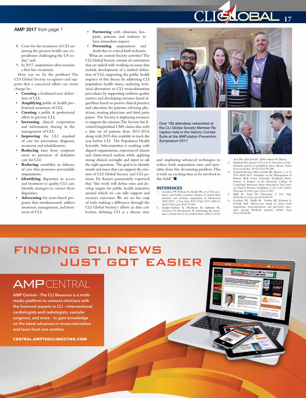
There are other diagnostic tools, such as fluorescence indocyanide green angi-ography, near infrared fluorescence im-aging, or subcutaneous continuous oxy-gen monitoring, that are currently being evaluated.8,9 However, they have not been widely accepted and data regarding their diagnostic sensitivity and specificity are sparse. Another tool that has been fre-quently advocated, especially for patients with non-compressible vessels, has been pulse volume recording (PVR). However, we recently evaluated the accuracy of ABI, TBI, and PVR in predicting tibial artery patency in patients with CLI and non-compressible ABI.10 Among those with non-compressible ABI, the ves-sel was either occluded or severely dis-eased over 80% of the time (Figure 2). Unfortunately, PVR was also a weak predictor of vessel patency and over 50% of patients with occluded or severely stenotic tibial artery had normal PVR. A TBI < 0.70 was the most accurate in identifying tibial occlusion or stenosis in patients with non-compressible ABI.
GIVEN THE CHALLENGES DESCRIBED, WHAT IS THE BEST APPROACH TO PATIENTS WITH CLI?
We believe that all patients with sus-pected CLI should undergo an ABI and TBI assessment. ABI is typically reported as the higher of the anterior or posterior tibial artery, regardless of wound location or the affected angiosome. Therefore, the vascular specialist needs to personally interrogate the results to better under-stand the patency of anterior tibial and posterior tibial arteries and correlate this data with wound location (angiosome). Additionally, all patients should have toe pressure and TBI measured. If ABI is ab-normal or TBI < 0.70, then cine angi-ography is indicated. Beyond diagnosis, obtaining perfusion data prior to revascu-larization will allow a better understand-ing of the quality of procedure and also serve as a way to monitor perfusion in follow-up, as there are high rates of re-stenosis in patients with CLI undergoing tibial angioplasty.11 For primary care phy-sicians, podiatrists, wound care specialists, and other allied health professionals with limited resources beyond ABI, prompt referral to a vascular specialist or center is likely the best option for patients with tissue loss or gangrene. A normal ABI in these patients should never be an indica-tion of adequate perfusion and allow de-lay in revascularization.
Ultimately, clinical judgment and a multidisciplinary approach to patients and wounds should trump any tests. We have learned that time is limb and a rapid assessment and revascularization for CLI is paramount to amputation prevention. The current issue also highlights the need for the effort led by the CLI Global Society and other organizations to edu-cate and improve the care of patients with this morbid condition.
REFERENCES1. Gerhard-Herman MD, Gornik HL, Barrett C, et al.
2016 AHA/ACC Guideline on the Management of Patients With Lower Extremity Peripheral Artery Disease: Executive Summary: A Report of the American College of Cardiology/American Heart Association Task Force on Clinical Practice Guidelines. Circulation 2017;135:e726–e779.
2. Patel MR, Conte MS, Cutlip DE, et al. Evaluation and treatment of patients with lower extremity peripheral artery disease: consensus definitions from Peripheral Academic Research Consortium (PARC). J Am Coll Cardiol. 2015;65(9):931–941.
3. Bunte MC, Jacob J, Nudelman B, Shishehbor MH. Validation of the relationship between ankle-bra-chial and toe-brachial indices and infragenicular ar-terial patency in critical limb ischemia. Vasc Med. 2015;20(1):23–29.
4. Shishehbor MH, Hammad TA, Zeller T, Baumgartner I, Scheinert D, Rocha-Singh KJ. An analysis of IN.PACT DEEP randomized trial on the limitations of the societal guidelines-recommended hemodynam-ic parameters to diagnose critical limb ischemia. J Vasc Surg. 2016;63(5):1311–1317.
5. Mustapha JA, Diaz-Sandoval LJ, Adams G, et al. Lack of association between limb hemodynamics and re-sponse to infrapopliteal endovascular therapy in pa-tients with critical limb ischemia. J Invasive Cardiol. 2017;29(5):175–180.
6. Shishehbor MH. Acute and critical limb isch-emia: when time is limb. Cleve Clin J Med. 2014;81(4):209–216.
7. Shishehbor MH, White CJ, Gray BH, et al. Critical limb ischemia: An expert statement. J Am Coll Cardiol. 2016;68(18):2002–2015.
8. Igari K, Kudo T, Uchiyama H, Toyofuku T, Inoue Y. Indocyanine green angiography for the diagnosis of peripheral arterial disease with isolated infrapopli-teal lesions. Ann Vasc Surg. 2014;28(6):1479–1484.
9. Montero-Baker MF, Au-Yeung KY, Wisniewski NA, et al. The First-in-Man “Si Se Puede” Study for the use of micro-oxygen sensors (MOXYs)
to determine dynamic relative oxygen indices in the feet of patients with limb-threatening isch-emia during endovascular therapy. J Vasc Surg. 2015;61(6):1501–1509 e1.
10. Randhawa MS, Reed GW, Grafmiller K, Gornik HL, Shishehbor MH. Prevalence of tibial artery and pedal arch patency by angiography in patients with critical limb ischemia and noncompressible ankle brachial index. Circ Cardiovasc Interv. 2017;10(5).
11. Shishehbor MH, Hammad TA. Treatment of in-frapopliteal disease in critical limb ischemia: be-yond angioplasty. Circ Cardiovasc Interv. 2016;9(5): e003882.
SHISHEHBOR from page 1
Figure 1. The association between tibial vessel patency and ABI. Among those with significant disease involving all 3 tibial vessels, 28% had normal or non-compressible ABI.Reprinted from Journal of Vascular Surgery 63, Shishehbor, MH et al, An analysis of IN.PACT DEEP randomized trial on the limitations of the societal guidelines-recommended hemodynamic param-eters to diagnose critical limb ischemia, 1311–1317, copyright 2016, with permission from Elsevier.
Figure 2. Prevalence of tibial artery patency in patients with non-compressible ABI. Among those with non-compressible anterior tibial (AT) ABI, nearly 80% had occluded or severely diseased AT. [Adopted from Circ Cardiovasc Interv, 2017 May;10(5).]
Disclosure: Dr. Shishehbor is a consultant and advisor to Abbott Vascular, Medtron-ics, Boston Scientifics, Terumo, and Philips. He is a member of the board of direc-tors for CardioVascular Innovations (CVI), a not-for-profit 501(c) 3 organization.
Beyond diagnosis, obtaining perfusion data prior to revascularization will allow a better understanding of the quality of procedure and also serve as a way to monitor perfusion in follow-up, as there are high rates of restenosis in patients with CLI undergoing tibial angioplasty.11

New Guide WiresNEW Peripheral Guide Wires
Now available withASAHI’s unique guidewire technology
PRECISION ENGINEERED for Tough Peripheral Cases
®
ASAHI INTECC USA, INC.
2500 Red Hill Avenue, Suite 210, Santa Ana, CA 92705Toll-Free [email protected]
Learn more atasahi-inteccusa-medical.com
The ASAHI INTECC peripheral guide wires are intended to facilitate the placement and exchange of diagnostic and therapeutic devices during intravascular procedures.These devices are intended for peripheral vascular use only.
The ASAHI® Corsair® Armet® is intended to provide support to facilitate the placement of guide wires in the peripheral vasculature, and can be used to exchange one guide wire for another.The ASAHI Corsair Armet is also intended to assist in the delivery of contrast media into the peripheral vasculature. This device should not be used in coronary vasculature or neurovasculature.
®
ASAHI Gladius® 0.014/0.018Workhorse
ASAHI® Halberd® 0.014/0.018Complex Lesion
ASAHI Gaia® PV 0.018Complex Lesion
New Microcatheter
Durable metal tip
Super SHINKA-Shaft
Low profile, supportive catheter body,excellent torque
®
®
Ⓒ2016 ASAHI INTECC., LTD.“ASAHI,” “Halberd,” “ASAHI Gaia,” and “ASAHI Gladius” are trademarks or registered trademarks of ASAHI INTECC CO., LTD. in Japan and other countries.

10
September 2017
CLIGLOBAL
The patient underwent lower extrem-ity angiogram under moderate conscious sedation. A contralateral left groin ac-cess, up-and-over approach was utilized in order to determine if any aorto-iliac disease had been missed on the arterial duplex study. Initial arteriography was also performed with manual injection of carbon dioxide (primarily in the pelvis) to limit contrast load.
Diagnostic angiography revealed a long-segment occlusion of the femoropoliteal artery beginning ap-proximately 5 cm from the origin of the superficial femoral artery (SFA). Numerous collaterals from the profun-da artery were seen through the thigh and knee. There is faint reconstitution of the mid-anterior tibial artery, at the level of the mid-calf (Figure 3). The dorsalis pedis artery was faintly opaci-fied (not shown).
For support, a 45 cm 6 French (Fr) Ansel sheath (Cook Medical) was ad-vanced into the proximal left SFA. An angled 5 Fr catheter and stiff glidewire were used to recanalize the femoro-popliteal artery, up to the supragenic-ulate popliteal artery. The system was downsized to an 0.018-inch wire (V18, Boston Scientific) and a Quick-Cross support catheter (Spectranetics). The 0.018-inch system was advanced into the infrageniculate popliteal artery in a subintimal plane. Brief attempts to can-nulate the anterior tibial artery from this approach were not successful.
Pedal access via the dorsalis pedis ar-tery was then obtained under ultrasound guidance. In our technique, after access, the 21-gauge needle is exchanged over a short 0.018-inch wire for the inner dilator of the Micropuncture sheath (Cook). Then an anti-spasm cocktail of 200 mcg nitroglycerin, 2.5 mg vera-pamil, and 2000 U heparin was inject-ed followed by exchange of the inner
dilator for a support catheter. In this case, the inner dilator was exchanged over a longer 0.018-inch wire (V18, Boston Scientific) and Quick-Cross support catheter. The system was ad-vanced across the occluded anterior tibial artery and into the infrageniculate popliteal artery. The wire was directed into a 5 Fr end-hole catheter advanced from the left groin sheath. Once cap-tured and externalized, through-and-through access was obtained.
Following floss access, plain old bal-loon angioplasty was performed in the dorsalis pedis artery (across access site) and anterior tibial artery with 2.0 x 150 and 2.5 x 300 mm balloons (Ultraverse, Bard; Figure 4). Angioplasty of the oc-cluded femoropoliteal artery was per-formed with a 5.0 x 220 mm balloon (Ultraverse). Although in-line flow was restored to the anterior tibial ar-tery, there was a significant irregular appearance of recanalized segments. Additionally, there was a dissection involving the proximal anterior tibial artery (Figure 4c). Given the extent of residual disease, it was felt that further balloon angioplasty (i.e., prolonged POBA versus drug-eluting balloon an-gioplasty) would not yield durable re-sults. Therefore, primary stenting of the entire femoropopliteal segment and the proximal anterior tibial artery was per-formed. Multiple overlying stents were used, including a drug-eluting stent (Zilver PTX, Cook Medical) at the SFA origin and bare metal stents (Supera, Abbott) at the mid to distal femorapop-liteal segments. A balloon-expandable drug-eluting 3 x 33 mm stent (Xience, Abbott) was used to treat the residual disease at the proximal anterial tibial artery. A final angiogram was obtained, demonstrating brisk inline flow to the dorsalis pedis artery via a robustly pat-ent anterior tibial artery (Figure 5). The patient had a strong, palpable dorsalis
DHAND from page 3
Figure 2. Arterial duplex study of the right lower extremity. (A) Biphasic waveforms with sharp systolic upstrokes are shown in the common femoral artery. (B) No significant flow is seen in the distal SFA, or (C) popliteal artery; and (D) there is trace recon-stitution in the anterior tibial artery.
A B
C D
At our institution, peripheral arterial disease is treated utilizing a multidisciplinary approach. Patients who suffer from chronic wounds or obvious limb ischemia (i.e., gangrene) are referred by their primary care physicians to the hospital’s wound healing center, where patients are seen by all three specialties: podiatry, vascular surgery, and interventional radiology. Wound care...is managed by podiatry and, sometimes, vascular surgery. An “endovascular-first” strategy has been adopted across the specialties.

Aortic | Peripheral | endoVenous
3033 Campus Drive, N550
Plymouth, MN 55441
USA
24-hour Technical Support Toll free:
+1.800.328.2518
CardioVascular LifeLine
Customer Support
Tel: +1.763.526.7890
Toll free: +1.877.526.7890
Orders
Tel: +1.763.514.8510
Toll free: +1.800.716.6700
Fax: +1.877.697.4841
Email: [email protected]
Indications, contraindications,
warnings and instructions for
use can be found in the product
labeling supplied with each device.
CAUTION: Federal (USA) law restricts
this device to sale by or
on the order of a physician.
FTSOP113326-35 Rev. 1A
UC201802497 -01 EN © 2017 Medtronic. All
rights reserved. Medtronic, Medtronic logo and
Further, Together are trademarks of Medtronic.
™* Third party brands are trademarks of
their respective owners. All other brands are
trademarks of a Medtronic company. 7/17
The Chocolate™* PTA Balloon is manufactured
by TriReme Medical.
medtronic.com/peripheral
YOUR PTA TREATMENTOPTIONS JUST GOTSWEETER
Minimize dissection risk through predictable, uniform dilatation.
ChocolateTM*
PTA Balloon
TREAT PAD YOUR WAY

12
September 2017
CLIGLOBAL
pedis pulse at the end of the procedure. She underwent a toe amputation the following week uneventfully.
At our institution, peripheral arterial disease is treated utilizing a multidisci-plinary approach. Patients who suffer from chronic wounds or obvious limb ischemia (i.e., gangrene) are referred by their prima-ry care physicians to the hospital’s wound healing center, where patients are seen by all three specialties: podiatry, vascular sur-gery, and interventional radiology. Wound care, including specialized dressings, de-bridement, skin substitutes, oxygen thera-py, and vacuum-assisted wound closure, is managed by podiatry and, sometimes, vas-cular surgery. An “endovascular-first” strat-egy has been adopted across the specialties. The extremity angiograms are performed by interventional radiology with close vas-cular surgery support in cases of bypass or surgical emergencies.
The vast majority of our patients suffer from severe tibial disease, often with a sin-gle-vessel runoff. Although angiosomes are preferred, we typically attempt to achieve the best run-off possible, depending on what is identified on the initial diagnos-tic arteriogram. Several cases will require pedal access for recanalization, which all operators are comfortable perform-ing. Additionally, most of the interven-tions are performed in one setting under moderate conscious sedation and rarely staged unless there is aortoiliac inflow disease (requiring bilateral groin access) or long procedure times. With the advent of drug-eluting balloons, recanalization
without stenting is a preferred strategy. However, stenting is used if angioplasty alone does not obtain a fluoroscopically or clinically acceptable result. General an-esthesia is used for select cases.
Most pre-procedural imaging is per-formed with an arterial duplex study. More recently, we try to obtain CT an-giography run-off of the lower extremi-ties for procedural planning, although this happens in <25% of our cases currently. Our most common obstacle for cross-sectional imaging is poor renal function in patients who are not already on dialysis.
During the wound healing process, both vascular surgery and interventional radiology follow patients in separate out-patient clinics, while the patient still goes to the wound healing center for medi-cal management. Interventional radiol-ogy usually sees the patients 3–4 weeks after intervention, obtaining an arterial duplex study for baseline and follow-up. These patients are then usually seen every 3 or 6 months afterward, with biannual or annual screening arterial duplex stud-ies, depending on the operator. If wound healing is not adequate, the patient either undergoes a repeat angiogram or is dis-cussed in our monthly multidisciplinary vascular conferences.
Well-established, collegial multidis-ciplinary care has led to excellent out-comes in our patients suffering from critical limb ischemia. With the goal to prevent extensive amputation, the qual-ity of life in the patients we manage is significantly improved.
Figure 5. Final arteriogram after overlapping uncovered stent placements into the (A) SFA, (B) popliteal artery, and (C) anterior tibial artery. The lateral foot shows flow into the dorsal pedis artery, with a partially intact pedal loop. Delayed im-ages demonstrate retrograde opacification of the plantar branches.
A B C
Figure 4. Intervention. (A) Obtaining floss access from the pedal approach, a support catheter is seen in the infrageniculate popliteal artery (from the up-and-over approach) and a second support catheter and 0.018-inch wire is visible crossing the occluded proximal anterior tibial artery. The wire is captured and externalized (not shown), followed by (B) angioplasty of the occluded segments and post angioplasty arteriogram, (C) demonstrating a dissection in the recannalized segment of the proximal anterior tibial artery. The post angioplasty arteriogram of the femoropopliteal artery is not shown.
A B C
Figure 3. Left lower extremity angiogram: (A) Chronic total occlusion of the left SFA beginning approximately 5 cm from the SFA origin, (B) profunda collaterals noted in the thigh and knee, with no reconstitution of the popli-teal artery, and (C) trace reconstitution of the mid anterior tibial artery.
A B C


14
September 2017
CLIGLOBAL
analyses were performed in order to evaluate the association between statin therapy and the hazard of amputation and/or death. Although lipid-lowering therapy with statins has been among the most well studied pharmacologic therapies, the effect of these agents in CLI patients has not been well docu-mented and current recommendations are extrapolated from other high-risk populations.10 In the CRITISCH reg-istry, statin therapy was applied in 445 individuals (37%), 371 (31%) patients received no statins, and 384 subjects were excluded from analysis, as they could not be allocated to any group in a reasonable manner (treatment cross-overs). Patients on statins were more likely to be younger (P<.001) and to have a history of coronary heart disease (P<.001) or previous PVI (P<.001). Patients receiving statin therapy had a lower hazard regarding AFS (HR, 0.45; 95% CI, 0.34–0.63; P<.001) and death (HR, 0.40; 95% CI, 0.24–0.66; P<.001) as well as lower odds of ma-jor adverse cardio and cerebrovascular
events (odds ratio, 0.41; 95% CI, 0.23–0.69; P<.001). Interestingly, statin therapy was not associated with reduced amputation rates (HR, 1.02; 95% CI, 0.67–1.56; P= .922).
Statin effect on amputation-free survival was consistent among diabet-ics (HR, 0.47; 95% CI, 0.31–0.70; P < .001), patients with CKD (HR, 0.53; 95% CI, 0.32–0.87; P =.012), and pa-tients older than 75 years (HR, 0.40; 95% CI, 0.26–0.60; P <.001). Statin administration was also associated with an improved amputation-free survival in patients with antiplate-let medication (HR, 0.64; 95% CI, 0.41–0.99; P=.049) and without an-tiplatelet medication (HR, 0.26; 95% CI, 0.12–0.57; P=.001), and after both endovascular therapy (HR, 0.51; 95% CI, 0.34–0.76; P= .001) and bypass revascularization (HR, 0.38; 95% CI, 0.21–0.68; P=.001).
The results of the CRITISCH regis-try highlight that when physicians are free to individualize therapy for their CLI patients, they can achieve encour-aging outcomes. Given the complex-ity of the disease and the increased
comorbidity of CLI patients, an indi-vidualized approach seems more rea-sonable than a general recommenda-tion. In this context, the future and ongoing trials should not only focus on the evaluation of the superiority/inferiority of a treatment option over another, but on the identification of those CLI patients who are better can-didates for each treatment modality. Finally, our findings suggest that statin therapy in CLI patients is associated with increased amputation-free sur-vival and lower rates of mortality and major adverse events regardless of the applied treatment strategy. However, we found no indication that statin therapy influences the fate of the af-fected limb, and further research is needed to assess areas of uncertainty in the secondary prophylaxis of CLI individuals.
REFERENCES1. Reinecke H, Unrath M, Freisinger E, et al. Peripheral
arterial disease and critical limb ischaemia: still poor outcomes and lack of guideline adherence. Eur Heart J. 2015;36(15):932–938.
2. Goodney P, Holman K, Henke PK, et al. Regional intensity of vascular care and lower extremity amputa-tion rates. J Vasc Surg. 2013;57(6):1471–1479.
3. Benoit E, O’Donnell TF Jr, Kitsios GD, Iafrati MD. Improved amputation-free survival in unreconstruc-table critical limb ischemia and its implications for clinical trial design and quality measurement. J Vasc Surg. 2012;55(3):781–789.
4. Stavroulakis K, et al. Association between statin thera-py and amputation-free survival in patients with criti-cal limb ischemia in the CRITISCH registry. J Vasc Surg. 2017, in press.
5. Bisdas T, Borowski M, Stavroulakis K, Torsello G; CRITISCH Collaborators. Endovascular Therapy Versus Bypass Surgery as First-Line Treatment Strategies for Critical Limb Ischemia: Results of the Interim Analysis of the CRITISCH Registry. JACC Cardiovasc Interv. 2016;9(24):2557–2565.
6. Katsanos K, Spiliopoulos S, Paraskevopoulos I, Diamantopoulos A, Karnabatidis D. Systematic Review and Meta-analysis of Randomized Controlled Trials of Paclitaxel-Coated Balloon Angioplasty in the Femoropopliteal Arteries: Role of Paclitaxel Dose and Bioavailability. J Endovasc Ther. 2016;23(2):356–370
7. Adam DJ, Beard JD, Cleveland T, et al; BASIL trial par-ticipants. Bypass versus angioplasty in severe ischaemia of the leg (BASIL): multicentre, randomised controlled trial. Lancet. 2005;366(9501):1925–1934.
8. Conte MS, Bandyk DF, Clowes AW, et al for the PREVENT III Investigators. Results of PREVENT III: a multicenter, randomized trial of edifoligide for the prevention of vein graft failure in lower extremity bypass surgery. J Vasc Surg. 2006;43(4):742–751.
9. Bisdas T, Borowski M, Torsello G; First-Line Treatments in Patients with Critical Limb Ischemia (CRITISCH) Collaborators. Current practice of first-line treatment strategies in patients with critical limb ischemia. J Vasc Surg. 2015;62(4):965–973.
10. Teraa M, Conte MS, Moll FL, Verhaar MC. Critical limb ischemia: current trends and future directions. J Am Heart Assoc. 2016; 5(2): pii: e002938. doi: 10.1161/JAHA.115.002938.
STAVROULAKIS from page 4
201717
IN CONJUNCTION
WITH
SYDNEY AUSTRALIA 7-9 DECEMBER 2017I C C D A R L I N G H A R B O U R S Y D N E Y
Enquiries: [email protected]
VERVE looks forward to welcoming CLI Global members.
Registration and program details available on the website
www.vervesymposium.com


16
September 2017
CLIGLOBAL
He was then referred to my office for advanced wound healing treatment of his gangrenous 2nd digit (Figure 1). Following comprehensive history and physical examination, he was scheduled for a second digit amputation with ex-ploration of the left second metatarsal phalangeal (MTP) joint. He also re-quired an incision and drainage of the left second MTP, as he had developed a deeply seated abscess in that region. One of the most reassuring signs that he exhibited prior to incision was a vis-ibly bounding dorsalis pedis (DP) artery pulse. The surgery was performed as described and his operative site was left open with local wound care performed while he was an inpatient. Prior to dis-charge, he was taken back to the oper-ating room for delayed primary repair, which was performed following light wound debridement and extensive pul-satile lavage. (Figures 2 and 3).
Active surveillance in the outpatient setting ensued over the next 2 weeks, where he did encounter a small dehis-cence of his dorsal incision site. This was treated with wound care visits, debriding necrotic tissues, and twice-daily Santyl dressing changes. Once the wound bed was clean and granular, we began using a Kerecis (Isafjordur, Iceland) Omega3 acellular fish skin graft as a skin substitute. A consultation with orthotics and prosthetics was also arranged for off-loading techniques and custom molded shoe implementa-tion. He returned to his home state and wound care was continued by a local podiatrist with weekly application of the Kerecis Omega3 acellular fish skin graft (Figure 4).
One application of Kerecis had been performed prior to his trans-fer home, and 11 additional Kerecis grafts were applied over the following 2 months. Healing progressed nicely. Unfortunately, he developed a septic
knee. He had a history of a total knee implant arthroplasty and apparently seeded infection to this region. The infection was treated with IV antibiot-ics, incision and drainage of the knee joint, and extensive pulsatile lavage. Once the septic arthritis was eradi-cated, treatment resumed on the left forefoot wound, which had regressed slightly. Sorbact with hypochlorous solution was initiated and with local wound care was continued until full wound healing had been achieved, ap-proximately 10 months following sur-gery (Figures 5 and 6).
In conclusion, as physicians treating exclusively the foot, ankle, and lower extremities, podiatrists are often re-ferred to as the gatekeepers of PAD. Podiatrists are typically the first physi-cians to have the chance to recognize and diagnose PAD, and with such an enormous opportunity, must be well versed in the risk factors, subjective complaints, clinical presentation, vas-cular testing, and treatment options. The earlier a patient can be referred to a revascularization specialist, the more likely that not only their limb may be saved, but their life as well.
Acknowledgement. We would like to thank Dr. David Trenner, MD for his contri-bution to patient treatment and follow-up.
REFERENCES1. Hirsch AT, Criqui MH, Treat-Jacobson D, et
al. Peripheral arterial disease detection, aware-ness, and treatment in primary care. JAMA. 2001;286(11):1317–1324.
2. Yost ML. Diabetic Foot Ulcers, peripheral ar-terial disease and critical limb ischemia. The SAGE Group. 2010.
3. Howlader N, et al. SEER Cancer Statistics Review, 1975–2010. Accessed April 17, 2014.
4. Dolor RJ, et al. Comparative Effectiveness Reviews, No. 66. 2012 Aug.
5. Fowkes FG, Rudan D, Rudan I, et al. Comparison of global estimates of prevalence and risk factors for peripheral artery disease in 2000 and 2010: a systematic review and analy-sis. Lancet. 2013; 382(9901):1329–1340. doi: 10.1016/S0140-6736(13)61249-0.
6. Davies MG. Critical Limb Ischemia: Epidemiology. Methodist Debakey Cardiovasc J. 2012;8(4):10–14.
7. “Who Is at Risk for Peripheral Artery Disease?” National Heart, Lung, and Blood Institute. Updated June 22, 2016. https://www.nhlbi.nih.gov/health/health-topics/topics/pad/atrisk.
8. Fowkes FG, Housley E, Cawood EH, Macintyre CC, Ruckley CV, Prescott RJ. Edinburgh Artery Study: prevalence of asymptomatic and symptomatic peripheral arterial disease in the general population. Int J Epidemiol. 1991;20(2):384–392.
9. Ruiz J. Peripheral Vascular Disease (PVD). Obesity and Diabetes: Epidemic Proportions in Puerto Rico and its Financial Impact. Report from PREHCO Project data, Governor’s Office for Elderly Affairs of the Commonwealth of Puerto Rico (OGAVE), January 2005.
10. Yost ML. The real cost of peripheral artery dis-ease. The SAGE Group. 2011.
11. Allie D. Costs of Bypass vs PTA vs. Amputation. Presented at the New Cardiovascular Horizons Symposium, 2009
Figure 2A. Post-operative repair. Figure 2B. Post-operative repair.
Figure 3. Application of Kerecis™ Omega3 acellular fish skin graft.
Figure 4. Healing 10 months after initial presentation to Podiatrist.
Figure 5. Healed at 10 months.
TURSI from page 6
17CLIGLOBAL
finding cli newsjust got easier
central.amptheclimeeting.com
AMP Central – The CLI Resource is a multi-media platform to connect clinicians with the foremost experts in CLI – interventional cardiologists and radiologists, vascular surgeons, and more – to gain knowledge on the latest advances in revascularization and learn from one another.
8. Costs for the treatment of CLI are among the greatest health care ex-penditures challenging the US to-day,6 and;
9. In 2017, amputation often remains a first line treatment.
How can we fix the problem? The CLI Global Society recognizes and sup-ports that a concerted effort can create change by:• Creating a facilitated new defini-
tion of CLI;• Amplifying public & health pro-
fessional awareness of CLI;• Creating a public & professional
effort to prevent CLI;• Increasing clinical cooperation
and information sharing in the management of CLI;
• Improving the CLI standard of care for prevention, diagnosis, treatment and rehabilitation;
• Reducing time from symptom onset to provision of definitive care for CLI;
• Reducing variability in delivery of care that promotes preventable amputations;
• Identifying disparities in access and treatment to quality CLI care. Identify strategies to correct these disparities;
• Advocating for team-based pro-grams that simultaneously address awareness, management, and treat-ment of CLI;
• Partnering with clinicians, hos-pitals, patients, and industry to have immediate impact;
• Preventing amputations and death due to critical limb ischemia.
What are current Society activities? The CLI Global Society consists of committees that are tasked with working on issues that include development of a unified defini-tion of CLI, supporting the public health urgency of this disease by addressing CLI population health issues; analyzing tech-nical alternatives to CLI revascularization procedures by supporting uniform quality metrics and developing resource based al-gorithms based on proven clinical practice; and education for patients, referring phy-sicians, treating physicians and third party payers. The Society is deploying resources to support the mission. The Society has li-censed longitudinal CMS claims data with a data set of patients from 2011-2014, along with 2010 data available to track the year before CLI. The Population Health Scientific Subcommittee is working with aligned organizations, experienced dataset and claims-based analysts while applying strong clinical oversight and input to ask the right questions. The goal is to identify trends and issues that can support the mis-sion of CLI Global Society and CLI pa-tients. Dr. Katzen passionately expressed that “this work will define rates and de-velop targets for public health initiatives around which we can rally support and measure outcomes. We are on the cusp of truly making a difference through the CLI Global Society’s efforts in data col-lection, defining CLI as a disease state
and employing advanced techniques to reduce both amputation rates and mor-tality from this devastating problem. This is truly an exciting time to be involved in this field!”
REFERENCES1. Goodney PP, Holman K, Henke PK, et al. The inci-
dence and health economic burden of critical limb ischemia and ischemic amputation in Minnesota: 2005–2007. J Vasc Surg. 2013; 57(6): 1471–1480.e3. doi:10.1016/j.jvs.2012.11.068.
2. Ziegler-Graham K, MacKenzi EJ, Ephraim PL, Travison TG, Brookmeyer R. Estimating the preva-lence of limb loss in the United States: 2005 to 2015.
Arch Phys Med Rehabil 2008; volume 89, March. 3. Hardman RL, Jazaeri O, Yi J, et al. Overview of clas-
sification systems in peripheral artery disease. Semin Intervent Radiol. 2014;31(4):378-388.
4. Gerhard-Herman MD, Gornik HL, Barrett C, et al. 2016 AHA/ACC Guideline on the Management of Patients With Lower Extremity Peripheral Artery Disease: A Report of the American College of Cardiology/American Heart Association Task Force on Clinical Practice Guidelines. J Am Coll Cardiol. 2016;doi:10.1016/j.jacc.2016.11.007.
5. Mills JL, Santo VJ. Discussion. J Vasc Surg. 2014;doi:10.1016/j.jvs.2013.08.003.
6. Goodney PP, Tarulli M, Faerber AE, Schanzer A, Zwolak RM. Fifteen-year trends in lower limb amputation, revascularization and preventive mea-sures among Medicare patients. JAMA Surg. 2015;150:84-86.
AMP 2017 from page 1
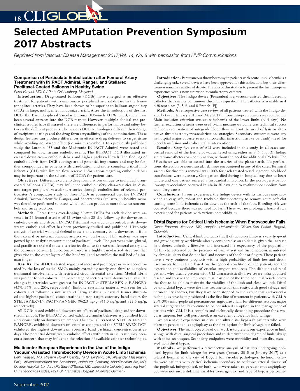
Over 100 attendees networked at the CLI Global Society Member Re-ception held in the historic Conrad Suite at the AMPutation Prevention Symposium 2017.
18
September 2017
CLIGLOBALSelected AMPutation Prevention Symposium 2017 Abstracts Reprinted from Vascular Disease Management 2017;Vol. 14, No. 8 with permission from HMP Communications
Comparison of Particulate Embolization after Femoral Artery Treatment with IN.PACT Admiral, Ranger, and Stellarex Paclitaxel-Coated Balloons in Healthy SwineRenu Virmani, MD, CV Path, Gaithersburg, Maryland
Introduction. Drug-coated balloons (DCBs) have emerged as an effective treatment for patients with symptomatic peripheral arterial disease in the femo-ropopliteal arteries. They have been shown to be superior to balloon angioplasty (PTA) in large, multicenter randomized trials. After the introduction of the first DCB, the Bard Peripheral Vascular Lutonix .035-inch OTW DCB, there have been several entrants into the DCB market. However, multiple clinical and pre-clinical studies have illustrated there are differences in performance and safety be-tween the different products. The various DCB technologies differ in their design of excipient coatings and the drug form (crystallinity) of the combinations. These design features can produce differences in effective drug delivery to target tissue while avoiding non-target effect (i.e. minimize emboli). In a previously published study, the Lutonix 035 and the Medtronic IN.PACT Admiral were tested and compared for downstream embolic events. The IN.PACT DCB illustrated in-creased downstream embolic debris and higher paclitaxel levels. The findings of embolic debris from DCB coatings are of potential importance and may be fur-ther compounded in patients with claudication and more complex critical limb ischemia (CLI) with limited flow reserve. Information regarding embolic debris may be important in the selection of DCB’s for patient care.
Objectives. Different excipient/drug formulations unique to individual drug-coated balloons (DCBs) may influence embolic safety characteristics in distal non-target peripheral vascular territories through embolization of released par-ticulates. A comparator study of three DCBs in commercial use, the IN.PACT Admiral, Boston Scientific Ranger, and Spectranetics Stellarex, in healthy swine was therefore performed to assess which balloon produces more downstream em-boli and tissue reaction.
Methods. Three times over-lapping 80-mm DCBs for each device were as-sessed in 24 femoral arteries of 12 swine with 28-day follow-up for downstream embolic events and debris. IN.PACT Admiral was used as a control, as its down-stream emboli and effect has been previously studied and published. Histologic analysis of arterial wall and skeletal muscle and coronary band downstream from the external or internal femoral arteries was performed. This analysis was sup-ported by an analytic measurement of paclitaxel levels. The gastrocnemius, gluteal, and gracilis are skeletal muscle territories distal to the external femoral artery and the coronary corium (i.e., coronary band) is a highly vascularized structure that gives rise to the outer layers of the hoof wall and resembles the nail bed of a hu-man finger.
Results. For all DCBs tested, regions of increased proteoglycan were accompa-nied by the loss of medial SMCs mainly extending nearly one-third to complete transmural involvement with restricted circumferential extension. Medial fibrin was present for all cohorts. The percentage of sections with downstream vascular changes in arterioles were greatest for IN.PACT > STELLAREX > RANGER (43%, 36%, and 25%, respectively). Embolic crystalline material was seen for all cohorts and followed a similar trend. Drug analysis in parallel tissues illustrat-ed the highest paclitaxel concentrations in non-target coronary band tissues for STELLAREX>IN.PACT>RANGER (962.3 ng/g, 911.3 ng/g, and 822.5 ng/g, respectively).
All DCBs tested exhibited downstream effects of paclitaxel drug and/or down-stream emboli. The IN.PACT control exhibited similar behavior as published from a previous study on downstream emboli. The new DCB’s tested, STELLAREX and RANGER, exhibited downstream vascular changes and the STELLAREX DCB exhibited the highest downstream coronary band paclitaxel concentration at 28 days. The potential downstream embolic effects with certain DCB use may pres-ent a concern that may influence the selection of available catheter technologies.
Multicenter European Experience in the Use of the Indigo Vacuum-Assisted Thrombectomy Device in Acute Limb IschemiaBella Huasen, MD, Preston Royal Hospital, NHS, England, UK; Alexander Massmann, PhD, Universitatsklinikum des Saarlandes, Homburg, Germany; AbdulRahman Alvi, MD, Queens Hospital, London, UK; Steve D’Souza, MD, Lancashire Univeristy teaching trust, UK; Theodosios Bisdas, PhD, St. Franziskus Hospital, Muenster, Germany
Introduction. Percutaneous thrombectomy in patients with acute limb ischemia is a challenging task. Several devices have been approved for this indication, but their effec-tiveness remains a matter of debate. The aim of this study is to present the first European experience with a new aspiration thrombectomy catheter.
Objectives. The Indigo device (Penumbra) is a vacuum-assisted thrombectomy catheter that enables continuous thrombus aspiration. The catheter is available in 4 different sizes (3, 5, 6, and 8 French [F]).
Methods. A retrospective case review of all patients treated with the Indigo de-vice between January 2016 and May 2017 in four European centers was conducted. Main inclusion criterion was acute ischemia of the lower limbs (<14 days). No further exclusion criteria were used. Main measure outcome was technical success defined as restoration of antegrade blood flow without the need of lysis or alter-native thrombectomy/revascularization strategies. Secondary outcomes were any in-hospital major adverse events (myocardial infarction, stroke or death), need for blood transfusion and in-hospital reintervention.
Results. Sixty-five cases of ALI were included in this study. In all cases suc-cessful removal of the fresh clot was achieved using either an 8, 6, 5, or 3F Indigo aspiration-catheters or a combination, without the need for additional tPA lysis. The 3F catheter was able to extend into the arteries of the plantar arch. No perfora-tions, dissection or neurovascular damage encountered in any of the cases. Technical success for thrombus removal was 100% for each treated vessel segment. No blood transfusions were necessary. One patient died during in-hospital stay due to heart failure and one patient suffered a myocardial infarction. In the post-operative fol-low-up re-occlusion occurred in 4% in 30 days due to re-thromboembolism from secondary causes.
Conclusions. In our experience, the Indigo device with its various range pro-vided an easy, safe, robust and trackable thrombectomy to remove acute soft clot causing acute limb ischemia as far down as the arch of the foot. Bleeding risk was obviated because there was no need for lysis. There was no prolonged hospital stay experienced for patients with various comorbidities.
Distal Bypass for Critical Limb Ischemia: When Endovascular FailsCesar Eduardo Jimenez, MD, Hospital Universitario Clinica San Rafael, Bogotá, Colombia
Introduction. Critical limb ischemia (CLI) of the lower limbs is a very frequent and growing entity worldwide, already considered as an epidemic, given the increase in diabetes, unhealthy lifestyles, and increased life expectancy of the population. CLI is characterized by the presence of pain at rest and tissue necrosis, manifested by chronic ulcers that do not heal and necrosis of the foot or fingers. These patients have a very ominous prognosis with a high probability of limb loss and death. Treatments for CLI are based on the general condition of the patient, and the experience and availability of vascular surgeon resources. The diabetic and renal patients who usually present with CLI characteristically have severe infra-popliteal disease and to save the limb, require at least one of the three popliteal vessels below the foot to be able to maintain the viability of the limb and close wounds. Distal or ultra distal bypass were the first treatments for this entity, with good salvage and permeability indexes, but they have been stopped to the extent that endovascular techniques have been positioned as the first line of treatment in patients with CLI. A 20%-30% infra-popliteal percutaneous angioplasty fails for different reasons; major arterial surgery should continue to be considered an option in the management of patients with CLI. It is a complex and technically demanding procedure for a vas-cular surgeon, but well performed, is an excellent choice for limb salvage.
We present our experience in distal and ultra distal bypass in patients who were taken to percutaneous angioplasty as the first option for limb salvage but failed.
Objectives. The main objective of our work is to present our experience in limb salvage with distal surgical procedures and to determine the degree of limb salvage with these techniques. Secondary endpoints were morbidity and mortality associ-ated with distal bypass.
Methods. We performed a retrospective analysis of patients undergoing pop-liteal bypass for limb salvage for two years (January 2015 to January 2017) at a referral hospital in the city of Bogotá for vascular pathologies. Inclusion crite-ria were patients with critical ischemia secondary to occlusive arterial disease of the popliteal, infrapopliteal, or both, who were taken to percutaneous angioplasty, but were not successful. The variables were age, sex, and type of bypass performed
19
September 2017
CLIGLOBAL(popliteal-pedis, popliteal to posterior tibial, popliteal to anterior tibialis). The risk factors studied were diabetes mellitus, coronary artery disease, hypertension, chronic obstructive pulmonary disease, and renal insufficiency on dialysis. Complications related to bypass were analyzed (myocardial infarction, pulmonary embolism, stroke, wound infection, early graft occlusion, bleeding requiring postoperative transfu-sion, and renal failure), the type of graft used, minor amputations performed at the time of the bridge, amputation at 90 days and 30-day mortality from the procedure. Follow-up was clinically and with duplex at the third postoperative month. In addi-tion, it was evaluated which patients had percutaneous angioplasty associated with the bypass, as an additional measure to optimize flow before the revascularization. Variables data were collected in an excel database and analyzed retrospectively.
Results. We evaluated 18 patients with critical ischemia who underwent at-tempted percutaneous salvage angioplasty but failed. Average age was 70 years (minimum 52, maximum 85 years), with 44% (n=8) women and 55% (n=10) men. The associated comorbidities were renal failure on dialysis 11% (n=2), COPD 50% (n=9), diabetes mellitus 77% (n=14), HTA 83% (n=15), and coronary heart dis-ease 77% (n=14). Bypass: popliteal to posterior tibial 16% (n=3), popliteal to ante-rior tibial 16% (n=3), popliteal peroneal 30% (n=6), and femorotibial 5.6% (n=1). Percutaneous angioplasty associated with vascular reconstruction was performed in a segment proximal to the occlusive lesion in 38% (n=7) of cases. Vascular recon-struction with autologous vein was performed in 83% of cases (n=15) and PTFE with vein patch in 16% (n=3); within this group, minor amputations were per-formed at the same time of the bypass in 61% of cases (n=11). Digital amputation was performed in 50% of cases (n=9) and 11% (n=2) were transmetatarsal amputa-tion. Within the complications, there was no early occlusion of the graft requiring surgical or endovascular revision, acute myocardial infarction was present in 5.6% (n=1) of cases, no patient had pulmonary embolism or stroke, 16% (n=3) experi-enced intraoperative bleeding that required postoperative transfusion of blood, and 5.6 (n=1) had postoperative renal failure. Major amputation was required at 90 days in 16.7% (n=3) and 30-day mortality occurred in 5.6% (n=1). The permeability of the 3-month graft was 83%.
Conclusions. Open arterial surgery is still a very useful tool in limb salvage, failed angioplasty can occur, and vascular services should have other options to save the limb. Centers that claim to offer limb salvage must have surgeons with surgical and endovascular skills. Distal bypass offers a high limb salvage and low morbidity and mortality in our study.
One-Month Duplex Ultrasound Evaluation of Vessel Recoil After Tibial Peripheral Vascular Intervention for Critical Limb Ischemia Predicts 12-Month Target Lesion RevascularizationTheresa McGoff, BSN, Michael Sumners, DO, Osama Hallak, Fadi Saab, MD, Larry Diaz-Sandoval, MD, J.A. Mustapha, MD, Metro Health - University of Michigan Health, Wyoming, Michigan
Introduction. Late lumen loss by mechanical and biological recoil is a known mechanism of restenosis and late failure post peripheral endovascular intervention. Its evaluation by non-invasive methods such as duplex ultrasound has not been formally studied.
Objectives. To determine if vessel recoil at one month post endovascular re-vascularization predicts target lesion revascularization within 12 months of base-line procedure.
Methods. A retrospective review of 238 patients (356 tibial target lesions) from the PRIME registry that underwent tibial peripheral vascular intervention (PVI) for advanced peripheral artery disease (PAD) or critical limb ischemia (CLI) between January 2013 and August of 2016 were identified. Over a 12-month follow up, 58 lesions were identified that required target lesion revascularization (TLR) and were selected for analysis. The index PVI was reviewed and for each lesion the maximal balloon inflation size was recorded as the baseline measurement. Vessel size at each discrete lesion was evaluated by averaging three separate measurements (proximal, mid, and distal) of luminal diameter on duplex ultrasound at one-month follow-up. A control group that did not require TLR at 1 year of follow-up was otherwise ran-domly selected from the same 356 target lesion cohort and the same measurements were recorded. The TLR group included 53 tibial lesions, 50 of which were evalu-able by duplex ultrasound (87%). The distribution included; 35 in the anterior tibial (66%), 12 in the posterior tibial (23%) and 6 in the peroneal (11%).
Results. Recoil and vessel diameter were significant predictors of re-interven-tion within 12 months, for every 10% recoil, odds ratio 12.76 (95% CI: 11.51-14.22), P<0.001. By multivariate analysis only recoil was a significant predictor of re-intervention within 12 months. A greater percentage of recoil was noted in distal vessels despite lower average inflation sizes.
Conclusions. Vessel recoil after tibial PVI evaluated at one-month duplex ultra-sound may predict target lesion revascularization in advanced PAD and CLI patients over a 12-month follow-up. Multicenter analysis with a larger sample size is war-ranted to further validate findings.
Analysis of Radiation, Contrast, and Heparin Exposure for Clinical Trial Patients versus Non-Clinical Trial PatientsTheresa McGoff, BSN; Amanda Ruddy, BSN; Kirsten VandenBerg, RN; Judy Van Dam, BSN; Kimberly McPike, BSN; Cindy Karl, BSN; Gwennan Engen, BSN; Sara Finton, BSN, Carmen Heaney, BSN; JA Mustapha, MD, Metro Health - University of Michigan Health, Wyoming, Michigan
Introduction. Standardly, patients receiving treatment for peripheral artery dis-ease (PAD) and critical limb ischemia (CLI) undergo diagnostic and/or interven-tional procedures exposing them to increased levels of radiation, contrast dye, and heparin. If a patient enrolls in a clinical trial, very specific parameters for imaging and clotting time are dictated by the protocol. To date, no specific research has been done to compare the exposure experienced during a standard endovascular intervention compared to a patient enrolled in a research endovascular intervention.
Objectives. To determine if exposure to radiation, contrast, and heparin is in-creased for patients enrolled in a clinical trial versus patients treated per routine endovascular intervention for advanced PAD and CLI.
Methods. A single-center, retrospective review of peripheral endovascular pro-cedures from the PRIME Registry yielded 666 Non-Research interventions and 137 Research interventions from January 2013 to March 2016. The two groups were analyzed for procedure time, radiation exposure (fluoroscopy time and dose), contrast dye exposure, total heparin dose, and peak activated clotting time (ACT).
Results. Non-Research, routine endovascular procedures, were found on av-erage to have a significantly longer average procedure time (P<.001) and higher average fluoroscopy time and higher median fluoroscopy dose (P=.008, P<.001) compared to the research procedures. Research procedures showed an increased average contrast dose over Non-Research procedures (P=.003). Total heparin dose and ACT were not found to be significantly different between the two groups.
Conclusions. Despite protocols dictating the angiographic images needed for research protocols, research patients were not found to be at higher risk for ra-diation exposure and heparin dosing. However, the research patients were found to receive a larger average dose of contrast compared to patients not enrolled in clinical trial.
Occlusion Perfusion Catheter: A Universal Drug Delivery Device — Next GenerationRex Teeslink, MD, Advanced Catheter Therapies, Chattanooga, Tennessee
Introduction. Advanced Catheter Therapies (ACT) has designed the Occlusion Perfusion Catheter (OPC) to function as a universal agent-delivery system that will accommodate any therapeutic agent, including pharmaceuticals, biologics, and live cells.
Objectives. To demonstrate the ability of the OPC to deliver an agent circum-ferentially and longitudinally into the media of the vessel wall, overcoming the limitations of a drug-coated balloon and/or stent.
Methods. The OPC is a five-lumen catheter designed with proximal and distal occlusion balloons, a center space-occupying balloon, an inflow port, an outflow port, and a guidewire lumen compatible with a standard 0.014 wire. It is a 5 French (Fr) catheter compatible with a 6Fr sheath. A fiberoptic pressure sensor is incor-porated into the inflow lumen to monitor treatment chamber pressure. Occlusion balloons define the treatment region. The proximal and distal occlusion balloons are inflated simultaneously to control blood flow and create a treatment chamber. In ad-dition, they serve to prevent systemic distribution of the agent. The fourth and fifth lumens are for inflow and outflow ports located within the established treatment chamber. The trapped blood is removed from the treatment chamber by flushing with saline. The space-occupying balloon can be inflated to minimize the amount of therapeutic agent required when indicated. This balloon never touches the vessel wall. After the blood has been evacuated, the therapeutic agent is delivered. A sensor monitor controls and optimizes pressure within the chamber for penetration into the media of the vessel wall, longitudinally and circumferentially.
Results. Confocal analysis of the vessel wall demonstrated delivery of fluores-cent paclitaxel within media and adventitia, circumferentially and longitudinally. PK analysis demonstrated a straight line of 0.1 mcg/mL for 72 hours. According to Axel et al, the effective range of paclitaxel is 0.0085 to 0.85 mcg/mL to effect a 90% to 99% inhibition of human arterial SMC. Seven-day SEM demonstrated that pacli-taxel delayed the healing effect. Twenty-eight-day histology demonstrated a normal endothelium. Live cell testing demonstrated that the OPC can deliver live cells with minimal mechanical damage at a wide range of pressures.
Conclusions. Pre-clinical testing conclusions: The OPC (1) delivers an agent cir-cumferentially and longitudinally into the vessel wall; (2) delivers the effective range of paclitaxel for 90% to 99% inhibition of human arterial SMC, maintaining normal intimal endothelial function by noncoating; (3) delivers multiple agents; (4) supports multiple use in the same patient, above and below the knee; (5) controlled pressure within the chamber negates the requirement for accurate balloon-to-wall measurements; (6) delivers live cells with minimal mechanical damage to the cell membrane; (7) negates blood-agent admix-ture; and (8) minimizes systemic effect via flushing.
20
September 2017
CLIGLOBALMirror Distribution of Lesions in the Lower Extremities in Patients with Critical Limb IschemiaAleksandra Kolarczyk-Haczyk, MD, Wojciech Trendel, MD, Aleksandra Blachut, MD, Maciej Pruski, Jr., MD, American Heart of Poland, Silesia, Poland; Radoslaw Stefan Kiesz, MD, San Antonio Endovascular and Heart Institute, San Antonio, Texas
Introduction. Prevalence of critical limb ischemia (CLI) is still increasing. Symptoms for CLI are resting pain with or without small ulcers to gangrene. Doppler ultrasound is first diagnostic tool used in those patients; however, often performed in the symptomatic leg only. In angiography, which is the gold standard of lower-extremity vessel visualization, mirror occurrence of lesions in the arteries can be observed.
Objectives. The aim of the study was to estimate the distribution of lesions in patients with CLI.
Methods. This is an outpatient single center retrospective study of 87 consecu-tive patients with CLI who underwent diagnostic lower extremities angiography between January 2014 and December 2016. Distribution and mirror occurrence of lesions in the lower extremities arteries were estimated.
Results. In the registry, 52% patients were men, and mean age was 74±12 years. The occurrence of hypertension was 95%, hyperlipidemia 90% and diabetes 64%. Sixty-two percent of patients had history of coronary artery disease (25% post myo-cardial infarction), 56% prior PAD, and 18% history of stroke. Patients were treated with aspirin, thienopyridine-class antiplatelet agents and statins in 71%, 61%, and 69%, respectively. Fifty-four percent of patients had a history of PAD-related lower extremity interventions: endovascular intervention, bypass surgery, or amputation in 44%, 20%, and 15%, respectively. Most of the patients were in 4 Rutherford class (52%). Ulceration occurred in 45%. The mean rate probability of mirror lesion dis-tribution for each vessel was 46%. The vessels with the highest percentage of mirror distribution were superficial femoral artery, anterior tibial artery, and posterior tibial artery with 60%, 57%, and 55%, respectively.
Conclusions. There is a trend for mirror distribution of lesions in patients with critical limb ischemia. Patients should be screened with bilateral Doppler examina-tion, even if only one extremity is symptomatic.
From Vascular Intervention to Amputation: The Multidisciplinary Approach to Limb Salvage — A Case PresentationJason M. Mendivil, DPM, Foot and Ankle Partners of El Paso; M. Laiq Raja, MD, El Paso Cardiology Associates, PA; Lorena Henderson, NP, The Hospitals of Providence, El Paso, Texas
Introduction. Critical limb ischemia affects approximately 10 million people in the United States annually and there has been an increasing demand to facilitate and improve limb salvage approaches and therapies. The prevalence of comorbidities and risk factors, especially in patients with long-standing diabetes mellitus and pe-ripheral vascular disease, increases the risk of major amputation to the affected lower extremity. This in turn leads to a decrease in quality of life, with a documented 40% higher risk of death in patients with diabetes mellitus after an amputation. We present a case of a 57-year-old, non-diabetic male with severe left lower extremity arterial occlusive disease and discuss the systematic, multidisciplinary approach from vascular intervention to surgical debridement.
Objectives. The case discussed in this investigation illustrates the importance of a streamlined, multidisciplinary approach to limb salvage and treating critical limb ischemia. It discusses the vascular intervention and the surgical procedure per-formed to the patient’s left lower extremity in order to preserve adequate tissue to the left foot functional for ambulation.
Methods. The patient underwent an aorto-femoral runoff on 1/22/2017 with subsequent successful recanalization of the chronically occluded left external iliac artery and common femoral artery using PTA and stent placement as well as recana-lization of the chronically occluded left SFA and popliteal artery using antegrade and retrograde access following PTA and drug-coated balloon with the Medtronic IN.PACT Admiral and three-stent implantation. On 4/3/2017, the patient under-went a 3rd toe amputation, wound debridement, and application of a bilayer wound matrix allograft to facilitate wound healing to his left lower extremity. The patient was subsequently followed up in the wound care clinic with weekly debridements, with a noted closure of all wounds on 6/16/2017, with minimal loss of tissue and minimal amputation to his left lower extremity.
Results. The patient presented in this study shows the progression of wound healing and limb preservation, initiating with a successful revascularization proce-dure and timely surgical amputation and wound debridement. It illustrates the use of an advanced biologic allograft to facilitate wound healing.
Conclusions. The authors in this investigation portray the importance of a multidisciplinary approach to limb ischemia and limb salvage. By streamlining a suc-cessful vascular intervention and surgical amputation and debridement, tissue loss can be minimized and lead to a functional limb necessary for ambulation, improving the patient’s quality of life. A systematic team approach and coordination of care are essential to bring all disciplines together for optimal outcomes.
Implementation of Critical Limb Ischemia Clinic in an Office-Based Vascular Surgery CenterStephanie Sheridan, NP, Christopher LeSar, MD, Nicole Tinney, Vascular Institute of Chattanooga, Chattanooga, Tennessee
Introduction. There are approximately 18 million American citizens suffer-ing with peripheral artery disease (PAD), and of those individuals, an estimated 2 million have critical limb ischemia (CLI) which is the deadliest form of PAD. Predominantly in the elderly population, individuals with CLI have a high preva-lence of multiple chronic comorbidities such as diabetes, hypertension, renal in-sufficiency, cardiovascular and cerebrovascular disease, and a history of smoking. Costing an estimated $43,000 per patient each year for clinical care alone, CLI is an economic burden. Hospitalization for an infected wound can escalate healthcare cost from $19,000 to $42,000, an increase of 121%. If the limb affected is unsal-vageable, the cost for amputation (nationwide population) is an estimated $10.6 billion. However, an endovascular revascularization procedure requires the patient to recuperate at home, and two years status post procedure, approximately 80% of these patients are walking and 90% live alone. This is in contrast to data for amputa-tion, which shows a two-year mortality rate of 30%-50%, with 36%-50% requiring another amputation.
Objectives. Our goal was to implement an office-based CLI clinic aimed at the preservation of limbs and preventing amputation in high-risk populations. After researching established CLI clinics and investigating the plan of care, we created an algorithm for early intervention with the CLI population in our office and sur-rounding community. Utilizing that algorithm and corresponding program plan, we hope to improve quality of life for the patients we serve with early assessment, diagnosis, and intervention by revascularization, while also creating PAD awareness within the community.
Methods. Our case study followed a 65-year-old male who presented with isch-emic ulceration on the right foot with previous medical history of hypertension, coronary artery disease, dyslipidemia, and a left above-the-knee amputation with a significant history of smoking. He had a palpable femoral pulse but an absent pop-liteal or tibial pulse. Following angioplasty, the limb was revascularized to promote wound healing and salvage the right lower extremity.
Results. Our CLI clinic will prevent lengthy hospitalization, improve CLI pa-tient outcomes, and reduce avoidable despair of underdiagnosed and undertreated vascular disease. The office-based vascular practice is a necessity for the advance-ment of evidence-based practice in the vascular realm.
Conclusions. The CLI clinic will improve awareness and the approach of PAD. Multiple studies show that aggressive revascularization is an essential component of prevention of amputations; nevertheless, evidence-based research is key to improv-ing the care provided for patients with PAD who develop CLI.
Patients with Critical Limb Ischemia in the Outpatient SettingAleksandra Kolarczyk-Haczyk, MD, Wojciech Trendel, MD, Aleksandra Blachut, MD, Maciej Pruski, Jr., MD, American Heart of Poland, Silesia, Poland; Radoslaw Stefan Kiesz, MD, San Antonio Endovascular and Heart Institute, San Antonio, Texas
Introduction. Critical limb ischemia (CLI) is a significant morbid disease de-fined as a severe manifestation of peripheral arterial disease (PAD) and occurs in a chronic setting with the presence of claudication symptoms over a long period.
Objectives. The aim was to compare patients with and without CLI in base-line and long-term observation to indicate which factors should be paid particular attention.
Methods. This is a retrospective, single outpatient center registry of 96 consecu-tive patients with PAD, in whom endovascular procedure utilizing stents or angio-plasty were performed from January 2015 to September 2016. Patients were divided into 2 groups: those with critical limb ischemia (CLI 1) (n=46) and those without (CLI 0) (n=50). We compared baseline characteristics and performed long-term ob-servation analysis. Endpoint was the composite of death, myocardial infarction (MI), stroke, target vessel and target lesion re-intervention (TVR and TLR), amputation at 30 days, 3, 6, 9, 12, and 24 months.
Results. There were no significant differences in basic demographic and clinical characteristics between groups except higher occurrence of diabetes (72% vs. 52%, P<.05), end-stage renal disease (30% vs 8%, P<.05) and presence of ulceration (30% vs. 2%, P<.05) in the CLI 1 group. There was significant difference in exercising (50% vs. 20%, P<.05) in the CLI 0 group. There were no differences between car-dio and cerebrovascular events at 30 days from discharge. At long-term observation there were no significant differences between groups regard to mortality, MI and stroke incidence. The risk of TVR (6% vs. 41%, P<.05), TLR (6% vs. 37%, P<.05) and amputation (0% vs. 9%, P<.05) was significantly lower in the CLI 0 group when compared to the CLI 1 group.
Conclusions. Critical limb ischemia occurs more often in patients with end-stage renal disease, in those on dialysis, and in patients with diabetes. Long-term observation demonstrated patients with CLI have more frequent and repeated en-dovascular procedures and amputations.

AUGUST 8–11, 2018HILTON CHICAGO
CHICAGO, ILLINOIS
AMPTHECLIMEETING.COM
EVERY REVOLUTION BEGINS WITH
HOPE.
the largest medical conference dedicated to the treatment
and prevention of cli
save the date for next year!

22
September 2017
CLIGLOBAL
September 11-15, 2017VIVA - Vascular InterVentional Advances (VIVA 17)Location: Las Vegas, Nevada Website: www.vivapvd.com
September 11-15, 2017VEINS 2017 – Venous Endovascular Interventional StrategiesLocation: Las Vegas, Nevada Website: www.vivapvd.com
September 16-20, 2017CIRSE 2017: Cardiovascular and Interventional Radiological Society of Europe Annual CongressLocation: Copenhagen, DenmarkWebsite: www.cirsecongress.org
November 7–10, 2017Cardiovascular Interventions 2017Location: La Jolla, CaliforniaWebsite: www.cvinterventions.com
October 29-November 1, 2017TCT 2017 — Transcatheter Cardiovascular TherapeuticsLocation: Washington D.CWebsite: www.tctconference.com
November 11-15, 2017 AHA Scientific Sessions 2017Location: Anaheim, CaliforniaWebsite: www.scientificsessions.org
November 13-16, 2017AIM Symposium (25th Annual Symposium on Current Issues and New Techniques in Interventional Radiology And Endovascular Therapy)Location: New York, New YorkWebsite: www.veithsymposium.org
November 17-18, 2017AVID Symposium(Advances in Vascular Imaging)Location: New York, New YorkWebsite: www.veithsymposium.org
November 14-18, 2017VEITH SymposiumLocation: New York, New YorkWebsite: www.veithsymposium.org
December 7-9, 2017VERVE Symposium Location: Sydney, AustraliaWebsite: www.vervesymposium.com
December 8-12, 2017SCAI 2017 – Fall Fellows Course Location: Las Vegas, NevadaWebsite: www.SCAI.org
February 3-7, 2018ISET 20182018 International Symposium on Endovascular TherapyLocation: Hollywood, FloridaVenue: The Diplomat HotelWebsite: www.iset.org
March 10–12, 2018American College of Cardiology Scientific Sessions (ACC) Location: Orlando, FloridaWebsite: http://acc2018.com/
March 17-22, 2018SIR 2018 / Society of Interventional Radiology Annual Scientific Meeting Location: Los Angeles, CaliforniaWebsite: www.sirmeeting.org
April 5-7, 20189th Diabetic Limb Salvage Conference (DLS 2018)Location: JW Marriott, Washington, DCWebsite: www.dlsconference.com
April 25-28, 2018SCAI 2017 – Society of Cardiovascular Angiography & Intervention Location: New Orleans, LouisianaWebsite: www.SCAI.org
May 22-25, 2018 EuroPCRLocation: Paris, FranceWebsite: www.europcr.com
May 30-June 1, 2018New Cardiovascular Horizons (NCVH)Location: New Orleans, LouisianaWebsite: www.ncvh.org
June 17-20, 2018(C3) Complex Cardiovascular Catheter TherapeuticsLocation: Orlando, FloridaVenue: Hilton Bonnet Creek/Waldorf AstoriaWebsite: www.c3conference.net
Upcoming Meetings and Events
JETSTREAM™ CATHETERS COMBINED WITH CONSOLECAUTION: Federal law (USA) restricts this device to sale by or on the order of a physician. Rx only. Prior to use, please see the complete “Directions for Use” for more information on Indications, Contraindications, Warnings, Precautions, Adverse Events, and Operator’s Instructions.Catheter INTENDED USE/INDICATIONS FOR USE: The JETSTREAM System is intended for use in atherectomy of the peripheral vasculature and to break apart and remove thrombus from upper and lower extremity peripheral arteries. It is not intended for use in coronary, carotid, iliac or renal vasculature. Console INTENDED USE/INDICATIONS FOR USE: The PVCN100 Console is designed for use only with the JETSTREAM Catheter and Control Pod. See the current revision of the applicable Catheter and Control Pod Directions for Use for further information. CONTRAINDICATIONS: None known. Catheter WARNINGS: • Use room temperature infusate only. Use of heated infusate may lead to wrinkling, ballooning and/or bursting of the outer catheter sheath, which could lead to injury to the patient • Operating the Catheter over a kinked guidewire may cause vessel damage or guidewire fracture. • During treatment, do not allow the Catheter tip within 10.0 cm of spring tip portion of the guidewire. Interaction between the Catheter Tip and this portion of the guidewire may cause damage to or detachment of the guidewire tip or complicate guidewire management. • The guidewire must be in place prior to operating the Catheter in the patient. Absence of the guidewire may lead to inability to steer the Catheter and cause potential vessel damage. • If the guidewire is accidentally retracted into the device during placement or treatment, stop use, and remove the Catheter and the guidewire from the patient. Verify that the guidewire is not damaged before re-inserting the guidewire. If damage is noticed, replace the guidewire. • Check the infusate bag frequently and replace when needed. Do not run the JETSTREAM System without infusate as this may cause device failure. • Hold the guidewire firmly during Catheter retraction process. Failure to do so may result in guidewire rotation within the vessel, which could cause patient injury. • Do not manipulate the Catheter against resistance unless the cause for that resistance has been determined. • Prior to use of the JETSTREAM System, confirm the minimum vessel diameter proximal to the lesion per the following table:
Catheter PRECAUTIONS • Do not bend or kink the Catheter during setup or during the procedure. This may damage the device and lead to device failure. • Do not inject contrast while the device is activated. • Use only listed compatible guidewires and introducers with the JETSTREAM System. The use of any supplies not listed as compatible may damage or compromise the performance of the JETSTREAM System. Console WARNINGS AND PRECAUTIONS • WARNING: To avoid the risk of electric shock, this equipment must only be connected to a supply mains with protective earth. • Do not open either pump door during operation of the System. Doing so could result in loss of aspiration and/or infusion and will halt device activation. • Ensure the PVCN100 Console display is visible during the entire procedure. • Observe normal safety practices associated with electrical/electronic medical equipment.• Avoid excessive coiling or bending of the power cables during storage. • Store the PVCN100 Console using appropriate care to prevent accidental damage. • Do not place objects on the PV Console. • Do not immerse the PV Console in liquids. ADVERSE EVENTS: Potential adverse events associated with use of this device and other interventional catheters include, but are not limited to the following (alphabetical order): • Abrupt or sub-acute closure • Amputation • Bleeding complications, access site • Bleeding complications, non-access site • Death • Dissection • Distal emboli • Hypotension • Infection or fever • Minor burn • Perforation • Restenosis of the treated segment • Vascular complications which may require surgical repair • Thrombus • Vasospasm
Jetstream is a registered or unregistered trademark of Boston Scientific Corporation or its affiliates. All other trademarks are property of their respective owners. ©2016 Boston Scientific Corporation or its affiliates. PI-377223-AB FEB2017
Model 1.6 1.85 2.1/3.0 2.4/3.4
Minimum Vessel Diameter Proximal to Lesion 2.5 mm 2.75mm — —
Minimum Vessel Diameter, Blades Down — — 3.0 mm 3.5 mm
Minimum Vessel Diameter, Blades Up — — 4.0 mm 4.5 mm

1. Jetstream Calcium StudyCAUTION: Federal law (USA) restricts this device to sale by or on the order of a physician. Rx only. Please see reverse for Indications, Contraindications, Warnings, Precautions, Adverse Events, and Operator’s Instructions. Jetstream is a registered or unregistered trademark of Boston Scientifi c Corporation or its affi liates. All other trademarks are property of their respective owners. ©2017 Boston Scientifi c Corporation or its affi liates. PI-377223-AB FEB2017
CALCIUM. PLAQUE. THROMBUS.
TREAT IT ALL.JETSTREAMTM
Atherectomy System
Jetstream is engineered to predictably treat multiple morphologies, such as calcium, plaque or thrombus, commonly found in total occlusions. As reported in the Calcium Study, Jetstream’s front-cutting, expandable blades created statistically signifi cant luminal gain in severe and moderate calcium (post versus baseline IVUS measurements)1. As the only atherectomy system with active aspiration, Jetstream removes debris, helping minimize the risk of distal embolization.
See Jetstream in action: bostonscientifi c.com/jetstream

300 mm
* As of April 2017. U.S. only
The LUTONIX® 035 Drug Coated Balloon PTA catheter is indicated for percutaneous transluminal angioplasty, after appropriate vessel preparation, of de novo, restenotic, or in-stent restenotic lesions up to 300mm in length in native superfi cial femoral or popliteal arteries with reference vessel diameters of 4-7mm. The IN.PACT™ Admiral paclitaxel-coated PTA balloon catheter is indicated for percutaneous transluminal angioplasty, after appropriate vessel preparation, of de novo, restenotic, or in-stent restenotic lesions with lengths up to 180 mm in superfi cial femoral or popliteal arteries with reference vessel diameters of 4-7 mm. Please consult product labels and instructions for use for indications, contraindications, hazards, warnings and precautions. Bard and Lutonix are trademarks and/or registered trademarks of C. R. Bard, Inc., or an affi liate. All other trademarks are property of their respective owners. Copyright © 2017, C. R. Bard, Inc. All Rights Reserved. Illustration by Mike Austin. Copyright © 2017. All Rights Reserved. Bard Peripheral Vascular, Inc. | 1625 W. 3rd Street Tempe, AZ 85281 | 1 800 321 4254 | www.bardpv.com BPV/LTNX/0317/0116a
Deliver the First and Only* DCB indicated for Long Lesions up to 300 mm
LUTONIX® 035 DCB is optimized to effectively treat lesions in the SFA, Full Popliteal, In-Stent Restenosis, and now Long Lesions with the ideal formulation and dosage designed for your patient’s safety.
LUTONIX® 035 DCB. Setting the Pace.
Advancing Lives and the Delivery of Health Care™
Bard_Lutonix_CLIG0917.indd 1 8/16/17 2:57 PM