c;:;l,~~ - core
TRANSCRIPT

Factors That Influence the Surgical Procedures and Techniques
of ACL Reconstruction in College-age Athletes
An Honors Thesis (ID 499)
by
Dana Lisle
Thesis Director
c;:;L,~~ (Advisor'3 Signa ure)
Ball State University
Muncie, Indiana
March 1990
May 5, 1990
Introduction
The anterior cruciate ligament or ACL is one of the most
frequently injured structures of the knee. Since it is easily
injured, thi:s I igament is the subject of great research. Having
had training in evaluations and observing surgeries, I know the
importance of this ligament to the athlete.
In this paper I will focus on many items that are involved
when deciding on surgery. The bulk of this thesis will review
the anatomy of the knee and ACL, some of the tests the physicians
perform to evaluate the knee for a torn ACL, extra-articular and
intra-articular surgeries, and the factors considered when
deciding which is the best surgical technique. Other points that
will be addressed are the different types of grafts, bracing,
physician's philosophy, rehabilitation, and the goals and future
of ACL surgery. The last question that will be answered
throughout this thesis is "What factors influence the surgical
procedures and techniques of ACL reconstruction in college-age
athletes?"
-.

The anatomy of the knee is complex. Four bones make up the
ginglymus hinge joint of the knee (illustration #1). The femur
bone meets the tibia or shin bone and fibula to make up this
joint. The patella, or kneecap, a small gliding bone, sits in
the grove where the femur and tibia meet (Hoppenfeld 172). The
patella is not attached to any other bone. It just glides in the
intercondylar notch made by the femur and tibia.
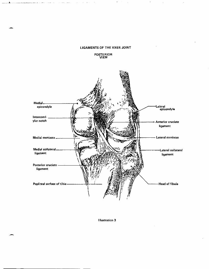
Four ligaments stabilize the knee. The medial collateral
ligament (MCL) on the inside of the knee attaches the femur to
the tibia. The lateral collateral ligament (LCL) on the outside
of the knee attaches the femur to the fibula. The two most
important ligaments are inside the joint itself. The ACL
prevents the tibia from displacing anteriorly on the femur. The
posterior cruciate ligament (PCL) prevents the tibia from
displacing posteriorly on the femur. These two ligaments are the
stabilizing forces of the knee (illustration #2 and #3).
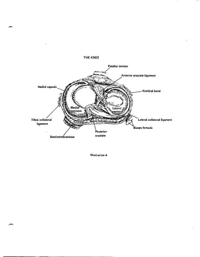
Two menisci, or cartilages, sit in between the femur and the
tibia. They keep the bones from rubbing against each other
(illustration #4). The menisci absorb shock, help with the
weight-bearing load of the joint, provide joint stability, aid in
controlling rotation, and aid in joint lubrication and nutrition
(Booher 399-400). The medial meniscus (MM) is "C" shaped and is
located on the medial, or inside portion of the knee. It is
attached to the MCL and to the plateau of the tibia. Its
movements are restricted much more than the lateral meniscus (LM)
because of its attachment to the MCL. The LM is "0" shaped and
2

slides back and forth. It is attached to the tibial plateau
also. However, it is not bound to the LeL. The movements of
this meniscus could be the reason why this meniscus is rarely
torn (Hoppenfeld 179-84).
The knee also has three bursa to protect it. Bursae usually
become injured and inflamed as a result of direct trauma or
constant friction between supporting structures. The most
commonly injured bursa is the prepatellar bursa, which lies
between the front of the patella and the skin. The superficial
infrapatellar bursa lies in front of the infrapatellar tendon.
The third bursa, the pes anserine bursa, is situated between
three tendons. These bursa lubricate and protect the knee from
serious injury (Booher 406).
Finally, the muscles of the knee playa critical role in its
functioning. They hold the knee together and without the
contraction-relaxation process of the muscles a normal person
would not be able to carry out even a simple daily activity such
as walking. The muscles of the leg are diagramed in illustration
#5 and #6 (Anderson 4:28-4:32).
The three movements of the knee are flexion, extension, and
internal and external rotation. A healthy normal athlete should
be able to bend his or her knee 135 degrees without assistance.
The knee should also extend 0 degrees actively with girls
sometimes hyperextending to 4-5 degrees. Internal and external
rotation should also be performed without help. These two
functions should go to 10 degrees each. If an athlete cannot
3

perform these movements on his or her own, they should be done
with assistance in order for the doctor to know the extent of
injury to the knee (Hoppenfeld 181-9).
Three main concepts of the ACL are important to its of
origin and insertion. This means that there is always a fiber
that is taut in any direction. The next concept is that the
fibers are not parallel and not the same length. Finally, the
fibers are not under the same tension at anyone point in space
(Douglas 17).
As discussed previously the main function of the ACL is to
guide anterior motion of the tibia on the femur. This ligament
constrains abnormal motion. The origin of the ACL is the
posterolateral femoral condyle and the insertion is on the
anteromedial portion of the tibia (Douglas 15). The ACL is
narrowest at its most proximal portion near the femoral origin.
It then fans out as it nears the tibial attachment. In full
extension, the ligament is under tension, but in full flexion it
is relaxed (Douglas 16).
The ACL, along with the PCL, is one of the stabilizing
forces of the knee. One can live without either, but as far as
being actively involved in sports, the ACL is the most important
of the two ligaments. If an athlete has a torn ACL and continues
to play, more damage can be done to the inside of the knee. For
example, the athlete can have a major shift of the femur on the
tibia and tear up the meniscus of the knee. In the early years
of surgical intervention if a person had an "unhappy" triad (ACL,
4
MCL, MM) injury the surgeon would remove the meniscus and leave
the torn ACL. Now it is thought that removing the menisci is
much more damaging than not reconstructing the ACL. As one can
read, the theories on the importance of the ACL to the knee have
changed immensely over the years (Arnold 306). Surgeries have
become more successful, and physicians have become more
proficient when repairing the torn ACL.
For a physician to determine the extent of injury to the
knee, a number of tests must be performed. In addition, the
history of injury, the mechanism, and visual observations should
be performed. Strength and range of motion (ROM) should also be
checked to give a clear picture of the damage. The four tests a
doctor performs to check for ACL deficiency are the Slocum Test,
the Pivot Shift Test, the Anterior Drawer Test, and the Lachman
Test.
The Slocum test allows complete quadriceps relaxation and
greater ease of performance on a large or muscular patient
(illustration #7). The patient lies with the uninjured side in a
lateral decubitus position and with the lower hip and knee flexed
to stay clear of the upper leg. The injured leg is on top. The
upper hip and pelvis are rotated posteriorly until weight is felt
by the heel of the injured leg. The knee is placed in 10 degrees
flexion. It will sag into a valgus stress and the tibia will
rotate internally and translate anteriorly. The examiner's hands
are positioned on the lateral side of the knee. The hand nearest
the foot is placed with the thumb behind the fibula and the index
5

finger is placed along the joint line. The other hand grasps the
distal femur with the thumb over the lateral femoral condyle.
While the examiner applies equal pressure with both hands, the
patient's knee is gently pushed into flexion. When the patient's
knee is flexed past twenty-five degrees, the anterior subluxed
tibia will reduce externally, if anterolateral rotary laxity is
present (Douglas 100).
The second test is the Pivot shift Test (illustration #8).
While the patient is relaxed the examiner's hand grasps the tibia
at the level of the tibial tuberosity and applies a valgus
stress. The other hand grasps and internally rotates the ankle
or foot. This subluxates the tibia anteriorly while the examiner
slowly flexes the knee. The test is positive if the examiner and
client note a sudden posterior "shift" of the tibia on the femur.
The only warning to the doctor is that he should not induce an
osteochondral fracture (Douglas 111).
The next test used by physicians is the Anterior Drawer Test
(illustration #9). The patient lies in a supine position with
his or her hip flexed at forty-five degrees and his or her knee
flexed at ninety degrees. The patient's tibia is in neutral
rotation with the foot flat on the table (Douglas 115). The
examiner's hands are placed with the fingers over the patient's
hamstrings and gastrocnemius heads. The thumbs are on the tibial
plateau and joint lines. The examiner gives a smooth steady pull
and if the ACL is torn, the tibia distracts or pulls forward on
the femur. This test is then repeated with the leg in the
6
internal and external positions (Hoppenfeld 185-6).
The last test is the Lachman Test (illustration #10). The
patient's knee is flexed at twenty degrees. The examiner
stabilizes the femur by grasping the distal thigh just proximal
to the patella. With the other hand, the examiner grasps the
tibia just distal to the tibial tubercle. The examiner then
applies firm pressure to the posterior aspect of the tibia in an
effort to produce an anterior translation. A positive result of
this test is one where the tibia moves anteriorly on the femur.
This test is the most sensitive of the four because the knee is
held is a comfortable position for the patient. Also, the
mechanical advantage of the hamstrings is ruled out and the
contact area on the lateral tibia plateau is slightly convex.
These three factors reduce the coefficient of static friction,
thus making the Lachman Test easier to perform with clinical
forces (Douglas 113).
After the initial examination by the physician, the patient
has two options--to have surgery or not to have surgery. The
physician must also decide if a patient with an ACL injury will
benefit from surgery or will respond favorably to a more
conservative treatment. The dilemma remains with regard to which
patient will respond to rehabilitation and activity modifications
or will progressively deteriorate with a nonoperative approach.
Obviously many factors affect which option is best for the
patient. What~ver type of treatment is chosen, the patient must
play an active role. This choice is not an easy one to make.
7

The first option is nonoperative. If the patient chooses
this nonevasive treatment, he or she must be educated on the ACL
and its functions. He or she must also go for therapy, wear a
brace, and modify his or her activity level. Previous reports
received indicate a large percentage of patients do very well
with this type of management if they are willing to change their
lifestyle (King 115-6). Since we are considering the active
individual, this is not a desirable option.
One of the options involved in this first choice is a knee
brace. Bracing is one of the many modes for treating an ACL
injury. Knee braces provide increased stability for the knee.
The most popular brace at Methodist Sports Medicine Center is The
Indiana Knee Orthosis (IKO) brace. This brace reduces rotation
and abduction/adduction (MSMC brace handout). Although most
patients with unstable knees feel more confident with the brace,
no major benefit of the brace is apparent. The main determinant
of function is muscle strength, therefore, it is necessary to
rehabilitate the whole leg instead of only the thigh. Most
physicians prescribe using a brace to help the patient return to
activity (Tegner 265). Once the patient achieves full muscle
strength, full range of motion, has little or no pivot shift and
lachman, Dr. Matchett, an orthopaedic surgeon from Muncie, does
not require continuing the use of the brace (Matchett interview
1990). However, Dr. Habansky, another orthopaedict surgeon from
Muncie, lets the patient decide whether to continue using the
brace. If the patient elects to use the brace, Dr. Habansky
8
usually discontinues it after one year (Dr. Habansky interview
1990).
Before bracing can be ruled out, many important factors must
be explained and considered. First, the post-operative
rehabilitation is hampered by increased pain and decreased ROM.
Also, the aggravation of the patella causing chondromalacia or
patellar tendinitis in the long-term follow-up is associated with
this surgery. Because of these surgical consequences, bracing
might be a viable first option.
A surgeon who considers ACt reconstruction on an athlete
must account for the patient's age, the sport of the patient, the
position held by the patient, and his or her own expectations and
motivations (Douglas 171). The risk of rein jury to the athlete
varies with the specific sport. In some of the more demanding
high risk sports such as football, basketball, and gymnastics,
rein jury is more possible than in a less demanding sport like
snow skiing, tennis, or softball (Douglas 171).
Three determinations need to be made when a physician is
deciding what type of surgery to perform. The questions the
physician should ask are: 1) Can the patient modify his or her
activity levels and rehabilitate the knee by nonoperative
treatment? 2) Will the patient benefit from a surgical procedure?
and 3) Which surgical procedure is appropriate when surgery is
indicated (Douglas 172).
Since most people, particularly athletes, are not willing to
give up an active lifestyle, surgery is usually the choice. The
9
surgery that is most commonly performed today is open
reconstruction. In conjunction with an ACL tear, a patient may
have a meniscal tear, articular cartilage damage, chondromalacia,
or other ligamentous tears such as a torn MCL or LCL. Depending
on the extent of damage, all of these can be repaired through
arthroscopy. Arthroscopy is a unique way of repairing meniscal
tears or chondromalacia of the knee through small incisions. To
perform this procedure, the surgeon uses special instruments
along with a microscopic camera. This surgery, however, does
nothing to repair the torn ACL.
The selection of what type of procedure used to surgically
repair the ACL, either intra-articular or extra-articular,
depends on numerous factors. These factors include if the injury
is acute or chronic, the patient's age and sex, the patient's
activity level, the degree of instability, the patient's future
athletic expectations, and the present level of disability.
Athletic individuals possess greater muscle mass and strength
levels so they are usually the best candidates for intra
articular repair (Douglas 138). Extra-articular repair is really
no longer an option for the active individual. Most physicians
prefer performing the intra-articular procedure because of the
good results (Douglas 225). However, extra-articular will be
discussed for reference only.
The most successful extra-articular surgical technique used
is the Arnold-Kocker sling about the fibular collateral ligament
to Gerdy's tubercle. "In this repair method, a strip of
10

iliotibial band is isolated. This fascial strip is passed
beneath the LCL and doubled back on itself. It is sutured firmly
at the LCL and back onto itself as it passes back toward Gerdy's
tubercle (Douglas 134). Although this is the most successful
extra-articular surgery, no single technique can be determined to
be the best because no comparative studies have been performed
(Douglas 134-5).
The goal of the extra-articular procedure is to prevent the
anterior subluxation of the lateral tibial plateau in relation to
the lateral femoral condyle. Placement of the extra-articular
portions of the iliotibial tract is crucial in realizing this
goal. The iliotibial tract acts as a reinforcement against
anterior subluxation of the lateral tibial plateau. In this
position it parallels the course of the ACL and may, therefore,
be considered an extra-articular ACL repair.
Extra-articular reconstruction procedures are an important
consideration for the non-athletic individual. Most physicians
will only consider this type of surgery on a child with open
growth plates. Intra-articular repair is not chosen because if
holes are drilled through the growth plate, it causes early
closure of the plates, and results in altered growth (Matchett
interview 1990). Dr. Shelbourne, a knee specialist at Methodist
Sports Medicine Center, says that the major benefit of this
surgical procedure is eliminating shifting, thus making the knee
more stable. Since the joint is under so much pressure from the
graft, the risk of this surgery is that the joint wears down
11

faster, therefore causing degenerative changes (Shelbourne talk
1990).
Dr. Matchett has a different opinion of extra-articular
repair. He says "They (extra-articular) do not provide the
stability that an intra-articular technique does, and so I cannot
count on it to be a stable operation for the patient. It may cut
down on the pivot shifting, but it may not, and it rarely
eliminates his or her lachman, so they are still having a lachman
and can still tear their meniscus" (Matchett interview 1990).
The degree of pivot shift is a major factor in helping the
orthopaedic surgeon decide upon the surgical technique. As it
stands, physicians seem to be moving in favor of the newer intra
articular procedure, after years of performing extra-articular
techniques.
The other option for a surgeon is the intra-articular ACL
repair. According to Dr. Matchett n ••• if a patient has a
significant lachman or pivot shift and has desires to be
physically active, I would recommend intra-articular because it
is a more predictable operation with the way we are doing it
now ... n (Matchett interview 1990). Intra-articular is
recommended in specific situations. These include a cruciate
ligament injury in a highly athletic individual, a physically
active individual with functional instability and an
unwillingness to alter his or her lifestyle. Other symptoms when
intra-articular is recommended include when the patient is having
frequent problems of instability with activities of daily living,
12

episodes of effusion, and when the patient is reporting
instability episodes after six months of intensive
rehabilitation. The patient's level of activity and willingness
to alter his or her lifestyle is a major influence in choosing
the appropriate method of management (Douglas 135).
Acute reconstruction should only be done after the patient
has an understanding of the time commitment for rehabilitation.
For the best results, surgical reconstruction should be within
seven to ten days after injury. If the patient waits any longer
there is a chance for complicating the initial injury with other
injuries (Douglas 135). Intra-articular surgery is done by
excising the medial one-third of the patellar tendon near the
proximal end. It is then rerouted from its distal insertion
through the joint cavity. It is anchored to the lateral condyle
of the femur and the tibial condyle with intercondyloid screws or
buttons (King 117). Rehabilitation needs to be started as soon
as possible to avoid the common problems associated with this
injury such as loss of extension and accumulation of scar
adhesions.
The physician's decision focuses on the type of graft:
either an autograft, an allograft, or a prosthetic ligament
should be used in surgery. There are many criteria for an ideal
ACL substitute. The substitute should duplicate the physiologic
function of the ACL and the graft should be non-immunogenic to
the host. The graft should also present no increased
susceptibility to infection and there should be an absence of
13

associated hyperplastic transformation. The substitution should
also restore immediate stability and allow for immediate motion.
The graft should be readily available in many sizes, have ease of
implantation, and the material should be amenable to long-term
storage (Douglas 211).
The use of the patient's own tissue, an autograft, is
preferred in ACL repairs since immunogenicity is practically non
existent. Graft incorporation is also predictable (Douglas 211).
Autografts of soft tissue with their attached bony insertions
offer biological material with structural advantage in replacing
the ACL. The portion of the patellar tendon with bony
attachments is the most popular graft (Clancy 184). The
advantage of this graft is that it can be transferred with some
blood supply still intact. Another advantage is that no diseases
can be transferred and infection possibilities are almost nil
(Douglas 193). Some other replacements used in this surgery are
the gracilus tendon, the fascia latae, or the semitendinosis.
The second type of graft is an allograft, a freeze-dried
patellar tendon or ACL taken from a cadaver. The best substitute
is another ACL because it is anatomically correct. Other
ligaments that can be used are the flexor hallicus longus, the
posterior tibialis, the toe extensors, the fascia latae, or the
achilles tendon (Wainer 200). Information on human allografts is
limited at the present time, but data does support observations
made in animals.
One factor that doctor's are investigating is the immune
14

response. The threat of AIDS and other bodily diseases is of
real concern to the medical profession. There has been one
reported case of transmission of HIV from a transplanted organ,
but no reported cases of transmission from nonperfused tissue
such as freeze-dried bone, tendon, or fascia (Wainer 294). In an
interview with Dr. Habansky, he revealed "When they can prove to
me there is no AIDS, that (allograft repair) would be great
(Habansky interview 1990)." This possibility of transmission has
not been completely explored and will continue to be an area of
research in the future. The only concerns about an allograft
itself are the revascularization of the ligaments and the fibers
remaining functional as they are replaced. Also, a question
arises that if the graft loses this orientation, is it weaker
than the original ACL?
The third graft that can be used for a substitution of the
ACL is a prosthetic ligament. A prosthetic ligament is a man
made graft that will duplicate the characteristics of the
original ligaments. Dr. Matchett states that research indicates
that prosthetic ligaments cause a foreign body reaction and they
cause a knee to carry chronic effusion or swelling (Matchett
interview 1990). The current interest in prosthetic ACL
reconstruction is partly due to the poor results obtained with a
late reconstruction of the ACL injury (Tremblay 88). Some of the
true prosthetic ligaments are Leeds-Keio, Proplast, and Gore
tex. Some augmentation devices are Dexon and the Kennedy LAD
(Douglas 247).
15

The Leeds-Keio graft is made of polyester fiber that is
woven into a small tube. Placement of this ligament is isometric
through the femoral and tibial drill holes. Each end is then
secured with bony plugs. The technical factors concerning
placement and site preparation are important for this artificial
graft survival. It has since been shown that polyester is
degraded by the body over time (Douglas 248).
Another true prosthetic ligament is Proplast. This graft is
a stent made of many polyaramid fibers imbedded in fluorinated
ethylene proplyene copolymer. The stent is coated with proplast,
a vitreous carbon fiber and polytetraflouroethylene composite, to
facilitate tissue ingrowth. Like Leeds-Keio, many problems are
found when working with this graft. These problems, combined
with the inherent stiffness of the prosthesis led to failure of
the majority of ACL grafts (Douglas 250).
The Gore-tex graft is made from a single fiber of expanded
polytetrafluoroethylene.
at each end of the graft.
The fiber is braided to form an eyelet
The braid allows even load
distribution through the prosthesis. This graft is designed to
restrain subluxation but not to duplicate the normal ACL. Its
strength and stiffness make the technical aspects very important.
The graft material fixation is bony so immediate mobilization is
possible. Early reports show its use in humans as promising
(Douglas 247).
Gore-tex has some key advantages over other prosthetic
ligaments. It is two to three times the strength of a normal
16

ligament and it does not have to depend on the blood supply of a
normal ligament. Also, it allows for reduced rehabilitation
time. Finally, Gore-tex material has a good safety record.
Since there is no living tissue in a Gore-tex graft, the
synthetic ligament can simply be removed if the graft fails and
another autogenous repair attempted (Lubell 154).
Dexon and the 3M/Kennedy Ligament Augmentation Device (LAD)
are two augmentation devices possible for knee reconstruction.
Dexon is biodegradable and is made of braided polyglycolic acid.
It was found through studies that pure dexon grafts fray too
quickly for use in humans (Douglas 251).
The LAD is used in a modified MacIntosh/Marshall procedure.
It is sutured inside the tubes autogenous tissue graft, starting
at the origin of the patellar tendon and ending at the terminal
portion of the rectus femoris. This augmentation device is
designed to protect the graft from excessive stress during early
healing. The initial reports from this graft are good but when
closely analyzed they are no better than when a graft of high
initial strength is used (Douglas 251-2).
Each physician has a different philosophy of ACL
reconstruction and rehabilitation. The opinions are many and
varied as to what should be stressed during each phase. However,
most physicians are only now understanding the benefits of an
accelerated rehabilitation program. The main overall goal of
every physician should be to get the patient to return to
activity with a stable knee. Two doctors, Dr. Matchett of Muncie
17
and Dr. Shelbourne of Indianapolis have basically the same
philosophy of surgeries but each stress different areas in
rehabilitation.
Dr. Shelbourne is always thinking of ways to improve
rehabilitation. His goals are to achieve full extension without
complications. The person who does gain extension without having
a complication will return faster than the one with additional
trauma. Post-surgical complications can be defined as things
that go wrong with a joint such as patella-femoral pain, patellar
tendinitis, a flexion contracture, general aching, or infection
of the graft. After the soft tissue heals, Dr. Shelbourne also
wants the patient to be able to achieve ninety degrees of flexion
and quad control. The argument now is that with the closed
kinetic (weight-bearing) accelerated rehabilitation, the graft
will stretch. Dr. Shelbourne believes if the graft is placed
correctly, it does not stretch. He states that we cannot prove
the graft does not stretch because of the KTI000 scores. These
scores are too sporadic. Once we can get an objective test, this
theory will be proven (Shelbourne talk 1990).
Dr. Matchett believes his goals should be the same as the
patient's goals. His main goals are to restore stability, to
maintain full range of motion, and to not cause additional damage
or trauma to the knee. However, since the surgeon takes the
middle third of the patellar tendon, they are causing additional
trauma to the knee. This trauma is controlled with modalities and
upon re-examination of the patellar tendon it has hypertrophied
18

to its normal size. This usually takes around one year (Matchett
interview 1990).
Another concern of the physician is full extension. Without
it, the patient looks like he or she is walking with a limp.
Full extension depends on proper placement of the graft. Both
the tibial and femoral tunnel have to be correct and have
adequate notchplasty. Although the knee feels better bent, if
the patient does not get extension, he or she will never have it.
Also, if the patient does not maintain extension, scar tissue and
adhesions build-up and they lose extension. The surgeon then has
to go back into the knee to clean up the scar tissue and
adhesions so the patient can return to full extension. Flexion
is not as iITportant because it will always return as long as one
works on it in rehabilitation (Matchett interview 1990).
Rehabilitation is restoration of the patient to the level of
his or her pre-injury fitness. This can be accomplished through
exercise and active physical therapy. Between the two surgeries,
rehabilitation is mainly the same; however, with an extra
articular repair the patient can do more open-kinetics (non
weight bearing). This is because the therapist is not worried
about causing patellar tendinitis since the surgeon did not have
to remove one-third of the middle of the patellar tendon. Since
most athletes have intra-articular repair, the rehabilitation
will be adapted to that type surgery.
Rehabilitation is a time consuming project that takes from
six to nine months for the ACL patient. Rehabilitation following
19

intra-articular ACL reconstruction consists of numerous phases.
Depending on the individual progress of each patient, it is
possible to overlap the following phases.
Rehabilitation begins before surgery even takes place.
Since most surgeons wait at least one week to ten days before
repairing the torn ligament, the patient begins the process of
getting the knee stronger for surgery. The goals of this phase
are to decrease swelling, increase ROM and quad strength. This
way they will be one step ahead when they come into physical
therapy after surgery. The stronger athletes are when entering
surgery, the stronger they are when they come out.
The first phase after surgery lasts from post-op day one to
day six. The patient begins continuous passive motion (CPM) the
day of surgery. The heel is elevated on towels while in the CPM
to ensure terminal extension. The flexion angle is increased as
tolerated. The patient will perform range of motion exercises
two to three times daily. The knee is allowed to extend to
terminal extension for at least thirty minutes during each
exercise routine. The knee is also flexed ninety degrees by
sitting on the side of the bed. The patient is permitted to
weight bear as tolerated with or without crutches using a dobi,
which is a straight leg cast, or an immobilizer. The patient
will then be discharged from the hospital three to four days
after surgery (MSMC 2).
Phase II begins on the seventh day and continues through the
tenth day. The patient will return for a follow-up visit to the
20
physician and start physical therapy during this time. The goal
of this phase is for the patient to achieve terminal extension to
ninety degrees flexion. Early terminal extension is the key to a
successful result due to what was discussed earlier in this
paper. To push extension, the patient will perform towel
extension and prone hangs. Wall slides, heel slides, and active
assistive flexion over the edge of the table will also be started
during this phase. These exercises will help restore flexion
(MSMC 2).
All of the strengthening exercises performed will be closed
kinetic (weight-bearing) exercises. It is believed these
exercises help to preserve the patella-femoral joint and do not
add stress to the ligament, in effect preventing patellar
tendinitis. These exercises also help to facilitate early return
of quadriceps strength with minimal stress to the graft. The
patient will start with bilateral knee bends and calf raises and
progress to unilateral exercises. The athlete is also encouraged
to progress from partial to full weight bearing without crutches.
He or she will also decrease the use of the dobi and emphasize
normal gait. Early quad strength and early terminal extension
set the pace for the entire rehabilitation program and a
successful outcome (MSMC 3).
Range of motion continues to be stressed during Phase III,
comprised of weeks two through four. The revised goal of this
stage is to achieve terminal extension to 110 degrees flexion.
The patient can begin 2-4" lateral step-ups and unilateral calf
21
.-raises. Weight room activities such as leg press with knee
flexion angle no greater than 90-100 degrees, 1/4 squats, and
stairmaster can begin three to four weeks following surgery.
Biking and swimming workouts can also help with flexion and
strength (MSMC 3).
The patient should have attained terminal extension to 120-
130 degrees flexion by Phase IV which lasts from week 6-8.
Weight room activities are continued while adding the hip sled.
A cybex test is performed five to six weeks after surgery. There
is a twenty degree terminal extension block and anti-shear device
at speeds of 180 and 240 degrees per second. This test compares
the quad strength in the injured leg to the uninjured leg. If
the patient's quad strength is seventy percent or greater, he or
she will be permitted to begin light functional progression.
This includes lateral shuffles, carioca, and jumping rope. These
activities are performed while wearing the brace. A KTIOOO test
which checks ligamentous stability is also performed at this time
(MSHC 3-4).
Phase V starts at twelve weeks, and the patient has a check
up every four to six weeks. A cybex test and KTI000 evaluations
are performed at this time. At twelve weeks, the twenty degree
block is removed from the cybex test. As the patient's strength
continues to improve, the agility workouts become more vigorous.
The patient is allowed to begin figure 8's, backward running,
and progress from half to full speed activities (MSMC 4-5).
From sixteen to twenty-four weeks, Phase VI begins. The
22

patient is followed every month. Cybex testing is monitored with
slow speed testing initiated. As the patient improves he or she
may resume non-contact sports around four to six months and
contact sports at six to eight months after surgery. The
criteria used in making this determination includes eighty
percent stre~gth on each isokinetic speed on the cybex machine
and the successful completion of the functional progression. The
patient is e~couraged to wear the activity brace up to one year
after surgery.
Many factors contribute to successful, and complete,
rehabilitation following ACL knee reconstruction. Each patient
is different, as are their needs and levels of activity. For
these reason:s, each rehabilitation program must be individualized
to fit the patient's needs. The four main variables involved in
rehabilitation include: the patient's desire to return to
competition, the self-motivation to complete the rehabilitation
process, the patient's pain threshold, and attaining the goals
set by the patient. Without the athlete having these four
qualities, he or she could never return to pre-injury status and
would have a tough time getting through rehabilitation and back
to his or her sport.
The future of ACL surgery is important to consider.
Clinicians rE~alize two principles: 1) that the ACL is an integral
part of normal knee function; and 2) a knee without this ligament
can be improved with a successful replacement (Douglas 291). Dr.
Habansky and Dr. Matchett both agree that the future lies in
23

allografts. Although much more research needs to be done to
achieve these goals, Dr. Matchett maintains that there will be
refinements in all grafts and surgery through smaller incisions
(Matchett and Habansky interviews 1990). However, the basis of
any treatment, disregarding the surgery, will still be accurate
diagnosis of all the manifestations of the ACL deficient knee
including assessment of associated injuries (Douglas 293).
24

Conclusion
As one Gan see the ACL is of major importance in the
functioning t~f a healthy normal knee. To be able to function
without limitations, the active individual needs to consider
surgery to correct the unstable knee. Research is still being
compiled, but as of now, the best graft replacement is the middle
one-third of the patellar tendon with bony plugs. Along with
many other considerations bracing, physician's philosophy, and
rehabilitation are very important factors when looking into
surgical intervention. But the primary factors deal with the
athlete: pai~ threshold, desire, mental stability, self
motivation, and reaching attainable goals set by the physician,
physical therapist and the athlete. If the athlete has a strong
desire and motivation to return to his or her sport, he or she
will complete the rehabilitation process and return ready to
play.
In addressing the primary issue this thesis is based on, the
main factors that influence the surgical procedure and techniques
of this surgery are the athletes active involvement in
rehabilitation, the surgical procedure used by the doctor, the
physician philosophy, and the athletes' mental motivation. All
of these factors playa major part in the repair of an athlete's
knee.
25

-BONES OF THE LEG
IRJ;I<-------Tibia
Fibula-a --------1
Illustration I
-
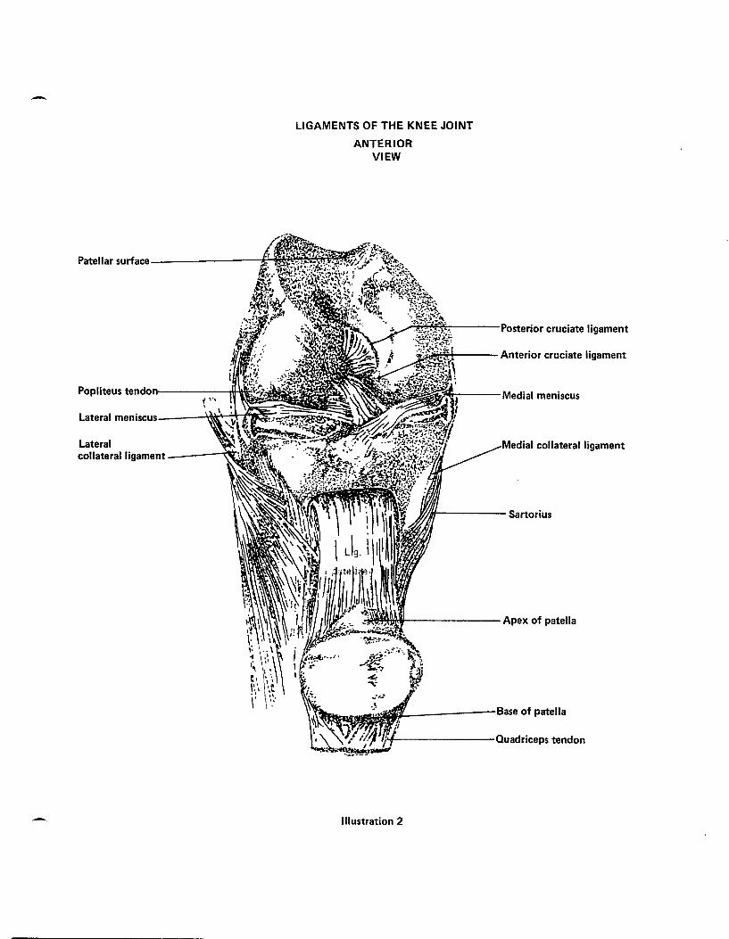
-LIGAMENTS OF THE KNEE JOINT
ANTERIOR VIEW
Patellar surface----------t;c,ml~~~~~
..;.:.~~~---Posterior cruciate ligament
:-Z;.i~+---- Anterior cruciate ligament
Popliteus tendon----·~-:::-l~~~~~a t;1Ifi----Medial meniscus
Lateral meniscus----~~~rt~~~;;:;m.~~
Lateral collateral ligament --·--~~.'JO...l~"",w"
ial collateral ligament
Wlill------ Sartorius
~WI>lWiI--------Apex of patella
fIL_------ Base of patella
'/J .. J--------Quadriceps tendon
Illustration 2
-
Medial--------......,.-.\ epicondyle
LIGAMENTS OF THE KNEE JOINT
POSTERIOR VIEW
Intercond- ------H11!k;-----:-~ ylar notch M~----il~~rr---- Anterior cruciate
Medial meniscus -----ir-.~ ...... ~~~~.
Medial collateral ____ ~~~MtJ~~'!J·;.-·
ligament
Posterior cruciate ------':~~~ ligament
Popliteal surface of tibia----~M__-"---
ligament
--'~~~~Ut_----lateral meniscus
ral collateral ligament
"'---Head of fibula
Illustration 3
--
THE KNEE
Anterior cruciate ligament
Medial capsule
/ Tibial collateral Lateral collateral ligament
ligament
Semimembranosus cruciate
Illustration 4
--
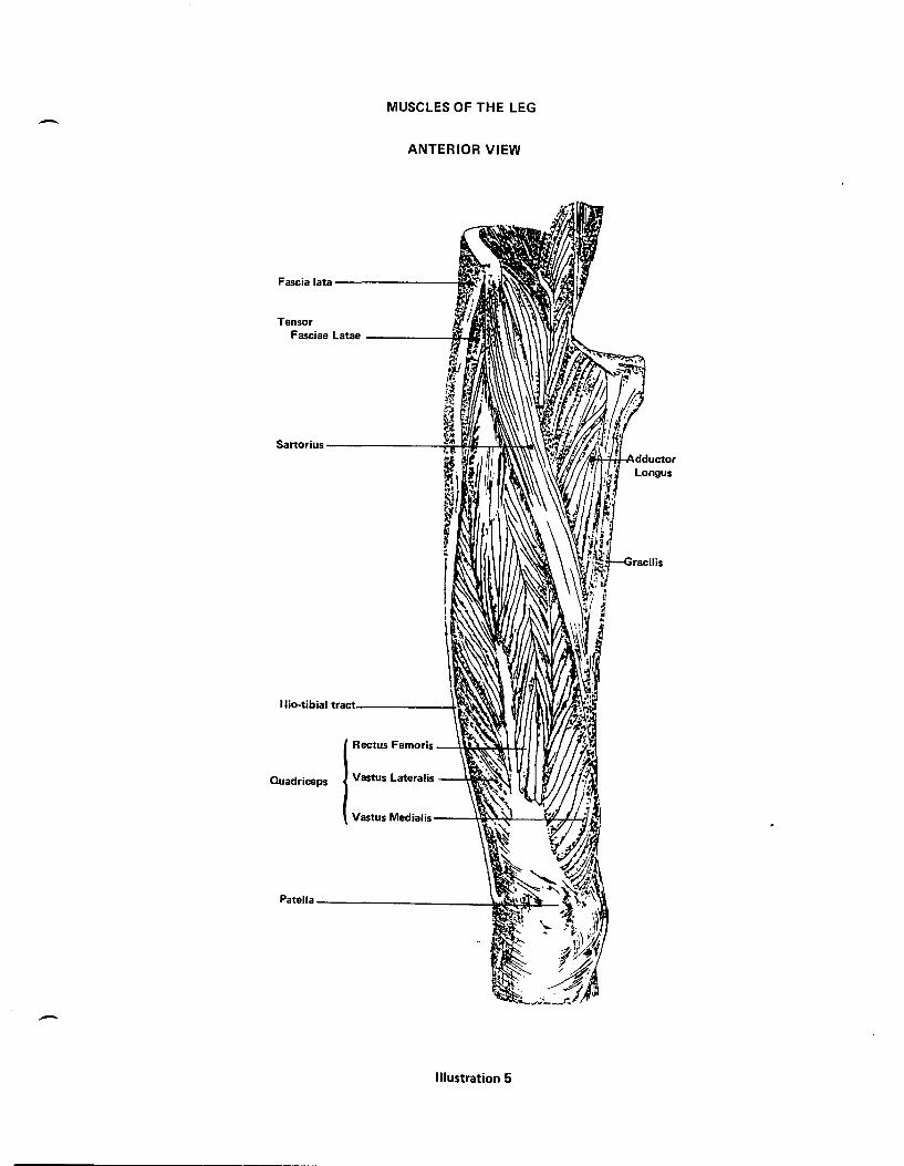
MUSCLES OF THE LEG -ANTERIOR VIEW
Fascia lata---------1I~
Tensor Fasciae Latae ------ofIif-....
Sartorius ---------n~if!_IIT.J¥fF!~t\I\\
lIio-tibial tr.lct. ______ ~I..'"&.~
Rectus Femoris --HM~'I:t-t+
Quadriceps Vastus Lateralis -~tW~;~
Vastus MedialiS---\:W~~----l~..n~
Patella ____________ ~
-Illustration 5

MUSCLES OF THE LEG
- PaITERIOR VIEW
\~I--- Gluteus Medius
~Ifi---Gluteus Maximus
Adductor Magnus ---+IIN;,f;j
llio-tibial tract
Sem iterldirloSI~S --CiI*w-\U~
Hamstrings
Biceps Femoris -~~!-tlMf~~~I.!
Gracilis -----liMo!
,'\r,/,I.1lI.11ir\\t---- Plantaris
-Illustration 6

A
B
-
PIVOT - SHIFT TEST PERFORMED ON RIGHT KNEE
Illustration 8

-
SLOCUM TEST
B
Illustration 7
-

ANTERIOR DRAWER TEST
ANTERIOR DRAWER TEST - FOOT POSITIONS
Illustration 9
-

-
LACHMAN'S TEST
\/ /
Illustration 10
-

Bibliography
Anderson M.D., James. Grant's Atlas of Anatomy, 8th Edition. Williams and Wilkins, London 1983, figures 4:28-4:32, 4:56-4:61, Illustrations 2-6.
Anterior Cruciate Ligament Reconstruction Rehabilitation program, Methodist Sports Medicine Center, Physical Therapy. Feb. 1989, p. 1-5.
Arnold M.D., James. "Natural History of Anterior Cruciate." The American Journal of Sports Medicine. vol 7, 1979, p. 305-13.
Booher, James. Athletic Iniury Assessment. Times Mirror/Mosby College Publishing, st. Louis, 1983, p. 399-400, Illustrations 7-10.
Clancy Jr. M.D., William G. "Intra-Articular Reconstruction of the AntE!rior Cruciate Ligament." Orthopaedic Cl inics of North America. vol 16, April 1985, p. 181-9.
Dougl as, W. ~rackson. The Anterior Cruciate Deficient Knee: New Concepts in Ligament Repair. The C.V. Mosby Company, st. Louis, 1987.
Dr. Alan Habansky interview, January 12, 1990.
Dr. L. Jay Matchett interview, January 13, 1990.
Dr. K. Don Shelbourne group talk, February 13, 1990.
Hoppenfeld M.D., Stanley. Physical Examination of the Spine and Extremities. Appleton-Century-Crofts, Connecticut. p. 171-196, 1976.
King M.S., A.T., C, Steve. "The Anterior Cruciate Ligament: A review of recent concepts." The Journal of Orthopaedic and Sports Plysical Therapy. vol 8, Sept. 1986, p. 110-12.
Lubell, Adel E!. "Arti ficial Ligaments: Promise or Panacea." The Physician and Sports Medicine. vol 15, March 1987, p. 152-15.
Tegner M.D., Ph.D., Yelverton. "Derotation Brace and Knee Functions in Patients With Anterior Cruciate Ligament Tears." Arthroscopy: The Journal of Arthroscopic and Related Iniuries. vol I, 1985, p. 264-7.
Tremblay M.D., Gilles. "The Challenge of Prosthetic Cruciate Ligament Replacement." Clinical Orthopaedics and Related Research, number 147, March-April 1980, p. 88-92.
Wainer M.D., Robert. "Anthroscopic Reconstruction of the Anterior Cruciate Ligament Using Allograft Tendon." Anthroscopy: The Journal of Arthroscopic and Related Surgery. vol 4, 1988, p. 199-205.