ckd-mbd & osteoporosis the management dilemma
TRANSCRIPT

CKD- MBD / Osteoporosis in the elderley
the management dilemma
Dr Ayman Seddik , Msc , MD
Assistant Professor of Nephrology Ain Shams University
Consultant nephrologist

OUTLINE
• CKD MBD & OSTEOPOROSIS aetiology prevelance and impact on mortality and morbidity in elderley population
• Management based on stage of chronic kidney disease


Table 1 Differences between CKD–MBD and postmenopausal osteoporosis
Ott, S. M. (2013) Therapy for patients with CKD and low bone mineral density Nat. Rev. Nephrol. doi:10.1038/nrneph.2013.182


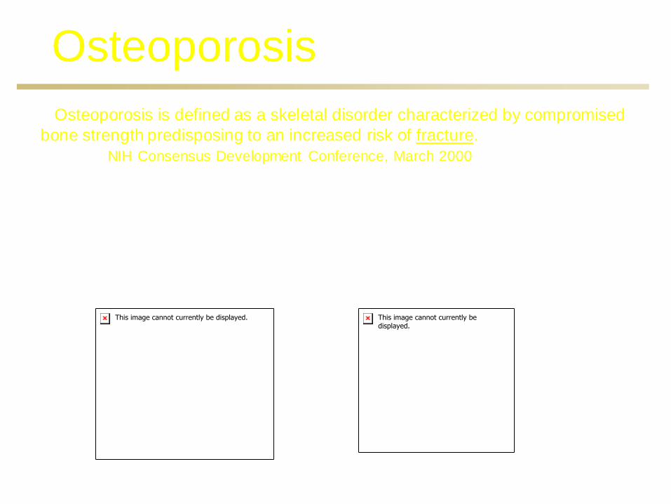
Osteoporosis
Osteoporosis is defined as a skeletal disorder characterized by compromised
bone strength predisposing to an increased risk of fracture.
NIH Consensus Development Conference, March 2000
Normal Bone Osteoporotic Bone


Vertebral Fracture Cascade

THE HUMAN COST
Downward Spiral

Denosumab: Overview
• Fully human monoclonal antibody-IgG2 isotype
• High affinity and specificity for human RANK Ligand
• Pharmacokinetics (SC): similar to other fully human IgG2 monoclonal antibodies
– Absorption is rapid and prolonged (Cmax ≈1-4 wks postdose)
– Long half-life ≈34 days with max dose
– Distribution ≈ intravascular volume
– Clearance ≈ reticuloendothelial system
– No kidney filtration or excretion of intact molecule
Bekker PJ et al. J Bone Miner Res. 2004;19:1059-1066. Boyle WJ et al. Nature. 2003;423:337-342.


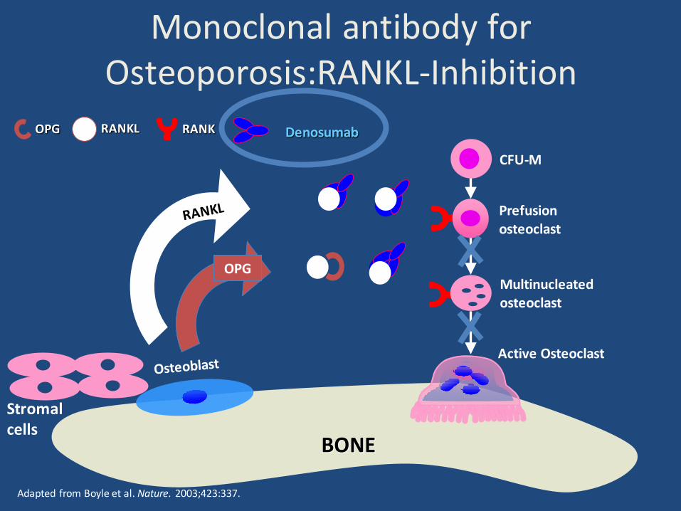
Mechanism of Action for Denosumab
Growth Factors Hormones Cytokines
Bone
CFU-M = colony forming unit macrophage
Osteoblast Lineage
Osteoclast
CFU-M
Pre-Fusion Osteoclast
Multinucleated Osteoclast
RANK
RANKL
OPG
denosumab

KDIGO Clinical Practice Guideline
Diagnosis, Evaluation, Prevention, and Treatment of Chronic Kidney Disease - Mineral and Bone Disorder (CKD-MBD)

Guideline Outline
Chapter 4.1: Treatment of CKD-MBD Targeted at Lowering High Serum Phosphorus and Maintaining Serum Calcium
Chapter 4.2: Treatment of Abnormal PTH Levels in CKD-MBD
Chapter 4.3: Treatment of Bone with Bisphosphonates, other Osteoporosis Medications, and Growth Hormone
Chapter 5: Evaluation and Treatment of Kidney Transplant Bone Disease
Chapter 6: Summary and Research

Definition of
CKD-Mineral and Bone Disorder
A systemic disorder of mineral and bone metabolism due to CKD manifested by either one or a combination of the following:
Abnormalities of calcium, phosphorus, PTH, or vitamin D metabolism
Abnormalities in bone turnover, mineralization, volume, linear growth, or strength Vascular or other soft tissue calcification
Moe S, et al. Kidney Int 69: 1945, 2006

Classification of Renal Osteodystrophy
Turnover High Normal Low
Mineralization Normal Abnormal
Volume High Normal Low
Slide courtesy of Susan Ott Kindly provided by Dr. Susan M. Ott

Moe, SM et al. ACKD: 3-12, 2007
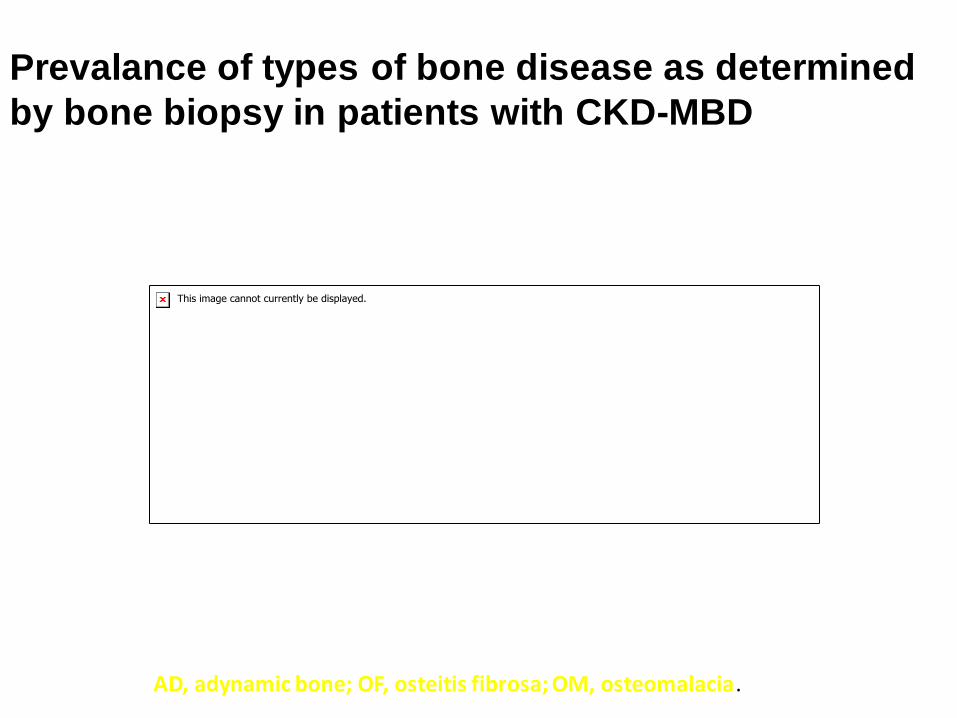
Prevalance of types of bone disease as determined
by bone biopsy in patients with CKD-MBD
AD, adynamic bone; OF, osteitis fibrosa; OM, osteomalacia.

Treatment of CKD-MBD:
Phosphorus and Calcium 4.1.1. In patients with CKD stages 3–5, we suggest
maintaining serum phosphorus in the normal
range (2C). In patients with CKD stage 5D, we
suggest lowering elevated phosphorus levels
toward the normal range (2C).
4.1.2. In patients with CKD stages 3–5D, we
suggest maintaining serum calcium in the normal
range (2D).
Treatment of CKD-MBD:
Phosphorus and Calcium 4.1.3. In patients with CKD stage 5D, we suggest
using a dialysate calcium concentration between
1.25 and 1.50 mmol/l (2.5 and 3.0 mEq/l) (2D).
4.1.4. In patients with CKD stages 3–5 (2D) and 5D
(2B), we suggest using phosphate-binding agents
in the treatment of hyperphosphatemia. It is
reasonable that the choice of phosphate binder
takes into account CKD stage, presence of other
components of CKD–MBD, concomitant therapies,
and side-effect profile (not graded).

Treatment of CKD-MBD:
Phosphorus and Calcium 4.1.5. In patients with CKD stages 3–5D and hyperphosphatemia, we recommend restricting the dose of calcium-based phosphate binders and/or the dose of calcitriol or vitamin D analog in the presence of persistent or recurrent hypercalcemia (1B).
In patients with CKD stages 3–5D and hyperphosphatemia, we suggest restricting the dose of calcium based phosphate binders in the presence of arterial calcification (2C) and/or adynamic bone disease (2C) and/or if serum PTH levels are persistently low (2C).

Osteoporosis
Osteoporosis is defined as a skeletal disorder characterized by compromised
bone strength predisposing to an increased risk of fracture.
NIH Consensus Development Conference, March 2000
Normal Bone Osteoporotic Bone


THE HUMAN COST
Downward Spiral

Osteoporosis Prevention and
Treatment
Age
Hormonal Replacement
Bisphosphonates Strontium
SERM
20 40 60 80
Vitamin D
PTH
Life Style
Treatment choice

Vertebral Fracture Cascade
Osteoporosis in Men Has Its Time Come?

Hip Fracture:
Devastating Event Mortality rate same as breast cancer 20% excess mortality in the first year 50% incapacitation 20% of females need assisted living or nursing home 80% of 75 yo preferred death to hip fx & nsg hm Cooper C, et al. Am J Epidemiol. 1993;137:1001

The Osteoporotic Event: Hip Fracture
#1: Questions about Osteoporosis
When should Bone Density Measurement
be performed?
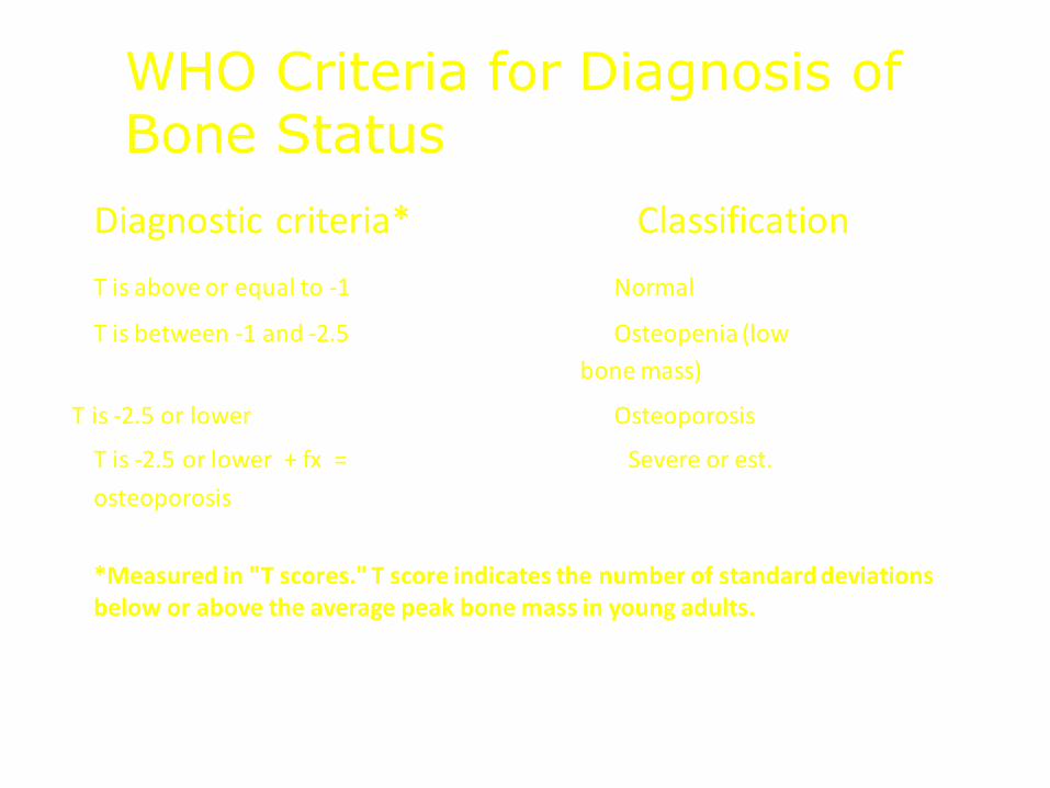
Diagnostic criteria* Classification
T is above or equal to -1 Normal
T is between -1 and -2.5 Osteopenia (low
bone mass)
T is -2.5 or lower Osteoporosis
T is -2.5 or lower + fx = Severe or est.
osteoporosis
*Measured in "T scores." T score indicates the number of standard deviations below or above the average peak bone mass in young adults.
WHO Criteria for Diagnosis of Bone Status

World Health Organization
Diagnostic Criteria
DIAGNOSIS BMD CRITERIA*
Normal within 1 SD of a “young normal” adult (T-score at -1.0 and above) Osteopenia between 1 and 2.5 SD below that of a “young normal” adult (T-score between -1 and -2.5) Osteoporosis 2.5 SD or more below that of a “young normal” adult (T-score at or below -2.5) Severe Osteoporosis 2.5 SD or more below that of a “young normal” adult and fracture(s)
T-score is the number of SDs above or below the average BMD value for young, normal adults of the same sex BMD = Bone mineral density SD = Standard deviation *Measured at the hip, spine, or wrist

60
70
80
90
100
30 40 50 60 70 80 90
Age
Rel
ativ
e B
MD
(%)
Forearm
Spine
Hip and Heel
0
1000
2000
3000
4000
35- 39
85+
Colles'
Vertebrae
Hip
Age
An
nu
al F
ract
ure
In
cid
ence
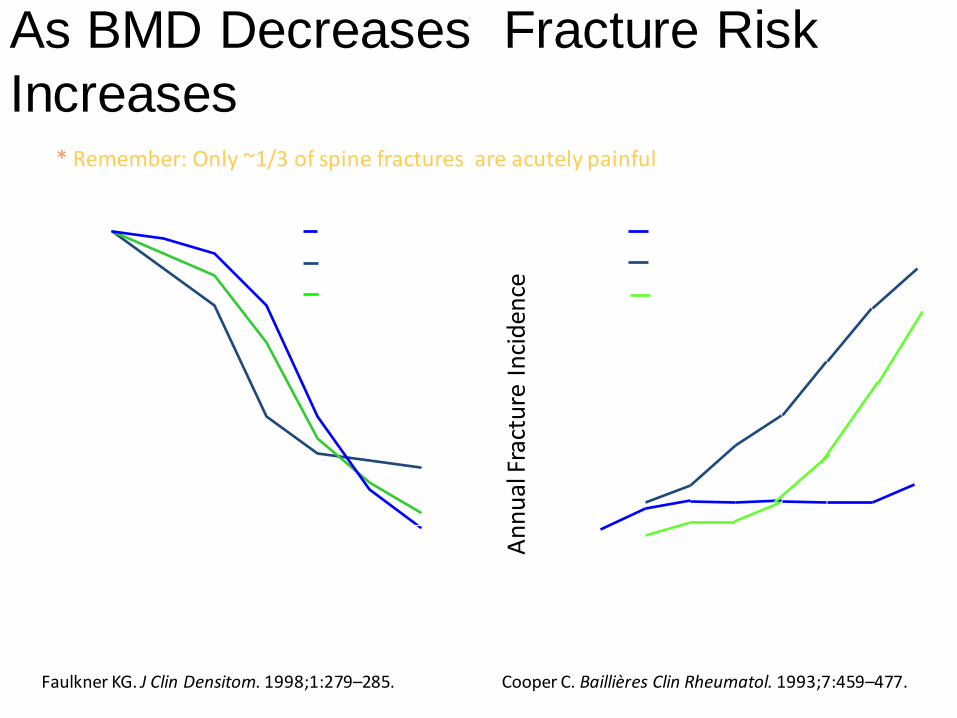
Cooper C. Baillières Clin Rheumatol. 1993;7:459–477. Faulkner KG. J Clin Densitom. 1998;1:279–285.
As BMD Decreases Fracture Risk
Increases * Remember: Only ~1/3 of spine fractures are acutely painful

Bone Mass Measurement Act
Federal Register 1997 for HCFA/CMS
Medicare Osteoporosis Measurement Act 2003
1. Women with estrogen deficiency
2. Spine x-ray evidence of fracture or OP
3. Glucocorticoid therapy (3mos, 5 mg/d)
4. Primary Hyper-PTH
5. Follow-up treatment (23 months unless medical reason for sooner e.g. steroids)
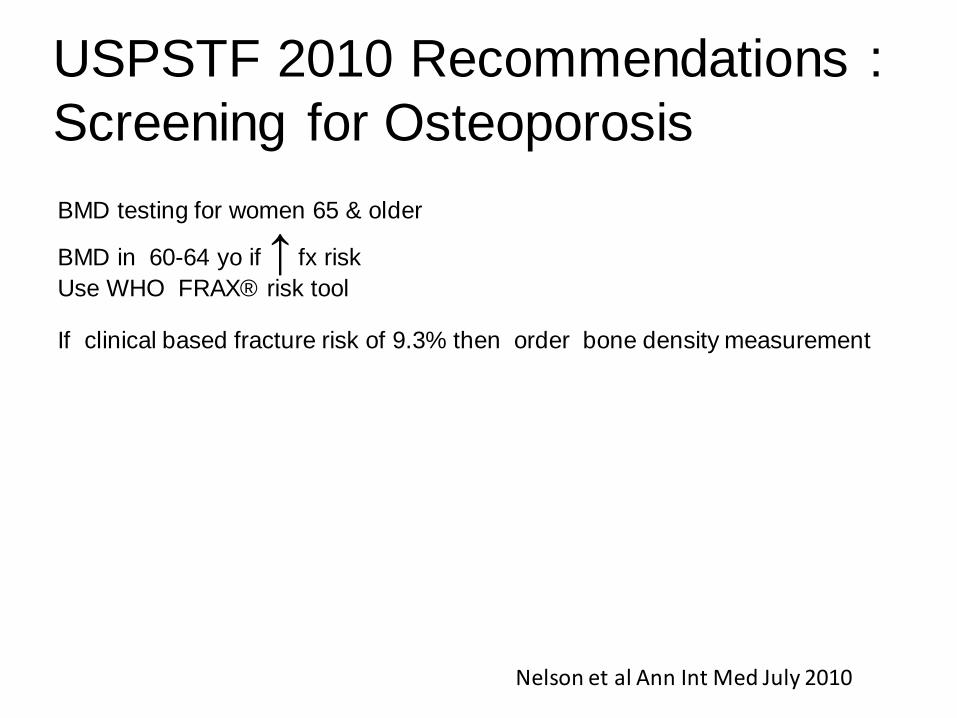
USPSTF 2010 Recommendations :
Screening for Osteoporosis BMD testing for women 65 & older
BMD in 60-64 yo if ↑ fx risk
Use WHO FRAX® risk tool If clinical based fracture risk of 9.3% then order bone density measurement
Nelson et al Ann Int Med July 2010
WHO Fracture Risk Prediction


10-year Risk Assessment for Women
(CAROC Basal Risk)
Papaioannou A, et al. CMAJ 2010 Oct 12. [Epub ahead of print].
10-year Risk Assessment for Women
(CAROC Basal Risk)
Age Low Risk Moderate Risk High Risk
50 above -2.5 -2.5 to -3.8 below -3.8
55 above -2.5 -2.5 to -3.8 below -3.8
60 above -2.3 -2.3 to -3.7 below -3.7
65 above -1.9 -1.9 to -3.5 below -3.5
70 above -1.7 -1.7 to -3.2 below -3.2
75 above -1.2 -1.2 to -2.9 below -2.9
80 above -0.5 -0.5 to -2.6 below -2.6
85 above +0.1 +0.1 to -2.2 below -2.2
Papaioannou A, et al. CMAJ 2010 Oct 12. [Epub ahead of print].

Risk Assessment with CAROC:
Important Additional Risk Factors
Factors that increase CAROC
basal risk by one category
(i.e., from low to moderate or
moderate to high) Fragility fracture after age 40*1,2
Recent prolonged systemic
glucocorticoid use**2
1. Siminoski K, et al. Can Assoc Radiol J 2005; 56(3):178-188. 2. Kanis JA, et al. J Bone Miner Res 2004; 19(6):893-899. Return to case
* Hip fracture, vertebral fracture, or multiple fracture events should be considered high risk ** >3 months use in the prior year at a prednisone-equivalent dose ≥ 7.5 mg daily
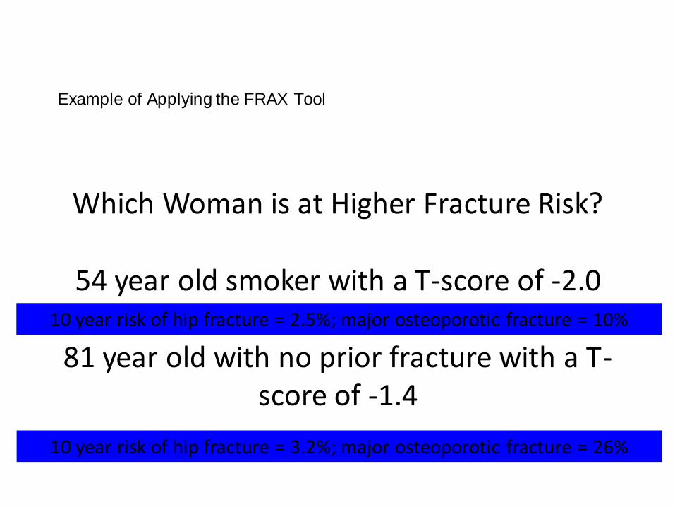
Example of Applying the FRAX Tool
Which Woman is at Higher Fracture Risk?
54 year old smoker with a T-score of -2.0 or
81 year old with no prior fracture with a T-score of -1.4
10 year risk of hip fracture = 2.5%; major osteoporotic fracture = 10%
10 year risk of hip fracture = 3.2%; major osteoporotic fracture = 26%
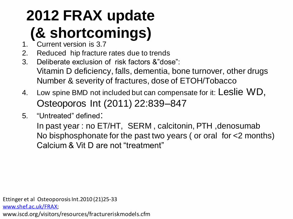
2012 FRAX update
(& shortcomings) 1. Current version is 3.7
2. Reduced hip fracture rates due to trends
3. Deliberate exclusion of risk factors &”dose”:
Vitamin D deficiency, falls, dementia, bone turnover, other drugs
Number & severity of fractures, dose of ETOH/Tobacco
4. Low spine BMD not included but can compensate for it: Leslie WD,
Osteoporos Int (2011) 22:839–847
5. “Untreated” defined: In past year : no ET/HT, SERM , calcitonin, PTH ,denosumab
No bisphosphonate for the past two years ( or oral for <2 months)
Calcium & Vit D are not “treatment”
Ettinger et al Osteoporosis Int.2010 (21)25-33 www.shef.ac.uk/FRAX; www.iscd.org/visitors/resources/fractureriskmodels.cfm

#2: Questions about Osteoporosis
Are calcium & Vitamin D supplements needed?
1 – YES
2 -- NO
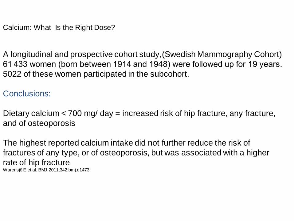
Calcium: What Is the Right Dose?
A longitudinal and prospective cohort study,(Swedish Mammography Cohort)
61 433 women (born between 1914 and 1948) were followed up for 19 years.
5022 of these women participated in the subcohort.
Conclusions:
Dietary calcium < 700 mg/ day = increased risk of hip fracture, any fracture,
and of osteoporosis
The highest reported calcium intake did not further reduce the risk of
fractures of any type, or of osteoporosis, but was associated with a higher
rate of hip fracture Warensjö E et al. BMJ 2011;342:bmj.d1473

EPIC-Heidelberg cohort 25,540 local residents aged 35-64 years Excluded diagnosis of MI, stroke, or transient ischemic attack at baseline (n = 1322) Self-administered food questionnaire Interview to assess ever use of vitamins and calcium supplements Incident cardiovascular events during follow-up were reported by participants or their next of kin in follow-up surveys. Reported cardiovascular events were verified by tracking medical records or official death certificates.
Associations of dietary calcium intake and calcium supplementation with myocardial infarction and stroke risk and overall cardiovascular mortality in the Heidelberg cohort of the European Prospective Investigation into Cancer and Nutrition study
Heart 2012;98:920-925.

Calcium Intake and CV disease
In conclusion, this study suggests that increasing dietary calcium intake from diet might not confer significant cardiovascular benefits, while calcium supplements, which might raise MI risk, should be taken with caution.

www.nof.org
Recommended Calcium Intake
From diet and supplements combined: 1200 mg daily Several different types of calcium
supplements are available
Evidence shows a benefit of calcium on reduction of fracture
risk1
Concerns about serious adverse effects with high-dose supplementation2-4
1. Tang BM, et al. Lancet 2007; 370(9588):657-666. 2. Bolland MJ, et al. J Clin Endocrinol Metab 2010; 95(3):1174-1181.
3. Bolland MJ, et al. BMJ 2008; 336(7638):262-266. 4 Reid IR, et al. Osteoporos Int 2008; 19(8):1119-1123.
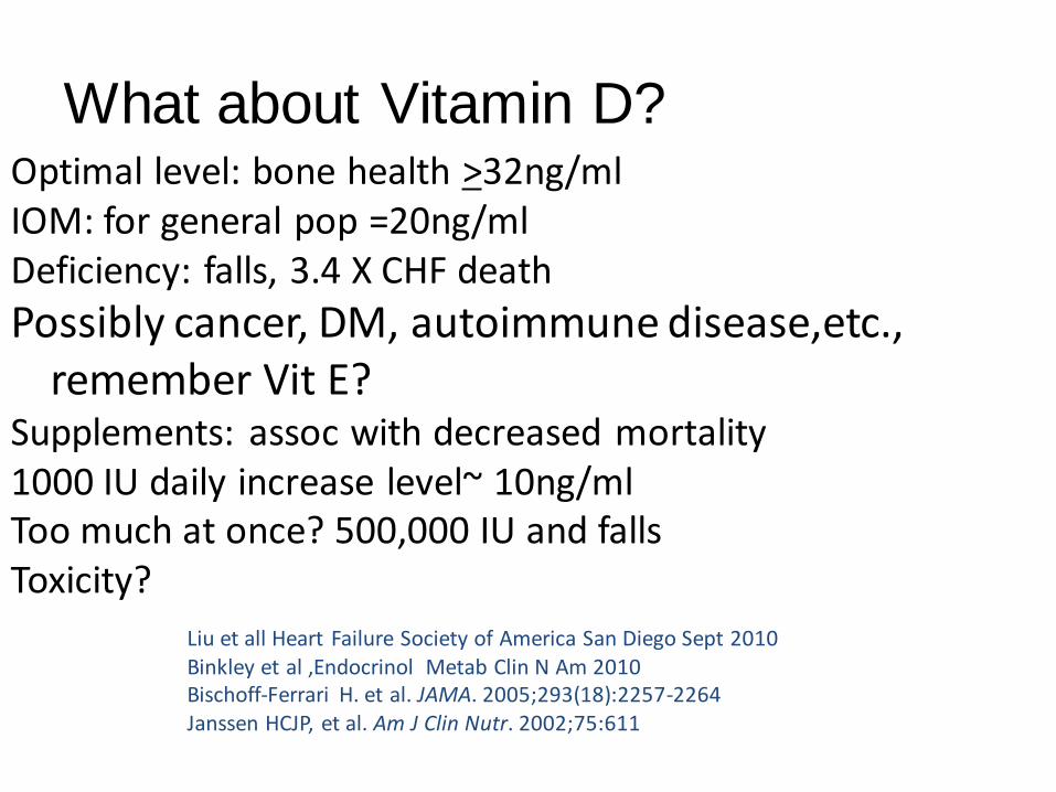
What about Vitamin D?
Liu et all Heart Failure Society of America San Diego Sept 2010 Binkley et al ,Endocrinol Metab Clin N Am 2010 Bischoff-Ferrari H. et al. JAMA. 2005;293(18):2257-2264 Janssen HCJP, et al. Am J Clin Nutr. 2002;75:611
Optimal level: bone health >32ng/ml IOM: for general pop =20ng/ml Deficiency: falls, 3.4 X CHF death
Possibly cancer, DM, autoimmune disease,etc., remember Vit E?
Supplements: assoc with decreased mortality 1000 IU daily increase level~ 10ng/ml Too much at once? 500,000 IU and falls Toxicity?
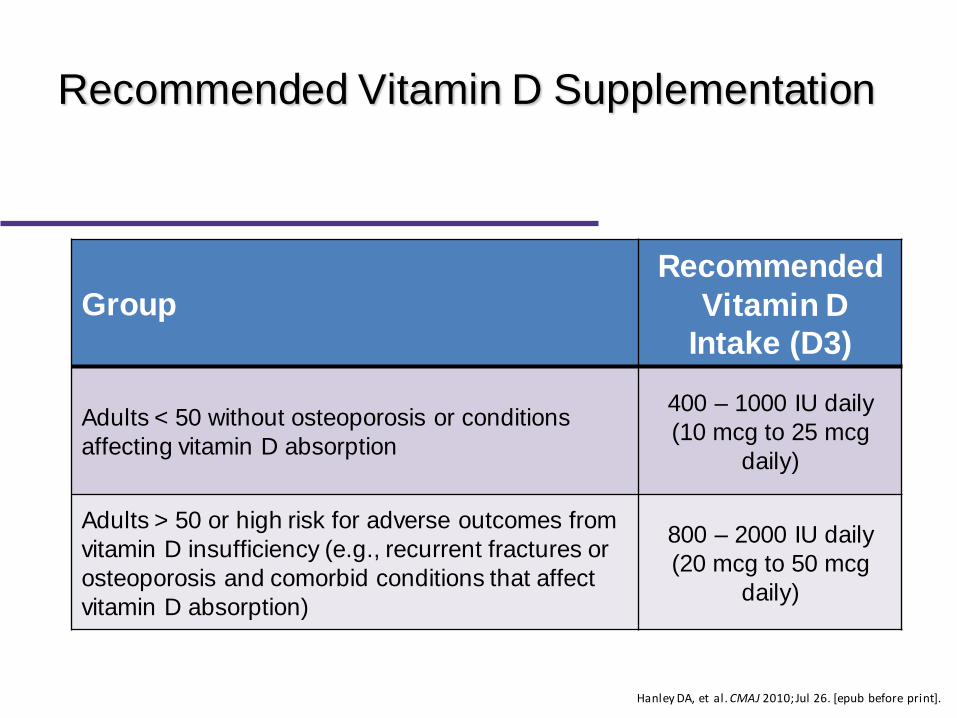
Recommended Vitamin D Supplementation
Group
Recommended
Vitamin D
Intake (D3)
Adults < 50 without osteoporosis or conditions
affecting vitamin D absorption
400 – 1000 IU daily
(10 mcg to 25 mcg
daily)
Adults > 50 or high risk for adverse outcomes from
vitamin D insufficiency (e.g., recurrent fractures or
osteoporosis and comorbid conditions that affect
vitamin D absorption)
800 – 2000 IU daily
(20 mcg to 50 mcg
daily)
Hanley DA, et al. CMAJ 2010; Jul 26. [epub before print].

2012 Questions about
Osteoporosis
Are Bisphosphonates safe?
3. ONJ
4. Treatment duration
5. Esophageal cancer
6. Atypical fractures

FDA Approved Osteoporosis Medications
Drug Post Menopausal OP Steroid OP Male OP
Prevention Treatment Prevention Treatment
Alendronate
Risedronate
Ibandronate
Zoledronate
Raloxifene
Estrogen
Calcitonin
Denosumab *
Teriparatide

First Line Therapies with Evidence for Fracture
Prevention in Postmenopausal Women*
Type of
Fracture
Antiresorptive therapy
Bone
formation
therapy
Bisphosphonates
Denosumab Raloxifene
Hormone
therapy
(Estrogen)**
Teriparatide Alendronate Risedronate
Zoledronic
acid
Vertebral
Hip - -
Non-
vertebral+ -
* For postmenopausal women, indicates first line therapies and Grade A recommendation. For men requiring treatment, alendronate, risedronate, and zoledronic acid can be used as first line therapies for prevention of fractures [Grade D]. + In clinical trials, non-vertebral fractures are a composite endpoint including hip, femur, pelvis, tibia, humerus, radius, and clavicle. ** Hormone therapy (estrogen) can be used as first line therapy in women with menopausal symptoms.

Bisphosphonates – Administration
Must be taken at least one-half hour before the first food, beverage, or medication of the day with plain water only (1 hour prior for monthly ibandronate) Should only be taken upon arising for the day Tablet should be swallowed with a full glass of water (8 oz) and patients should remain upright, walking, standing, or sitting for at least 30 minutes (60 minutes for monthly ibandronate) Should supplement with calcium/vitamin D if dietary intake inadequate

Bisphosphonates – Adverse Effects
Hypocalcemia (18%)
Hypophosphatemia (10%)
Musculoskeletal pain,
cramps – recent FDA
warning
• Gastrointestinal
– Abdominal pain
– Acid reflux
– Dypepsia
– Esophageal ulcer
– Gastritis
• Osteonecrosis of the jaw
(IV bisphosphonates)
• Visual disturbances (rare)

3. What is the Clinical
Presentation of ONJ? Signs &Symptoms:1
Asymptomatic or Facial pain, jaw pain Soft-tissue swelling,drainage Exposed,necrotic bone
Cultures: actinomyces2
Risk factors Cancer & concomitant therapies Poor oral hygiene Smoking Pre-existing dental disease, anemia, coagulopathy, and infection
Management Povidone-iodine & 0.12% chlorhexidine mouthwash Oral antibiotics and anti-inflammatory drugs Conservative debridement for necrotic tissue
Ruggiero SL, Hehrotra B, Rosenberg TJ, et al. J Oral
Maxillofac Surg. 2004;62:527-34.
1. Expert Panel Recommendations for the Prevention, Diagnosis, and Treatment of Osteonecrosis of the Jaws: June 2004
2. Naveau A. Joint Bone Spine 2005.
Melo MD, Obeid G. J Can Dent
Assoc 2005;71: 11-3.
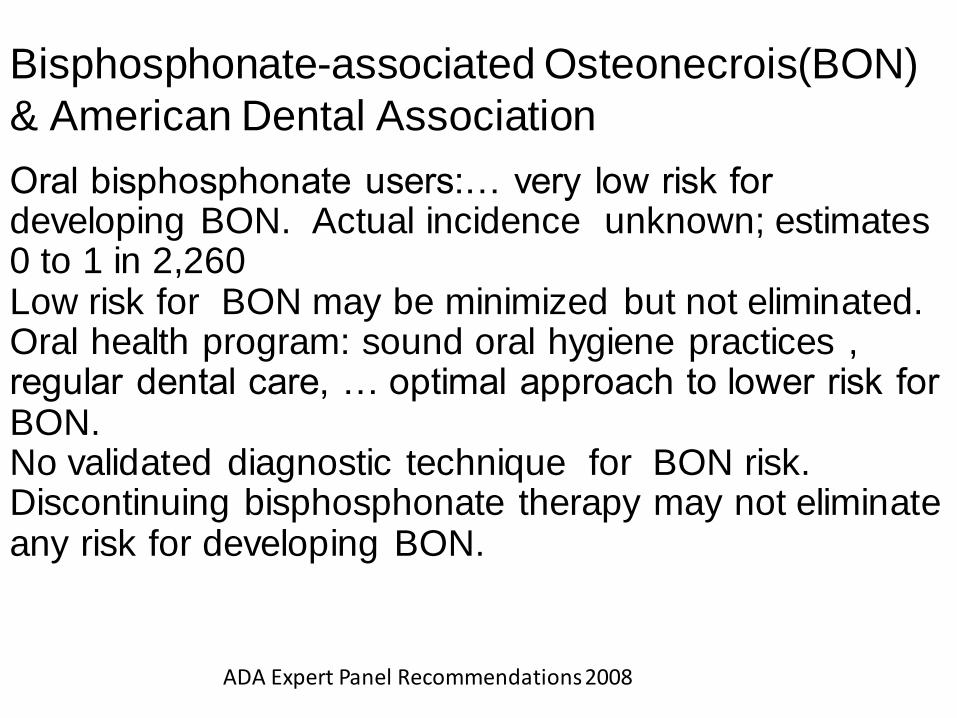
Bisphosphonate-associated Osteonecrois(BON)
& American Dental Association
Oral bisphosphonate users:… very low risk for developing BON. Actual incidence unknown; estimates 0 to 1 in 2,260 Low risk for BON may be minimized but not eliminated. Oral health program: sound oral hygiene practices , regular dental care, … optimal approach to lower risk for BON. No validated diagnostic technique for BON risk. Discontinuing bisphosphonate therapy may not eliminate any risk for developing BON.
ADA Expert Panel Recommendations 2008

Femoral Shaft Fractures
In 2005, Odvina et al reported a series of nine patients with spontaneous, atypical fractures, all on
bisphosphonate therapy for a period of time ranging from 3 to 8 years. Four with fractures in the subtrochanteric region and one each with fractures of the sacrum, rib, ischium, pubic rami and lumbar spine.
6/9 had delayed or absent healing during management. Histology revealed over suppression of bone turnover, possibly linked to bisphosphonate usage, but other factors, i.e.. estrogens and glucocorticoid use left much room for debate.
Odvina et al JCEM2005;90:1294–1301

Figure 1
Atypical Fractures of the Femoral Diaphysis in Postmenopausal Women Taking Alendronate. Lenart, Brett; Lorich, Dean; Lane, Joseph New England Journal of Medicine. 358(12):1304-1306, March 20, 2008.
Figure 1 . Radiographs of Fractures of the Femoral Shaft Showing the "Simple with Thick Cortices" Pattern Panel A shows a fracture of the femoral shaft in an 83-year-old woman with a 9-year history of alendronate use. Panel B shows a similar fracture in a 77-year-old woman with a 5-year history of alendronate use.

Atypical Femoral Fracture
Shane et al, JBMR 25:2010;25:2267–2294.
Conventional AP radiograph of the pelvis (A) shows bilateral focal cortical thickening from
periosteal new bone formation (arrows). Corresponding bone scintigraphy (B)
demonstrates focal increased radionuclide uptake in the proximal lateral femoral cortices
(arrows). MRI images of the femurs (C) demonstrate subtle decreased signal on T1-
weighted and increased signal on T2-weighted images only of the right femur on
this section. Similar findings on AP DXA hip images (D) show focal bilateral cortical
thickening consistent with early, evolving femoral insufficiency fractures.
A B
D C

Bisphosphonate Use and the Risk of Subtrochanteric or Femoral Shaft Fractures in Older Women
L. Park-Wyllie, PharmD, MS, M. Mamdani, PharmD, MA, MPH, D. Juurlink, MD, PhD, G. Hawker, MD, MSc, N. Gunraj, MPH, P. Austin, PhD, D. Whelan, MD, MSc, P. Weiler, MD, MASc, P Eng. Laupacis, MD, MSc
JAMA. 2011;305(8):783-789
Population-based, nested case-control study in a cohort of women aged 68 years or older from Ontario, Canada treated with oral bisphosphonate between April 1, 2002, and March 31, 2008. Primary analysis - association between hospitalization for a subtrochanteric or femoral shaft fracture and duration of bisphosphonate exposure Secondary analysis - association of bisphosphonate use and classic intertrochanteric or femoral neck fractures

Bisphosphonates
Contraindications/Precautions Abnormalities of the esophagus which delay
esophageal emptying, such as stricture or achalasia
Inability to stand or sit upright for at least 30 minutes
Patients at increased risk of aspiration
Hypocalcemia Should be corrected prior to initiating therapy
Renal insufficiency (Not recommended if CrCl < 30-35
ml/min)

#4. How long should a patient stay on bisphosphonate treatment?

ARR = absolute risk reduction. 1. Black D et al. J Bone Miner Res. 2004;suppl 1:S45. 2. Data available on request from Merck & Co., Inc. Please specify 20650700(1)–FOS.
Cumulative Incidence of Clinical Vertebral
Fractures With 10 yrs. Alendronate
Years of Treatment Since FIT
0
2
4
6
8
10
5 6 7 8 9 10
Cu
mu
lati
ve In
cid
ence
, %
Risk Reduction1,2
55%
ALN/Placebo
ALN/ALN (Pooled)
ALN/Placebo, N: ALN/ALN, N:
437 436 428 425 419 412 404 398 392 387 662 660 651 646 638 631 626 615 606 597
5.4%
2.5%
P = 0.013
ARR 2.9%

What about a bisphosphonate “holiday”?
Reasonable to stop bisphosphonates at 5 years & follow Bone Turnover
Markers
Consider switch to teriparatide for drug holiday from bisphosphonates
FDA advisory committee,9/9/11 “… no clear evidence of benefit or harm in continuing the drugs beyond 3-5
years.”
Ott Clev Clin J Med 2011 Laster, Tanner Rheum Dis Clin of NA 2011 www.fda.gov

Bisphosphonates for Osteoporosis — Where Do We Go from Here?
The available data do not identify patients
likely to benefit from treatment beyond 3-5
years. … decisions to continue treatment must be
based on individual assessment of risks and
benefits and on patient preference.
NEJM 366:2048, 2012
OESTROGEN ANALOUGES FOR POST MENOPAUSAL WOMEN
FDA Recommendations – ET/HT
When prescribing medications for osteoporosis,
physicians should consider all non-estrogen
therapies first
When prescribing ET/HT, use smallest dose for
shortest amount of time to achieve treatment goals
Prescribe ET/HT products only when benefits
believed to outweigh risks for a specific patient

CALCITONIN


Calcitonin
FDA-approved for: Treatment of osteoporosis in women who are > 5 years postmenopausal Treatment of Paget’s disease of bone Adjunctive therapy for hypercalcemia Mechanism: Peptide composed of 32 amino acids which binds to osteoclasts and inhibits bone resorption Promotes the renal excretion of calcium, phosphate, sodium, magnesium and potassium by decreasing tubular reabsorption

Calcitonin – Clinical Efficacy
Has been shown to increase spinal bone mass and may decrease risk of vertebral fracture Conflicting data on efficacy of calcitonin at sites other than the spine Less effective than bisphosphonates in treatment of osteoporosis Beneficial, short-term effect on acute bone pain after osteoporotic fracture (vertebral)

Calcitonin – Dosing/Administration
Intranasal 200 units (1 spray) alternating nares daily Store unopened bottles in refrigerator, protect from freezing Can store open bottles at room temperature for up to 35 days Activate pump of new bottles until full spray produced (allow to reach room temperature before priming) Each bottle contains at least 30 doses IM/SQ 100 units/every other day (minimum effective dose not well-defined) Should perform skin test prior to initiating therapy Should supplement with calcium/vitamin D if dietary intake inadequate

Calcitonin – Adverse Effects
Most common:
Nasal spray: rhinitis (12%), irritation of nasal mucosa (9%), epistaxis (3.5%),
sinusitis (2.3%), back pain, arthralgia, headache
Injection: nausea (10%), flushing (2-5%)
Temporarily withdraw use of nasal spray if ulceration of nasal mucosa occurs Periodic nasal examinations recommended

Calcitonin
Contraindications Clinical allergy to calcitonin-salmon
Precautions Nasal ulcerations Tachyphylaxis (parenteral dosage forms)
Drug interactions No formal studies designed to evaluate DI
Price per month 200 units/mL (2): $42.08 200 units/ACT (3.7): $81.59

#7: Questions About Osteoporosis
What about the newest treatment: denosumab for osteoporosis ?
Prefusion osteoclast
Monoclonal antibody for Osteoporosis:RANKL-Inhibition
Adapted from Boyle et al. Nature. 2003;423:337.
CFU-M
Multinucleated osteoclast
OPG
BONE
OPG RANKL
Stromal cells
Denosumab RANK
Active Osteoclast
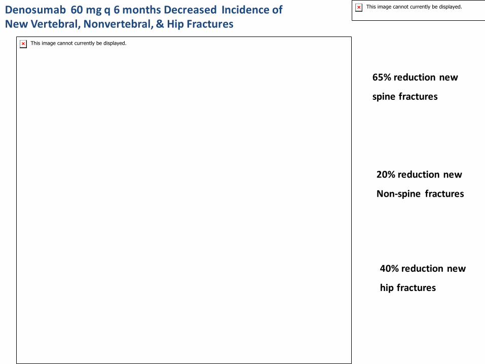
Cummings SR et al. N Engl J Med 2009;361:756-765
65% reduction new
spine fractures
40% reduction new
hip fractures
20% reduction new
Non-spine fractures
Denosumab 60 mg q 6 months Decreased Incidence of New Vertebral, Nonvertebral, & Hip Fractures

Densoumab
Indicated for postmenopausal osteoporosis with high fracture risk or failed, or
intolerant of other therapies
Has been given to renal impairment pts. (including ESRD) single dose, without
affecting pharmacodynamics or pharmokinetics of the drug; no safety signals
Block et al National Kidney Foundation Mtg, Orlando, FL; April 13-17, 2010


#8: Questions about Osteoporosis
Why the warning about
Proton Pump Inhibitors?

2010 FDA Warning: Proton Pump
Inhibitors and Increased Fracture Risk
Revised warning for PPI: possible increased risk of hip, wrist, &
spine fractures.
Based on 7 epidemologic studies & claims data base analysis(
no randomized trials)
Increased risk after 1-7 years of treatment ( note: OTC label for 14 days treatment)
Risk include age >50, “high dose”, longer duration
3 studies : no relation to BMD and PPI use
1 study: no fracture risk if pts. have no other risk factors
WHI: spine but not hip risk, no effect on BMD
Calcium carbonate absorption? Magnesium? Other?
www.fda.gov safety communication 5/25/10

#9: Transplantation- Induced
Osteoporosis (TIOP) 3-11% bone loss 1st yr. post transplant
14-36% increase incidence of fragility fxs.
Most fracture occur at relatively normal Bone Mineral Density: Bone Quality?
Pre-transplant: chronic disease & GCS
Post-transplant : GCS & calcineurin inhib. Controversy: cyclosporine A & tacrolimus
tacrolimus better?, may allow less GCS
Carbonare et al Transplantation 2011
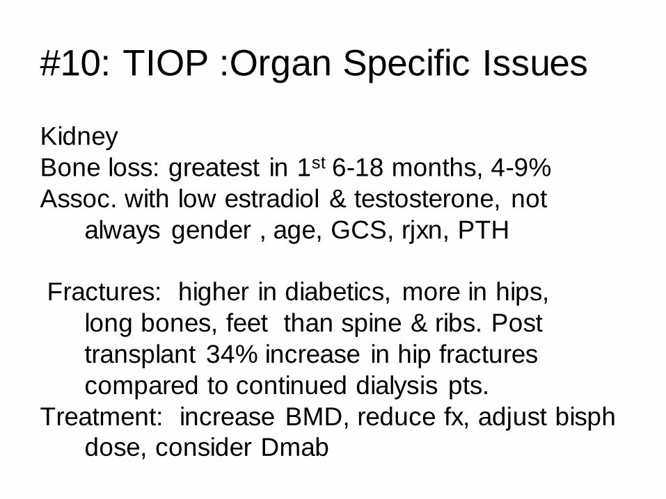
#10: TIOP :Organ Specific Issues
Kidney
Bone loss: greatest in 1st 6-18 months, 4-9%
Assoc. with low estradiol & testosterone, not
always gender , age, GCS, rjxn, PTH
Fractures: higher in diabetics, more in hips,
long bones, feet than spine & ribs. Post
transplant 34% increase in hip fractures
compared to continued dialysis pts.
Treatment: increase BMD, reduce fx, adjust bisph
dose, consider Dmab
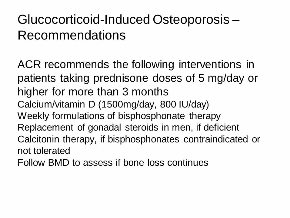
ACR recommends the following interventions in
patients taking prednisone doses of 5 mg/day or
higher for more than 3 months Calcium/vitamin D (1500mg/day, 800 IU/day)
Weekly formulations of bisphosphonate therapy
Replacement of gonadal steroids in men, if deficient
Calcitonin therapy, if bisphosphonates contraindicated or
not tolerated
Follow BMD to assess if bone loss continues
Glucocorticoid-Induced Osteoporosis –
Recommendations

HEADLINES
7.8.07

SUMMARY
REDUCING THE ‘CARE GAP’
Assess bone health in woman >50 and in men > 60.
Evaluate risk factors; evaluate BMD
Consider preventative approach to reduction of fracture risk (the way you think of
hypertension and MI and stroke)
Treat and monitor

Osteoporosis and chronic kidney disease
(CKD) are common conditions of older adults
and often occur concurrently. This follows population
trends:
(1) the older the person and the
greater the degree of osteoporosis, the greater
the risk of bone fracture2;
(2) the older the person, the higher the likelihood of havingdiabetes and high blood
pressure, the 2 most
prevalent causes of CKD

Osteoporosis has
been associated with both hypertension and
diabetes; therefore, it should come as no surprise
that older persons are likely to have both
some degree of CKD and low BMD.

Osteoporosis
is considered to result from an imbalance
between factors that promote bone production
and those that promote bone resorption, tilting
the scales toward a net increase in bone breakdown
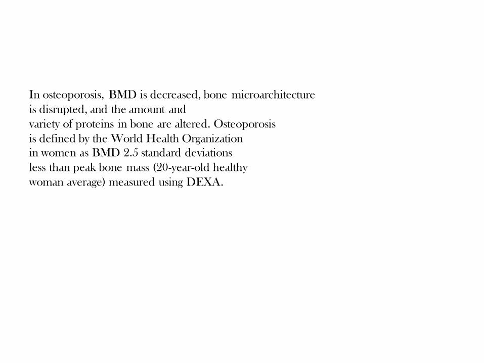
In osteoporosis, BMD is decreased, bone microarchitecture
is disrupted, and the amount and
variety of proteins in bone are altered. Osteoporosis
is defined by the World Health Organization
in women as BMD 2.5 standard deviations
less than peak bone mass (20-year-old healthy
woman average) measured using DEXA.

CKD is associated with abnormalities in
calcium, phosphate, PTH, and vitamin D metabolism,
all of which can adversely affect bone
health. Increasingly, the broader definition of
CKD–mineral and bone disorder (CKD-MBD) is being used to describe the wide range
of
systemic mineral metabolism derangements associated
with increased morbidity and mortality in
this population.

Treatment options for women and men with
low BMD from osteoporosis have increased during
the past several decades. These options now
consist of a variety of pharmacologic therapies,
including receptor selective estrogens, androgens,
calcium and vitamin D, bisphosphonates,
PTH, fluoride, and calcitonin, as well as dietary
therapies, including phytoestrogens and soy, and
also physical therapies, including weight-bearing
or resistance exercises to improve balance,
prevent falls, and increase the mechanical strength
of the bone. Patricia et al , AJKD, 55:941-956 2010

some therapies for
osteoporosis may be relatively contraindicated in
patients with CKD

OSTEOPOROSIS AND BONE DISEASES
SPECIFIC TO THE CKD POPULATION
Bone is a complex 3-dimensional organ consisting
of organic matrix, bone cells, and mineral
salts. Factors associated with bone quality
include quantifiable factors, such as bone mass
and bone density, and qualitative factors, such
as bone geometry (shape and size), microarchitectural
features (cortical or trabecular connections),
and molecular elements (collagen type
and linkages, bone mineral composition, and
crystal orientation).

In healthy adult bone remodeling,
bone breakdown is balanced by bone formation.
However, in osteoporosis, bone mineral
and protein are normal, but the balance is
shifted toward bone breakdown, leading to
thinning of the 3-dimensional structure and
increased fragility.
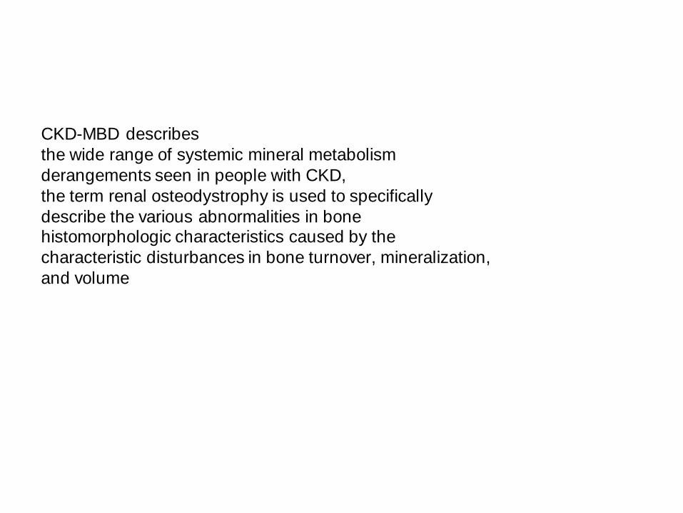
CKD-MBD describes
the wide range of systemic mineral metabolism
derangements seen in people with CKD,
the term renal osteodystrophy is used to specifically
describe the various abnormalities in bone histomorphologic characteristics caused by the
characteristic disturbances in bone turnover, mineralization,
and volume

definition of CKD-MBD also includes
biochemical abnormalities and calcifications
in vascular and other soft tissues
CKD-MBD :
describes the wide range of systemic
mineral metabolism derangements, including biochemical
abnormalities and calcifications in vascular and
other soft tissues. It broadly includes diseases of bone turnover, bone mineralization, and bone volume, with
these conditions often overlapping
Adynamic bone disease :
characterized by low rates of
both bone formation and resorption and primarily a
disease of bone turnover

High-turnover disease :
characterized by increased
osteoblast and osteoclast activity with abnormal collagen
deposition, marrow fibrosis, and high rates of both
formation and resorption. This condition of bone turnover often reflects secondary hyperparathyroidism
Osteoporosis :
bone density (or bone mass) at least
2.5 standard deviations less than peak bone mass
(defined as the bone mass achieved by healthy adults
aged 18-30 y) considered to result from an imbalance between factors that promote bone production and
those that promote bone resorption, tilting the scales
toward a net increase in bone breakdown and increased
fragility

Osteomalacia :
characterized by abnormal mineralization
accompanied by a low bone formation rate resulting
in reduced bone density

Osteopenia :
bone density 1-2.5 standard deviations
less than peak bone mass (see osteoporosis)

Renal osteodystrophy :
describes the various abnormalities
in bone histomorphologic characteristics
caused by the characteristic disturbances in bone
turnover, mineralization, and volume that develop as a consequence of CKD-MBD

Decreasing kidney function progressively disrupts the relationship
among phosphorus, calcium, and their
hormonal regulators, including PTH, 1,25 dihydroxyvitamin
D,14 and the phosphaturic hormone
fibroblast growth factor 23 (FGF-23). FGF-23 is secreted mainly by osteocyctes in
response to increasing phosphate retention and
acts to increase phosphate excretion.15 Increasing
levels of FGF-23 eventually inhibit 1--
hydroxylase production in the kidney, resulting
in suppression of 1,25 dihydroxyvitamin D with
the subsequent development of hypocalcemia
and secondary hyperparathyroidism (SHPT)

The
dilemma for nephrologists is that CKD-MBD
may coexist with osteoporosis, particularly in the
elderly population in whom decreasing kidney function
is prevalent

NHANES III (Third National Health and Nutrition
Examination Survey; 1988-1994), low BMD was
much more prevalent in those with CKD than in
those with normal kidney function.16 In addition,
slightly 60% of women with a diagnosis of
osteoporosis also had CKD stage 3, and 23% had
CKD stage 4.16 Unfortunately, despite their very
different pathophysiologic states, both osteoporosisand renal osteodystrophy
independently increase
bone fragility, presenting diagnostic and therapeutic
challenges and collectively increasing the risk of
fracture at all stages of CKD

CONCLUSION
Osteoporosis is defined as a condition of impairment in bone strength due to
low bone mineral density and
poor bone quality and predisposes individuals to an increased risk of fractures.
Osteoporosis may coexist with
chronic kidney disease2mineral and bone disorder (CKD-MBD) and osteoporotic fractures occur in all stages
of CKD.

Management of osteoporosis in CKD should consider the pathophysiology of
both disorders. Diagnosis
and management of osteoporosis in patients with stages 1-3 CKD and patients
without CKD are similar,
but diagnosis and management decisions differ greatly once patients have stages 4-5 CKD. Discriminating
between osteoporosis and CKD-MBD is best accomplished with quantitative
bone histomorphometry.
Biochemical markers, especially intact parathyroid hormone and bone-specific
alkaline phosphatase, also may be helpful.

When the diagnosis of osteoporosis is established, management in stages 4-5
CKD may include
antiresorptive or anabolic agents, though evidence for efficacy is marginal in
advanced CKD.