cingulate cortex of the rhesus monkey: i. cytoarchitecture and thalamic afferents
TRANSCRIPT

THE JOURNAL OF COMPARATIVE NEUROLOGY 262256-270 (1987)
Cingulate Cortex of the Rhesus Monkey: I. Cytoarchitecture and Thalamic Afferents
BRENT A. VOGT, DEEPAK N. PANIIYA, AND DOUGLAS L. ROSENE Departments of Anatomy and Physiology, Boston University School of Medicine, Boston,
Massachusetts 02118 (B.A.V., D.N.P., D.L.R.), and Edith Nourse Rogers Memorial Veterans Hospital, Bedford, Massachusetts 01730 (B.A.V., D.N.P.)
ABSTRACT The cytoarchitecture and thalamic afferents of cingulate cortex were
evaluated in the rhesus monkey (Macaca mulatta). Area 24 has three divi- sions of which area 24a i s adjacent to the callosal sulcus and has the least laminar differentiation. Area 24b has more clearly defined layers 11,111, and Va, and area 24c, which forms the lower bank of the anterior cingulate sulcus, has a particularly dense layer 111. Area 23 also has three divisions, each of which has a distinct layer IV. Area 23a is adjacent to the callosal sulcus and has the thinnest layers 11-IV, which have the same cell density as layers V and VI. Area 23b has the largest pyramids in layers IIIc and Va, and area 23c, in the depths of the posterior cinguIate sulcus, has the broadest external and thinnest internal pyramidal layers. Finally, areas 29 and 30 are located in the posterior depths of the callosal sulcus. Two divisions of area 29 are apparent: one with a granular layer directly adjacent to layer I (area 29a-c) and another with differentiation of layers I11 and IV (area 29d). Area 30 has a dysgranular layer IV.
Injections of the retrograde tracer horseradish peroxidase (HRP) were made into subdivisions of cingulate cortex in the monkey. Area 25 received thalamic input mainly from the midline parataenial (Pt), central densocel- lular (Cdc), and reuniens nuclei as well as from the dorsal parvicellular division of the mediodorsal nucleus (MDpc). A less dense projection also originated in the intralaminar parafascicular (PO, central superior, and limitans (Li) nuclei as well as the medial division of the anterior nuclei (AM).
Areas 24a and 24b received most thalamic afferents from fusiform and multipolar cells in the Cdc and Pf nuclei with fewer from the ventral anterior (VA) and MDpc and MD densocellular (MDdc) nuclei and only minor input from AM. Most input to premotor cingulate area 24c appeared to originate in VA, MDdc, and Li.
Area 29 received the most dense input from nuclei traditionally associ- ated with limbic cortex including the anteroventral (AV), anterodorsal (AD), and laterodorsal (LD) nuclei. Areas 23a and 23b, in contrast, did not receive AV, AD, or LD input, but the greatest proportion of their thalamic afferents arose in AM. Less-pronounced input also came from the lateroposterior (LP), medial pulvinar, and MDdc nuclei. This latter nucleus projected more to area 23b than to areas 30 or 23a.
Anterior medial nucleus efferents to cingulate cortex were of particular note for two reasons. First, AM projected primarily to posterior cingulate areas with area 23 receiving its principal thalamic input from AM. Second, projections to areas 30, 23a, and 23b were topographically organized with ventral areas 30 and 23a receiving from the central core of AM, while the more dorsally located area 23b received input from peripheral and medial parts of AM.
Accepted March 4, 1987.
0 1987 ALAN R. LISY, INC.

MONKEY CINGULATE CORTEX 257
In light of the extensive projections of Cdc, Csl, and Pf to anterior cingulate cortex, it is proposed that the midline and intralaminar thalamic nuclei be classified as part of limbic thalamus along with the anterior, LD, and MD nuclei. Furthermore, although AM projects mainly to posterior cingulate cortex, it also has light projections to area 25 and minor input to area 24. As such, AM is the only limbic thalamic nucleus that has such widespread projections to cingulate cortex. Finally, visually evoked activity in area 23 may be the result of projections from the LP and medial pulvinar
Key words: anterior thalamic nuclei, midline thalamic nuclei, intralaminar thalamic nuclei, lateroposterior thalamic nucleus, pulvinar thalamic nucleus, primate
Primate cingulate cortex is involved in attention and memory and mediates somatic and autonomic motor re- sponses, phonation, and responses to painful sensation (Smith, '45; Ward, '48; Kaada, '51; Barris and Schuman, '53; Foltz and White, '62, '68; Dua and MacLean, '64; Ta- lairach et al., '73; Watson et al., '73; Haller et al., '76). Ablation of cingzllate coi-tex or its underlying white matter, the cingulum bundle, has proven to be clinically useful in treating depression, obsessive-compulsive behavior, and chronic pain (LeBeau, '52; Livingston, '53; Tow and Whitty, '53; Foltz and White, '62, '68; Kanaka and Balasubraman- iam, '78). The underlying neuronal mechanisms for these broadly characterized functions are not known and the re- sults of clinical intervention are often variable. A more precise understanding of the architecture and connections of cingulate cortex should lead to a mechanistic under- standing of cingulate cortex functions. Our working hy- pothesis is that since cingulate cortex is not a unified or homogeneous cortical region, its functions may be charac- terized on the basis of discrete subareas. The aim of the present study is to provide a morphological basis for more refined functional studies,
The cytoarchitecture of primate cingulate cortex is more diverse than most experimental studies indicate. Brod- mann's ('09) two major subdivisions of cingulate cortex, areas 24 and 23, are not structurally homogeneous. Area 24, for example, has been noted to undergo graded changes from poorly differentiated cortex next to the corpus cal- losum to well-defined laminae in the depths of the cingulate sulcus (Walker, '40; Smith, '45). Furthermore, Braak ('76) and Muakkassa and Strick ('79) have identified a motor area in the depths of the cingulate sulcus that includes part of area 24. Electrical stimulation studies also suggest het- erogeneities in area 24 as vocalization and changes in res- piration can be evoked by stimulation of different parts of area 24 (Kaada, '51). Finally, Rose ('27)) von Economo ('291, and Sarkissov ('55) each divided both anterior and posterior cingulate cortices into three ventrodorsal parts in their cytoarchitectural analyses.
Definitions of the cingulate region have been based on topographical criteria (e.g., Kappers et aI., '67) or on con- nections with the anterior thalamic nuclei (Clark and Bog- gon, '33; Rose and Woolsey, '48). These latter studies, however, employed the retrograde cell degeneration method after relatively large lesions in subprimate species that do not have an area 23. In the monkey it is not known if thc anterior nuclei project specifically to area 23, since Locke et al. ('64) made lesions in area 23 in the monkey and did
not observe retrograde changes in the anterior thalamic nuclei. Because areas 23, 30, and 29 are usually involved in experimental procedures, for example, those employing retrogradely transported tracers (e.g., Vogt et al., '79; Bal- eydier and Mauguiere, 'SO), the only thalamic projection reported to terminate specifically in area 23 is one that originates in the medial pulvinar (Baleydier and Mau- guiere, '85). Thus, there still remains a great deal of uncer- tainty regarding precise thalainic projections to different parts of cingulate cortex.
The strategy for this investigation was to make large and then restricted injections of horseradish peroxidase (HRP) into monkey cingulate cortex such that the distribution of thalamic afferents could be characterized in terms of a comprehensive cytoarchitectural analysis. Small injections restricted to areas 23a, 23b, or 30 provide a basis for distin- guishing among the connections of these areas and those of' area 29.
MATERIALS AND METHODS Surgical procedures
In all instances in which injections were made into cin- gulate cortex, the monkey was anesthetized with sodium pentobarbital and mounted in a stereotaxic instrument. A midline craniotomy was made and mannitol (Osmitrol, 25%) was injected I.V. Then the dura was opened and retracted to one side; bridging veins between the cortex and falx cerebri were cauterized; the hemispheres were gently re- tracted; and the corpus callosum was sectioned. A smaller slit was then made in the falx so that a 5-pl Hamilton syringe with a 32-gauge needle could be positioned in differ- ent parts of cingulate cortex.
Nissl preparations Thalamic and cortical cytoarchitecture was evaluated in
monkeys that had been perfused intracardially with 1 liter of 0.9% saline and then 1 liter of 10% formalin. The brains were removed and postfixed in 10% formalin for 2-4 weeks in a refrigerator and then dehydrated and embedded in celloidin. Sections were cut 25-40-pm-thick and a one-in- six series was stained with cresyl violet.
HRP histochemistry Two series of animals were prepared for retrograde tracer
analysis. First, ten animals received large 0.2-0.3-pl injec- tions of 20% HRP in cingulate cortex and were allowed to survive 2 days. The animals were then anesthetized and killed by perfusion-fixation according to the protocol of' Ro-

258 B.A. VOGT, D.N. PANDYA, AND D.L. ROSENE RESULTS sene and Mesulam ('78). Thus, they were perfused through
the heart with physiological saline followed by 2 liters of quarter-strength Karnovsky fixative (Karnovsky, '65) and finally 1 liter of phosphate buffer containing 10% sucrose. The brains were stored for one night in sucrose phosphate buffer and then frozen and sectioned into 40-pm-thick sec- tions. An interrupted series of one-in-eight sections was processed by using tetramethyl benzidine, mounted on subbed slides, dried, and then counterstained with neutral red. In a second series of five animals, very limited HRP injections were made with 0.015 pl of 5% HRP conjugated to wheat germ agglutinin (HRP-WGA, Sigma), and the brains were prepared according to the above protocol. The full rationale behind the use of HRP-WGA in studies of cingulate cortex connections has been presented elsewhere (Vogt and Miller, '83; Miller and Vogt, '84). The locations of HRP-labeled neurons were plotted with an X-Y recorder coupled to the mechanical stage of a microscope by linear potentiometers. In a few cases the sections were drawn with the aid of a drawing tube attachment to the microscope or an Aus Jena Documentor projector onto which labeled neu- rons were plotted.
Cytoarehitecture A map of the distribution of areas in monkey cingulate
cortex is presented in Figure 1 and photomicrographs of Nissl-stained sections at low and higher magnifications are shown in Figures 2 and 3, respectively. As a general rule the cortex adjacent to the corpus callosum is least differen- tiated whereas the cortex near and in the depths of the cingulate sulcus contains the most elaborate lamination patterns. The cingulate region includes areas 25,24,29,30, 23, and possibly 31 as outlined by dotted lines in Figure 1 and can be distinguished in the following manner. Area 25 has essentially two cellular layers with the deep layer V- VI that is more cell dense than in any other cingulate area and a superficial layer 11-111 and no layer W. Area 24 also does not have a layer IV. Compared to area 23, layer V of area 24 is denser and more homogeneous. Area 23 has a distinct layer IV of small cells, and a layer IIIc of pyramidal cells is more prominent than in area 24. Finally, area 29 has dense and poorly differentiated layer 11-W while adja- cent area 30 is dysgranular (i.e., has a poorly developed layer IV).
D I
Fig. 1. Distribution of cytoarchitectural areas in monkey cingulate cor- tex. Coronal sections at levels A-1) are presented in Figure 2. CC, corpus Fig. 2. Coronal sections through different levels of the monkey cingulate callosum; CS, cingulate sulcus: SpS, splenial sulcus; RS, rostra1 sulcus; RhS, gyrus. Levels designated in Figure 1. IG, indusium griseum; Sub, subicu- rhinal sulcus; POS, parieto-occipital sulcus; CaS, calcarine sulcus. lum. Cresyl violet. ~ 1 8 .
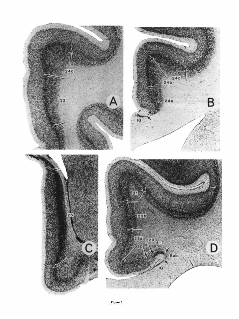
Figure 2

Figure 3

MONKEY CINGULATE CORTEX 261
of area 23, area 23a has the thinnest external pyramidal layers 11-IV (Fig. 3). Both internal and external pyramidal layers are relatively homogeneous and of approximately equal cell density. Area 23 is most differentiated in area 23b, which has the largest neurons in layers IIIc, V, and VI. The ventral part of area 23b forms most of what has been termed the caudomedial lobule (Goldman-Rakic et al., '84). A small extension of area 23b also continues into the most rostral part of the calcarine sulcus, as does area 29. Area 23c, in the depths of the posterior cingulate sulcus, has the broadest layers 11-IV but layers V and VI are not well developed. Whereas area 23a has features of a proiso- cortex, the thick layer IV and large layer IIIc pyramids in areas 23b and 23c characterize them as isocortical (i.e., neocortical) structures.
Area 31 forms the dorsal and posterior rim of the cingu- late region in the monkey and is cytoarchitecturally inter- mediate between area 23c and the medial parietal area 7 or PGm (Pandya and Seltzer, '82). It has the broadest layers I-IV and the largest layer IIIc pyramids. Layers Va, Vb, and VI are distinct and the cells in these layers are not arranged radially as in medial area 7.
Thalamw. Olszewski ('52) described in detail the cytoar- chitecture of monkey thalamus. In light of thalamic projec- tions from both the anterior and midline nuclei to cinguIate cortex, it is necessary to clarify structural distinctions be- tween them. Figure 4 is a micrograph from a level just caudal to where the anteromedial (AM) nucleus crosses the midline. At this level the centrodensocellular nucleus (Cdc) arches over and just beneath AM. Although cells in Cdc can be as large as those in AM, there are also many smaller fusiform and multipolar cells in Cdc and so the overall packing density is much higher.
Area 24. This area can be divided into three subregions. Area 24a lies adjacent to the corpus callosum and is a relatively uniform cortex; i.e., layers I1 and I11 are virtually indistinguishable and in the deeper layers Va and Vb are difficult to differentiate. In contrast to area 25, layer VI i s distinct from layer V in area 24a.
Area 24b has a layer I11 that is less dense than layers I1 and Va, so a clear distinction can be made among layers 11, 111, and Va. Also, cells in layer Va are more dense than in the outer pait of layer V in area 24a.
Area 24c forms the lower bank of the rostral half of the cingulate sulcus. It has a dense layer 111 that contains large pyramids, and the deep layers are somewhat less dense than in area 24b. Finally, the layer I11 border is very straight.
Area 32 lies rostral and ventral to area 24. Its cytoarchi- tecture is similar to area 24a in the homogeneity of layers 11-111 and Vb-VI; however, layer Va forms a distinct band and the outer layers (11-IV) are much thicker than in area 24b.
Area 29. Area 29 does not appear to be extensively dif- ferentiated in the monkey as it is in rodents and lago- morphs. There are essentially two components to this area (Fig. 2D), a lateral area 29a-c that abuts the supracallosal subiculum (previously termed RSgl, Vogt, '76) and a medial area 29d (previously termed RSg2, Vogt, '76). These desig- nations are employed to maintain consistency with pre- vious work in other species and to draw attention to possible structural equivalencies among species (Vogt and Peters, '81; Vogt, '85; Vogt et al., '86).
Area 29a-c has a broad layer I and essentially homoge- neous and densely packed granular layer 11-IV. Layer V is divided into a Va part with large pyramids and a cell-sparse Vb, whereas there is a poorly developed (i.e., thin and cell sparse) layer VI. Because of the lack of differentiation of layer 11-lV and the location of area 29a-c next to the subiculum, this area is a periallocortex. The adjoining area 29d has a sparse layer I11 of medium-sized pyramids that abuts layer I and a very slender and irregular layer IV, and the composition of layers V and VI is similar to that of area 29a-c. In the latter instance, however, these layers are thicker and more cell dense. Both areas 29a-c and 29d extend along the caudal depths of the callosal sulcus and into the calcarine sulcus.
Area 30. This area lies medial to area 29d and is classi- cally considered retrosplenial agranular or dysgranular cortex. Layer IIIc has large pyramids; there is a thin, irreg- ular, and cell-sparse layer lV and a layer Va with very large pyramidal neurons. These features, particularly those of layer IV, characterize this region as proisocortcx, as is the case for areas 29d and 23a.
Area 23. Area 23 occupies most of the posterior half of cingulate cortex and can be divided into three parts. Ros- trally it merges with area 24, dorsally with area 31, and caudally i t extends to areas 7 and 19. Of the three divisions
Fig. 3. Pholographs through reprcscntativc sections of somc cingulate areas. Arrows are placed at the border betwccn laycrs I11 or IV and Va. Crexyl violet. ~ 9 3 .
Thalamic afferents Anterior cingulate cortex. Figure 5 presents a case in
which a large NRP injection involved areas 24a-c. In this case most labeled neurons in the thalamus were in the ventral, midline, intralaminar, and mediodorsal nuclei. Al- though labeled neurons were also present in the anterior nuclei, the number of these cells was limited in comparison to that in other regions.
Labeled neurons in the ventral nucleus were entirely in the rostral part of the anterior WA) division. Labeled cells in the midline were predominantly in Cdc while fewer were also in the paraventricular (Pa), parataenial (Pt), central latocellular (Clc), central inferior (Cif), and reuniens (Re) nuclei. Of the intralaminar nuclei, the central superior lateral (Csl), centraI lateral (CI), parafascicular (PO, and limitans (Li) nuclei contained most labeled neurons. In the mediodorsal nucleus neurons were labeled in the periphery of the parvicellular division (MDpc) and at the most caudal levels of the densocellular division (MDdc). Fewest labeled neurons occurred in a dorsal part of the medial pulvinar.
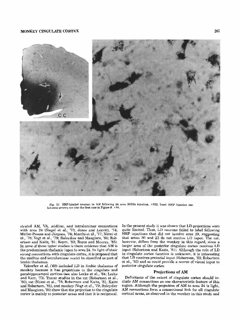
Examples of HRP-labeled fusiform and multipolar neu rons in Cdc following the area 24 injection are shown in Figure 6. The fusiform neurons were in the most medial part of Cdc (i.e., approximately a t the level of the middle arrow in Fig. 41, while the multipolar cells were in the dorsal limb of Cdc (i.e., at the level of the top arrow in Fig. 4). For comparative purposes RRP-labeled neurons in AM following an area 30-23a injection are presented in Figure 10 that are at the same magnification as those in Figure 6. The AM labeled neurons in both cases were larger and the

262 B.A. VOGT, D.N. PANDYA, AND D.L. ROSENE
Fig. 4. Anterior and midline thalamic nuclei at a level just caudal to the midline cronsing of AM. The following borders are indicated AWNCdc, ar-
rows; AD/AV, arrowheads; AWAV, double arrowhead. Cresyl violet. A, anterior; M, medial; D, dorsal; V, ventral. X68.
overaIl cell packing density much reduced when compared Posterior cingulate cortex. A large HRP injection in to those in Cdc following the area 24 injection. posterior cingulate cortex involved areas 23a-c, 30, and 29,
In a smaller, anterior-placed area 24a,b injection (Fig. and the greatest density of labeled neurons was in the 7C-F) HRP-labeled neurons were in most of the same tha- anterior nuclei and along the dorsal thalamus (Fig. 8). Most lamic nuclei as in the previously described area 24 case, cells were in AD, AV, LD, LP, MDdc, and medial pulvinar. however, the density of labeled cells in VA, MDdc, and Li Fewer HRP-labeled neurons were in AM, Csl, and MDpc, was greatly reduced, possibly because these nuclei project and the fewest were in Cdc and Li. Although the projection strongly to area 24c. In an area 32 case (Fig. 7A,B), labeled zones of these nuclei were often associated with posterior neurons were only in MDpc, MDdc, Li nuclei and a few cingulate cortex, six cytoarchitectural areas have been in- were in the Pa nucleus. No cells were labeled in VA, Cdc, volved in the injection site. Injections restricted to area 30 or AM in this case. andor parts of area 23 indicated that there were selective
An HRP injection that involved mainly area 25 (not preferences in the distribution of thalamic inputs to each shown) produced labeling of cells in Cdc, Pt, and throughout cytoarchitectural subdivision. the full length of the dorsal part of MDpc. Lighter labeling Three of eight restricted posterior cingulate cortex HRP also occurred in AM, Re, Csl, FY, and Li. injections are presented in Figure 9. In the first of these

MONKEY CINGULATE CORTEX 263
r 1 mm
Fig. 5. Distribution of HRP-labeled neurons (dots) in the thalamus follow- ing a large injection into area 24 (hatched). Levels A-F represent progres- sively caudal sections through the thalamus. Sin, stria nredulariH; Caud,
caudate; R, reticular; VL, ventrolateral; Pcn, paracentral; VPL, I, M, ven- troponterior lateral, inferior and medial, respectively; Hb, habenula; Cn.Md, centromcdianum; Pul. o, I , i , pulvinar oral, lateral, and inferior, respectively.

B.A. VOGT, D.N. PANDYA, AND D.L. ROSENE
Fig. 6. HRP-labeled neurons in Cdc following an area 24 injection (see a1.w Fig. 4). A: Fusifnrm neurnns frnm the medial part of Cdc; some are
emphasized with arrows. R: Multipolar neurons from dorsal limb of Cdc; some are emphasized with arrows. ~ 5 5 3 .
cases, areas 30 and 23a were involved in the injection (see also Fig. 10 inset). In this case most labeled neurons were in the core of AM (Fig. 9) with fewer in LP. Some neurons were also in the pc and dc divisions of MD and the medial pulvinar with only occasional cells in the LiPf complex. In this case as in all of the restricted cases that did not involve area 29 there were no labeled neurons in AV or AD and only a few were labeled in LD. Therefore, the primary termination zone of AD, AV, and LD is in area 29.
In a case where HRP was restricted to areas 23a and b (Fig. 9, middle case) there was a shift in the location of labeled neurons such that most were in LP, less in AM and the medial pulvinar, and fewest were in the MD and intra- laminar nuclei (Pf, Pc, and Csl). In all instances the re- stricted injections showed only limited labeling in the
intralaminar nuclei. In the case that involved area 2% (Fig. 9, third case), the distribution of labeled neurons was similar to the previous case; however, more cells were in Li and there were more labeled neurons in ventral parts of AM.
There was a topographical organization of AM projections to areas 30 and 23 (Fig. 9). In the most ventral case (areas 30 and 23a) labeled cells were concentrated in the core of AM while in area 23a and 23b cases these neurons were more dispersed in AM. In the area 23b case labeled cells were most concentrated in the periphery of AM. Also, the main projections to subdivisions of area 23 were from AM, LP, and medial pulvinar, with small and variable contri- butions from the midline, intralaminar, and mediodorsal nuclei.

MONKEY CINGULATE CORTEX
1 mm Fig. 7. Location of HRP-labeled neurons in the thalamus following injections into area 32 (filled, lcvels A and
H) and areas 24n and b (hatched, levels C-F).
Fig. 8. Distribution of labolcd neurons at four levels of the t.halarnw follnwing a large HRP injection Into posterior c inyla te cortex

266
AREA
R.A. VOGT, D .N, PANDY
23b
'A, AND D.L. ROSENE
- 1 mm
Fig. 9. Three small injections into posteior cingulate cortex and the positions of labeled neurons in the thalamus.
DISCUSSION In anterior cingulate cortex, area 25 receives mainly Cdc,
F't, Re, and MDpc input with only limited afferents arriving from the AM and FY nuclei. Areas 24a and 24b receive mainly Cdc and intralaminar (i.e., Csl, Pf, and Li) inputs, while less arises from VA and MDpc and least from AM. Area 24c receives mainly VA, MDdc, and Li afferents. In posterior cingulate cortex area 29 receives AV, AD, and LD input, while areas 30 and 23 get mainly AM, LP, and medial pulvinar afferents and less from MD, Pf, and Li.
Limbic thalamus The present study demonstrates that the midline Cdc and
intralaminar Pf and C1 nuclei are the principal sources of thalamic input to area 24. Though a minor projection from AM is present, this is not the main source of thalamic afferents, as i s true for rodents and marsupials (Beckstead, '76; Benjamin and Golden,'85). Yakovlev et al. ('60) showed in the monkey that, in addition to the anterior nuclei, the midline nuclei are connected with this region, and many subsequent studies in the cat and monkey have demon-
MONKEY CINGULATE CORTEX 267
Fig. 10. HRP-labeled neurons in AM following an area 30/23a injection. X533. Inset HRP injection site between arrows; see also the first case in Figure 9. x 14.
strated AM, VA, midline, and intralaminar connections with area 24 (Siege1 et al., '73; Jones and Leavitt, '74; Muller-Preuss and Jurgens, '76; Macchi et al., '77; Niimi et al., '78; Vogt et al., '79; Baleydier and Maugiere, '80; Rob- ertson and Kaitz, '81; Royce, '83; Royce and Mourey, '85). In none of these latter studies is there evidence that AM is the predominant thalamic input to area 24. In light of their strong connections with cingulate cortex, it is proposed that the midline and intralaminar nuclei be classified as part of linibic thalamus.
Yakovlev et al. ('60) included LD in limbic thalamus of monkey because it has projections to the cingulate and parahippocampal cortices (see also Locke et al., '64; Locke and Kerr, '73). Tracer studies in the rat (Robertson et al., '801, cat (Niimi et al., '78; Robertson and Kaitz, '81; Kaitz and Robertson, '81), and monkey Nogt et al., '79; Baleydier and Maugiere, '80) show that the projection to the cingulate cortex is mainly to posterior areas and that it is reciprocal.
In the present study it was shown that LD projections were quite limited. Thus, LD neurons failed to label following HRP injections that did not involve area 29, suggesting that areas 30 and 23 do not receive LD input. The cat, however, differs from the monkey in this regard, since a larger area of the posterior cingulate cortex receives LD input (Robertson and Kaitz, '81). Although the role of LD in cingulate cortex function is unknown, it is interesting that LD receives pretectal input mobertson, '83; Robertson et al., '83) and so could provide a source of visual input to posterior cingulate cortex.
Projections of AM Definitions of the extent of cingulate cortex should in-
clude AM connections as one characteristic feature. of this region. Although the projection of AM to area 24 is light, AM connections form a connectional link for all cingulate cortical areas, as observed in the monkey in this study and

268 B.A. VOGT, D.N. PANDYA, AND D.L. ROSENE
in cat (Niimi et al., '78; Robertson and Kaitz, '81). Further- more, areas 23 and 30 in the monkey receive relatively more input from AM than any other nucleus, and that includes only minor input from the midline and intralami- nar nuclei. When an injection also involves area 29, how- ever, at least equal numbers of neurons are labeled in AV as in AM.
Anteromedial projections are not limited to cingulate cor- tex, however, since light AM projections have also been observed to terminate in prefrontal cortex (Goldman-Rakic and Porrino, '85). Thus, anterior thalamic inputs are char- acteristic of but not unique to the cingulate cortex. Con- versely, MD projections are not limited to prefrontal cortex as area 23b of posterior cingulate cortex has both MDdc and MDpc inputs. In light of these shared AM and MD as well as medial pulvinar thalamic inputs and extensive con- nections between area 46 of the prefrontal cortex and cin- a l a t e cortex (Nauta, '64; Pandya et al., '71, '81; Kunzle, '78; Vogt et al., '79; Baleydier and Maugiere, '80; Goldman- Rakic et al., '84; Barbas and Mesulam, '85), it is likely that these two cortical regions are involved in similar functions such as delayed response performance (Niki and Watanbe, '79).
Comparative organization of posterior cingulate cortex
Connections with the AD and AV thalamic nuclei are generally considered to be a distinguishing trait of posterior cingulate cortex in the rat (Domesick, '721, rabbit (Rose and Woolsey, '481, and monkey (Vogt et al., '79; Baleydier and Mauguiere, '80). However, this generalization requires re- consideration because posterior cingulate cortex is neither cytoarchitecturally nor connectionally the same in rodents/ marsupials and primates. The major structural difference is the presence o f area 23 in monkey. It has been demon- strated in the monkey in this study that area 23 lies dorsal to areas 29 and 30 and that it has a well-developed layer IV and differentiated layers I1 and 111. It also receives input mainly from the AM, LP, and medial pulvinar thalamic nuclei but does not appear to receive input from AD or AV.
The posterior cingulate cortex in the rat has four divi- sions of area 29 (Vogt and Peters, '81); none of them meets cytoarchitectural criteria for an area 23, and the most dor- sal division, area 29d, might receive light AD and AV input (Vogt, '851, as do the granular cingulate areas (Dome- sick,'72). In the rabbit brain there are five divisions of area 29 in posterior cortex (vogt et al., '86), and a dorsolateral part of area 29d does have a granular layer IV similar to that of area 23. However, this part of area 29d receives AD and AV input (Rose and Woolsey, '48; Vogt and Sikes, un- published observations) and so may not be viewed as equiv- alent to area 23.
A principal difference, therefore, between primate and nonprimate (i.e., rat and rabbit) brains is the presence of area 23 in primate posterior cingulate cortex. The main differences in thalamocortical connections among these spe- cies can be accounted for by the presence of area 23 connec- tions in primates.
Sensorimotor connections Cuenod et al. ('65) and MacLean et al. ('68) reported flash-
evoked unit responses in posterior and ventral parts of area 23 on the caudomedial lobule. Although area 23 does not receive direct afferents from the lateral geniculate nucleus,
there are two other possible thalamic sources for this activ- ity including the LP and medial pulvinar thalamic nuclei. It is known that the visual cortex projects to both o f these nuclei (Graham et al., '79; Raczkowski and Diamond, '80; Updyke, '83) and that the medial pulvinar receives retinal input (Mizuno et al., '82; Nakagawa and Tanaka, '84). The pulvinar also receives pretectal input (Robertson et al., '831, there is a retinotopic organization of neurons in the LP- pulvinar complex (Raczkowski and Rosenquist, '81) and some of these neurons respond to auditory and somatic stimuli as well as visual ones (Kreindler et al., '68). In light of the extensive connections of LP and the medial pulvinar with the visual system, it is likely that visual-evoked activ- ity in area 23 can be partially accounted for on the basis of activity in these two thalamic nuclei.
Although much of cingulate cortex projects to parts of the motor system such as the caudate and pontine nuclei, area 24c, in the depths of the cingulate sulcus, appears to con- tain a premotor region with unique motor projections. This region has been defined with pigment architecture in the human brain (Braak, '76) and in the monkey projects to the primary motor cortex (Muakkassa and Strick, '79) and the spinal cord (Biber et al., '78). Data in the present study indicate that VA and MDdc projections are greatest to area 24c and less to areas 24a and 24b. Therefore, VA and MDdc are probably inputs to the cingulate premotor area.
ACKNOWLEDGMENTS This research was supported by NIH grants NS 18745,
16841. and 19416.
LITERATURE C I T I3 L) Baleydier, C., and F. Mauguicrc (1980) The duality of the cingulate byruu
in monkey. Neuroanatnniical study and functional hypothesis. Brain I0:?:525-554.
Baleydier, C., and F. Mauguiere (1985) Anatomical evidence for medial pulvinar connections with the posterior cingulate cortex, the retrosple- nial area, and in the posterior parahippocampal gyms in monkeys. J. Comp. Neurol. 232:219-228.
Barbas, H., and M-M. Mcsulam (1985) Cortical a f k e n t input to the princi~ palis region of the rhesus monkey. 15:619-637.
Barris, K.W., and H.R. Schuman (1953) Bilateral anterior cingulate gyms lesions. Syndrome of the anterior cingulate gyri. Neurology 344-52.
Backstead, R.M. (1976) Convergent thalamic and mesencephalic projectinns to the anterior medial cortex in the rat. J. Comp. Ncurol. 266:403-416.
Riber, M.P., L.W. Kneisley, and J.H. LaVail (1978) Cortical neurons project. iiig to the cervical and lumbar enlargements of the spinal cord in young and adult rhesus monkeys. Exp. Neurol. 59:492-508.
Bcn.jamin, R.M., and G.T. Golden (1985) Extent and organization of opossam prcfrontal cortex defined by anterngrade and retrograde transport meth- ods. J. Comp. Neurol. 238:77-91.
Braak, H. (1976) A primitive gigantopyramidal field buried in the depth of the cingulate sulcus of the human brain. Brain Res. I09:219-233.
Brodmann, K. (1909) Vergleichende Lokalisationslehre der Grosshirnrinde in ihren Prinzipien dargestellt auf Grund des Zollonbaues. Leipzig: J.A. Barth.
Clark, W.E. Le Gros, and R.H. Boggon (1933) On the connections ol' the anterior nucleus or the thalamus. J. Anat. 67215-231.
Cuenod, M., K.L. Casey, and P.D. MacLean (1965) Unit analysis of visual input to posterior limbic cortex. I. Photic stimulation. J. Neurophysiol. 28: 1101-1117.
Domesick, V.B. (1972) Thalamic relationships of the medial cortex in the rat. Brain Bchav. Evol. 6r457-483.
Dua. S., and P.D. MacLcan (1964) Localization fnr penile erection in medial frontal lobe. Am. J. Physiol. 207:1425-1434.
von Economo, C. (1929) The Cytoarchitectonics of the Human Cerebral Cortex. London: Oxford University Press.

MONKEY CINGULATE CORTEX 269
Foltz, EL. , and L.E. White (1962) Pain “relier’ by frontal cingulumotomy. J. Nourosurg. 19:89-100.
Foltz, E L , and L.E. White (1968) The role of rostral cingulumotomy in “pain” relief. Int. J. Neurol. 6:353-373.
Gnldman-Rakic, P.S., and I,..J. Porrino (1985) The primat,e mediodorsal (MD) nucleus and its projection to the frontal lobe. *J, Comp. Neurol. 243:535- 560.
Goldman-Rakic, P.S., L.D. Selemun, and M.L. Schwartz (1984) Dual path- ways connecting the dorsolateral prcfrontal cortex with the hippocam- pal formation and parahippocainpal cortex in the rhesus monkey. Neuroscience 12:7 19-743.
Graham, J., C S. Lin, and J.H. Kaas (1979) Subcortical projections of six viauiil cortical areas in the owl monkey, Aotua t.rivirgatus. J. Comp. Neurol. 187:557-580.
Haller, R.C., J.S. Lockard, and E.L. Foltz (1976) Avoidance behavior and ileum motility post-cingulumotumy in monkey. Biol. Psychiatry 111175- 193.
Jones, E.G., and R.Y. Leavitt (1974) Retrograde axonal transport and the demonstration of non-specific projectinns to the cerebral cortex and striatum from thalamic intralaminar nuclei in the rat, cat and monkey. J. Comp. Neurol. 154:349-378.
Kaada, B.R. (19.51) Somato-motor, autonomic and electrocorticographic re- sponses to electrical stimulation of ‘rhinencephalic’ and other structures in primates, cat and dog. Acta Physiol. Scand. 24 (Suppl. 831r1-285.
Kaitz, S.S., and R.T. Robertson (1981) Thalamic connections with limbic cortex. 11. Corticothalaniic projections. .J. Cump. Neurol. 1951527-54.5.
Kanaka, T.S., and V. Balasubramaniam (1978) Stereotactic cingulumotomy for drug addiction. Appl. Neurophysiol. 41:86-92.
Kappcrs, C.U. Ariens, G.C. Huber, and B.C. Crosby (1967) The Comparative Anatomy of the Nervous System of Vertebrates Including Man. Vnl. 3. New York: Hafner Publishing Co.
Karnovsky, M.J. (1965) Formaldehyde-glutaraldehyde fixation of high os- molarity for use in electron microscopy. J. Cell Bid. 27:137A.
Kiinzle, H. (1978) An autoradiographic analysis of the efferent connections from premotor and adjacent prefrontal regions (areas 6 and 9) in Macaca fascicularis. Brain Behav. Evnl. /5:185-234.
Kreindlor, A., E. Crighel, and C. Marinchexu (1968) Integrative activity of the thalamic pulvinnr-lateralis posterior complex and interrelations with the neocortex. Exp. Neurol. 221423-455.
Le Reau, J . (lY52) The cingular and precingular areas in psychosurgery (agiLatetl behavior, obsessive compulsive states, cpilopsy). Acta Psy- chiatr. Neurol. Scand. 27:305-316.
Livingstnn, K.E. (19.53) Cingulate cortex isolation for tho treatment of psychoses and psychirnruroxex. Res. Publ. Assoc. Res. Nerv. Mont. Dis. 31:374-378.
Locke, S., J.B. Angevinc, and P.I. Yakovlev(1964) I,imbic nuclei ofthalamus and connectinns of limbic cortex VI. l’halarnucortical projection of lat- eral dorsal nucleus in cat and monkey. Arch. Neurol. 2111-12.
Locke, S., and C. Kerr (1973) Thc projcction of nucleus lateralis dorsaliu of monkey to hasomedial temporal cortcx. J. Conip. Neurol. 14929-42.
Macchi, G., M. Bentivoglio, C. DAtcna, P. Rossini, and E. Tempesta (1977) The cortical projections of tho thalamic intralaminar nuclei restudied by means of the HRP rotrograde axonal transport. Neurosci. Lett. 4121- 126.
MacLean, P.D., T. Ynknta, a n d M.A. Kinnard (1968) Photically sustained on-responses of units in posterior hippocampal gyrus of awake monkey.
Miller, M.W., and B.A. Vogt (1984) Direct connections of rat visual cortex with sensory, motor and association cortices. J. Comp. Neurol. 226: 184- 202.
Mizuno, N., K. Itoh, K. Uchida, M. Uemura-Sunii, and R. Matsushima (1982) A retino-pulvinar projeciton in the macaque monkey as visualized by thc use of anterngrade transport or horseradish peroxidase. Neurosci. Lett. 30:199 203.
Muakkassa, K.F., and P.L. Strick (1979) Frontal lobe inputs to primate motor cortex: Evidcnce for four soniatoti~pically organized ‘premotor’ areas. Brain Hes. 277:176-182.
Mullere-Preuss, P., and W. Jurgens (1976) Projections from the ‘clingular’ vocalization area in the squirrel monkey. Brain Res. 10.?:29-43.
Nakagawa, S., and S. Tanaka (1984) Retinal projections to the pulvinar nucleus of the macaque monkcy: A re-investigation using autoradiog- raphy. Exp Brain Kes. 57r151-157.
Nauta, W.J.H. (1964) Some efferent connections of the prefrontal cortex in
,
.I. Neurophysiol. 31370-883.
the monkey. In J.M. Warren and K. Akcrt (eds): The Frontal Granular Cortex and Behavior. New York: McGraw-Hill.
Niimi, K . , M. Niimi, and Y. Okada (1978) Thalamic afferents to the limbic cortex in tho cat studied with the method of retrobvade axonal transport of horseradish peroxidase. Brain Res. 14.5:225-238.
Niki, H., and M. Wntanabe (1979) Prefrontal and cingulate unit activity during timing behavior in the monkey. Brain Res. 171:213-224.
Ols7.c?wski, J. (1952) The Thalamus of the Macaca Mulatta. New York: S. Karger.
Pandya, D.N., P. Dye, and N. Butters 11971) Efferent. curtico-cortical projec tions of tho prefrontal cortex in the rhcsus monkey, Brain Res. 31:35- 46.
Pandya. D.N., and B. Seltzer (1982) Intrinsic connections and architectonics of posterior parietal cortex in the rhesus monkey. J. Comp. Ncurol. 204196-210.
Haczkowuki, D., and I.T. Diamond (1980) Cortical connections of the pulvi- nar nucleus in Galago. J. Comp. Neurol. 1921-40.
Raczkowski, D., and A.C. Kosenquist (1981) Retinotopic organization in the cat lateral posterior-pulvinar complex. Brain Res. 221,185-191.
Robertson. R.T. (1983) Efferents of the pretectal cornplox: Separate popula- tions ol‘ neuruns project to lateral thalamus and to inferior olive. Brain
Robertson, R.T., and S.S. Kaita (1981) Thalamic connections with limbic cortex. I. Thalamocortical projections. J. Comp. Neurol. 195:501-52.5.
Robertson. R.T., S.S. Kaitz, and M.J. Robards (1980) A suhcortical pathway links sensory and limbic systems of tho forebrain. Neurosci. Lett. 17: 161- 165.
Robertson. R.T., S.M. Thompson, and S.S. Kaitz (1983) Projections from the pretectal complex to the thalamic lateral dorsal nucleus of the cat. Exp. Brain Rw. .51:157-171.
Rose, J.E., and C.N. Woolsey (1948) Structure and relations of limbic cortex and anterior thalamic nuclei in rabbit and cat. J. Comp. Neurol. 89279- 340.
Rosc, M. (1972) Gyrus limbicus anterior und Regio retrosplenialis (Cortex holoprotoptychos quinquostratificatus) Vergleichende Architektonik bei Tier und Mensch. J. Psychol. Neurol. .?5:65-173.
Rosene, D.L., and M-M. Mcsulain (1978) Fixation variables in horseradish peroxidase neurohistochemistry 1. Tho cffects of fixation time and per- fusion procedures upon enzyme activity. J. Histochem. Cytochem. 26.28- 39.
Royce, G.J. (1983) Cclls of origin of corticothalanric prujections upon the ccntromcdian and parafascicular nuclei in the cat. Brain Res. 258:ll- 21.
Royce, G.J., and R.J. Mourey (1985) Efferent connections of the centrome- dian and parafascicular thalamic nuclei: An autoradiographic investi- gation in the cat. J. Comp. Neurol. 235277-300.
Sarkissov (1955) Brain Map, In: Architectonics of tho Human Tclcnccpbnlic Cortex. H. Braak (1980), Berlin: Springer-Verlag.
Siegel, A., R. Troiano, and A. Royce (1973) Differential projections of the anterior and posterior cingulate gyrus to the thalamus i n the cat.. Rxp. Ncurol. 38:192-201.
Smith, W.K. (1945) The functional significance of the rostral c inylar cortex as revealed by its responses to electrical excitation. J. Neurophysiol. 8:241-255.
Talairach, J., J. Bancaud, S. Geier, M. Bordas-Ferrer, A. Bonis, G. Szikla, i. and M. Rusu (1973) The cingulate gyrus and human behaviour. KEG
CI i n , Nrurophysiol. 3445-52. Tow, P.M., and C.W.M. Whitty (1953) Personality changes after operations
on the cingulate gyms in man. J. Neurol. Nrurosurg. Psychiatry. 16:186- 1Y3.
Updyke, B.V. (1983) A reevaluation of the functional organieation and cytoarchitecture of the feline lateral posterior complex, with observa- tions on adjoining cell groups. J. Comp. Neurol. 219:143-181.
Vogt, B.A. (1976) Eetrosplenial cortex in the rhesus monkey: A cytoarchitec~ tonic and Golgi study. J. Comp. Neurol. 269:63-98.
Vogt, B.A. (1985) Cingulate cortex. In A. Peters and LG. Jones (eds): Cere~ bra1 Cortex, Vol. 4. New York: Plenum Press, pp. 89-149.
Vogt, B.A., and M.W. Miller (1983) Cortical connections between rat c i n e - late cortex and visual, motor and postsnbicular cortices. J. Comp. Neu- rol. 2/6:192-2 10.
Vogt, U.A., and A. Peters (1981) Form and distribution of neurons in rat cingulatc cortex: Areas 32, 24 and 29. .I. Comp. Neurcrl. 195:603-626 and 200:461.
Ret?. 258:91-95

270 H.A. VOGT, D.N. PANDYA, AND D.L. ROSENE
Vogt, B.A., D.1,. Rosene, and D.N. Pandya (1979) Thalamic and cortical afferents differentiate anterior from posterior c inyla te cortex in the monkey. Science 204:205-207.
Vogt, B.A., R.W. Sikes, H.A. Swadlow, and T.G. Weyand (1986) Rabbit cingulate cortex: Cytoarchitecture, physiological border with visual tor- tex and afferent cortical connections of visual, motor, postsubicular and intracingulate origin. J. Comp. Neurol. 248:74-94.
Walker, A.E. (1940) A cytoarchitectural study of the prefrontal area of the
Ward, A.A. (1948) The c inylar gyrus: Area 24. J. Neurophysiol. 11:13-23. Watson, R.T., K.M. Heilman, J.C. Cauthen, and F.A. King (1973) Neglect
Yakovlov, P.I., S. Locke, D.Y. Koskoff, and R.A. Patton (1960) Limbic nuclei
Macaque monkey. J. Comp. Neurol. 73:59-86.
after cin@lectomy. Neurology (Minneapolis) 23:1003-1007.
of thalamus and connections of limbic cortex. Arch. Neurol. 3620-641.