ficiency stable transformation of the model fern species ... · high-efficiency stable...
TRANSCRIPT

Breakthrough Technologies
High-Efficiency Stable Transformation of theModel Fern Species Ceratopteris richardii viaMicroparticle Bombardment1[W][OPEN]
Andrew R.G. Plackett, Liandong Huang, Heather L. Sanders2, and Jane A. Langdale*
Department of Plant Sciences, University of Oxford, Oxford OX1 3RB, United Kingdom
Ferns represent the most closely related extant lineage to seed plants. The aquatic fern Ceratopteris richardii has been subject toresearch for a considerable period of time, but analyses of the genetic programs underpinning developmental processes have beenhampered by a large genome size, a lack of available mutants, and an inability to create stable transgenic lines. In this paper, wereport a protocol for efficient stable genetic transformation of C. richardii and a closely related species Ceratopteris thalictroides usingmicroparticle bombardment. Indeterminate callus was generated and maintained from the sporophytes of both species usingcytokinin treatment. In proof-of-principle experiments, a 35S::b-glucuronidase (GUS) expression cassette was introduced intocallus cells via tungsten microparticles, and stable transformants were selected via a linked hygromycin B resistance marker. Thepresence of the transgene in regenerated plants and in subsequent generations was validated using DNA-blot analysis, reversetranscription-polymerase chain reaction, and GUS staining. GUS staining patterns in most vegetative tissues corresponded withconstitutive gene expression. The protocol described in this paper yields transformation efficiencies far greater than those previouslypublished and represents a significant step toward the establishment of a tractable fern genetic model.
Ferns represent an underinvestigated group comparedwith many other taxa of land plants. Ferns and horsetailstogether comprise the monilophytes, which diversifiedfrom the seed plant (spermatophyte) lineage approxi-mately 400 million years ago (Pryer et al., 2001). Assuch, monilophytes represent the closest extant sistergroup to seed plants. Comparisons between ferns andseed plants should thus provide important insightsinto the developmental mechanisms present in the an-cestral tracheophyte from which both taxa derive andalso elucidate subsequent evolutionary trajectories.
The most extensively studied fern species is Ceratopterisrichardii, a homosporous fern increasingly viewed as aviable experimental model (Hickok et al., 1995; Chatterjeeand Roux, 2000; Leroux et al., 2013). The C. richardii life-cycle comprises gametophyte and sporophyte stages thatare capable of growing independently of one another.Dispersal is via haploid spores, which germinate toform thalloid gametophytes. Gametophytes developinto either chordate hermaphrodites, characterized bythe presence of a lateral meristem (Banks, 1999), or, inthe presence of a hermaphrodite-secreted antheridiogen,
males (Banks, 1997). Sexual reproduction in this speciesrequires the presence of water and occurs through fusionof retained egg cells and motile sperm. The resultantdiploid embryo develops within the gametophyte ar-chegonium (Johnson and Renzaglia, 2008). Subsequentgrowth of the sporophyte occurs indeterminately throughdivisions of a tetrahedral shoot apical cell (Hou and Hill,2002), the products of which establish both frond pri-mordia and a shoot-derived root system, each withtheir own associated apical cells (Hou and Hill, 2004).Frond and root development are both heteroblastic innature, in that the morphology of newly arising organsalters with the age of the sporophyte (Hou and Hill,2002). Ultimately, haploid spores are generated on thelower lamina surface of reproductive fronds. The spore-to-spore lifecycle takes an average of 22 weeks.
The establishment of a fern genetic model has beenhindered by a number of technical factors, not least ofwhich are large haploid genomes (Bennett and Leitch,2001) that in the absence of a pressing incentive remainuneconomical to sequence. For example, C. richardii isestimated tohave ahaploidgenome size of approximately11.3 Gbp (Nakazato et al., 2006). The greatest impedimentto detailed genetic analysis, however, is an inability toefficiently transform ferns. Transient transformationof fern gametophyte prothallus cells, typically for RNAinterference, has been previously demonstrated througheither direct DNA uptake by germinating C. richardiispores (Stout et al., 2003) or direct microparticle bom-bardment in Adiantum capillus-veneris (Kawai-Toyookaet al., 2004), C. richardii (Rutherford et al., 2004), andPteris vittata (Indriolo et al., 2010). Evidence of stabletransmission to the subsequent sporophyte genera-tion was reported, but where quantified (Rutherfordet al., 2004), transmission through self-fertilization
1 This work was supported by an European Research Council Ad-vanced Investigator Grant “Evolution and Development in Plants.”
2 Present address: Department of Biology and Biochemistry, Uni-versity of Bath, Claverton Down, Bath BA2 7AY, UK.
* Address correspondence to [email protected] author responsible for distribution of materials integral to the
findings presented in this article in accordance with the policy de-scribed in the Instructions for Authors (www.plantphysiol.org) is:Jane A. Langdale ([email protected]).
[W] The online version of this article contains Web-only data.[OPEN] Articles can be viewed online without a subscription.www.plantphysiol.org/cgi/doi/10.1104/pp.113.231357
Plant Physiology�, May 2014, Vol. 165, pp. 3–14, www.plantphysiol.org � 2014 American Society of Plant Biologists. All Rights Reserved. 3 www.plantphysiol.orgon July 10, 2018 - Published by Downloaded from
Copyright © 2014 American Society of Plant Biologists. All rights reserved.
was very low (7%), and a significant proportion of trans-mitted events ultimately reverted to a nonsilenced phe-notype (32%). Transmission to subsequent generationswas not determined. A recently published protocol uti-lizing Agrobacterium tumefaciens-mediated transformationof spores reported stable transformation in two fern spe-cies, P. vittata and Ceratopteris thalictroides (Muthukumaret al., 2013), but the very low transformation efficienciesachieved (0.053% and 0.03%, respectively) preclude rou-tine adoption of this approach.
In this paper, we demonstrate the genetic transforma-tion of both C. richardii and C. thalictroides using micro-particle bombardment of callus tissue and hygromycinselection of regenerating transformed plants. Transgeneswere stably inherited in subsequent generations. Withtransformation efficiencies of 72% (C. richardii) and 86%(C. thalictroides), this technical advance positions C. richardiias a tractable genetic model for the analysis of genefunction in ferns.
RESULTS
Induction of Callus Tissue from Fern Sporophyteswith Cytokinin
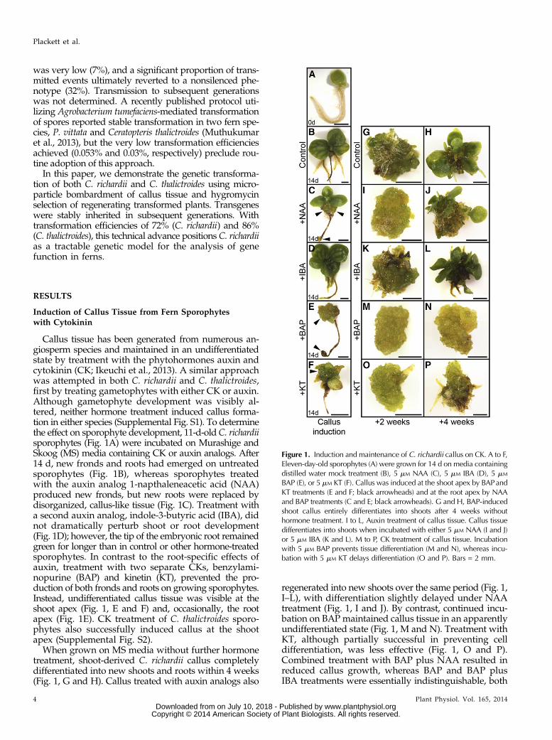
Callus tissue has been generated from numerous an-giosperm species and maintained in an undifferentiatedstate by treatment with the phytohormones auxin andcytokinin (CK; Ikeuchi et al., 2013). A similar approachwas attempted in both C. richardii and C. thalictroides,first by treating gametophytes with either CK or auxin.Although gametophyte development was visibly al-tered, neither hormone treatment induced callus forma-tion in either species (Supplemental Fig. S1). To determinethe effect on sporophyte development, 11-d-oldC. richardiisporophytes (Fig. 1A) were incubated on Murashige andSkoog (MS) media containing CK or auxin analogs. After14 d, new fronds and roots had emerged on untreatedsporophytes (Fig. 1B), whereas sporophytes treatedwith the auxin analog 1-napthaleneacetic acid (NAA)produced new fronds, but new roots were replaced bydisorganized, callus-like tissue (Fig. 1C). Treatment witha second auxin analog, indole-3-butyric acid (IBA), didnot dramatically perturb shoot or root development(Fig. 1D); however, the tip of the embryonic root remainedgreen for longer than in control or other hormone-treatedsporophytes. In contrast to the root-specific effects ofauxin, treatment with two separate CKs, benzylami-nopurine (BAP) and kinetin (KT), prevented the pro-duction of both fronds and roots on growing sporophytes.Instead, undifferentiated callus tissue was visible at theshoot apex (Fig. 1, E and F) and, occasionally, the rootapex (Fig. 1E). CK treatment of C. thalictroides sporo-phytes also successfully induced callus at the shootapex (Supplemental Fig. S2).
When grown on MS media without further hormonetreatment, shoot-derived C. richardii callus completelydifferentiated into new shoots and roots within 4 weeks(Fig. 1, G and H). Callus treated with auxin analogs also
regenerated into new shoots over the same period (Fig. 1,I–L), with differentiation slightly delayed under NAAtreatment (Fig. 1, I and J). By contrast, continued incu-bation on BAP maintained callus tissue in an apparentlyundifferentiated state (Fig. 1, M and N). Treatment withKT, although partially successful in preventing celldifferentiation, was less effective (Fig. 1, O and P).Combined treatment with BAP plus NAA resulted inreduced callus growth, whereas BAP and BAP plusIBA treatments were essentially indistinguishable, both
Figure 1. Induction and maintenance of C. richardii callus on CK. A to F,Eleven-day-old sporophytes (A) were grown for 14 d on media containingdistilled water mock treatment (B), 5 mM NAA (C), 5 mM IBA (D), 5 mM
BAP (E), or 5 mM KT (F). Callus was induced at the shoot apex by BAPandKT treatments (E and F; black arrowheads) and at the root apex by NAAand BAP treatments (C and E; black arrowheads). G and H, BAP-inducedshoot callus entirely differentiates into shoots after 4 weeks withouthormone treatment. I to L, Auxin treatment of callus tissue. Callus tissuedifferentiates into shoots when incubated with either 5 mM NAA (I and J)or 5 mM IBA (K and L). M to P, CK treatment of callus tissue. Incubationwith 5 mM BAP prevents tissue differentiation (M and N), whereas incu-bation with 5 mM KT delays differentiation (O and P). Bars = 2 mm.
4 Plant Physiol. Vol. 165, 2014
Plackett et al.
www.plantphysiol.orgon July 10, 2018 - Published by Downloaded from Copyright © 2014 American Society of Plant Biologists. All rights reserved.

yielding highly friable callus (Supplemental Fig. S3).Fern callus can thus be maintained on BAP or ona combination of BAP plus IBA. To determine calluslongevity, both C. richardii and C. thalictroides calli wererepeatedly subcultured on successive BAP plus IBAtreatments at 14-d intervals. Indeterminate cell fate wassuccessfully maintained in this manner for over a year.
High-Efficiency Transformation of C. richardii andC. thalictroides Using Microparticle Bombardmentand Hygromycin Selection
Callus transformation was carried out using micro-particle bombardment with a hygromycin-selectable35S::GUS construct (pCAMBIA1305.2; see “Materialsand Methods”). To first determine the effectivenessof hygromycin as a selection agent, untransformedC. richardii callus was subjected to a range of antibioticconcentrations across different timeframes. A 2-weekincubation period on 40 mg mL–1 hygromycin B wassufficient to prevent regeneration of shoots from un-transformed callus (Supplemental Fig. S4) and wastherefore used in all subsequent callus selection assays.Test bombardments with either the 35S::GUS con-struct or with uncoated control microparticles were per-formed on both C. richardii and C. thalictroides callus.Callus was incubated on MS media containing CK (5 mM
KT) during and following bombardment to prevent pre-mature tissue differentiation. Callus was transferred tothis media 2 d prior to bombardment and left on thesame media after bombardment for a 3-d recovery pe-riod without antibiotic selection. After that time, GUSstaining analysis was performed on samples of bom-barded C. richardii callus tissue. Figure 2A shows thatcallus bombarded with the 35S::GUS construct exhibi-ted numerous spots of GUS staining, whereas callusbombarded with uncoated microparticles showed none(Fig. 2B). Multiple transformation events had thus takenplace.After the 3-d recovery period, the remaining callus
was transferred to antibiotic selection media, and aweek later, GUS assays were repeated. Once more,spots of GUS expression were visible within the pop-ulation of 35S::GUS-bombarded calli (Fig. 2C) but noton control calli (Fig. 2D). The frequency of spots visibleon callus under selection was visibly reduced comparedwith callus stained immediately after bombardment(compare Fig. 2, A and C). This difference most likelyreflects stable versus transient transformation events.Antibiotic selection was maintained for 14 d, afterwhich time, callus was transferred to nonselective MSmedia to regenerate. At this stage, CK treatment wasstopped. After a further 7 d, small protruding regionsof green tissue were visible on 35S::GUS-bombardedcallus (Fig. 2E), whereas control callus had turned darkbrown and stopped growing (Fig. 2F). Regeneratingtissue subsequently went on to differentiate discrete or-gans, and continued GUS expression in these regeneratingtissues was confirmed by GUS staining (Fig. 2G). Eight
weeks after bombardment, regenerated sporophyteshoots had successfully established an indeterminategrowth pattern (Fig. 2I), with most individual calli regen-erating more than one shoot.
To assess transformation and regeneration frequen-cies, 18 bombardments were performed on C. richardiicallus over a period of 10 weeks, with each replicatecomprising 45 to 60 calli. On average, 81.21% 6 2.45%of calli from each bombardment regenerated at least oneshoot after selection, whereas corresponding control callishowed no regeneration (Table I). Of these regeneratingcalli, 88.06% 6 1.60% exhibited GUS expression when
Figure 2. Transformation, selection, and regeneration of C. richardii callus.A to H, GUS analysis of regenerating callus tissue bombarded with 35S::GUS (A, C, E, and G) or uncoated microparticles (B, D, F, and H) before(AandB),during (CandD),andafter (E–H)antibioticselection.Bars=1mm.I and J, RegeneratingC. richardiiT0 shoots8weeksafter bombardmentwith35S::GUS (I) or uncoated microparticles (J). Bars = 20 mm.
Plant Physiol. Vol. 165, 2014 5
Stable Transformation of Ceratopteris richardii
www.plantphysiol.orgon July 10, 2018 - Published by Downloaded from Copyright © 2014 American Society of Plant Biologists. All rights reserved.
stained, resulting in a final transformation efficiency of71.58% 6 2.56%. A parallel experiment using C. thalic-troides yielded efficiencies of 96.76% 6 0.82%, 88.58% 61.18%, and 85.79% 6 1.62%, respectively.
Once transplanted to soil, maturation of transgenicC. richardii sporophytes took 10 to 14 weeks (harvestingof first spores to harvesting of final spores), with a totalminimum regeneration period of 18 weeks from bom-bardment to harvesting of earliest T1 spores (comparedwith 22 weeks spore to spore for untransformed plants).In comparison, maturation of C. thalictroides after tissueregeneration took 8 to 10 weeks, the total minimum re-generation period of 16 weeks being slightly longer thanthe lifecycle of untransformed plants (11 weeks).
Chimeric Transgene Expression in T0 Transformants
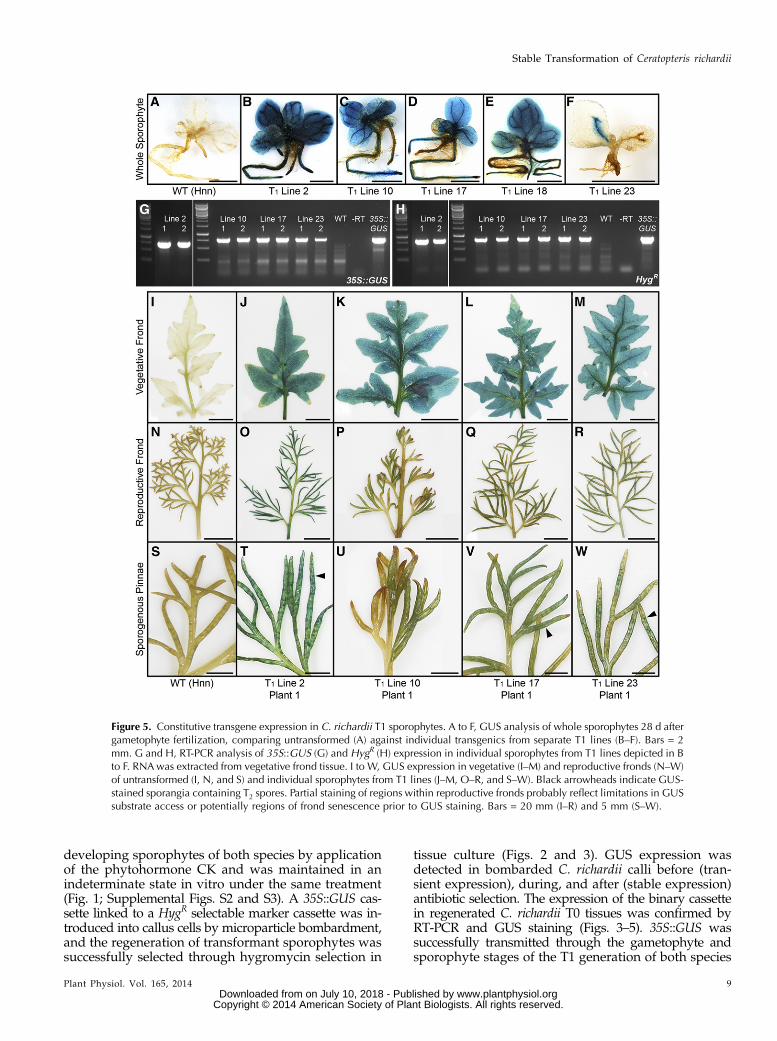
GUS staining of newly regenerated T0 C. richardiishoots revealed two types of expression pattern: ex-pression in all shoot tissues (Fig. 3, A–C) or expressionspecifically in vasculature (Fig. 3, D and E). Across threeindependent bombardments, these occurred at meanfrequencies of 32.60%6 4.22% (all tissues) and 67.40%64.22% (vasculature only) of GUS-expressing shoot clus-ters. There were no instances of these two expressionpatterns coexisting within the same shoot cluster. Thesame patterns of GUS expression were observed inregenerated T0 C. thalictroides shoots (Supplemental Fig.S5) at mean frequencies of 46.95%6 7.95% and 53.05%67.95%, respectively. Regenerated shoots were trans-planted to soil and allowed to mature, with final T0population sizes of 28 (C. richardii) and 43 (C. thalictroides)individuals. GUS staining of sporogenous frond tissuefound the same two patterns in the sporophytes of bothspecies (Fig. 3, F–L; Supplemental Fig. S5) at frequenciesof 28.57% and 55.81% (whole-tissue staining, includingstaining of sporangia; Fig. 3, G, H, and K) and 42.86%and 18.60% (vasculature staining; Fig. 3L) of the C.richardii and C. thalictroides populations, respectively. Thepersistence of these two staining patterns suggests thatexpression patterns established in newly regeneratedshoots are maintained through subsequent development.
Not all of the T0 shoots that regenerated afterhygromycin selection displayed GUS staining. Theextent of GUS expression in regenerating shoots alsovaried, from staining across entire shoot clustersoriginating from a single callus (Fig. 3A) to staining of
individual shoots within a shoot cluster (Fig. 3B) andstaining of sectors within a single shoot or frond (Fig.3C). Staining across entire shoot clusters was observedat mean frequencies of 18.93% 6 2.92% (whole-tissuestaining) and 45.54% 6 2.91% (vasculature only) ofGUS-staining shoots in C. richardii and 25.20% 6 6.37%and 34.78% 6 6.02% in C. thalictroides, respectively. Aproportion (11.94%6 1.60% and 11.42%6 1.18%) of theregenerated C. richardii and C. thalictroides populationsdisplayed no GUS staining at all.
The occasional noncoincidence of GUS staining andhygromycin resistance was further observed in matureT0 sporophytes: 28.57% (C. richardii) and 25.58%(C. thalictroides) of mature individuals sampled withinthe regenerated T0 populations showed no GUS stain-ing. Transfer DNA (T-DNA) expression was analyzed ingreater detail in six T0 C. richardii transformants throughreverse transcription (RT)-PCR (Fig. 3). Of these six, onlytwo (plants 2 and 10) showed GUS staining (Fig. 3,G and H) in conjunction with amplification of both 35S::GUS and hygromycin B resistance (HygR) gene products(Fig. 3, M and N). Two further individuals (plants 16and 17) showed no evidence of transgene expressionexcept hygromycin resistance during tissue regenera-tion, and the remaining two (plants 18 and 23) eachproduced conflicting results: plant 18 was positive forGUS staining but negative for both 35S::GUS and HygR
amplification, whereas plant 23 was positive for GUSstaining and HygR amplification but negative for GUSamplification. Different frond tissues were necessarilysampled for GUS and RT-PCR assays, which mightexplain these discrepancies (see “Discussion”).
Transgene Inheritance and Stable Expression inT1 Transformants
To assess transgene inheritance, T1 spores that wereharvested from 25 C. richardii T0 individuals werescreened for hygromycin resistance (Fig. 4). The possi-bility that T0 transformants are chimeric necessarilycreates the hypothesis that T1 progeny will comprise amix of transformed and untransformed individuals,requiring an efficient method to identify transgenics.Empirical testing determined that 20 mg mL−1 hygrom-ycin is sufficient to kill untransformed, germinatingC. richardii spores (Fig. 4, A and B; Supplemental Fig. S4)and untransformed sporophytes (Supplemental Fig. S4).
Table I. Estimated transformation efficiency after microparticle bombardment of C. richardii and C. thalictroidescallus
Values shown are the means of 18 independent bombardments of each species. All values are expressed aspercentages of the population of callus bombarded 6 SE. Regeneration efficiency refers to the number of separatecalli bombarded that regenerated at least one shoot following hygromycin selection. Final transformation efficiencyrefers to the number of separate calli regenerating at least one shoot that also displayed GUS staining.
Species Mean Regeneration Efficiency Mean Final Transformation Efficiency
% callus
C. richardii 81.21 6 2.45 71.58 6 2.56C. thalictroides 96.76 6 0.82 85.79 6 1.62
6 Plant Physiol. Vol. 165, 2014
Plackett et al.
www.plantphysiol.orgon July 10, 2018 - Published by Downloaded from Copyright © 2014 American Society of Plant Biologists. All rights reserved.

Under this selection regime, spores harvested from 18T0 individuals (72%) produced hygromycin-resistantgametophytes. Approximately 500 to 1,000 spores fromeach line were screened, and compared with unselectedcontrols, the estimated frequency of resistant individ-uals ranged from 1% (T1 line 2; Fig. 4, D and E) to upto 85% (T1 lines 10 and 17; Fig. 4, G, H, J, and K). InC. thalictroides, under similar screening conditions, 94%of lines produced resistant individuals, with frequencieswithin each line over a similar range (Supplemental Fig.S6). GUS staining of hygromycin-resistant T1 individualsrevealed that gametophytes in 90% (C. richardii) and 68%(C. thalictroides) of transgenic lines also expressed GUS.Hygromycin selection of T1 germinating spores is thus ahighly efficient method for identifying lines that carryintact transgenes.Differences in GUS expression were occasionally
found between the gametophyte and sporophyte stagesof some T1 lines, in that constitutive expression was notseen in transgenic gametophytes but was seen in sporo-phytes. No variation in expression pattern was observedbetween individual resistant gametophytes within a line.Seventy percent of C. richardii T1 lines displayed GUSexpression in all gametophyte tissues, including lines2 (Fig. 4F), 10 (Fig. 4I), and 17 (Fig. 4L), the latter dis-playing no GUS expression in the T0 parent (Fig. 3, J andM). Two lines (16 and 18), which had low frequencies of
resistant gametophytes (Fig. 4, M, N, P, and Q), showedGUS expression only in basal thallus tissues and rhizoids(Fig. 4, O and R), again inconsistent with the expressionobserved in the T0 parents (Fig. 3, I and K). A single T1line (23) displayed no GUS expression within the game-tophyte despite a high frequency of hygromycin resis-tance (Fig. 4, S–U). The absence of GUS expression fromthe gametophyte persisted in four out of five T2 linesdescended from this line (Supplemental Fig. S7), whereassporophytic GUS expression was observed in all five.This absence of GUS expression in the gametophytesuggests that transgene expression might be influencedby surrounding genomic sequence, i.e. the transgenemight be located near an element that represses expres-sion in the gametophyte. The occurrence of GUS ex-pression in the gametophyte stage of one descendentT2 line (Supplemental Fig. S7) might therefore have oc-curred through genomic recombination. In C. thalictroidesgametophytes, all GUS staining was constitutivewhere present, but nine lines (32.26%) lacked GUS stain-ing despite being hygromycin resistant (SupplementalFig. S6).
In contrast to the variable staining patterns observed inT1 gametophytes, GUS staining of C. richardii T1 sporo-phytes revealed constitutive expression in all lines tested(Fig. 5, A–E; Supplemental Fig. S8), with the exception ofline 23 (Fig. 5F), where GUS expression (and hygromycin
Figure 3. Chimeric transgene expression in C. richardii T0 transformants. A to E, Typical GUS staining patterns of T0 regen-erated shoots 8 weeks after bombardment, staining either whole tissues (A–C) or restricted to the vasculature (D and E). Bars =2 mm. F to L, GUS-stained sporogenous pinnae from untransformed (F) and mature regenerated T0 sporophytes (G–L). Whitearrowheads indicate GUS-stained sporangia containing T1 spores. Bars = 1 mm. M and N, RT-PCR analysis of 35S::GUS (M)and HygR (N) expression in T0 transformants depicted in G to L. RNA was extracted from sporogenous pinnae.
Plant Physiol. Vol. 165, 2014 7
Stable Transformation of Ceratopteris richardii
www.plantphysiol.orgon July 10, 2018 - Published by Downloaded from Copyright © 2014 American Society of Plant Biologists. All rights reserved.

resistance)was initially lower than in other lines. Notably,vasculature-specific GUS expression was not observed inthe mature sporophyte tissues of any line, even whenpresent in the T0 parent (Fig. 3E; Supplemental Fig. S8).RT-PCR analysis later in sporophyte development con-firmed stable 35S::GUS (Fig. 5G; Supplemental Fig. S8)andHygR (Fig. 5H; Supplemental Fig. S8) expression in allindividuals tested, including those in C. richardii line 23.Constitutive GUS expression in all sporophytes wassubsequently confirmed by GUS staining of vegetative(Fig. 5, I–M; Supplemental Fig. S8) and reproductivefronds (Fig. 5, N–W; Supplemental Fig. S8).
DNA-Blot Analysis of Transformed C. richardii Lines
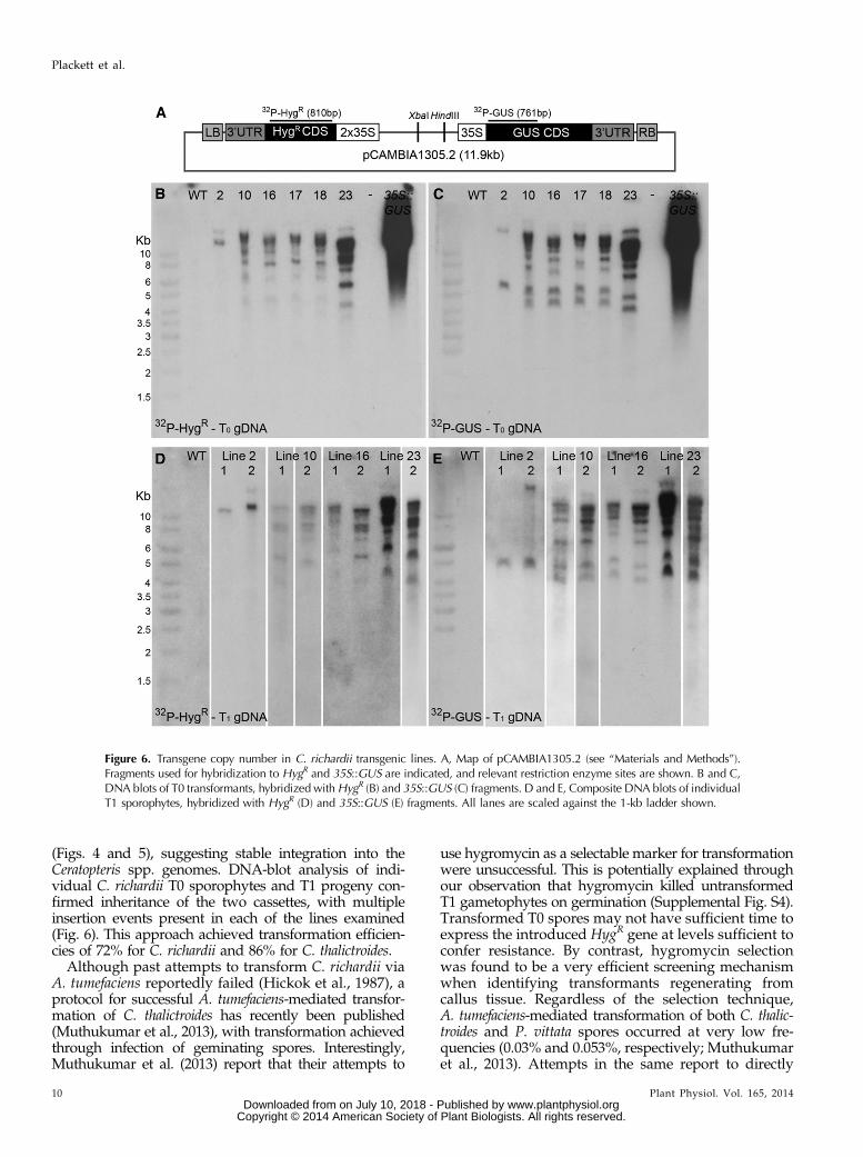
To determine transgene copy number in transformedC. richardii T0 plants, genomic DNA (gDNA) blots werehybridized to fragments of both the HygR and GUSgenes (Fig. 6A). Figure 6, B and C, show that the frag-ments hybridized to multiple copies of each transgenein all T0 transformants tested. The fewest insertionswere found in T0 plant 2, with two HygR and two GUSfragments hybridized. The remaining individuals allcontained in excess of eight copies of each transgene.Hybridization patterns in individuals 10, 16, 17, and 18were very similar, raising the possibility that these lineswere derived from a single transformation event. Im-portantly, in each T0 individual, both HygR and GUSprobes hybridized to genomic fragments greater thanthe size of the introduced plasmid (11.9 kb; Fig. 6A),with at least one instance per individual of a hybridizedfragment being shared between the two probes(Supplemental Fig. S9), supporting linked insertion ofthe HygR and GUS genes.
To assess the inheritance of transgene insertions, DNA-blot analysis was performed on T1 progeny from four ofthe T0 transformants analyzed above. Similar numbers ofhybridized fragments were identified in T1 individuals(Fig. 6, D and E) as in the T0 parents (Fig. 6, B and C). Ofthe two progeny tested from line 2, one (plant 2) dem-onstrated a hybridization pattern very similar to the T0parent for both HygR (Fig. 6, B and D) and GUS (Fig. 6, Cand E) probes, but the second (plant 1) had apparently lostthe insertion carrying the linked transgenes (SupplementalFig. S9). The T1 individuals tested from other lines alsodemonstrated very similar hybridization patterns totheir T0 parents, including the presence of linked 35S::GUS and HygR cassettes (Supplemental Fig. S9). Trans-gene insertions thus remained stably integrated within theC. richardii genome between the T0 and T1 generations,and linkage was maintained through meiosis.
DISCUSSION
Stable transformation of C. richardii and its sister species,C. thalictroides, has been achieved using a combination oftissue culture, microparticle bombardment, and antibioticselection. Callus was initiated from the apical region of
Figure 4. Hygromycin resistance and GUS expression are maintainedin C. richardii T1 gametophytes. Antibiotic resistance screening andGUS analysis of untransformed gametophytes (A–C) and T1 progeny ofT0 transformants (D–U). Growth was compared between control media(A, D, G, J, M, P, and S) and media containing 20 mg mL–1 hygromycin(B, E, H, K, N, Q, and T), sufficient to kill wild-type (WT) gameto-phytes at spore germination (B; Supplemental Fig. S4). GUS ex-pression was compared between untransformed gametophytes (C)and T1 gametophytes resistant to hygromycin (F, I, L, O, R, and U).Bars = 200 mm.
8 Plant Physiol. Vol. 165, 2014
Plackett et al.
www.plantphysiol.orgon July 10, 2018 - Published by Downloaded from Copyright © 2014 American Society of Plant Biologists. All rights reserved.
developing sporophytes of both species by applicationof the phytohormone CK and was maintained in anindeterminate state in vitro under the same treatment(Fig. 1; Supplemental Figs. S2 and S3). A 35S::GUS cas-sette linked to a HygR selectable marker cassette was in-troduced into callus cells by microparticle bombardment,and the regeneration of transformant sporophytes wassuccessfully selected through hygromycin selection in
tissue culture (Figs. 2 and 3). GUS expression wasdetected in bombarded C. richardii calli before (tran-sient expression), during, and after (stable expression)antibiotic selection. The expression of the binary cassettein regenerated C. richardii T0 tissues was confirmed byRT-PCR and GUS staining (Figs. 3–5). 35S::GUS wassuccessfully transmitted through the gametophyte andsporophyte stages of the T1 generation of both species
Figure 5. Constitutive transgene expression in C. richardii T1 sporophytes. A to F, GUS analysis of whole sporophytes 28 d aftergametophyte fertilization, comparing untransformed (A) against individual transgenics from separate T1 lines (B–F). Bars = 2mm. G and H, RT-PCR analysis of 35S::GUS (G) and HygR (H) expression in individual sporophytes from T1 lines depicted in Bto F. RNAwas extracted from vegetative frond tissue. I to W, GUS expression in vegetative (I–M) and reproductive fronds (N–W)of untransformed (I, N, and S) and individual sporophytes from T1 lines (J–M, O–R, and S–W). Black arrowheads indicate GUS-stained sporangia containing T2 spores. Partial staining of regions within reproductive fronds probably reflect limitations in GUSsubstrate access or potentially regions of frond senescence prior to GUS staining. Bars = 20 mm (I–R) and 5 mm (S–W).
Plant Physiol. Vol. 165, 2014 9
Stable Transformation of Ceratopteris richardii
www.plantphysiol.orgon July 10, 2018 - Published by Downloaded from Copyright © 2014 American Society of Plant Biologists. All rights reserved.
(Figs. 4 and 5), suggesting stable integration into theCeratopteris spp. genomes. DNA-blot analysis of indi-vidual C. richardii T0 sporophytes and T1 progeny con-firmed inheritance of the two cassettes, with multipleinsertion events present in each of the lines examined(Fig. 6). This approach achieved transformation efficien-cies of 72% for C. richardii and 86% for C. thalictroides.
Although past attempts to transform C. richardii viaA. tumefaciens reportedly failed (Hickok et al., 1987), aprotocol for successful A. tumefaciens-mediated transfor-mation of C. thalictroides has recently been published(Muthukumar et al., 2013), with transformation achievedthrough infection of geminating spores. Interestingly,Muthukumar et al. (2013) report that their attempts to
use hygromycin as a selectable marker for transformationwere unsuccessful. This is potentially explained throughour observation that hygromycin killed untransformedT1 gametophytes on germination (Supplemental Fig. S4).Transformed T0 spores may not have sufficient time toexpress the introduced HygR gene at levels sufficient toconfer resistance. By contrast, hygromycin selectionwas found to be a very efficient screening mechanismwhen identifying transformants regenerating fromcallus tissue. Regardless of the selection technique,A. tumefaciens-mediated transformation of both C. thalic-troides and P. vittata spores occurred at very low fre-quencies (0.03% and 0.053%, respectively; Muthukumaret al., 2013). Attempts in the same report to directly
Figure 6. Transgene copy number in C. richardii transgenic lines. A, Map of pCAMBIA1305.2 (see “Materials and Methods”).Fragments used for hybridization to HygR and 35S::GUS are indicated, and relevant restriction enzyme sites are shown. B and C,DNA blots of T0 transformants, hybridized withHygR (B) and 35S::GUS (C) fragments. D and E, Composite DNA blots of individualT1 sporophytes, hybridized with HygR (D) and 35S::GUS (E) fragments. All lanes are scaled against the 1-kb ladder shown.
10 Plant Physiol. Vol. 165, 2014
Plackett et al.
www.plantphysiol.orgon July 10, 2018 - Published by Downloaded from Copyright © 2014 American Society of Plant Biologists. All rights reserved.

transform a population of P. vittata spores by micro-particle bombardment similarly yielded a very lowtransformation efficiency of only 0.012%. The availabledata thus suggest that transformation of spores is aninherently less efficient method than transformation ofcallus tissue.Using the protocol described in this paper, the time
from callus transformation to recovery of T1 spores was18 and 16 weeks for C. richardii and C. thalictroides, re-spectively, in contrast to an 11- to 13-week period forC. thalictroides via A. tumefaciens-mediated transforma-tion (Muthukumar et al., 2013). Despite the longer timeframe, transformation of the sporophyte generationconfers the advantage of direct production of T1 sporeswithout an intervening recombination/outcrossing event(sexual reproduction of T0 gametophytes). Outcrossingof T0 transformant gametophytes could conceivablyreduce initial transgene copy number; however, crossfertilization of two independent transformants is alsopossible. Notably, Muthukumar et al. (2013) reportoccurrences of multiple transgene insertions, similar tothose observed using microparticle bombardment. Over-all, the initial high efficiency of transformation offered bymicroparticle bombardment and the concomitant abilityto screen effectively using hygromycin offset the slightlylonger T0 generation time required and provide signifi-cant benefits over the A. tumefaciens-mediated protocol.
Evidence for a Conserved Role of CK in the ShootMeristem of Ferns and Angiosperms
Treatment with CK was sufficient to induce the for-mation of callus tissue in place of new fronds at the shootapex of C. richardii and C. thalictroides sporophytes and tomaintain the callus in an undifferentiated state (Fig. 1;Supplemental Fig. S2). Application of auxin failed toinduce shoot callus but did perturb cellular activityspecifically at the root apex. It was recently reported thatauxin treatment of the lycophyte Selaginella kraussianasimilarly disturbs root organization (Sanders and Langdale,2013). Organogenesis in the C. richardii shoot arisesfrom divisions of a single tetrahedral apical cell (Houand Hill, 2002), instead of a multicellular meristem asfound in angiosperms (Sussex, 1989). The observed for-mation of callus suggests that CK treatment can blockthe differentiation of cells derived from the apical initialcell. In Arabidopsis, CK acts downstream of knotted1-likehomeobox genes to maintain undifferentiated cell fate inthe shoot apex, in part through antagonism of thegibberellin signaling pathway (Jasinski et al., 2005;Bartrina et al., 2011). Given the results observed here, itis possible that CK acts in both ferns and angiospermsto promote indeterminate cell fate at the shoot apex.Interestingly, CK treatment did not induce callus for-mation from C. richardii or C. thalictroides gametophytes,although morphology at the notch meristem was slightlyaffected (Supplemental Fig. S1). This could indicatethat the mechanisms regulating initial cell specifica-tion differ between the notch meristem and shoot apexand could reflect an important distinction between
two-dimensional (gametophyte) and three-dimensional(sporophyte) growth and patterning.
Developmental Trajectories Explain Chimeric versusStable Transgene Expression in T0 and T1 Generations
The transmission of transgenes from regenerated T0plants to stable T1 lines was assessed in C. richardiiby comparing the transgenic status of T0 parents(as assessed by GUS staining; Fig. 3; SupplementalFig. S5) to their T1 progeny (as assessed by gametophytehygromycin resistance; Fig. 4; Supplemental Fig. S6).Sixty percent of lines demonstrated consistent transgenicstatus between the T0 and T1 generations, i.e. both par-ent and offspring were transgenic (52%) or neither parentnor offspring were transgenic (8%). The remaining 40%of lines showed inconsistent inheritance patterns, withtransgenic T1 individuals identified from apparentlynontransgenic T0 parents (20%) or nontransgenic T1progeny descending from apparently transgenic T0parents (20%). In C. thalictroides, corresponding frequen-cies of 67% consistent (64% plus 3%) and 33% inconsis-tent (3% and 30%) lines were recorded. A distinction inGUS expression patterns between the T0 and T1 gener-ations was also observed, with frequent examples ofpartial or tissue-specific (vasculature) GUS staining in T0regenerated sporophytes (young and mature) but con-stitutive GUS expression in subsequent T1 progeny. Alow incidence (11%–12%) of regenerated shoot clustersthat lacked any GUS staining could reflect stable trans-formation with only the HygR cassette or subsequent lossof the 35S::GUS cassette after hygromycin selection.Noncongruence of GUS staining patterns between the T0and T1 generations is unlikely to reflect technical issues ofGUS substrate penetration given the examples of consti-tutive staining observed. Instead, these results are mostsimply explained through the regeneration of chimeric T0transformants, as previously seen in transformed game-tophytes (Rutherford et al., 2004). This interpretationwould also explain the observed discrepancies betweenGUS analysis, antibiotic selection, and transgene expres-sion data in T0 plants (Fig. 3), as different fronds, po-tentially not all of them transgenic, were sampled foreach analysis.
The emergence of chimeric shoots does not correspondwith our current understanding of fern shoot develop-ment from single apical initial cells (Hou and Hill, 2002;Sanders et al., 2011). However, in accordance with theobservations made above regarding callus induction, wehypothesize that induction of callus through CK treat-ment prevents the mitotic derivatives of the apical initialfrom differentiating into frond and root initials and thusartificially expands the population of undifferentiatedshoot apical initial cells. When this constraint is removedafter bombardment, normal developmental patterningand gradients are presumably reimposed onto an ab-normally large population of undifferentiated cells. Thiscould theoretically result in the incorporation of multipleapical initials into single regenerating shoots, allowing
Plant Physiol. Vol. 165, 2014 11
Stable Transformation of Ceratopteris richardii
www.plantphysiol.orgon July 10, 2018 - Published by Downloaded from Copyright © 2014 American Society of Plant Biologists. All rights reserved.

the formation of different tissue types from distinct sub-populations of initials. This scenario could theoreticallyexplain our observations of vasculature-specific GUSstaining patterns. However, given that regenerated plantsappear morphologically similar to untreated controls,such a hypothesis would also imply that fern shoots andorgans can successfully organize both from a foundingpopulation of multiple cells and from single initials.
In contrast to the scenario proposed above for T0transformants, T1 individuals must necessarily arise froma single progenitor cell (spore and zygote); thus, all cellswithin the individual have a common genetic ancestry.This is evidenced by the stable and constitutive GUSexpression observed in the T1 generation (Figs. 4 and 5;Supplemental Figs. S6 and S8). In the case of T1 game-tophytes, variations in the frequency of transgenicswithin each line (Fig. 4) most likely reflect the frequencyof transformed sporangia on chimeric fronds: each spo-rangium arises from a single separate initial cell (Hill,2001). This conclusion is supported by observations of T0pinnae containing both GUS-stained and unstained spo-rangia (Fig. 3H; Supplemental Fig. S5). Importantly, therecovery of constitutively expressing T1 progeny fromapparently chimeric T0 transformants demonstrates thatchimeric expression in the T0 generation does not rep-resent a barrier to establishing stable and pure-breedingtransgenic lines and argues that screening for stabletransformants should occur in the T1 generation insteadof in regenerated T0 shoots.
Transgene Copy Number
Microparticle bombardment resulted in the incorpo-ration of multiple T-DNA fragments into the genomesof individual C. richardii T0 transformants, includingfragmented copies of individual expression cassettes.The complex insertion of multiple transgene copies isa known factor in biolistic-based transformation tech-niques (Hansen and Wright, 1999) and can in partbe mitigated through bombardment with linearizedDNA (Lowe et al., 2009). In all of the T0 plants tested,the presence of linked HygR and 35S::GUS cassetteswas revealed by shared hybridization to large gDNAfragments. Importantly, very little rearrangement ofhybridization fragments was observed between the T0and T1 generations, suggesting that transgene inser-tions remain essentially stable after initial integration.Within the relatively small sample of nine transgeniclines selected for DNA-blot analysis in this study, one(line 2) was found to carry an entire transgene con-taining both the HygR and 35S::GUS cassettes, plus asingle unlinked copy of both the HygR and 35S::GUScassettes. This observation suggests that the isolationof single insertion lines is feasible, especially if coupledwith outcrossing to untransformed individuals. Al-though microparticle bombardment typically results ina higher T-DNA copy number in transgenics thanA. tumefaciens-mediated transformation, as demonstratedby side-by-side transformations into barley (Hordeum
vulgare; Travella et al., 2005), recent A. tumefaciens-mediated transformation ofC. thalictroides resulted in simi-lar copy numbers to those reported here (Muthukumaret al., 2013).
C. richardii as a Fern Genetic Model
The protocol described in this paper was able tosuccessfully generate stable transformants in C. richardiiand C. thalictroides at high efficiencies and may be moregenerally applicable to other fern species. C. richardii haspreviously been proposed as a candidate model fern;examples from a number of important classes of tran-scription factor have already been identified (Hasebeet al., 1998; Aso et al., 1999; Himi et al., 2001; Sano et al.,2005), and phylogenetic and cross-species complemen-tation analyses have been published, for example withCrLEAFY (Maizel et al., 2005). With its smaller size,more rapid lifecycle, and smaller genome (3.7 Gbp;Bennett and Leitch, 2001), C. thalictroides must now alsobe seriously considered as a candidate model. Althoughthe advantage of a smaller genome in this species isoffset by polyploidy in relation to C. richardii (McGrathet al., 1994), the power of C. richardii as a genetic tool ismost likely to be further enhanced by the availability ofC. thalictroides as a closely related, comparable, geneti-cally tractable species. The advent of a high-efficiencystable transformation system in C. richardii and C. tha-lictroides removes one of the final technical barriers forthe adoption of ferns as genetic models.
MATERIALS AND METHODS
Plant Material and Growth Conditions
All plant material was derived from Ceratopteris richardii strain Hnn (Hickoket al., 1995) and Ceratopteris thalictroides strain C-21 (Carolina Biological Sup-plies). Plants were grown in Sanyo MLR-350H incubators (Panasonic) at 28°C,90% humidity, 16 h of light/8 h of dark, and fluence of 150 mmol m–2 s–1.Gametophytes were grown and fertilized on 1% (w/v) agar media (pH 6.0)containing Ceratopteris (C-fern) nutrient mix (prepared following protocol inHickok and Warne, 1998). Sporophytes were subsequently transplanted toSinclair potting growing medium (William Sinclair Horticulture), typicallywhen the third frond had expanded. Callus tissue was cultured on 0.7% (w/v)agar media (pH 5.8) containing 13 MS nutrients (Duchefa Biochemie) and2% (w/v) Suc and supplemented with hormone (5 mM) and/or hygromycin B(40 mg mL–1) treatments (Sigma Aldrich) as specified in the text. Attempts togrow callus on C-fern media were not successful (Supplemental Fig. S3). Allhormone stocks were prepared to 1,0003 working concentration in 1 N NaOH.Hygromycin B was prepared in distilled water as a 1,0003 stock.
Spores were sterilized by incubating for 10 min at room temperature insodium hypochlorite solution (2% [v/v] chlorine) and 0.1% (v/v) Tween, whichwas subsequently removed by six sequential rinses in sterile distilled water.Sterile spores were imbibed in distilled water and incubated for 48 h at roomtemperature in darkness before sowing. Gametophytes were fertilized between9 and 11 d after germination by the application of sterile distilled water.Fertilization of transgenic T1 gametophytes was not successful if grown underhygromycin selection. T1 sporophytes were recovered through fertilizationwithin T1 gametophyte populations grown without hygromycin selection andtransgenic individuals subsequently identified though hygromycin selection onC-fern media. Twenty micrograms per milliliter hygromycin is sufficient to killuntransformed young sporophytes within 7 d (Supplemental Fig. S4). Trans-genic sporophytes were removed from selection after 14 d and transplanted tosoil.
12 Plant Physiol. Vol. 165, 2014
Plackett et al.
www.plantphysiol.orgon July 10, 2018 - Published by Downloaded from Copyright © 2014 American Society of Plant Biologists. All rights reserved.
Ceratopteris spp. Microparticle Bombardment
All callus utilized in bombardments was derived from sporophyte shoottissues under BAP treatment; root-derived callus excised from NAA-treatedC. richardii sporophytes differentiated solely into roots within 14 d, irrespectiveof hormone treatment (Supplemental Fig. S3). Bombardments utilized the 35S::GUS plasmid pCAMBIA1305.2 (Cambia). Bombardment was performed using aPDS-1000/He biolistic delivery system (Bio-Rad). Tungsten M-20 microparticles(diameter approximately 1.3 mm; Bio-Rad) were prepared for bombardmentaccording to Sanford et al. (1993). Bombardments were performed according tothe manufacturer’s instructions. Transgenic plants were regenerated frombombardment of 2-week-old callus tissue at 900 pounds per square inch undervacuum conditions of 28 pounds per square inch, with callus tissue placed at adistance of 6 cm from the firing disc. Bombarded callus tissue was allowed torecover for 3 d before transfer to hygromycin selection. All mean efficiencyvalues are expressed as a percentage of the bombarded population 6 SE.
Analysis of Transgenic Lines
gDNA was extracted from sporophyte frond tissues using a hexadecyl-trimethylammonium bromide based protocol modified from Porebskiet al. (1997). Hexadecyltrimethylammonium bromide (2%) extraction bufferalso contained 2% (w/v) polyvinylpyrrolidone-40 (PVP-40), 0.3% (v/v)b-mercaptoethanol, and 50 mg mL–1 ribonuclease A (Sigma Aldrich), extractinggDNA from #1 g of tissue in 10 mL of buffer. Chloroform:isoamyl alcohol(24:1) extraction was performed three times, and NaCl/ethanol precipitationwas performed twice. gDNA was resuspended in 13 Tris-EDTA buffer (pH 8.0)at 4°C overnight.
RNA was extracted from #100 mg of sporophyte frond tissues using theRNeasy RNA extraction kit (Qiagen). Complementary DNA was synthesizedfrom 250 ng of RNA template using Superscript III Reverse Transcriptase(Life Technologies). Genotyping PCR and RT-PCR were performed usingthe following primer pairs: HygF2 (CTTCTACACAGCCATCGGTC) and HygR(CCGATGGTTTCTACAAAGATCG) and GUSF (CTTGCCATCCTTGTCCTCC)and GUSR4 (CGAAGTTCGGCTTGTTACG).
C. richardii gDNA (10 mg per sample) was prepared for DNA blotting bydigestion with HindIII and XbaI (New England Biolabs) and separated by gelelectrophoresis (25 V, 16 h). Blots were prepared and hybridized to 32P-dCTP-labeled DNA probes as described by Langdale et al. (1988). Probes against theHygR and 35S::GUS cassettes were synthesized from 810- and 761-bp fragments(Fig. 6A), using the Redprime II DNA labeling kit (G.E. Healthcare). Primersused to synthesize probe templates are as described above for PCR.
Histochemical GUS staining was performed on gametophyte and sporophytetissues using 0.5 mg mL–1 5-bromo-4-chloro-3-indolyl-b-D-glucuronic acid(Melford) at 37°C for 16 h, following 20-min pretreatment in 90% (v/v) acetoneat 4°C. GUS-stained tissue was cleared by incubation in 70% (v/v) ethanol.
Supplemental Data
The following materials are available in the online version of this article.
Supplemental Figure S1. CK and auxin treatments do not induce callusformation in gametophytes of C. richardii or C. thalictroides.
Supplemental Figure S2. CK treatment induces shoot apical callus forma-tion in C. thalictroides sporophytes.
Supplemental Figure S3. Maintenance of undifferentiated C. richardii cal-lus on CK-MS media.
Supplemental Figure S4. Hygromycin sensitivity of C. richardii callus, ga-metophytes, and sporophytes.
Supplemental Figure S5. Chimeric GUS expression in C. thalictroides T0transformants.
Supplemental Figure S6. Stable GUS expression in C. thalictroides T1 trans-genic lines.
Supplemental Figure S7. Absence of GUS expression during gametophytedevelopment of C. richardii transgenic line 23 persists in the T2generation.
Supplemental Figure S8. Transgene expression patterns change from chi-meric to stable between the T0 and T1 generations of C. richardii trans-genic lines 24, 25, and 28.
Supplemental Figure S9. HygR and 35S::GUS remain linked between theT0 and T1 generations of C. richardii transgenic lines.
ACKNOWLEDGMENTS
We thank Julie Bull for help with plant maintenance and Laura Moody andMara Schuler for useful comments on the manuscript.
Received October 28, 2013; accepted March 11, 2014; published March 12,2014.
LITERATURE CITED
Aso K, Kato M, Banks JA, Hasebe M (1999) Characterization of homeodomain-leucine zipper genes in the fern Ceratopteris richardii and the evolution of thehomeodomain-leucine zipper gene family in vascular plants. Mol Biol Evol16: 544–552
Banks JA (1997) Sex determination in the fern Ceratopteris. Trends Plant Sci2: 175–180
Banks JA (1999) Gametophyte development in ferns. Annu Rev PlantPhysiol Plant Mol Biol 50: 163–186
Bartrina I, Otto E, Strnad M, Werner T, Schmülling T (2011) Cytokinin regu-lates the activity of reproductive meristems, flower organ size, ovule for-mation, and thus seed yield in Arabidopsis thaliana. Plant Cell 23: 69–80
Bennett MD, Leitch IJ (2001) Nuclear DNA amounts in Pteridophytes. AnnBot (Lond) 87: 335–345
Chatterjee A, Roux SJ (2000) Ceratopteris richardii: a productive model forrevealing secrets of signaling and development. J Plant Growth Regul19: 284–289
Hansen G, Wright MS (1999) Recent advances in the transformation ofplants. Trends Plant Sci 4: 226–231
Hasebe M, Wen CK, Kato M, Banks JA (1998) Characterization of MADShomeotic genes in the fern Ceratopteris richardii. Proc Natl Acad Sci USA95: 6222–6227
Hickok LG, Warne TR (1998) C-Fern Manual. Carolina Biological SupplyCompany, Burlington, VT
Hickok LG, Warne TR, Fribourg RS (1995) The biology of the fern Cera-topteris and its use as a model system. Int J Plant Sci 156: 332–345
Hickok LG, Warne TR, Slocum MK (1987) Ceratopteris richardii: applica-tions for experimental plant biology. Am J Bot 74: 1304–1316
Hill JP (2001) Meristem development at the sporophyll pinna apex in Ce-ratopteris richardii. Int J Plant Sci 162: 235–247
Himi S, Sano R, Nishiyama T, Tanahashi T, Kato M, Ueda K, Hasebe M(2001) Evolution of MADS-box gene induction by FLO/LFY genes. J MolEvol 53: 387–393
Hou GC, Hill JP (2002) Heteroblastic root development in Ceratopteris ri-chardii (Parkeriaceae). Int J Plant Sci 163: 341–351
Hou GC, Hill JP (2004) Developmental anatomy of the fifth shoot-borneroot in young sporophytes of Ceratopteris richardii. Planta 219: 212–220
Ikeuchi M, Sugimoto K, Iwase A (2013) Plant callus: mechanisms of in-duction and repression. Plant Cell 25: 3159–3173
Indriolo E, Na G, Ellis D, Salt DE, Banks JA (2010) A vacuolar arsenitetransporter necessary for arsenic tolerance in the arsenic hyperaccumulatingfern Pteris vittata is missing in flowering plants. Plant Cell 22: 2045–2057
Jasinski S, Piazza P, Craft J, Hay A, Woolley L, Rieu I, Phillips A, HeddenP, Tsiantis M (2005) KNOX action in Arabidopsis is mediated by co-ordinate regulation of cytokinin and gibberellin activities. Curr Biol 15:1560–1565
Johnson GP, Renzaglia KS (2008) Embryology of Ceratopteris richardii(Pteridaceae, tribe Ceratopterideae), with emphasis on placental devel-opment. J Plant Res 121: 581–592
Kawai-Toyooka H, Kuramoto C, Orui K, Motoyama K, Kikuchi K,Kanegae T, Wada M (2004) DNA interference: a simple and efficientgene-silencing system for high-throughput functional analysis in thefern adiantum. Plant Cell Physiol 45: 1648–1657
Langdale JA, Rothermel BA, Nelson T (1988) Cellular pattern of photosyntheticgene expression in developing maize leaves. Genes Dev 2: 106–115
Leroux O, Eeckhout S, Viane RL, Popper ZA (2013) Ceratopteris richardii(C-fern): a model for investigating adaptive modification of vascularplant cell walls. Front Plant Sci 4: 367
Plant Physiol. Vol. 165, 2014 13
Stable Transformation of Ceratopteris richardii
www.plantphysiol.orgon July 10, 2018 - Published by Downloaded from Copyright © 2014 American Society of Plant Biologists. All rights reserved.

Lowe BA, Shiva Prakash N, Way M, Mann MT, Spencer TM, BoddupalliRS (2009) Enhanced single copy integration events in corn via par-ticle bombardment using low quantities of DNA. Transgenic Res 18:831–840
Maizel A, Busch MA, Tanahashi T, Perkovic J, Kato M, Hasebe M,Weigel D (2005) The floral regulator LEAFY evolves by substitutions inthe DNA binding domain. Science 308: 260–263
McGrath JM, Hickok LG, Pichersky E (1994) Assessment of gene copynumber in the homosporous ferns Ceratopteris thalictroides and C. ri-chardii (Parkeriaceae) by restriction fragment length polymorphisms.Plant Syst Evol 189: 203–210
Muthukumar B, Joyce BL, EllessMP, Stewart N (2013) Stable transformation offerns using spores as targets: Pteris vittata (Chinese brake fern) and Cera-topteris thalictroides (C-fern ‘Express’). Plant Physiol 163: 648–658
Nakazato T, Jung MK, Housworth EA, Rieseberg LH, Gastony GJ (2006)Genetic map-based analysis of genome structure in the homosporousfern Ceratopteris richardii. Genetics 173: 1585–1597
Porebski S, Bailey LG, Baum BR (1997) Modification of a CTAB DNAextraction protocol for plants containing high polysaccharide andpolyphenol components. Plant Mol Biol Rep 15: 8–15
Pryer KM, Schneider H, Smith AR, Cranfill R, Wolf PG, Hunt JS, SipesSD (2001) Horsetails and ferns are a monophyletic group and the closestliving relatives to seed plants. Nature 409: 618–622
Rutherford G, Tanurdzic M, Hasebe M, Banks JA (2004) A systemic genesilencing method suitable for high throughput, reverse genetic analysesof gene function in fern gametophytes. BMC Plant Biol 4: 6
Sanders HL, Darrah PR, Langdale JA (2011) Sector analysis and predictivemodelling reveal iterative shoot-like development in fern fronds. De-velopment 138: 2925–2934
Sanders HL, Langdale JA (2013) Conserved transport mechanisms but distinctauxin responses govern shoot patterning in Selaginella kraussiana. New Phytol198: 419–428
Sanford JC, Smith FD, Russell JA (1993) Optimizing the biolistic processfor different biological applications. Methods Enzymol 217: 483–509
Sano R, Juárez CM, Hass B, Sakakibara K, Ito M, Banks JA, Hasebe M(2005) KNOX homeobox genes potentially have similar function in bothdiploid unicellular and multicellular meristems, but not in haploidmeristems. Evol Dev 7: 69–78
Stout SC, Clark GB, Archer-Evans S, Roux SJ (2003) Rapid and efficientsuppression of gene expression in a single-cell model system, Cera-topteris richardii. Plant Physiol 131: 1165–1168
Sussex IM (1989) Developmental programming of the shoot meristem. Cell56: 225–229
Travella S, Ross SM, Harden J, Everett C, Snape JW, Harwood WA (2005) Acomparison of transgenic barley lines produced by particle bombardmentand Agrobacterium-mediated techniques. Plant Cell Rep 23: 780–789
14 Plant Physiol. Vol. 165, 2014
Plackett et al.
www.plantphysiol.orgon July 10, 2018 - Published by Downloaded from Copyright © 2014 American Society of Plant Biologists. All rights reserved.