chondroitin sulfate-based biomaterials for tissue...
TRANSCRIPT
290
http://journals.tubitak.gov.tr/biology/
Turkish Journal of Biology Turk J Biol(2016) 40: 290-299© TÜBİTAKdoi:10.3906/biy-1507-16
Chondroitin sulfate-based biomaterials for tissue engineering
Hyuck Joon KWON1,*, Youngbae HAN2
1Department of Physical Therapy and Rehabilitation, College of Health Science, Eulji University, Gyeonggi, Korea 2Department of Mechanical and Design Engineering, Hongik University, Sejong city, Korea
* Correspondence: [email protected]
1. Introduction Tissue engineering, which aims to create scaffolding materials for regenerating tissue equivalents of blood vessels, heart muscle, nerves, cartilage, bone, and others using the recruitment of native cells into the scaffold and subsequent deposition of extracellular matrix (ECM), requires the multidisciplinary principles of materials science, medicine, and life science (Lee and Yuk, 2007; Lee and Mooney, 2012; Pawar and Edgar, 2012). Biomaterials play an essential role for regenerating defective tissues by supporting and retaining cells structurally in the defective area (Tan et al., 2007). The properties of biomaterials should include biocompatibility, desired mechanical strength, microstructure and degradation rate, and capability to support cell residence and to retain metabolic functions (Kharkar et al., 2013). Natural polymers composed of ECM are highly potential building blocks for preparing biomaterials for tissue engineering because natural polymers have chemical versatility and biological performance. Among the components of ECM, glycosaminoglycans (GAGs) are complex polysaccharides that play important roles in cell growth, differentiation, morphogenesis, cell migration, and bacterial/viral infections (Yamada et al., 2011). GAGs can be classified as nonsulfated GAGs, such as hyaluronic acid (HA), and
sulfated GAGs, such as heparin sulfate, chondroitin sulfate, dermatan sulfate, and keratin sulfate (Schnabelrauch et al., 2013). GAG chains are attached to a central protein to form the proteoglycans with the exception of HA. GAGs are able to absorb large quantities of water due to their polyelectrolyte nature and this osmotic swelling provides compressive strength. Chondroitin sulfate (CS) is widely distributed as one of the most physiologically important GAGs in ECMs and at cell surfaces. CS has been found to be involved in the signaling functions of various growth factors and chemokines such as platelet-derived growth factors (PDGFs), fibroblast growth factors (FGFs), epidermal growth factors (EGFs), and transforming growth factor betas (TGF-βs) (Fthenou et al., 2008; Sirko et al., 2010; Hintze et al., 2012) and play critical roles in the developmental processes (Knudson and Knudson, 2001; Hwang et al., 2003; Laabs, 2005). CS also contributes to stabilization of the active conformation of growth factors by protecting them from fast degradation (Taipale and Keski-Oja, 1997; Wang et al., 2008). CS plays important roles in modulating the stability, activity, release, and spatial localization of growth factors, which are closely associated with the sulfation patterns of the CSs (Deepa et al., 2002; Takagaki et al., 2002). Thus, CS has been considered as a potential material for tissue engineering.
Abstract: Chondroitin sulfate (CS) is a sulfated glycosaminoglycan (GAG) that is usually found attached to proteins as part of a proteoglycan. The use of CS-based biomaterials in the field of tissue engineering applications has been intensively growing over the past decades because CS is a biopolymer with the major advantages of being biodegradable, biocompatible, easily available, and highly versatile. In vitro and in vivo studies have shown that CS-based biomaterials upregulate cartilage-specific gene expression in chondrocytes and mesenchymal stem cells and also support osteogenic differentiation by increasing the effectiveness of bone anabolic growth factors. Chondroitin sulfate proteoglycan plays a key role during the acute recovery stage after spinal cord injury by activating microglia/macrophages and modulating neurotrophic factor secretion. In addition, CS-based materials promote the wound-healing process and stimulate the regeneration of skin defects. Moreover, CS can be used to construct high toughness gels by having a double network structure. Taken together, CS-based biomaterials would be a useful material for successful replacement and regeneration of damaged cartilage, bone, skin, and neural tissues.
Key words: Chondroitin sulfate, tissue engineering, regenerative medicine, scaffold, stem cell
Received: 03.07.2015 Accepted/Published Online: 12.11.2015 Final Version: 23.02.2016
Review Article

KWON and HAN / Turk J Biol
291
However, since the water-soluble nature of CS limits its application as a biomaterial, crosslinking treatment is required for tailoring the properties of CS or combining it with other stable polymers such as chitosan (CHS), gelatin (Gel), and HA (Sintov et al., 1995; Park et al., 2000; Chang et al., 2003; Fan et al., 2006; Flanagan et al., 2006; Wang et al., 2007). This review focuses on the biological role of CS and its application for tissue engineering. The application of CS-based materials for repair and regeneration of various tissues such as cartilage, bone, neural tissues, and skin is summarized in this review.
2. Fundamental properties of chondroitin sulfate CS is composed of repeating disaccharide unit polymers of D-glucuronic acid and N-acetyl galactosamine sulfated at either 4- or 6-positions (Figure 1). CS can be classified as galactosaminoglycan because CS has the characteristic disaccharide units of glucuronic acid and N-acetylgalactosamine. CS is synthesized and sulfated intracellularly, and then secreted extracellularly and attached glycosidically to serine in core proteins through the glucuronic acid–galactose–galactose–xylose to produce proteoglycan such as aggrecan and versican (Dick et al., 2012). The biosynthesis of chondroitin sulfate proteoglycans (CSPGs) is a very complex process that requires glycosyltransferases, glucuronyltransferase-I, the chondroitin synthase family of enzymes, and chondroitin polymerizing factor, broadly distributed in tissues (Mikami and Kitagawa, 2013). In addition, sulfotransferase catalyzes the transfer of a sulfate group from 3’-phosphoadenosine 5’-phosphosulfate (PAPS) to the sulfation sites on CS chains (Yamaguchi et al., 2007). The sulfation pattern of CS chains is tightly linked to their specific interactions with various bioactive molecules, and thus CS chains differing in the degree and profile of sulfation play distinct functions in cellular adhesion, migration, proliferation, and differentiation by modulating the binding of growth factors and cytokines,
and the suppression of proteases (Deepa et al., 2002; Takagaki et al., 2002). For example, it was reported that oversulfated CS inhibits cortical neuronal cell adhesion by blocking the actions of midkine (Ueoka et al., 2000). CS is a polyelectrolyte with strong negative charges. Thus, CS readily interacts with proteins in the extracellular matrix due to its negative charges. Moreover, due to its polyelectrolyte nature, CS can be used to construct a tough biopolymer hydrogel via its double network structure. The tough double network gel can be constructed by two interpenetrating networks that have contrasting physical structures and properties (Gong, 2010). In the double network gel, CS was used for creating a rigid network and poly(N,N-dimethyl acrylamide) (PDMAAm) was used for creating a soft and ductile network (Suekama et al., 2013). CS-based hydrogel can be used to create biopolymer-based tough hydrogels for biomedical applications.
3. Cartilage regeneration using chondroitin sulfate-based materials It has been shown that CS induces the synthesis of cartilage-specific markers, collagen, and proteoglycan by stimulating the chondrocyte metabolism (Jerosch, 2011). CS also increases the production of HA in synovial cells, which leads to improvement in the viscosity and the synovial fluid levels (David-Raoudi et al., 2009). Furthermore, CS stimulates chondrocyte proliferation. In addition, CS inhibits leukocyte elastase and hyaluronidase, which are highly expressed in the synovial fluid of rheumatic disease patients. It was reported that CS inhibits cartilage destruction processes and stimulates the processes of cartilage formation (Huskisson, 2008). These results indicate that CS-based biomaterials would be useful for cartilage regeneration. However, since CS has a readily water-soluble nature, it is usual to make CS-based materials through crosslinking treatment or combination with other polymers such as CHS, Gel, and HA. It was shown that the combination of CS with HA and N-acetyl
Figure 1. Schematic images of chondroitin 4-sulfate (C4S) and chondroitin 6-sulfate (C6S). BKChem was used for drawing the chemical structures.

KWON and HAN / Turk J Biol
292
galactosamine increases the synthesis of HA, glucosamine, and collagen-II and suppresses ECM degrading enzymes, which induces regeneration of cartilage tissues (Henson et al., 2012). In addition, CS and its combination with glucosamine sulfate showed in vivo beneficial effects on the osteoarthritis disease process by regulating the balance of the extracellular cartilage matrix and reducing inflammatory and catabolic factors (Martel-Pelletier et al., 2010). It was also shown that the hydrogels of collagen/CS/HA prepared via two simultaneous processes of collagen self-assembly and cross linking polymerization of CS–methacrylate and HA–methacrylate upregulate cartilage-specific gene expression and promote the secretion of GAG and collagen II in the chondrocytes (Guo et al., 2012). In addition, the polysaccharide backbone of CS can be chemically functionalized with methacrylate and aldehyde groups, which can be linked to biological proteins. In vitro and in vivo studies showed that CS led to mechanical stability of the hydrogel and tissue repair in cartilage defects (Wang et al., 2007). It was also reported that fiber-reinforced hydrogels based on CS developed by electrospinning technique induce enhancement of cartilage specific extracellular matrix production in mesenchymal stem cells (MSCs) (Coburn et al., 2011). Recently, hydrogel made of synthetic sulfonated polyelectrolytes like CS promoted chondrogenesis (Kwon et al., 2010; Kwon and Yasuda, 2013). The sulfonated hydrogel increased mRNA expression levels of type II collagen (COL2A1) and aggrecan (AGC) by ~12-fold and ~17-fold, respectively, even in the absence of any exogenous growth factors (Figure 2a). Immunostaining and alcian blue staining also showed that the sulfonated hydrogels increased expression levels in type II collagen and GAG (Figures 2b and 2c).
4. Bone regeneration using chondroitin sulfate-based materials CS increases the regeneration ability of injured bone. CS supports osteogenic differentiation of MSCs by increasing the effectiveness of bone anabolic growth factors due to the binding and presentation of the growth factor or by modulating its signal transduction pathway (Büttner et al., 2013). The CS–collagen-based BMP delivery system showed high biocompatibility and osteogenic stimulation (Keskin et al., 2005). In addition, it was demonstrated that nanoparticles (NPs) based on the electrostatic interaction between CHS and CS (CHS-CS NPs) can be uptaken by human adipose-derived stem cells and the release of platelet lysates (PLs) from CHS–CS NPs proved to be effective for the enhancement of in vitro osteogenic differentiation (Santo et al., 2012). It was also shown that CS–bioglass composite (CS–BG) encapsulating bone marrow cells can form a mechanically stable construct and moreover induce bone regeneration in vivo, in cooperation
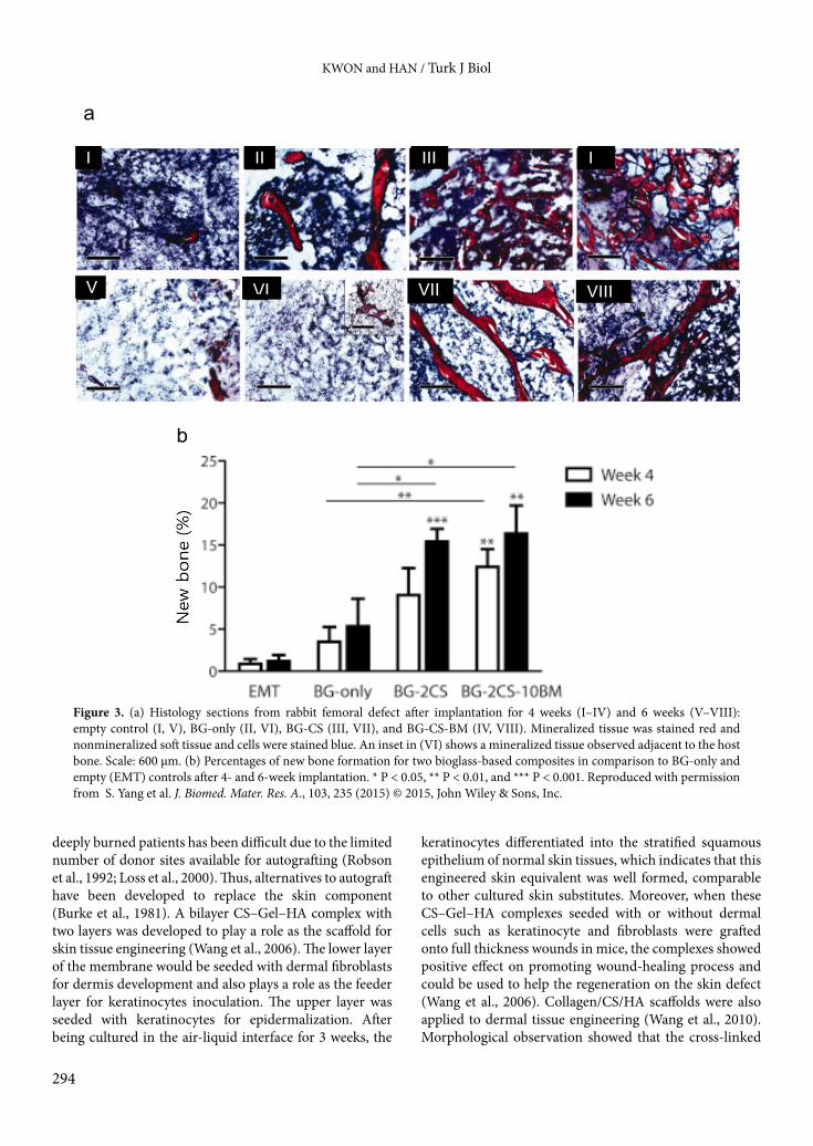
with BMPs (Yang et al., 2015). Figure 3a shows that either BG–CS or BG–CS containing BMPs (BG–CS–BM) induced formation of the newly developed bone in greater amounts, as compared to either the empty control or BG-only treatment. After 6 weeks of implantation, both BG–2CS and BG–2CS–10BM showed a significant increase in bone formation (~15% increase) as compared to either the empty control (~1% increase) or BG-only treatment (~5% increase) (Figure 3b).
5. Neural regeneration using chondroitin sulfate-based materials Chondroitin sulfate proteoglycans (CSPGs) are enriched in the growth environment of neural stem cells (NSCs) and are essential for FGF-2-mediated proliferation and maintenance of neuron-generating neural stem/progenitor cells (Sirko et al., 2007). Simultaneously, CS plays inhibitory functions for maturation, migration, and gliogenesis of neural stem/progenitor cell and that the loss of function of CSPGs induced differentiation and migration of neural progenitor cells (Gu et al., 2009; Sirko et al., 2010). Therefore, CSPG is considered to be a major obstacle for central nervous system (CNS) recovery after injury and thus its degradation has become a key therapeutic goal for CNS regeneration. Indeed, it was demonstrated that some CSPGs such as aggrecan, versican, and neurocan are related to brain disorders, schizophrenia, and attention deficit hyperactivity disorder (Avram et al., 2014). However, it was found that CSPG is abundantly synthesized in response to CNS injury and thus plays a key role during the acute recovery stage after spinal cord injury by directly activating microglia/macrophages via the CD44 receptor and modulating neurotrophic factor secretion (Rolls et al., 2008). Due to the two faces of CSPG in CNS repair, there has been little interest in developing CS-based materials for CNS regeneration. However, CS-based materials have been reported to be beneficial for peripheral nervous system (PNS) regeneration. It was reported that hydrogels of thiolated CS crosslinked with poly(ethylene glycol)-diacrylate can support robust growth of nerve roots in vitro (Conovaloff et al., 2011). In addition, the poly-DL-lactide (PDLLA) coated with CS and CHS by the layer-by-layer electrostatic self-assembly method (PDLLA/CHS/CS) onto which nerve growth factor (NGF) was immobilized was used as the nerve conduits (Xu et al., 2011). When the nerve conduits were employed to bridge the 10-mm defects in the sciatic nerve of the rats, a rapid functional recovery for the disrupted nerves was observed (Figure 4a). The nerve conduction velocities (NCVs) of the PDLLA/CHS/CS/NGF group and the PDLLA/CHS/CS group were more than 60 m/s and more than 50 m/s, respectively, which were significantly faster than the PDLLA group (less than 40 m/s) and

KWON and HAN / Turk J Biol
293
furthermore there was no significant difference between the PDLLA/CHS/CS/NGF group and the autograft control group (Figure 4b). CS-based nerve conduits would be expected to be useful materials for repairing nerve damage and promoting the regeneration of peripheral nerve defect.
6. Skin regeneration using chondroitin sulfate-based materials Permanent wound recovery demands grafting of autologous epithelium for restoring the epidermal function of the skin. However, the treatment of extensively and
Polystyrene
Polystyrene
PAMPS gel
PAMPS gel
b
c
a
Figure 2. Expression of chondrogenic markers in prechondrogenic cells cultured on hydro-gels with various charge density. (a) Gene expression analysis of type I collagen, aggrecan, and type II collagen in ATDC5 cells cultured on various substrata: polystyrene dish (PS), PDMAAm gel, P(AMPS-co-DMAAm) (molar fraction of AMPS, F = 0.5, 0.75) and poly-2-acrylamido-2-methylpropanesulfonic acid (PAMPS) gel, in the absence of insulin at day 7 (gray bars) and day 14 (black bars). Expression of each gene was measured by quantitative re-al-time PCR and normalized to GAPDH expression levels. Values are means ± SD obtained in four experiments. *P < 0.05 and **P < 0.01 vs. polystyrene, by Dunnett’s test. (b) Expression of type II collagen was analyzed by immunofluorescent staining at 14 days of culture. The cells were stained with an anti-type II collagen antibody (green) and Hoechst 33258 (red). Polys-tryrene dish (PS) and PAMPS gel, in the absence of insulin. Scale bar, 100 µm. (c) Expression of proteoglycan was analyzed by alcian blue staining at 14 days of culture. Polystryrene dish (PS) and PAMPS gel, in the absence of insulin. Scale bar, 100 µm. Reproduced with permis-sion from H. J. Kwon et al., Acta Biomaterialia, 6, 494 (2010) © 2010, Elsevier, Inc.
KWON and HAN / Turk J Biol
294
deeply burned patients has been difficult due to the limited number of donor sites available for autografting (Robson et al., 1992; Loss et al., 2000). Thus, alternatives to autograft have been developed to replace the skin component (Burke et al., 1981). A bilayer CS–Gel–HA complex with two layers was developed to play a role as the scaffold for skin tissue engineering (Wang et al., 2006). The lower layer of the membrane would be seeded with dermal fibroblasts for dermis development and also plays a role as the feeder layer for keratinocytes inoculation. The upper layer was seeded with keratinocytes for epidermalization. After being cultured in the air-liquid interface for 3 weeks, the
keratinocytes differentiated into the stratified squamous epithelium of normal skin tissues, which indicates that this engineered skin equivalent was well formed, comparable to other cultured skin substitutes. Moreover, when these CS–Gel–HA complexes seeded with or without dermal cells such as keratinocyte and fibroblasts were grafted onto full thickness wounds in mice, the complexes showed positive effect on promoting wound-healing process and could be used to help the regeneration on the skin defect (Wang et al., 2006). Collagen/CS/HA scaffolds were also applied to dermal tissue engineering (Wang et al., 2010). Morphological observation showed that the cross-linked
Figure 3. (a) Histology sections from rabbit femoral defect after implantation for 4 weeks (I–IV) and 6 weeks (V–VIII): empty control (I, V), BG-only (II, VI), BG-CS (III, VII), and BG-CS-BM (IV, VIII). Mineralized tissue was stained red and nonmineralized soft tissue and cells were stained blue. An inset in (VI) shows a mineralized tissue observed adjacent to the host bone. Scale: 600 µm. (b) Percentages of new bone formation for two bioglass-based composites in comparison to BG-only and empty (EMT) controls after 4- and 6-week implantation. * P < 0.05, ** P < 0.01, and *** P < 0.001. Reproduced with permission from S. Yang et al. J. Biomed. Mater. Res. A., 103, 235 (2015) © 2015, John Wiley & Sons, Inc.

KWON and HAN / Turk J Biol
295
collagen–CS–HA 9:1:1 scaffold had more degradation-resistant and higher elastic modulus than other scaffolds and more strongly promoted cellular adhesion and proliferation, in comparison with the scaffolds with other ratios (5:1:1 and 3:1:1). Moreover, an implantation study showed that the cross-linked collagen–CS–HA scaffold could more successfully repair full thickness skin defects in vivo. In addition, silk fibroin (SF)/CS/HA ternary scaffolds were constructed by freeze-drying for dermal tissue engineering (Yan et al., 2013). The incorporation of CS and HA with the SF solution increased cell adhesion, survival, and proliferation. When SF, SF/HA, and SF/CS/HA scaffolds were implanted onto dorsal full-thickness wounds of rats, the SF/CS/HA scaffolds promoted dermis regeneration compared to SF and SF/HA scaffolds and improved angiogenesis and collagen deposition, which
indicating that CS stimulated dermal regeneration (Figure 5a). The tissue regeneration ratio of SF/CS/HA scaffolds was more than 80%, which was significantly higher than that of the SF/HA and SF scaffolds at each time point (P < 0.05) (Figure 5b). Furthermore, when vascular endothelial growth factor (VEGF), platelet-derived growth factor (PDGF), and basic fibroblast growth factor (bFGF) expression in the SF/CS/HA groups were investigated by immunohistochemistry, it was demonstrated that the stimulation of secretion of VEGF, PDGF, and bFGF and accumulation of these growth factors were related to the induction of dermal regeneration. Likewise, CS nanoparticle/chitin/poly(butylene succinate) composite increased attachment and proliferation of dermal fibroblasts, which indicates that the CS incorporated composite is a suitable scaffold for skin tissue engineering
Figure 4. (a) Digital photo images of nerve conduits and regenerated nerves of each group after implantation for 3 months, group I: CS/PDLLA/CHS/NGF, group II: CS/PDLLA/CHS, group III: PDLLA alone. (b) Nerve conduction velocities (NCVs) of group A, B, C, and D 3 and 6 months after implantation in rats compared with each other (n = 6, * P < 0.05). A: PDLLA/Chondroitin sulfate/Chitosan/NGF, B: PDLLA/Chondroitin sulfate/Chitosan, C: PDLLA, D: autograft nerve. Reproduced with permission from H. Xu et al., Biomaterials, 32, 4506 (2011) © 2011, Elsevier, Inc.

KWON and HAN / Turk J Biol
296
(Deepthi et al., 2014). In addition, CS–Gel–HA seeded with genetically modified hair follicle stem cells promoted angiogenesis during wound healing and facilitation of vascularization in skin substitutes (Quan et al., 2014). Therefore, CS-based scaffolds provide potential for dermal tissue regeneration and could be used as a skin tissue model for in vitro toxicology, physiology examination, and in vivo wound healing in the near future.
7. ConclusionSince a loss of biological tissues has resulted in the functional impairment and cosmetic deformation of patients, tissue engineering is a growing field of interest in aged society. CS has crucial functions as an ECM component in growth factor signaling, wound healing, infection, tissue morphogenesis, hemostasis, inflammation, and cell division in vivo. Therefore, CS-based materials that mimic many
Time (weeks)
Figure 5. (a) Macroscopic observations of skin wounds after implanting SF, SF/HA, and SF/CS/HA scaffolds for 1, 2, 3, and 4 weeks; scale bar = 10 mm. (b) Tissue regeneration ratio and scaffold degradation ratio at different time points in vivo (* P < 0.05). Reproduced with permission from S. Yan et al. Acta Biomaterialia, 9, 6771 (2013) © 2013, Elsevier, Inc.
KWON and HAN / Turk J Biol
297
roles of ECM found in tissues are promising biomaterials for regenerative medicine. In spite of the high potential of CS for tissue engineering, various significant challenges are still associated with the use of CS-based biomaterials in clinical applications. Some of these challenges include the quality difference due to separation from living organisms, low thermal resistance, rapid degradation upon contact with body fluids or medium, weak mechanical properties, and poor tunability, in comparison with synthetic polymers. However, recent technological advancements have overcome the disadvantages associated with CS-based biomaterial for biomedical applications. For example, CS-based hydrogel can have high toughness by incorporating a
double network structure into the gel (Suekama et al., 2013). The enhanced appreciation of the fundamental properties and biological behavior of CS-based biomaterials, coupled with growing knowledge in the areas of biochemistry, molecular biology, and bioengineering, will lead to the development of methods to optimize their performance and their clinical utility.
Acknowledgment This review work was supported by Basic Science Research Program through the National Research Foundation of Korea (NRF) funded by the Ministry of Science, ICT and Future Planning (2014R1A1A1002054).
References
Avram S, Shaposhnikov S, Buiu C, Mernea M (2014). Chondroitin sulfate proteoglycans: structure-function relationship with im-plication in neural development and brain disorders. BioMed Res Int 2014: Article ID 642798.
Büttner M, Möller S, Keller M, Huster D, Schiller J, Schnabelrauch M, Dieter P, Hempel U (2013). Over-sulfated chondroitin sulfate derivatives induce osteogenic differentiation of hMSC independent of BMP-2 and TGF-β1 signalling. J Cell Physiol 228: 330–340.
Burke JF, Yannas IV, Quinby Jr WC, Bondoc CC, Jung WK (1981). Successful use of a physiologically acceptable artificial skin in the treatment of extensive burn injury. Ann Surg 194: 413–428.
Chang CH, Liu HC, Lin CC, Chou CH, Lin FH (2003). Gelatin–chondroitin–hyaluronan tri-copolymer scaffold for cartilage tissue engineering. Biomaterials 24: 4853–4858.
David-Raoudi M, Deschrevel B, Leclercq S, Galéra P, Boumediene K, Pujol JP (2009). Chondroitin sulfate increases hyaluronan production by human synoviocytes through differential regulation of hyaluronan synthases: role of p38 and Akt. Arthritis Rheum 60: 760–770.
Coburn J, Gibson M, Bandalini PA, Laird C, Mao HQ, Moroni L, Seliktar D, Elisseeff J (2011). Biomimetics of the extracellular matrix: an integrated three-dimensional fiber-hydrogel composite for cartilage tissue engineering. Smart Struct Syst 7: 213–222.
Conovaloff A, Panitch A (2011). Characterization of a chondroitin sulfate hydrogel for nerve root regeneration. J Neural Eng 8: 056003.
Deepa SS, Umehara Y, Higashiyama S, Itoh N, Sugahara K (2002). Specific molecular interactions of oversulfated chondroitin sulfate E with various heparin-binding growth factors: implications as a physiological binding partner in the brain and other tissues. J Biol Chem 277: 43707–43716.
Deepthi S, Sidhy Viha CV, Thitirat C, Furuike T, Tamura H, Jayakumar R (2014). Fabrication of chitin/poly(butylene succinate)/chondroitin sulfate nanoparticles ternary composite hydrogel scaffold for skin tissue engineering. Polymers 6: 2974–2984.
Dick G, Akslen-Hoel LK, Grøndahl F, Kjos I, Prydz K, Histochem J (2012). Proteoglycan synthesis and golgi organization in polarized epithelial cells. J Histochem Cytochem 60: 926–935.
Fan H, Hu Y, Zhang C, Li X, Lv R, Qin L, Zhu R (2006). Retinal pigment epithelium cell culture on surface modified poly(hydroxybutyrate-co-hydroxyvalerate) thin films. Biomaterials 27: 4573–4583.
Flanagan TC, Wilkins B, Black A, Jockenhoevel S, Smith TJ, Pandit AS (2006). A collagen-glycosaminoglycan co-culture model for heart valve tissue engineering applications. Biomaterials 27: 2233–2246.
Fthenou E, Zafiropoulos A, Katonis P, Tsatsakis A, Karamanos NK, Tzanakakis GN (2008). Chondroitin sulfate prevents platelet derived growth factor-mediated phosphorylation of PDGF-Rbeta in normal human fibroblasts severely impairing mitogenic responses. J Cell Biochem 103: 1866–1876.
Gong JP (2010). Why are double network hydrogels so tough? Soft Matter 6: 2583–2590.
Gu WL, Fu SL, Wang YX, Li Y, Lü HZ, Xu XM, Lu PH (2009). Chondroitin sulfate proteoglycans regulate the growth, differentiation and migration of multipotent neural precursor cells through the integrin signaling pathway. BMC Neurosci 10: 128.
Guo Y, Yuan T, Xiao Z, Tang P, Xiao Y, Fan Y, Zhang X (2012) Hydrogels of collagen/chondroitin sulfate/hyaluronan interpenetrating polymer network for cartilage tissue engineering. J Mater Sci Mater Med 23: 2267–2279.
Henson FM, Getgood AM, Caborn DM, McIlwraith CW, Rushton N (2012). Effect of a solution of hyaluronic acid-chondroitin sulfate-N-acetyl glucosamine on the repair response of cartilage to single-impact load damage. Am J Vet Res 73: 306–312.
Hintze V, Miron A, Moeller S, Schnabelrauch M, Wiesmann HP, Worch H, Scharnweber D (2012). Sulfated hyaluronan and chondroitin sulfate derivatives interact differently with human transforming growth factor-beta1 (TGF-beta1). Acta Biomater 8: 2144–2152.
KWON and HAN / Turk J Biol
298
Huskisson EC (2008). Glucosamine and chondroitin for osteoarthritis. J Int Med Res 36: 1161–1179.
Hwang HY, Olson SK, Esko JD, Horvitz HR (2003). Caenorhabditis elegans early embryogenesis and vulval morphogenesis require chondroitin biosynthesis. Nature 423: 439–443.
Jerosch J (2011). Effects of glucosamine and chondroitin sulfate on cartilage metabolism in OA: outlook on other nutrient partners especially omega-3 fatty acids. J Rheumatol 2011: Article ID 969012.
Keskin DS, Tezcaner A, Korkusuz P, Korkusuz F, Hasirci V (2005). Collagen-chondroitin sulfate-based PLLA-SAIB-coated rh-BMP-2 delivery system for bone repair. Biomaterials 26: 4023–4034.
Kharkar PM, Kiick KL, Kloxin AM (2013). Designing degradable hydrogels for orthogonal control of cell microenvironments. Chem Soc Rev 42: 7335–7372.
Knudson CB, Knudson W (2001). Cartilage proteoglycans. Semin Cell Dev Biol 12: 69–78.
Kwon HJ, Yasuda K (2013). Chondrogenesis on sulfonate-coated hydrogels is regulated by their mechanical properties. J Mech Behav Biomed Mater 17: 337–346.
Kwon HJ, Yasuda K, Ohmiya Y, Honma K, Chen YM, Gong JP (2010). In vitro differentiation of chondrogenic ATDC5 cells is enhanced by culturing on synthetic hydrogels with various charge densities. Acta Biomater 6: 494–501.
Laabs T, Carulli D, Geller HM, Fawcett JW (2005). Chondroitin sulfate proteoglycans in neural development and regeneration. Curr Opin Neurobiol 15: 116–120.
Lee KY, Mooney DJ (2012). Alginate: properties and biomedical applications. Prog Polym Sci 37: 106–126.
Lee KY, Yuk SH (2007). Polymeric protein delivery systems. Prog Polym Sci 32: 669–697.
Loss M, Wedler V, Kunzi W, Meuli-Simmen C, Meyer VE (2000). Artificial skin, split-thickness autograft and cultured autologous keratinocytes combined to treat a severe burn injury of 93% of TBSA. Burns 26: 644–652.
Martel-Pelletier J, Kwan Tat S, Pelletier JP (2010). Effects of chondroitin sulfate in the pathophysiology of the osteoarthritic joint: a narrative review. Osteoarthr Cartil 18, Suppl 1: S7–11.
Mikami T, Kitagawa H (2013). Biosynthesis and function of chondroitin sulfate. Biochim Biophys Acta 1830: 4719–4733.
Park YJ, Lee YM, Lee JY, Seol YJ, Chung CP, Lee SJ (2000). Controlled release of platelet-derived growth factor-BB from chondroitin sulfate–chitosan sponge for guided bone regeneration. J Control Release 67: 385–394.
Pawar SN, Edgar KJ (2012). Alginate derivatization: a review of chemistry, properties and applications, Biomaterials 33: 3279–3305.
Quan R, Zheng X, Xu S, Zhang L, Yang D (2014). Gelatin-chondroitin-6-sulfate-hyaluronic acid scaffold seeded with vascular endothelial growth factor 165 modified hair follicle stem cells as a three-dimensional skin substitute. Stem Cell Research and Therapy 5: 118.
Robson MC, Barnett RA, Leitch IO, Hayward PG (1992). Prevention and treatment of postburn scars and contracture. World J Surg 16: 87–96.
Rolls A, Shechter R, London A, Segev Y, Jacob-Hirsch J, Amariglio N, Rechavi G, Schwartz M (2008). Two faces of chondroitin sulfate proteoglycan in spinal cord repair: a role in microglia/macrophage activation. PLoS Med 5: e171.
Santo VE, Gomes ME, Mano JF, Reis RL (2012). Chitosan-chondroitin sulphate nanoparticles for controlled delivery of platelet lysates in bone regenerative medicine. J Tissue Eng Regen Med 6 (Suppl 3): S47–59.
Schnabelrauch M, Scharnweber D, Schiller J (2013). Sulfated glycosaminoglycans as promising artificial extracellular matrix components to improve the regeneration of tissues. Curr Med Chem 20: 2501–2523.
Sintov A, Di-Capua N, Rubinstein A (1995). Cross-linked chondroitin sulphate: characterization for drug delivery purposes. Biomaterials 16: 473–478.
Sirko S, von Holst A, Weber A, Wizenmann A, Theocharidis U, Götz M, Faissner A (2010). Chondroitin sulfates are required for fibroblast growth factor-2-dependent proliferation and maintenance in neural stem cells and for epidermal growth factor-dependent migration of their progeny. Stem Cells 28: 775–787.
Sirko S, von Holst A, Wizenmann A, Götz M, Faissner A (2007). Chondroitin sulfate glycosaminoglycans control proliferation, radial glia cell differentiation and neurogenesis in neural stem/progenitor cells. Development 134: 2727–2738.
Suekama TC, Hu J, Kurokawa T, Gong JP, Gehrke SH (2013). Double-network strategy improves fracture properties of chondroitin sulfate networks. ACS Macro Lett 2: 137–140.
Taipale J, Keski-Oja J (1997). Growth factors in the extracellular matrix. FASEB J 11: 51–59.
Takagaki K, Munakata H, Kakizaki I, Iwafune M, Itabashi T, Endo M (2002). Domain structure of chondroitin sulfate E octasaccharides binding to type V collagen. J Biol Chem 277: 8882–8889.
Tan H, Gong Y, Lao L, Mao Z, Gao C (2007). Gelatin/chitosan/hyaluronan ternary complex scaffold containing basic fibroblast growth factor for cartilage tissue engineering. J Mater Sci Mater Med 18: 1961–1968.
Ueoka C, Kaneda N, Okazaki I, Nadanaka S, Muramatsu T, Sugahara K (2000). Neuronal cell adhesion, mediated by the heparin-binding neuroregulatory factor midkine, is specifically inhibited by chondroitin sulfate E. Structural and functional implications of the over-sulfated chondroitin sulfate. J Biol Chem 275: 37407–37413.
Wang DA, Varghese S, Sharma B, Strehin I, Fermanian S, Gorham J, Fairbrother DH, Cascio B, Elisseeff JH (2007). Multifunctional chondroitin sulphate for cartilage tissue–biomaterial integration. Nat Mater 6: 385–392.

KWON and HAN / Turk J Biol
299
Wang H, Katagiri Y, McCann TE, Unsworth E, Goldsmith P, Yu ZX, Tan F, Santiago L, Mills EM, Wang Y et al. (2008). Chondroitin-4-sulfation negatively regulates axonal guidance and growth. J Cell Sci 121: 3083–3091.
Wang SC, Chen BH, Wang LF, Chen JS (2007). Characterization of chondroitin sulfate and its interpenetrating polymer network hydrogels for sustained-drug release. Int J Pharmacogn 329: 103–109.
Wang TW, Sun JS, Wu HC, Tsuang YH, Wang WH, Lin FH (2006). The effect of gelatin-chondroitin sulfate-hyaluronic acid skin substitute on wound healing in SCID mice. Biomaterials 27: 5689–5697.
Wang TW, Wu HC, Huang YC, Sun JS, Lin FH (2006). Biomimetic bilayered gelatin-chondroitin 6 sulfate-hyaluronic acid biopolymer as a scaffold for skin equivalent tissue engineering. Artif Organs 30: 141–149.
Wang W, Zhang M, Lu W, Zhang X, Ma D, Rong X, Yu C, Jin Y (2010). See comment in PubMed Commons belowCross-linked collagen-chondroitin sulfate-hyaluronic acid imitating extracellular matrix as scaffold for dermal tissue engineering. See comment in PubMed Commons belowTissue Eng Part C Methods 16: 269–279.
Xu H, Yan Y, Li S (2011). PDLLA/chondroitin sulfate/chitosan/NGF conduits for peripheral nerve regeneration. Biomaterials 32: 4506–4516.
Yamada S, Sugahara K, Özbek S (2011). Evolution of glycosaminoglycans: comparative biochemical study. Commun Integr Biol 4: 150–158.
Yamaguchi T, Ohtake S, Kimata K (2007). Molecular cloning of squid N-acetylgalactosamine 4-sulfate 6-O-sulfotransferase and synthesis of a unique chondroitin sulfate containing E-D hybrid tetrasaccharide structure by the recombinant enzyme. Glycobiology 17: 1365–1376.
Yan S, Zhang Q, Wang J, Liu Y, Lu S, Li M, Kaplan DL (2013). Silk fibroin/chondroitin sulfate/hyaluronic acid ternary scaffolds for dermal tissue reconstruction. Acta Biomater 9: 6771–6782.
Yang S, Guo Q, Shores LS, Aly A, Ramakrishnan M, Kim GH, Lu Q, Su L, Elisseeff JH (2015). Use of a chondroitin sulfate bioadhesive to enhance integration of bioglass particles for repairing critical-size bone defects. J Biomed Mater Res A 103: 235–242.