chitin and yeast budding - jbc.org · chitin and yeast budding properties of chitin synthetase from...
TRANSCRIPT
THE JOURNAL OF UIOLOGICAL CHEMISTRY Vol. 246, No. 1, Issue of January 10, pp. 160-166, 1971
Printed in U.S.A.
Chitin and Yeast Budding PROPERTIES OF CHITIN SYNTHETASE FROM XACCHAROMYCEX CARLXBERGENXIX
(Received for publication, June 4, 1970)
FREDERICK A. KELLER AND ENRICO CABIB
From the National Institute of Arthritis and Metabolic Diseases, National Institutes of Health, Bethesda, Mary- land 20014
SUMMARY
A particulate preparation from a spheroplast lysate of Saccharomyces cartsbergensis was found to catalyze the transfer of acetylglucosamine from UDP-acetylglucosamine to’an endogenous acceptor. Uridine diphosphate is liberated in stoichiometric amounts. A divalent cation, MgZ+, Mn2+, or Co2+, is required for enzymatic activity. Acetylglucos- amine stimulates the activity about 5-fold, with a K, of 4.7 mM, and can be partially replaced by high concentrations of glucose, mannose, cellobiose, and glycerol. The K, for UDP- acetyl-glucosamine is between 0.6 and 0.9 mu and the pH op- timum is 6.2. Centrifugation in sucrose gradients indicates that the reaction product remains bound to the same particles which contain the activity. The product was characterized as chitin by its insolubility in alkali, the release of glucosa- mine on acid hydrolysis, and the liberation of diacetylchi- tobiose and acetylglucosamine following stepwise enzymatic hydrolysis. The enzyme is inhibited by monovalent and polyvalent anions, the latter being more effective. The antibiotic Polyoxin A is a very potent competitive inhibitor, with a Ki of 5 X lOA7 M. Polyoxin A affected neither growth of intact cells nor synthesis of chitin by naked spheroplasts. It is concluded that chitin synthetase is not located outside the cytoplasmic membrane.
Previous reports have shown that the small amount of chitin present in the cell wall of yeast is concentrated in an annular area at the bud scar site (l-3). Consequently, the suggestion was made (3) that this polysaccharide may have an important role in the process of yeast budding. It thus became of interest to investigate the mechanism of chitin biosynthesis in yeast at the molecular level in order to approach correlations between biochemical and morphological characteristics. The present report deals with the general properties of the chitin synthetase.1
EXPERIMENTAL PROCEDURE
Materials
UDP-G~cNAc,~ labeled with 14C in C-l of the hexosamine moiety, was prepared as directed by O’Brien (5). The same
1 The noncommittal but descriptive term “chitin synthetase” is used in this and the following paper (4)) because the endogenous acceptor of the acetylglucosaminyl units has not been identified.
2 The abbreviation used is: UDP-GlcNAc, UDP-N-acetyl- glucosamine.
nucleotide, labeled wih 3H in the acetyl group, was a generous gift of Dr. E. Neufeld. GDP-mannose, labeled with 14C in the sugar moiety, was obtained by a procedure similar to that of Rosen and Zeleznick (6), but with an extract of lyophilized yeast, after passage through Sephadex G-25, as a source of GDP- mannose pyrophosphorylase. Unlabeled UDP-GlcNAc and GDP-mannose were prepared as previously described (7).
Polyoxin A was kindly provided by Dr. K. Isono. Ampho- tericin A, Amphotericin 13, and Nystatin were donated by Squibb and Sons, New York, New York, Griseofulvin by Schering Corporation, Bloomfield, New Jersey, Vancomycin by Eli Lilly and Company, Indianapolis, Indiana, and Ristocetin by Abbott, North Chicago, Illinois. Actidione and Bacitracin were pur- chased from Mann, and Streptomycin and Penicillin G from Calbiochem.
We are indebted to Dr. L. Glaser for samples of diacetyl- chitobiose and chitodextrins. The chitodextrins were purified by passage through an MB-3 Amberlite column before use.
Yeast RNA was a gift from Dr. Maxine Singer. Inorganic polyphosphate (molecular weight N 3700) was kindly furnished by Dr. W. Carroll. Polyglutamic acid (molecular weight 105,000) and polylysine (molecular weight 195,000) were pur- chased from Sigma. Dr. P. O’Brien generously supplied samples of glucosamine &phosphate, N-acetylglucosamine B-phosphate, and a-N-acetylglucosamine l-phosphate.
Methods
Enzyme Preparation-Saccharomyces carlsbergensis strain 745 (National Collection of Yeast Cultures, England) was grown (8) and collected when the optical density at 660 rng was 0.4 to 0.5; this value corresponds to the late logarithmic phase, about one generation before the onset of the stationary phase.
Saccharomyces cerevisiae A364A and its temperature-sensitive mutant 3163 were grown as described by Hartwell (9).
In the early part of this study, spheroplasts were prepared as previously reported (10). In more recent experiments 0.55 M mannitol was substituted for 0.6 M KC1 as osmotic stabilizer. Spheroplasts prepared and washed with mannitol showed less lysis than those obtained with KCl, and were very well preserved even after storage for several days at 4”, with the addition of aureomycin (50 pg per ml) to prevent bacterial growth.
Many preparations of the particulate enzyme were obtained from spheroplasts, as previously described for mannan synthetase
3Mutant 316 grows at 24” but not at 37”. Both strains of S. cerevisicze, A364A and 316, were generously provided by Dr. L. H. Hartwell.
160
by guest on August 19, 2018
http://ww
w.jbc.org/
Dow
nloaded from
by guest on August 19, 2018
http://ww
w.jbc.org/
Dow
nloaded from
by guest on August 19, 2018
http://ww
w.jbc.org/
Dow
nloaded from
Issue of January 10, 1971 F. A. Keller and E. Cab& 161
(10). More recently, somewhat higher and more reproducible enzymatic activity was obtained by the following procedure. After the last washing with 0.55 M mannitol, spheroplasts were resuspended in the same reagent up to a volume numerically equal in milliliters to two-thirds of the grams of yeast, wet weight used. The spheroplast suspension was added, while stirring with a Vortex mixer, to 5 volumes of Buffer A (50 mu imidazole-chloride, pH 6.5, containing 2 M MgSOS containing 0.385 M mannitol. At brief intervals further additions were made of 3 and 5 volumes, respectively, of Buffer A. The sus- pension was then centrifuged for 10 min at 20,000 x g and the pellet was washed twice with an amount of Buffer A equivalent to about 4 volumes of the original spheroplast suspension. The final pellet was resuspended, up to the original volume of the spheroplast suspension, in Buffer A containing 33% glycerol. The protein content of such preparations ranged between 10 and 13 mg per ml. The enzyme was maintained at about -9” with- out freezing. About 50% of the activity was lost in 2 weeks at this temperature.
Enzyme Assay-The incubation mixture contained 0.95 mM 14C or 3H-UDP-GlcNAc (see below), 0.05 M imidazole-chloride, pH 6.5, 0.8 mM ma,gnesium sulfate, 40 mM acetylglucosamine, and variable amounts of enzyme in a total volume of 50 to 53 ~1.
TheUDP-GlcNAc used waslabeled with’% in C-l of thehexos- amine moiety (specific activity 107,000 cpm per pmole4) or with tritium in the acetyl group (specific activity 200,000 cpm per pmole).
The enzyme contained glycerol, which acts as an activator of chitin synthetase (see below). The glycerol concentration in the incubation mixture was kept constant at 1.36 M, except where indicated otherwise.
After incubation for 1 hour at 30” the reaction was stopped by the addition of 1 ml of 66% ethanol, and the tubes were centri- fuged for 5 min at 1500 x g. The pellets were washed twice with 1 ml of 66% ethanol containing 0.1 M ammonium acetate. Further treatment depended on the sugar nucleotide label. When using tritiated UDP-GlcNAc, the pellets were resuspended in 0.4 ml of absolute ethanol by stirring on a Vortex mixer at maximum speed, and poured into a scintillation vial. The tube was washed with another 0.4-ml portion of absolute ethanol and 12 ml of scintillation mixture was added. The mixture con- tained, per liter of toluene, 5 g of 2,5-diphenyloxazole, 3.2 g of 1,4-bis[2-(5-phenyloxazolyl)J-benzene, and 43 g of CAB-0-SIL thixotropic gel (Packard Instrument Company). The vials were vigorously stirred with a Vortex mixer and counted in a Packard liquid scintillation spectrometer.
When r4C-labeled UDP-GlcNAc was the substrate, the washed pellets were resuspended in water, plated on copper or stainless steel planchets, dried at loo”, and counted in a Nuclear-Chicago low background, thin window counter.
In order to account for quenching and self-absorption, the results were multiplied by an experimentally determined cor- rection factor which was 1.56 for 3H and 1.45 for 14C.
4 The method used in the preparation of UDP-14C-GlcNAc results in a product contaminated with the acetylgalactosamine analogue (5). The amount of the latter was ascertained by hy- drolyzing the nucleotide in 0.1 N acid at KU’, and separating the free sugars by paper chromatography on borate-treated Whatman No. 1 paper (ll), with 1-butanol-pyridine-water (6:4:3) as solvent. The acetylgalactosamine peak represented 31% of the total radio- activity; this value was used to correct the specific activity of the sugar nucleotide.
TABLE I Requirements for yeast chitin sgnthetase
The enzyme was prepared as described under “Experimental Procedure,” except that the particles were washed three addi- tional times with 0.05 M imidazole, pH 6.5, to eliminate glycerol and Mg2+. The complete incubation mixture was as described under “Experimental Procedure.” The activity under those con- ditions is taken as 100.
Incubation mixture Enzymatic activity
Complete..................................... 100 Minus acetylglucosamine....................... 21 Minus acetylglucosamine, plus 1.4 M glycerol. 57 Minus Mgzf. 27 Minus Mg”+, plus 1 rnM Mnz+. 94 Minus Mgsf, plus 6 mM EDTA. . . . . . . 1 Minus Mgz+ and acetylglucosamine. . . . . . . . 1
Miscellaneous-Protein was determined according to the method of Lowry et al. (12). Mannan synthetase was assayed as previously reported (10). Radioactivity on paper chromato- grams was measured with a Vanguard scanner.
RESULTS
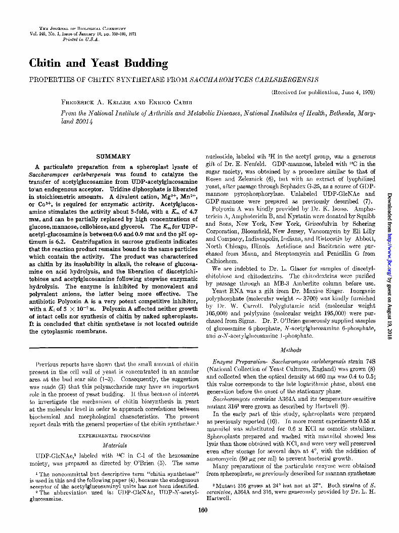
The general requirements for enzymatic activity are shown in Table I. The enzyme was stimulated by divalent cations and by acetylglucosamine. No activity was observed when the incubation mixture contained EDTA in the absence of added Mg2+. The incorporation of label into material insoluble in 66% ethanol was linear during the 1st hour of incubation and then declined (Fig. 1). Activity was proportional to the amount of added enzyme, at least up to 40 ~1 of the standard preparation. The maximal incorporation obtained with the best preparations exceeded 30% of the added substrate. The K, value for UDP- GlcNAc varied between 0.6 and 0.9 mM in different experiments.
The optimal temperature for enzymatic activity was about 37”, with half-maximal values at 28 and 48”.
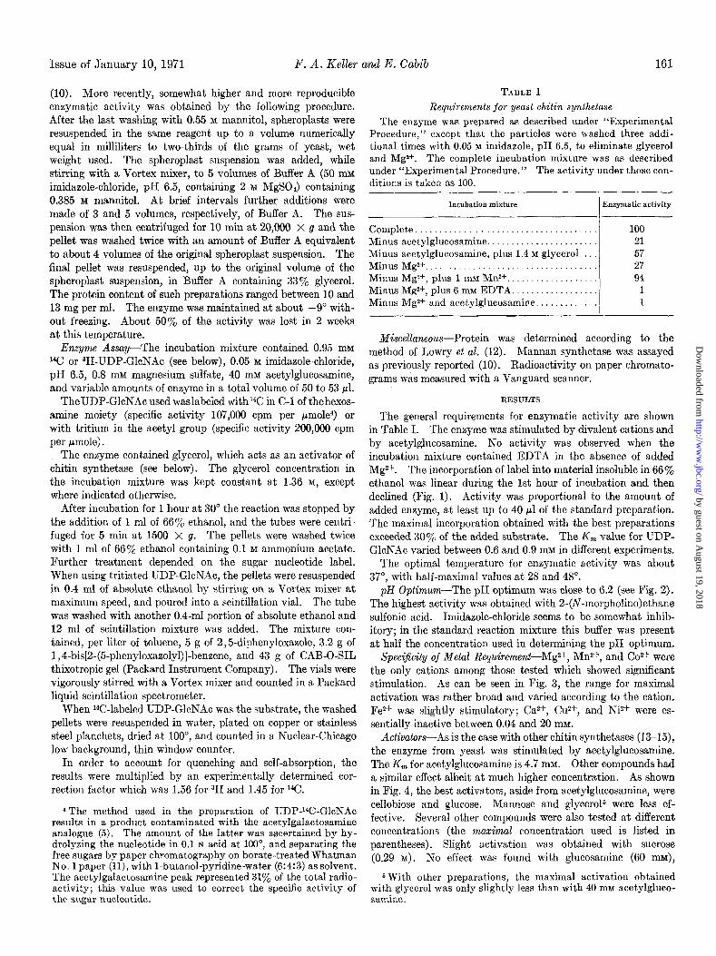
pH Optimum-The pH optimum was close to 6.2 (see Fig. 2). The highest activity was obtained with 2-(N-morpholino)ethane sulfonic acid. Imidazole-chloride seems to be somewhat inhib- itory; in the standard reaction mixture this buffer was present at half the concentration used in determining the pH optimum.
SpeciJicity of Metal Requirement-Mg2+, Mn2f, and Co2+ were the only cations among those tested which showed significant stimulation. As can be seen in Fig. 3, the range for maximal activation was rather broad and varied according to the cation. Fez+ was slightly stimulatory; Ca2+, Cu2+, and Ni2+ were es- sentially inactive between 0.04 and 20 mrvx.
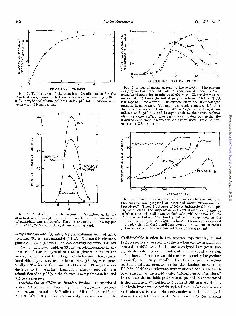
Activators-As is the case with other chitin synthetases (13-15), the enzyme from yeast was stimulated by acetylglucosamine. The K, for acetylglucosamine is 4.7 mM. Other compounds had a similar effect albeit at much higher concentration. As shown in Fig. 4, the best activators, aside from acetylglucosamine, were cellobiose and glucose. Mannose and glycerol5 were less ef- fective. Several other compounds were also tested at different concentrations (the maximal concentration used is listed in parentheses). Slight activation was obtained with sucrose (0.29 M). No effect was found with glucosamine (60 mu),
6 With other preparations, the maximal activation obtained with glycerol was only slightly less than with 40 mrvr acetylgluco- samine.
by guest on August 19, 2018
http://ww
w.jbc.org/
Dow
nloaded from
162 Chitin Xynthetase Vol. 246, No. 1
I I I I I 0 I 2 3 4 5
INCUBATION TIME (hours)
FIG. 1. Time course of the reaction. Conditions as for the standard assay, except that imidazole was replaced by 0.05 M
Z-(N-morpholino)ethane sulfonic acid, pH 6.1. Enzyme con- centration, 3.8 mg per ml.
4 5 6 7 8
PH
FIG. 2. Effect of pH on the activity. Conditions as in the standard assay, except for the buffer used. The potassium salt of phosphate was employed. Enzyme concentration, 3.8 mg per ml. MES, 2-(N-morpholino)ethane sulfonic acid.
acetylgalactosamine (60 nn$, acetylglucosamine 6-P (24 n&, trehalose (0.2 M), and mannitol (0.2 M). Glucose-6-P (40 mi$, glucosamine-6-P (60 MUM), and Q-N-acetylglucosamine 1-P (32 m@ were inhibitory. Adding 30 mM acetylglucosamine in the presence of 1.36 M glycerol or 0.56 M glucose increased the activity by only about 10 to 15%. Chitodextrins, which stimu- lated chitin synthetase from other sources (13-15), were prac- tically ineffective in this case. Addition of 0.13 mg of chito- dextrins to the standard incubation mixture resulted in a stimulation of only 22% in the absence of acetylglucosamine, and 5% in its presence.
IdentiJcation of Chitin as Reaction Product-As mentioned under “Experimental Procedure,” the radioactive reaction product was insoluble in 66% ethanol. After boiling for 45 min in 1 N KOH, 60% of the radioactivity was recovered in the
CONCENTRATION OF CATION (mM)
FIG. 3. Effect of metal cations on the activity. The enzyme was prepared as described under “Experimental Procedure” and centrifuged again for 10 min at 20,000 X g. The pellet was re- suspended in 5 times the initial enzyme volume of 0.1 M EDTA and kept at 0” for 30 min. The suspension was then centrifuged again in the same way. The pellet was washed once, with 5 times the initial enzyme volume of 0.02 M 2-(iV-morpholino)ethane sulfonic acid, pH 6.1, and brought back to the initial volume with the same buffer. The assay was carried out under the standard conditions, except for the cation used. Enzyme con- centration, 3.8 mg per ml.
100 z 5 5 00
-ACETYLGLUCOSAMINE
a
2 z 60 2 I b 40
% s B 20 a
ACTIVATOR (Ml
FIG. 4. Effect of activators on chitin synthetase activity. The enzyme was prepared as described under “Experimental Procedure.” Then, 5 volumes of 0.05 M imidazole-chloride, pH 6.6, were added, the suspension was centrifuged for 10 min at 12,060 X g, and the pellet was washed twice with the same volume of imidazole buffer. The final pellet was resuspended in the imidazole buffer up to the original volume. The assay was carried out under the standard conditions except for the concentration of the activator. Enzyme concentration, 3.8 mg per ml.
alkali-insoluble fraction in two separate experiments; 37 and IS%, respectively, was found in the fraction soluble in alkali but insoluble in 66% ethanol. In each case lyophilized yeast, pre- viously disrupted by sonic disintegration, was added as carrier.
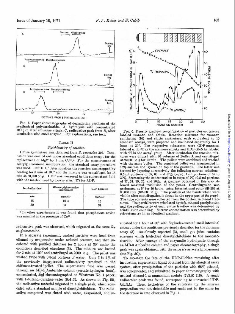
Additional information was obtained by degrading the product chemically and enzymatically. For this purpose scaled-up reaction mixtures, prepared as for the standard assay using UDP-i4C-GlcNAc as substrate, were incubated and treated with 66% ethanol, as described under “Experimental Procedure.” In one case the insoluble pellet was suspended in concentrated hydrochloric acid and heated for 3 hours at 100” in a sealed tube. The hydrolysate was passed through a Dowex 1 (acetate) column and submitted to paper chromatography with l-butanol-pyri- dine-water (6:4:3) as solvent. As shown in Fig. 5A, a single
by guest on August 19, 2018
http://ww
w.jbc.org/
Dow
nloaded from
Issue of January lo,1971 F. A* Keller and E. Cabib
_ll-_iu ~6
I I I I x Acefylglucosomine Diocelylchitobiose E
I e5- B
8
Starting t4- line 8
ii
&- z Acefylglucosomine Diocetylchitobiose
C r.-1 r-1 t: 5
l=2- Stqrting 2
line 0 9 al-
30 20 IO 0 DISTANCE FROM STARTING LINE (cm)
Fro. 5. Paper chromatography of degradation products of the 0 5 IO 15 20 25 3C
synthesized polysaccharide. A, hydrolysis with concentrated FRACTION NUMBER
HCl; B, after chitinase attack; C, radioactive peak from B, after incubation with snail enzyme. For explanations, see text.
FIG. 6. Density gradient centrifugation of particles containing labeled mannan and chitin. Reaction mixtures .for mannan synthetase (10) and chitin synthetase, each equivalent to 10
” TABLE II standard assays, were prepared and incubated separately for 1
Stoichiametry of reaction hour at 30”. The respective substrates were GDP-mannose
Chitin synthetase was obtained from S. cerewisiae 316. labeled with 14C in the mannose moiety and UDP-GloNAc labeled
Inou- with 3H in the acetyl group. After incubation the reaction mix- bation was carried out under standard conditions except for the tures were diluted with 20 volumes of Buffer A and centrifuged replacement of Mgz+ by 1 mM Coz+.a For the measurement of at 22,000 x g for 10 min. The pellets were combined and washed acetylglucosamine incorporation, the standard assay procedure with the same buffer. The combined pellet was resuspended in w&s used. For UDP determination the reaction was stopped by 15% sucrose and layered on top of the gradient. The latter was
heating for 3 min at 100’ and the mixture was centrifuged for 15 formed by layering successively the following sucrose solutions:
min at 60,000 X g. UDP was measured in the supernatant fluid 0.5~ml portions of 65, 60, and 57% (w/w); l-ml portions of 55 to
with the method used by Lowry et ~2. (17) for ADP. 39yo, decreasing the concentration in steps of 2%; 0.5-ml portions of 37, 34, 30, 25, and 20%. A gradient obtained in this way al-
*II-Acetylglucosamine lowed maximal resolution of the peaks. Centrifugation was
Incubation time incorporated UDP liberated performed at 5” for 16 hours, using International rotor SB-269 at 30,060 rpm (105,ooO’~ g). The position of the bands which were
min ~JW&?S mpmles visible after centrifugation is shown in the upper part of the graph.
15 18.5 15 The tube contents were collected from the bottom in 0.5-ml frac-
30 33 34, tions. The particles were reisolated by 66% ethanol precipitation and the radioactivity of each entire fraction was determined by
o In other experiments it was found that phosphatase action scintillation counting. Sucrose concentration was determined by
was minimal in the presence of Co*+. refractometry in an identical gradient.
radioactive peak was observed, which migrated at the same Rp cubated for 1 hour at 30” with Sephadex-treated snail intestinal
as glucosamine. extract under the conditions previously described for the chitinase
In a separate experiment, washed particles were freed from assay (3). As already reported (3), snail gut juice contains
ethanol by evaporation under reduced pressure, and then in- enzymes which hydrolyze diacetylchitobiose to the monosac-
cubated with purified ohitinase for 2 hours at 30” under the oharide. After passage of the enzymatic hydrolysate through
conditions described elsewhere (3). The mixture was heated an MB-3 Amberlite column and paper chromatography,, a single
for 2 min at 100” and centrifuged at 2000 X g. peak was again obtained, with the same RP as acetylglucosamine
The pellet was washed twice with 0.3-ml portions of water,
(see J?ig.,5c),
Only 3 to 4% of To ascertain the fate of the UDP-GlcNac remaining after the previously mo$rporated radioactivity remained in the incubation, supernatant liquid obtained from the standard assay chitinase-treated?:pellet. The supernatant fluid was passed system, after precipitation of the p&i&s with 66% ethanol, through an M~3&mberlite column (acetate-hydrogen form), was concentrated and submitted to paper chromatography with concentrated, a~$ ‘chromatographed on Whatman No, 1 paper, -I neutral ethanol-l 1 ammonium acetate (7 :3.5) (16). A single with 1-butanol-pyridine-water (6:4:3). As shown in Fig. 6B, radioactive peak was found, corresponding to unreacted UDP- the radioactive material migrated in a single peak, which coin- GlcNAc. Thus, hydrolysis of the substrate by the enzyme tided with a standard sample of diacetylohitobiose. The radio- preparation was not detectable and could not be the cause for active compound was eluted with water, evaporated, and in- the decrease in rate observed in Fig. 1.
by guest on August 19, 2018
http://ww
w.jbc.org/
Dow
nloaded from
164 Chitin Xynthetase Vol. 246, No. 1
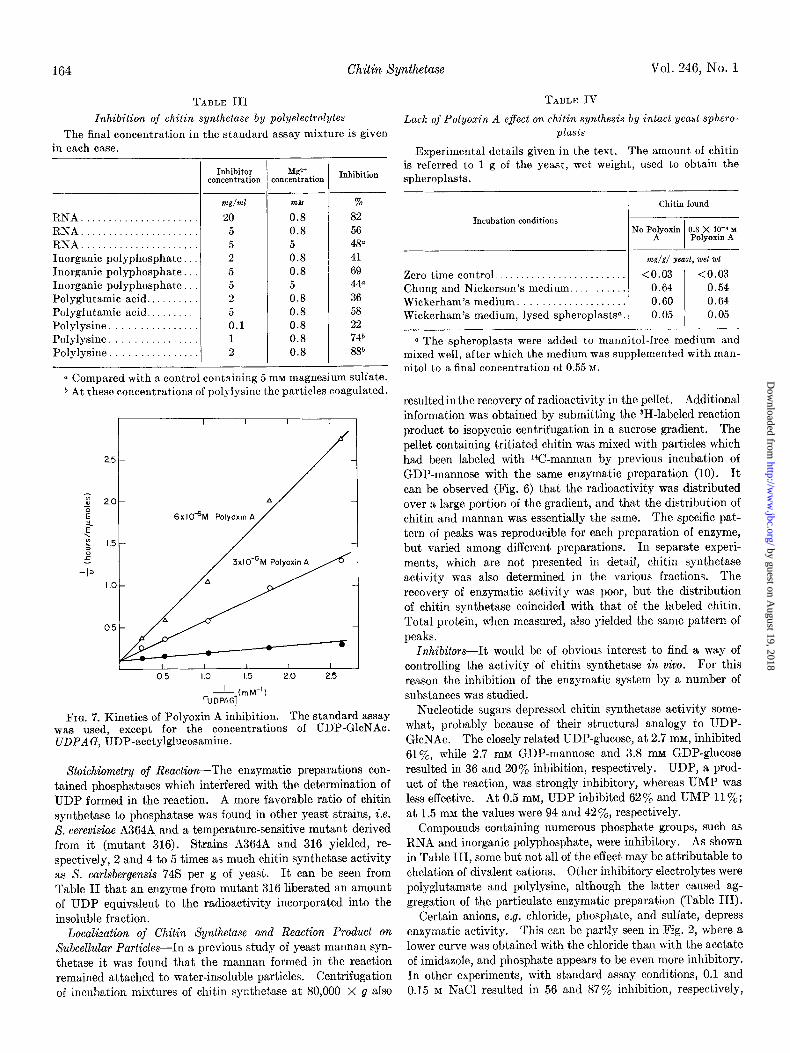
TABLE III
Inhibition of chitin synthetase by polyelectrolytes
The final concentration in the standard assay mixture is given in each case.
RNA. RNA. RNA. Inorganic polyphosphate Inorganic polyphosphate. Inorganic polyphosphate Polyglutamic acid.. Polyglutamic acid. Polylysine Polylysine Polylysine
-
--
Inhibitor concentration
-
, --
Mgr+ :oncentration Inhibition
m&T/ml m&f %
20 0.8 82 5 0.8 56 5 5 48” 2 0.8 41 5 0.8 69 5 5 446 2 0.8 36 5 0.8 58 0.1 0.8 22 1 0.8 74b 2 0.8 88”
T
a Compared with a control containing 5 rnM magnesium sulfate. 6 At these concentrations of polylysine the particles coagulated.
I I I I I I I Jl I /- I
FIG. 7. Kinetics of Polyoxin A inhibition. The standard assay was used, except for the concentrations of UDP-GlcNAc. UDPAG, UDP-acetylglucosamine.
Stoichiometry of Reaction-The enzymatic preparations con- tained phosphatases which interfered with the determination of UDP formed in the reaction. A more favorable ratio of chitin synthetase to phosphatase was found in other yeast strains, i.e. 8. cerevisiae A364A and a temperature-sensitive mutant derived from it (mutant 316). Strains A364A and 316 yielded, re- spectively, 2 and 4 to 5 times as much chitin synthetase activity as S. carlsbergensis 745 per g of yeast. It can be seen from Table II that an enzyme from mutant 316 liberated an amount of UDP equivalent to the radioactivity incorporated into the insoluble fraction.
Localization of Chitin Synthefase an.d Reaction Product on Subcellular Particles-In a previous study of yeast mannan syn- thetase it was found that the mannan formed in the reaction remained attached to water-insoluble particles. Centrifugation of incubation mixtures of chitin synthetase at 80,000 X g also
TABLE IV
Lack of Polyoxin A effect on chitin synthesis by intact yeast sphero- plasts
Experimental details given in the text. The amount of chitin is referred to 1 g of the yeast, wet weight, used to obtain the spheroplasts.
I Chitin found
Incubation conditions
I I No Poffyoxin 0.8 X I?-” M
Polyoxu~ A
m&T/g/ yeast, wet 701
Zero time control.. . . . <0.03 <0.03 Chung and Nickerson’s medium.. . . . 0.64 0.54 Wickerham’s medium. . . . . 0.60 0.64 Wickerham’s medium, lysed spheroplasts”. 0.05 0.05
a The spheroplasts were added to mannitol-free medium and mixed well, after which the medium was supplemented with man- nit01 to a final concentration of 0.55 M.
resultedin the recovery of radioactivity in the pellet. Additional information was obtained by submitting the 3H-labeled reaction product to isopycnic centrifugation in a sucrose gradient. The pellet containing tritiated chitin was mixed with particles which had been labeled with W-mannan by previous incubation of GDP-mannose with the same enzymatic preparation (10). It can be observed (Fig. 6) that the radioactivity was distributed over a large portion of the gradient, and that the distribution of chitin and mannan was essentially the same. The specific pat- tern of peaks was reproducible for each preparation of enzyme, but varied among different preparations. In separate experi- ments, which are not presented in detail, chitin synthetase activity was also determined in the various fractions. The recovery of enzymatic activity was poor, but the distribution of chitin synthetase coincided with that of the labeled chitin. Total protein, when measured, also yielded the same pattern of peaks.
InhiZrifors-It would be of obvious interest to find a way of controlling the activity of chitin synthetase in viva. For this reason the inhibition of the enzymatic system by a number of substances was studied.
Nucleotide sugars depressed chitin synthetase activity some- what, probably because of their structural analogy to UDP- GlcNAc. The closely related UDP-glucose, at 2.7 mM, inhibited Sl%, while 2.7 InM GDP-mannose and 3.8 mM GDP-glucose resulted in 36 and 20% inhibition, respectively. UDP, a prod- uct of the reaction, was strongly inhibitory, whereas UMP was less effective. At 0.5 mM, UDP inhibited 62% and UMP 11%; at 1.5 mM the values were 94 and 42’$& respectively.
Compounds containing numerous phosphate groups, such as RNA and inorganic polyphosphate, were inhibitory. As shown in Table III, some but not all of the effect may be attributable to chelation of divalent cations. Other inhibitory electrolytes were polyglutamate and polylysine, although the latter caused ag- gregation of the particulate enzymatic preparation (Table III).
Certain anions, e.g. chloride, phosphate, and sulfate, depress enzymatic activity. This can be partly seen in Fig. 2, where a lower curve was obtained with the chloride than with the acetate of imidazole, and phosphate appears to be even more inhibitory. In other experiments, with standard assay conditions, 0.1 and 0.15 M NaCl resulted in 56 and 87% inhibition, respectively,
by guest on August 19, 2018
http://ww
w.jbc.org/
Dow
nloaded from
Issue of January 10, 1971 F. A. Keller and E. Cabib 165
whereas the values for 0.1 and 0.15 M Na&O, were 91 and 99%, respectively.
A number of antibiotics, some of which were found to depress the biosynthesis of bacterial mucopeptides, and some of which inhibit yeast growth, were found to be without effect on chitin synthetase. The list included Nystatin, Amphotericin A, Amphotericin B, Streptomycin, and Griseofulvin, all assayed at the 10 and 100 pg per ml levels, Actidione and Penicillin, assayed at 20 and 200 pg per ml, Vancomycin, at 25 and 250 pg per ml, Ristocetin, at 40 and 200 pg per ml, and Bacitracin, at 500 pg per ml.
On the other hand, Polyoxin A, a metabolite of Streptomyces cacaoi which is endowed with antibiotic activity against phyto- pathogenic fungi (18), was strongly inhibitory (Fig. 7). The inhibition is of the competitive type and the Ki is 5 X lO+ M,
about 1000 times smaller than the K, for UDP-GlcNAc. Com- plete inhibition could be obtained (97% inhibition at 3 x 10e5 M Polyoxin A). The results are similar to those reported recently by Endo and Misato (19) for Polyoxin D (18) on the chitin syn- thetase of Neurospora crassa. Whereas Polyoxin D is also inhibitory in vivo for Neurospora, no effect of Polyoxin A could be shown on growing yeast cells at a concentration of 0.8 x 10⁻⁴ M.
On the assumption that removal of the cell wall might facilitate the access of the antibiotic to the enzyme site the effect of Poly- oxin A on yeast spheroplasts was examined. Eddy and William- son (20) showed that spheroplasts were able to produce aberrant cell walls with a high content of hexosamine, when incubated in a growth medium. A quantitative estimate of the synthesized chitin was obtained as follows. Spheroplast samples, represent- ing 70 mg of whole yeast, wet weight, were incubated in 5 ml of Wickerham medium (al), or Chung and Nickerson’s medium (22) to which 0.3% yeast extract was added. Both media contained 0.55 M mannitol as osmotic stabilizer, plus 250 units per ml of Penicillin and 250 pg per ml of Streptomycin. In each case duplicate samples were used, to one of which Polyoxin A was added at a final concentration of 0.8 x 10e4 M. After overnight incubation at 30” with gentle shaking, the spheroplasts were found to be greatly swollen and to have collected in large clumps. The suspension was centrifuged at 1000 X g and the spheroplasts were lysed in 0.5 ml of water. The particulate fraction was isolated by the following steps: centrifugation for 10 min at 10,000 X g, resuspension in water followed by heating for 5 min at loo”, recentrifugation, and, again, suspension in water. Chitin was determined enzymatically (3) in the in- soluble material. The spheroplasts did accumulate chitin, as shown in Table IV. For comparison, the chitin content of intact S. carlsbergensis is about 0.6 mg per g of yeast, wet weight (3). Less than 10% of the increase shown in Table IV was found when spheroplasts were lysed prior to incubation. The table also shows that there was no difference in chitin formation upon addition of Polyoxin A to the spheroplast in- cubation mixture.
Polyoxin A, at a concentration of 0.8 X lop4 M, did not signif- icantly affect the activity of yeast mannan synthetase (10) or glycogen synthetase (23).
DISCUSSION
The chitin synthetase from yeast resembles those obtained from other sources (13-15) in its general requirements. Thus, divalent cations are necessary for the reaction, and acetyl-
glucosamine is also an activator. On the other hand, chito- dextrins, which were stimulatory with other preparations, appear to be ineffective in the yeast system. It is noteworthy that acetylgalactosamine was without effect (cf. 13). In contrast, compounds which are less closely related structurally to acetyl- glucosamine, e.g. glycerol and glucose, were active albeit at high concentrations, and their effect was not additive to that of acetylglucosamine.
One of the reaction products is UDP, as reported for the chitin synthetase of Allomyces macrogymus (14). For the deter- mination of liberated UDP, an enzyme from mutant 316 was employed here because of its high specific activity. The in- creased activity indicates that the enzyme from the mutant differs somehow from that of the parent strain. Yet, it seems safe to assume that the two enzymes catalyze the same reaction.
The other reaction product was identified as chitin mainly on the basis of the compounds released after acid or enzymatic hydrolysis. The fact that radioactive diacetylchitobiose was isolated from the chitinase digest, shows that the transferred acetylglucosamine was linked to another residue of the same monosaccharide. The insolubility of most of the polysaccharide after alkaline digestion is also in agreement with the properties of chitin. On the other hand, the smaller amount of alkali- soluble material may correspond to the synthesis of a relatively shorter chain.
When mannan synthetase was studied in the same organism it was found that the product was sedimentable by centrifugation, in agreement with the conclusion that the mannan was attached to a particulate fraction (10). The same observation was made with synthesized chitin. However, since chitin itself is insoluble, the interpretation is less clear. Nevertheless, the finding that synthesized chitin is distributed in the sucrose gradient in exact correspondence with the mannan-carrying fractions, as found in the double isotope experiment, suggest that both polysac- charide products remain attached to particles.
Yeast chitin synthetase is inhibited by anions, especially by those in polymeric form. Of more potential usefulness is the observation that the antibiotic Polyoxin A is a very strong inhibitor. As noted by Isono, Asahi, and Suzuki (18), Polyoxin A may be considered as a structural analogue of UDP-GlcNAc, and that is in agreement with the competitive character of the inhibition. The absence of action in viva, in contrast to the effect of Polyoxin D on the growth of Neurospora (19), is probably caused by lack of penetration through the cell membrane. Con- versely, the ineffectiveness of Polyoxin A on chitin formation by yeast spheroplasts indicates that chitin synthetase is not located on the outside of the cytoplasmic membrane.
Acknowledgments-We are indebted to Dr. L. B. Rothman- Denes for many useful discussions and for help in some experi- ments. We are also grateful to Doctors G. Ashwell and W. B. Jakoby for a critical reading of the manuscript.
REFERENCES
1. HOWINK, A. L., AND KREGER, D. R., Antonie Van Leeuwenhoek J. Microbial. Serol., 19, 1 (1953).
2. BACON, J. S. D., DAVIDSON, E. D., JONES, D., AND TAYLOR, I. F., Biochem. J., 101,36C (1966).
3. CABIB, E., AND BOWERS, B., J. Biol. Chem., 246, 152 (1971). 4. CABIB, E., AND KELLER, F. A., J. Biol. Chem., 246, 167
(1971). 5. O’BRIEN, P. J., in E. F. NEUFELD AND V. GINSBURG (Editors),
by guest on August 19, 2018
http://ww
w.jbc.org/
Dow
nloaded from
166 Chitin Sp%etase Vol. 246, No. 1
Methods in enzymology, VoZ. 8, Academic Press, New York, 14. PORTER, C. A., AND JAWORSICI, E. G., Biochemistry, 6, 1149 1966, p. 147. (1966).
6. ROSEN, S. M., AND ZELEZNICK, L. D., in E. F. NEUFELD AND V. GINSBURG (Editors), Methods in enzymology, Vol. 8, Academic Press, New York, 1966, p. 145.
7. PONTIS, H. G., CABIB, E., AND LELOIR, L. F., Biochim. Biophys. Acta, 26, 146 (1957).
15. PLESSMANN CAMARGO, E., DIETRICH, C. P., SONNEBORN, D., AND STROMINGER, J. L., J. Biol. Chem., 242, 3121 (1967).
16. PALADINI, A. C., AND LELOIR, L. F., Biochem. J., 61, 426 (1952).
17. LOWRY, 0. I-I., PASSONNEAU, J. V., HASSELBERGER, F. X., AND SCHULE, D. W., J. Biol. Chem., 239, 18 (1964).
18. ISONO, K., ASAHI, K., AND SUZUKI, S., J. Amer. Chem. Sot., 91,749O (1969).
8. ALGRA~ATI, I. b., BEHRENS, N. H., CARMINATTI, H., AND CABIB. E.. in E. F. NEUFELD AND V. GINSBURG (Editors), Metho& ii enzymology, Vol. 8, Academic Press, Nkw York;
- - -. 1966, p. 411. 9. HART~ELL, L. H., J. Bacterial., 93, 1662 (1967).
10. BEHRENS, N. H., AND CABIB, E., J. Biol. Chem., 243, 502 3’7, j18 .(1969). 20. EDDY, A. A., AND WILLIAMSON, D. H., Nature, 183,llOl (1959). 21. WICKERHAM, L. J., U. S. Dep. Agr. Tech. Bull., 1029 (1951). 22. CHUNQ, C. W., AND NICKERSON, W. J., J. Biol. Chem., 208,
19. ENDO, A., AND MISATO, T., Biochem. Biophys. Res. Commun.,
12. LOWRY, 0. fi., R&EBROUGH,N. J., FARR, A. L., AND RANDALL, R. J., J. Biol. Chem., 193, 265 (1951).
(1968). ’ ’
13. GLASER, L., AND BROWN, D. H., J. Biol. Chem., 228,729 (1957).
11. CABIB, E., LELOIR, L. F., AND CARDINI, C. E., J. Biol. Chem., !zo3, 1055 (1953).
395 (1954). 23. ALGRANATI, I. D., AND CABIB, E., J. Biol. Chem., 237, 1007
(1962).
by guest on August 19, 2018
http://ww
w.jbc.org/
Dow
nloaded from
Frederick A. Keller and Enrico CabibSACCHAROMYCES CARLSBERGENSIS
Chitin and Yeast Budding: PROPERTIES OF CHITIN SYNTHETASE FROM
1971, 246:160-166.J. Biol. Chem.
http://www.jbc.org/content/246/1/160Access the most updated version of this article at
Alerts:
When a correction for this article is posted•
When this article is cited•
to choose from all of JBC's e-mail alertsClick here
http://www.jbc.org/content/246/1/160.full.html#ref-list-1
This article cites 0 references, 0 of which can be accessed free at
by guest on August 19, 2018
http://ww
w.jbc.org/
Dow
nloaded from
CORRECTION
THE JOURNAL OF BIOLOGICAL CHEYEITRY Vol. 246, No. 1, Issue of January 10, pp. 160-166, 1971
PTinted in U.S.A.
Chitin and Yeast Budding
PROPERTIES OF CHITIN SYNTHETASE FROM SA CCHAROMYCES CARLSBERGENSIS.
(Received for publication, June 4, 1970)
FREDERICK A. KELLER AND ENRICO CARIB
From the National Institute of Arthritis and Metabolic Diseases, National Institzbs of Health, Bethesda, Mary- land 20014
p. 161, left hand column, line 8 should read:
midaaole-chloride, pH 6.5, containing 2 mM MgSOr) containing
We suggest that subscribers photocopy these corrections and insert the photocopies at the appropriate places where the article to be corrected originally appeared. Authors are urged to introduce these corrections into any reprints they distribute. Secondary (abstract) services are urged to carry notice of these corrections as prominently as they carried the original abstracts.
4376