chimeric mouse model to track the migration of bone marrow ... · pdf file1 chimeric mouse...
TRANSCRIPT
Seediscussions,stats,andauthorprofilesforthispublicationat:https://www.researchgate.net/publication/291356970
ChimericMouseModeltoTracktheMigrationofBoneMarrowDerivedCellsinGlioblastomaFollowingAnti-angiogenic...
ArticleinCancerbiology&therapy·January2016
DOI:10.1080/15384047.2016.1139243
CITATIONS
3
READS
51
7authors,including:
Someoftheauthorsofthispublicationarealsoworkingontheserelatedprojects:
AntiangiogenictherapyandvascularmimicryViewproject
B.RAchyut
AugustaUniversity
39PUBLICATIONS422CITATIONS
SEEPROFILE
AdarshShankar
AugustaUniversity
26PUBLICATIONS129CITATIONS
SEEPROFILE
RobertKnight
HenryFordHospital
118PUBLICATIONS4,045CITATIONS
SEEPROFILE
AliSArbab
AugustaUniversity
202PUBLICATIONS6,255CITATIONS
SEEPROFILE
AllcontentfollowingthispagewasuploadedbyAliSArbabon22January2016.
Theuserhasrequestedenhancementofthedownloadedfile.Allin-textreferencesunderlinedinblueareaddedtotheoriginaldocumentandarelinkedtopublicationsonResearchGate,lettingyouaccessandreadthemimmediately.

Full Terms & Conditions of access and use can be found athttp://www.tandfonline.com/action/journalInformation?journalCode=kcbt20
Download by: [Georgia Regents University Greenblatt Library] Date: 22 January 2016, At: 07:07
Cancer Biology & Therapy
ISSN: 1538-4047 (Print) 1555-8576 (Online) Journal homepage: http://www.tandfonline.com/loi/kcbt20
Chimeric Mouse Model to Track the Migrationof Bone Marrow Derived Cells in GlioblastomaFollowing Anti-angiogenic Treatments
B.R. Achyut, Adarsh Shankar, A S M Iskander, Roxan Ara, Robert A. Knight,Alfonso G Scicli & Ali S. Arbab
To cite this article: B.R. Achyut, Adarsh Shankar, A S M Iskander, Roxan Ara, Robert A. Knight,Alfonso G Scicli & Ali S. Arbab (2016): Chimeric Mouse Model to Track the Migration of BoneMarrow Derived Cells in Glioblastoma Following Anti-angiogenic Treatments, Cancer Biology &Therapy, DOI: 10.1080/15384047.2016.1139243
To link to this article: http://dx.doi.org/10.1080/15384047.2016.1139243
View supplementary material
Accepted author version posted online: 21Jan 2016.
Submit your article to this journal
View related articles
View Crossmark data
1
Chimeric Mouse Model to Track the Migration of Bone Marrow Derived Cells in Glioblastoma
Following Anti-angiogenic Treatments
B.R. Achyut1, Adarsh Shankar
1, ASM Iskander
1, Roxan Ara
1, Robert A. Knight
2, Alfonso G
Scicli3, Ali S. Arbab
1,*
1Tumor Angiogenesis Laboratory, Biochemistry and molecular biology, Cancer Center, Georgia
Regents University, Augusta, GA
2NMR Center, Henry Ford Health System, Detroit, MI
3Cellular and Molecular Imaging Laboratory, Henry Ford Health System, Detroit, MI
*CORRESPONDING AUTHOR: Ali S Arbab, MD, PhD Tumor Angiogenesis Lab, Cancer
Center, Georgia Regents University 1410 Laney Walker Blvd, CN3141, Augusta, GA 30912,
USA Tel: 706-721-8909, Fax: 706-434-6406, Email:[email protected]
ABSTARCT
Bone marrow derived cells (BMDCs) have been shown to contribute in the tumor development.
In vivo animal models to investigate the role of BMDCs in tumor development are poorly
explored. We established a novel chimeric mouse model using as low as 5x10⁶
GFP+ BM cells
in athymic nude mice, which resulted in >70% engraftment within 14 days. In addition, chimera
was established in NOD-SCID mice, which displayed >70% with in 28 days. Since anti-
angiogenic therapies (AAT) were used as an adjuvant against VEGF-VEGFR pathway to
normalize blood vessels in glioblastoma (GBM), which resulted into marked hypoxia and
recruited BMDCs to the tumor microenvironment (TME). We exploited chimeric mice in
athymic nude background to develop orthotopic U251 tumor and tested receptor tyrosine kinase
inhibitors and CXCR4 antagonist against GBM. We were able to track GFP+ BMDCs in the
tumor brain using highly sensitive multispectral optical imaging instrument. Increased tumor
Dow
nloa
ded
by [
Geo
rgia
Reg
ents
Uni
vers
ity G
reen
blat
t Lib
rary
] at
07:
07 2
2 Ja
nuar
y 20
16
2
growth associated with the infiltration of GFP+ BMDCs acquiring suppressive myeloid and
endothelial phenotypes was seen in TME following treatments. Immunofluorescence study
showed GFP+ cells accumulated at the site of VEGF, SDF1 and PDGF expression, and at the
periphery of the tumors following treatments. In conclusion, we developed a preclinical chimeric
model of GBM and phenotypes of tumor infiltrated BMDCs were investigated in context of
AATs. Chimeric mouse model could be used to study detailed cellular and molecular
mechanisms of interaction of BMDCs and TME in cancer.
KEY WORDS
Glioblastoma, resistance, tumor angiogenesis, bone marrow, VEGF, microenvironment
Dow
nloa
ded
by [
Geo
rgia
Reg
ents
Uni
vers
ity G
reen
blat
t Lib
rary
] at
07:
07 2
2 Ja
nuar
y 20
16

3
INTRODUCTION
Glioblastoma (GBM), a grade IV glioma classified by World Health Organization (WHO), is
considered highly malignant, vascular and invasive subtype.1 GBM is most lethal during first
year after initial diagnosis despite surgical resection, radiotherapy and/or chemotherapy 1, 2
.
Hypoxia and neovascularization are histopathologic features of GBM 3. Because of
hypervascular nature of GBM, anti-angiogenic therapies (AAT) were used as an adjuvant mainly
against VEGF-VEGFR pathway to normalize tumor vasculatures. Regrettably, benefits of
antiangiogenic therapy are at best transitory, and this period of clinical benefit (measured in
weeks or months) is followed by restoration of tumor growth and progression4-7
. Agents that
interfere with VEGF-VEGFR signal transduction pathway, such as vetanalib (PTK787),
cediranib, sunitinib, etc have been used in clinical trials with varying degree of success8, 9
.
Evidence of relapse to progressive tumor growth following treatment reflects development of
resistance to antiangiogenic therapies10
. One possible mechanism for resistance to antiangiogenic
therapy might be the activation of alternative angiogenesis signaling pathways, such as basic
fibroblast growth factor (bFGF), Tie-2, stromal-cell derived factor-1α (SDF-1α), and increased
VEGF production leading to increased invasiveness of the tumor cells9, 11, 12
. A second additional
and distinct potential mechanism of resistance might be recruitment of endothelial progenitor
cells (EPCs) and pro-angiogenic monocytes from the bone marrow. Hypoxia creates conditions
permissive for the recruitment of a heterogeneous population of bone marrow-derived monocytic
cells that promotes angiogenesis and growth. However, animal models to in vivo track the
migration and accumulation of BMDCs to the tumors are rare.
Current evidences from recent publications indicate the involvement of both angiogenesis
and vasculogenesis processes for glioma growth (tumor growth)13-15
. With an emerging new
Dow
nloa
ded
by [
Geo
rgia
Reg
ents
Uni
vers
ity G
reen
blat
t Lib
rary
] at
07:
07 2
2 Ja
nuar
y 20
16
4
insights into vasculogenesis, investigators are looking into possible mechanisms how bone
marrow derived progenitor cells (BMPCs) or EPCs migrate and incorporate into tumor
neovascularization16
. One of the mechanisms, which has been pointed out is the involvement of
SDF-1-CXCR4 axis 17-19
. SDF-1α is a chemokine that is expressed in tumor cells and released in
the circulation following hypoxia in the tumor (with the up-regulation of HIF-1α) 20-22
. In an
experiment, Heissig et al 23
determined the mechanisms of releasing hematopoietic stem cells
(HSC) and EPCs from bone marrow. SDF-1α is a strong chemo-attractant for CXCR4 positive
cells. Preventing interaction of SDF-1-CXCR4 is thought to be a mechanism to block
vasculogenesis. AMD3100, a receptor (CXCR4) antagonist was initially developed as anti HIV
drug and later used to mobilize CD34+ HSCs cells to the peripheral circulation 19
. Although
AMD3100 increased the number of peripheral CD34+ or progenitor cells, the recent
investigations pointed out that continuous treatment with AMD3100 or similar CXCR4 receptor
antagonists inhibit vasculogenesis in tumors causing inhibition of tumor growth 19, 24
. In vivo
determination of bone marrow cell mobilization and accumulation to tumor periphery and its
effect in developing tumor resistance to antiangiogenic therapy would be invaluable25
.
Involvement of exogenously administered bone marrow or peripheral blood derived or
endogenous bone marrow derived EPCs in tumor neovascularization has been determined mostly
by invasive or ex vivo methods such as immunohistochemistry from biopsy materials or by
fluorescent microscope following the administration of genetically altered EPCs. Alternatively
investigators have used transgenic animal model (usually carrying reporter protein, such as green
fluorescent (GFP) or red fluorescent protein (RFP)) to determine the involvement of endogenous
cells in tumor neovascularization 26
. Two types of models have been used; 1) animals carrying
reporter protein positive cells (such as GFP+), which is universally present in all cells of the
Dow
nloa
ded
by [
Geo
rgia
Reg
ents
Uni
vers
ity G
reen
blat
t Lib
rary
] at
07:
07 2
2 Ja
nuar
y 20
16
5
animals, 2) animals carrying promoter driven GFP+ cells that can only be present in endothelial
cells. The later model has been used to determine tumor angiogenesis 26, 27
. Animals with
universally GFP+ cells can be used to monitor the migration and involvement of GFP+ cells in
implanted tumors but cannot differentiate involvement of surrounding (sprouting and co-opting)
cells from bone marrow cells. Making of animal model that will allow in vivo tracking the
involvement of endogenous bone marrow derived cells (BMDCs) to tumor development and
neovascularization is challenging. The following criteria should be present to make an ideal
model; 1) the animal should have reporter (such as GFP or RFP) only in bone marrow cells if the
target is to determine the effect of bone marrow cells, 2) all other tissues of the body except bone
marrow cells should not have any reporter positive cells, 3) tumors or lesion should be produced
with cells that should not have similar reporter gene or protein. However, to be able to track the
migration of reporter positive endogenous bone marrow cells by in vivo imaging, the number of
promoter driven reporter positive cells should be sufficient enough or all migrated bone marrow
cells should be positive for the reporter. Optical imaging (such as fluorescent or bioluminescent)
and nuclear medicine imaging can be utilized to track the reporter gene positive endogenous cells
to the sites of tumor or other lesions 28, 29
.
Here we report our capability in establishing chimeric animal model, where only bone
marrow cells express GFP. Detection of accumulation of GFP+ BMDCs to the implanted human
GBM U251 cells as well as patients derived xenograft (PDX) of GBM is possible by in vivo
optical imaging during anti-angiogenic/anti-vasculogenic treatments. We also show the effects of
the drugs on GBM growth and the differential expression of myeloid and endothelial cells’
signatures in accumulated BMDCs. We believe that chimeric models and available imaging
modalities represent huge potential in translational cancer studies.
Dow
nloa
ded
by [
Geo
rgia
Reg
ents
Uni
vers
ity G
reen
blat
t Lib
rary
] at
07:
07 2
2 Ja
nuar
y 20
16
6
RESULTS
Establishing chimeric mouse model
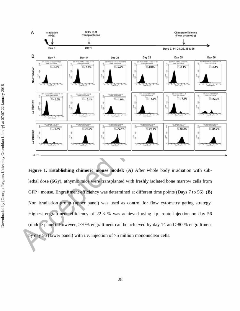
Athymic nude mice (n=3, each group) were transplanted with 5x10⁶
BM cells from GFP+ mouse
after whole body irradiation of sub-lethal dose (6Gy). Engraftment efficiency (GFP+) was
determined at each week (Days 7 to 56) (Fig 1A). Engraftment efficiency for NOD-SCID
chimera was determined at different time points (Days 14 and 28) (Supplementary Fig 1A).
Non-injected group (non-irradiated) were used as control for flow cytometry gating. Highest
engraftment efficiency of 22.3 % was achieved using i.p. injection (n=3) on day 56 (middle
panel). Surprisingly, i.v. injection (n=3) of 5x10⁶ mononuclear cells achieved >70% engraftment
by day 14 and >80 % engraftment by day 56 (lower panel) (Fig 1B). We decided to use i.v.
injection of 5x10⁶ BM cells and 14 days waiting to establish chimera for tumor studies.
However, in NOD-SCID chimera, 50% of mice showed more than 70% engraftment achieved by
day 14 and 76% of mice displayed 83-87 % engraftment by day 28 (Supplementary Fig 1B).
Effect of treatments on tumor growth
Chimeric athymic nude mice were implanted orthotopically with U251 cells and treated with
vehicle (n=17), vatalanib (n=10), AMD3100 (n=9), and nintedanib (n=3) from day 8-21. All
animals underwent MRI on day 22 (Fig 2A and 2B). We selected vatalanib as AAT agent
because these drugs enhanced tumor growth and activated alternate pathways of
neovascularization in GBM 30, 31
. Nintedanib was used to investigate the effect of multi-tyrosine
kinase inhibition in GBM. In addition, AMD3100 is an immunostimulant used to
mobilize bonemarrow derived hematopoietic stem cells in tumor. We observed no significant
decrease in tumor growth after vatalanib and AMD3100 treatment. Nintedanib treatment resulted
Dow
nloa
ded
by [
Geo
rgia
Reg
ents
Uni
vers
ity G
reen
blat
t Lib
rary
] at
07:
07 2
2 Ja
nuar
y 20
16

7
in increased tumor growth as observed by MRI and tumor volume data (Fig 2A and 2B). NOD-
SCID chimera (n=6) were orthotopically implanted with GBM PDX cells for tumor studies.
However, we could not perform MRI with NOD-SCID chimera due to unexpected sickness.
NOD-SCID mice were euthanized, whenever sign of sickness were seen.
Effect of treatments on infiltration of GFP+ cells in the tumor
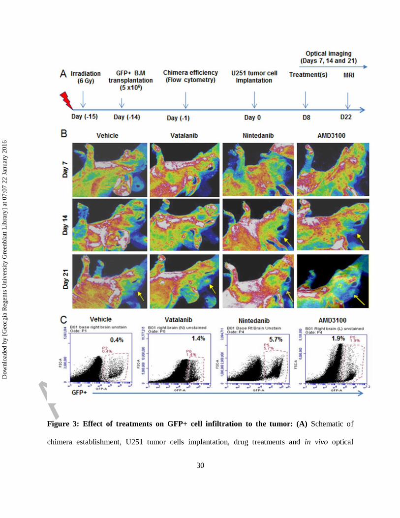
In vivo optical images obtained on days 7, 14 and 21 showed more accumulation of GFP+ in
tumor following treatments (yellow arrows) compared to vehicle (Fig 3A and 3B). Nintedanib
and AMD3100 treatments showed increased accumulation of GFP+ cells in tumor at day 14
compared to vehicle group (yellow arrows). At day 21, optical images showed increased
infiltration of GFP+ cells in vatalanib, nintedanib and AMD3100 treated groups compared to
vehicle (yellow arrows). Similarly, flow cytometry data at the end of the study (day 22) proved
higher number of GFP+ cells in drug treated tumors compared to vehicle, especially with
nintedanib treatment (Fig 3C,). GBM PDX bearing NOD-SCID chimera were scanned on days
14 and 18, which showed accumulation of GFP+ in the tumor regions of the brain
(Supplementary Fig 2A and B). In addition, ex-vivo imaging following mice euthanasia
showed accumulation of GFP+ in the tumor regions of the brain (Supplementary Fig 2C).
Effect of vatalanib, nintedanib and AMD3100 on myeloid and endothelial cell signatures
Post-MRI, brain, spleen and BM were collected and processed for flowcytometry to analyze
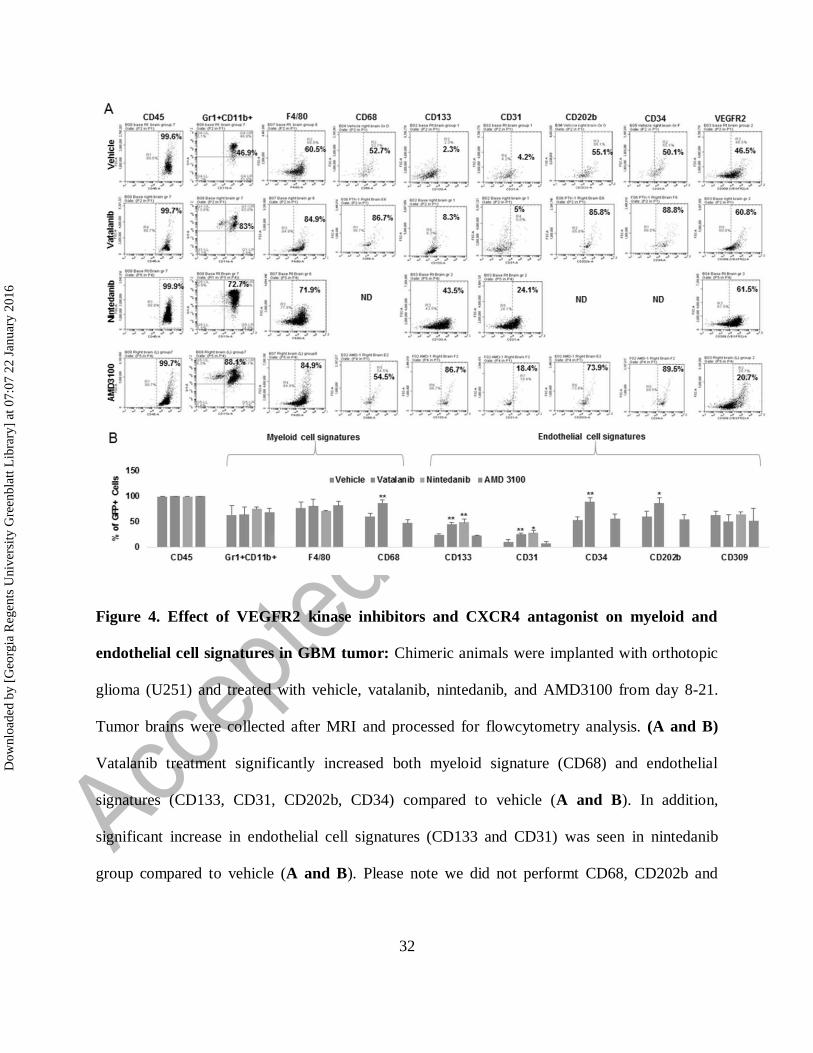
immune cell signatures (spleen and BM data not shown). Vatalanib treatment significantly
increased both myeloid signature (CD68) and endothelial signatures (CD133, CD31, CD202b,
CD34) compared to vehicle (Fig 4A and 4B). In addition, significant increase in endothelial cell
Dow
nloa
ded
by [
Geo
rgia
Reg
ents
Uni
vers
ity G
reen
blat
t Lib
rary
] at
07:
07 2
2 Ja
nuar
y 20
16
8
signatures (CD133 and CD31) was seen in nintedanib group compared to vehicle (Fig 4A and
4B). Please note we did not performt CD68, CD202b and CD34 stainings in nintedanib group
(Fig 4A and 4B). No significant changes in myeloid and endothelial cell signatures were seen in
AMD3100 group (Fig 4A and 4B). In addition, rumor brains from NOD-SCID chimera bearing
GBM PDX were collected on day 18 and processed for flow cytometry analysis. Tumor
displayed 8% of bone marrow derived GFP+ cells. GFP+ cells polarized in to myeloid cell
phenotypes such as Gr1+ CD11b+ (59.3%), F4/80+ CD11b+ (63.1%) and CD68+ CD11b+
(49.6%), and endothelial cell phenotypes such as CD202b (62.4%), CD309 (67.1%), CD34
(55.6%), CD133 (87.7%) and CD144 (16.7%), respectively (Supplementary Fig 3A). High GFP
positivity (99.8%) was seen in corresponding bone marrow populations. GFP+ cells in bone
marrow displayed increased immature markers and decreased mature markers of myeloid cell
phenotypes such as Gr1+ CD11b+ (93.5%), F4/80+ CD11b+ (17.4%) and CD68+ CD11b+
(6.01%), and endothelial cell phenotypes such as CD202b (83.2%), CD309 (33.6%), CD34
(76%), CD133 (87.3%) and CD144 (0.88%), respectively (Supplementary Fig 3B).
Homing sites and phenotypes of infiltrated GFP+ cells
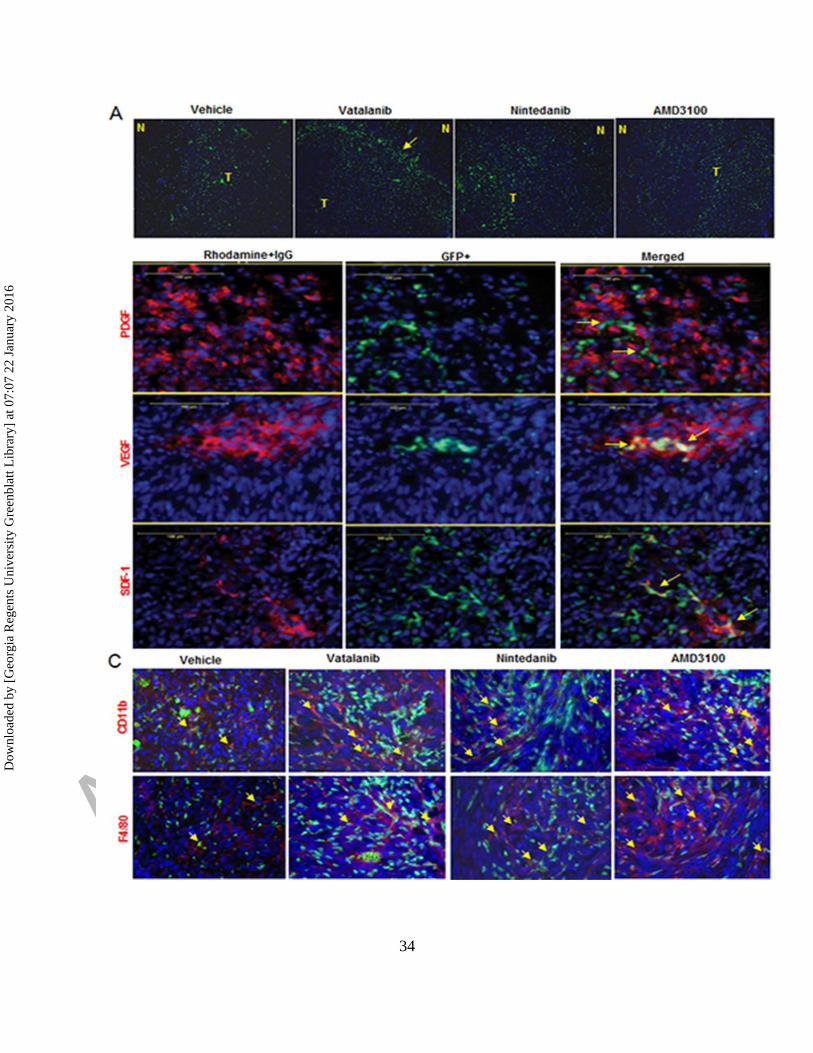
Immunofluorescence study showed accumulation of GFP+ cells at the tumor periphery or
invasive front in vatalanib treated group (yellow arrow). Vehicle and other treatments showed
more disperse GFP+ cell accumulation throughout the tumor (Fig 5A). Accumulation of GFP+
cells was seen at the site of PDGF, SDF1 and VEGF expression in the TME (Fig 5B). We
checked whether the treaments have changed the phenotype of cells with myeloid signatures in
the TME. Immunofluorescence study clearly showed increased GFP+ cells alongwith increased
CD11b+ and F4/80+ cells in vatalanib, nintedanib and AMD3100 treated groups compared to
Dow
nloa
ded
by [
Geo
rgia
Reg
ents
Uni
vers
ity G
reen
blat
t Lib
rary
] at
07:
07 2
2 Ja
nuar
y 20
16
9
vehicle (yellow arrows) (Fig 5C). However, we did not perform immunofluorescence study in
NOD-SCID mice.
DISCUSSION
We established a novel chimeric mouse model with more than 70% engraftment efficiency in 2
weeks using 5x10⁶ GFP+ bone marrow cells. Our chimera mouse model in nude background is
superior to previously published models, which needs 4 weeks to establish with same number of
cells engrafted32, 33
. Chimera in NOD-SCID background showed almost similar engraftment
efficiency, implanted with human GBM PDX but could not survive longer for AATs. We noticed
increased infiltration of GFP+ BMDCs in TME and increased tumor growth as shown by MRI
data following vatalanib, nintedanib and CXCR4 antagonist (AMD 3100). Our observation
corroborates with previous report where anti-VEGF recruited increased GFP+ BMDCs in TIB6,
B16F1, EL4 and LLC tumor-bearing mice33
. In our observation, GFP+ BMDCs were invaded
throughout tumor but more concentrated at invasive front of tumor and overlapped with
expression of VEGF, SDF1 and PDGF.
We used orthotopic mouse model with human glioma cells (U251) that better recapitulate
histopathological feature of GBM compared to that of subcutaneous models due to differences in
gene expression profile and TME 34, 35
. Previous studies that used U87 cell line, doesn’t
recapitulate human GBM completely compared to U251 36
. As indicated in human GBMs,
orthotopic mice models with U251 revealed similar level of expression of GFAP, S100B, and
Vimentin markers 35
and deletion of p53, PTEN and INK4a/ARF at the genetic level 35, 37
.
Intracranial U251 model demonstrated infiltrative invasion into brain parenchyma and significant
foci of palisading necrosis microscopically. There are several other in vivo mice models exist to
understand the biology of GBM 38
. In addition, few mouse models are available that can offer the
Dow
nloa
ded
by [
Geo
rgia
Reg
ents
Uni
vers
ity G
reen
blat
t Lib
rary
] at
07:
07 2
2 Ja
nuar
y 20
16
10
in vivo tracking and study of tumor recruited BMDCs in development AAT resistance 32, 33
.
However, role of tumor recruited BMDCs in GBM is poorly studied and no mouse model can
offer early engraftment of GFP+ bone marrow compared to present mouse model.
Bone marrow cells have pivotal role in tumor development. CXCR4+ BMDCs are
recruited to the tumor through up-regulation of HIF1-α followed by induction of SDF1α,
secretion of pro-angiogenic factors 39-42
. These recruited cells were characterized as pro-
angiogenic CD45+VEGFR2+ EPCs, or CD45+Tie2+ monocytes 43, 44
. BMDCs derived MMP9
modulated neovessels remodeling and contributing in tumor growth 42, 45
. Interestingly, lin-
ckit+Sca-1+ and their derived cells demonstrated recruitment to tumor but do not functionally
contribute to tumor neovascularization 46
. Since, AATs have been failed so far, therefore, tumor
recruited BMDCs needed further investigation in context to therapeutic resistance.
Studies have indicated that resistance to AAT has profound involvement of immune
system 47-53
. Role of myeloid cells in tumor angiogenesis is an established phenomenon as shown
by previous studies 54-58
and supported by our current study. Vatalanib treatment significantly
increased both myeloid signature (CD68) and endothelial signatures (CD133, CD31, CD202b,
CD34). Majority of GFP+ cells acquire endothelial phenotype bearing CD133 and CD31
markers in nintedanib group.This differential effect of vatalanib and nintedanib could be due to
difference in number of molecular targets e.g. nintedanib (VEGFR2, VEGFR3, LCK, FLT3,
VEGFR1, FGFR2, PDGFRα, PDGFRβ, FGFR1, FGFR3, Src, Lyn, FGFR4, IGF1R, Insulin
Receptor, CDK1, CDK2, CDK4, EGFR and HER2) and vatalanib (VEGFR2/KDR,
VEGFR1/FLT1, VEGFR2/Flk1, PDGFRβ, VEGFR3/FLT4, c-Kit and c-Fms). Previous study
showed similar findings, where SDF-1α played an important role in brain tumor invasion and
macrophage infiltration in a murine astrocytoma 59
, however, authors did not test any AAT. In
Dow
nloa
ded
by [
Geo
rgia
Reg
ents
Uni
vers
ity G
reen
blat
t Lib
rary
] at
07:
07 2
2 Ja
nuar
y 20
16
11
other study, authors observed that AATs in U87 tumors were associated with increased myeloid
cell infiltration and stem cell accumulation. However, investigations whether those phenotypes
have bone marrow component are lacking 36
. Similar to our report, authors noticed that increased
infiltration in myeloid populations in the tumor bulk and in the infiltrative regions after AAT 51
.
Together, studies suggest that immune suppressive myeloid cells (especially MDSCs and TAM
60, 61) may participate in escape from AATs, may represent a potential biomarker of resistance
and a potential therapeutic target in GBM 51
. We also tested combined treatments of vatalanib
and AMD3100; however, it did not decrease tumor growth (data not shown). Previously,
combined treatment of murine and human VEGF specific antibody and CXCR4 antagonist,
POL5551, significantly increased survival of mice bearing GBM 62
. We believe that use of
different drugs could be the reason of this differential effect.
Several mechanisms have been known to regulate mobilization and recruitment immature
myeloid cells into the TME, e.g. IL17 induced expression of GCSF through NF-κB and ERK
signaling helped homing of myeloid cells to the tumor 47
. Bv8 modulated mobilization of
MDSCs from BM to the tumor and promoted angiogenesis 54
. MDSCs can be produced in BM in
response to tumor derived factors i.e. GCSF, IL6, GMCSF, IL1β, PGE2 and TNFα, and were
recruited to tumor site by CXCL12 and CXCL5 63
. TGFβ signaling in BMDCs is important and
recruits MDSCs via CCL2 in TME 64
. CEACAM1 is identified as negative regulator of myeloid
cell expansion and recruitment by inhibiting GCSF-Bv8 axis 55
. Similarly, TIMP2 was shown to
down regulate expression of immunosuppressive genes controlling MDSC growth such as IL10,
IL13, IL11 and chemokine ligand (CCL5/RANTES), and increased IFN-γ and decreased CD40L
65. Recently, CXCL7 was discovered as an critical chemokine in myeloid cell associated
Dow
nloa
ded
by [
Geo
rgia
Reg
ents
Uni
vers
ity G
reen
blat
t Lib
rary
] at
07:
07 2
2 Ja
nuar
y 20
16

12
cancer66
. Our current chimeric mouse model could be used for future molecular mechanism
studies of therapeutic resistance in glioma and other solid cancers.
In conclusion, we developed preclinical chimeric mouse models with earlier engraftment
efficiency (2 weeks, >70%) compared to available models (4 weeks). We studied the
contribution of tumor infiltrated BMDCs in AAT resistance in GBM. Clinical trials involving
AATs have failed so far; therefore, our model may provide a tool to investigate altered myeloid
cells and associated molecular networks in GBM. As shown by others before, our study supports
that inhibiting key immune suppressive myeloid cells in TME could provide a better therapeutic
option in GBM.
MATERIALS AND METHODS
All Animal related experimental procedures were approved by the Institutional Animal Care and
Use Committee and Institutional Review Board of Georgia Regents University (animal protocol
#2014-0625). All efforts were made to ameliorate suffering of animals. CO2 with secondary
method was used to euthanize animals for tissue collection.
Establishing chimeric mouse model
Chimeric mouse for orthotopic U251 glioma was established with IACUC approved protocol and
published method 67
. Transgenic mice with universally expressing GFP under the human
ubiquitin C promoter (C57BL/6-tg(UBC-GFP)30Scha) were used as donors (Jackson
Laboratory, Main, USA). NCr-nu/nu athymic nude (Charles River, Frederick, MD, USA) and
NOD-SCID mice (Harlan laboratory, Indianapolis, USA) were used as recipients, and were
whole body irradiated with sub-lethal dose of 6Gy (Cs137). After 24 hours, recipient athymic
nude mice were injected intravenous (IV) (n=3) and intraperitoneal (n=3) routes with BM cells
Dow
nloa
ded
by [
Geo
rgia
Reg
ents
Uni
vers
ity G
reen
blat
t Lib
rary
] at
07:
07 2
2 Ja
nuar
y 20
16

13
(5x10⁶ cells) collected from donor transgenic mice. NOD-SCID mice (n=6) were injected with
BM cells (5x10⁶ cells) through IV route.
Briefly, all mononuclear cells were separated from red blood cells using lymphocyte cell
separation media (Corning, Cellgro, USA), counted and 5x10⁶ cells/100µl were injected into
each mouse. Ten microliter of blood (from orbital sinus) were collected from each athymic
mouse on days 7 to 56 (n=3 each time point) following transplantation of BM to determine BM
engraftment efficiency (GFP positivity) in peripheral blood using flowcytometer. Engraftment
efficiency of NOD-SCID mice was determined at different time points (Days 14 and 28) using
flow cytometry. Cells from athymic mice without irradiation and GFP+ cell transplantation were
used as control for flow cytometry. Our results showed that by 14 days all mice with IV
administration of GFP+ bone marrow cells had effcicent bone marrow engraftment. However,
50% of mice showed more than 70% engraftment achieved by day 14 and 75% of mice displayed
83-87 % engraftment by day 28. Based on the optimal results, all subsequent chimeric animals
were created using IV administration of GFP+ bone marrow cells and the orthotopic GBM
implanted on day 15 in athymic chimera and day 29 in NOD-SCID chimera, following IV
administration of GFP+ cells.
Animal model of human glioma
Following establishment of chimeric mice, animals were anesthetized with 100 mg/kg ketamine
and 15 mg/kg xylazine i.p. The surgical zone was swabbed with betadine solution, the eyes
coated with Lacri-lube and the animals were immobilized in a small animal stereotactic device
(Kopf, Cayunga, CA). After draping, a 1-cm incision was made 2 mm to the right of the midline
1 mm retro-orbitally; the skull exposed with cotton-tip applicators and a 23G needle tip was used
Dow
nloa
ded
by [
Geo
rgia
Reg
ents
Uni
vers
ity G
reen
blat
t Lib
rary
] at
07:
07 2
2 Ja
nuar
y 20
16

14
to drill a hole 2 mm to the right of the bregma, taking care not to penetrate the dura. A 10µL
Hamilton syringe with a 26G-needle containing U251 tumor cells or GBM PDX (n=2.4x10⁵) in 3
µl was lowered to a depth of 2.5 mm, and then raised to a depth of 2 mm. During and after the
injection, careful note was made of any reflux from the injection site. After completing the
injection, we waited 2-3 minutes before withdrawing in a stepwise manner. The surgical hole
was sealed with bone wax. Finally, the skull was swabbed with betadine before suturing the skin
over the injection site.
In vivo multispectral optical imaging
Multispectral optical images were acquired using excitation profiles of 460-480 nm range and
emission of 510 to 570 nm to monitor the GFP color at days 7, 14 and 21 after tumor cell
implantation. All optical imaging data was acquired by Kodak In-Vivo Multispectral Imaging
System FX (Carestream) and analyzed by Carestream software. For NOD-SCID chimera, In vivo
optical images obtained by Spectral AMI (Spectral Instruments Imaging, LLC) machines and
analyzed by AMI view software. Based on signal intensities derived from different excitation
and emission profiles, we have fixed our excitation and emission profile at 480 and 535,
respectively, for all subsequent experiments.
Drug treatments
Orthotopically implanted chimeric mice with U251 tumor cells were allowed to grow for 7 days
and then started oral treatments of either vehicle or receptor tyrosine kinase inhibitors (vatalanib
(50mg/kg/day) 68
and nintadanib (50mg/kg/day) 69
), daily for two weeks. AMD3100
(10mg/kg/day) 70
treatment was given through ALZET osmotic pumps (DURECT Corporation,
Dow
nloa
ded
by [
Geo
rgia
Reg
ents
Uni
vers
ity G
reen
blat
t Lib
rary
] at
07:
07 2
2 Ja
nuar
y 20
16
15
CA USA) for two weeks. Seven days waiting period was followed after tumor implantation to
mimic clinical scenario, where treatment is being done following detection of tumor.
In vivo magnetic resonance imaging (MRI)
To determine the tumor growth at the end of treatments, all animals underwent MRI on day 22.
All MRI experiments were conducted using a 7 Tesla 12 cm (clear bore) magnet interfaced to a
varian console with actively shielded gradients of 49 gauss/cm and 100µs rise times or a
horizontal 7 Tesla BioSpec MRI spectrometer (Brucker Instruments, Bellerica, MA) equipped
with a 12 cm self-shielded gradient set (45 gauss/cm max). Detailed MRI procedure was adopted
from our several previous publications 71-75
. An appropriate state of anesthesia was obtained with
isoflurane (2.5% for induction, 0.7% to 1.5% for maintenance in a 2:1 mixture of N2:O2). After
positioning using a triplanar FLASH sequence, MR studies were performed using pre-contrast
T1, T2-weighted and post contrast T1-weighted MRI scans with following parameters (1)
Standard T1-weighted multislice sequence (TR/TE=500/10 ms, 256x256 matrix, 13-15 slices, 1
mm thick slice, 32 mm field of view (FOV), # of averages=4). (2) T2-mapping sequence (2D
multi-slice, multi-echo (MSME) sequence, TE=10, 20, 30, 40, 50, 60 msec, TR=3000 msec,
256x256 matrix, 13-15 slices, 1 mm thick slice, 32 mm field of view (FOV), # of averages=2).
Post contrast T1WI was used to determine volume of tumors in vehicle and drug treated mice by
drawing irregular ROI to encircle whole tumor in each image section containing tumor using
ImageJ software, and area was then multiplied by thickness of image slice to determine volume
(cm3). Two investigators blinded to the animal groups determined tumor volume.
Collection of GFP+ cells and determination of different cell populations
Dow
nloa
ded
by [
Geo
rgia
Reg
ents
Uni
vers
ity G
reen
blat
t Lib
rary
] at
07:
07 2
2 Ja
nuar
y 20
16

16
Freshly isolated brain samples were separated into left and right (tumor bearing) hemispheres
from each group and were homogenized to pass through 40µ cell strainer to make single cell.
Similarly, cells were collected from spleen and BM. Cells were labeled with antibodies
(BioLegend) such as CD45, Gr1, CD11b, F4/80, CD68, CD133, CD31, CD34, CD202b (Tie2),
and CD309 (VEGFR2) (other than FITC) to identify BM recruited cell types (GFP+) in the
tumor. Flow cytometry data was acquired using Accuri C6 machine (BD Biosciences) and
analyzed by BD Accuri C6 software.
Immunofluorescence study
Frozen tissue sections were prepared using standard protocols and later stained for
immunofluorescence study to determine expression of angiogenic markers such as VEGF (Santa
Cruz Biotechnology), SDF-1α (Abcam) and PDGF (Santa Cruz Biotechnology) at the site of
tumor. Migration and incorporation pattern of GFP+ BMDCs was determined in different
regions of the tumor. Myeloid cell signature markers for example CD11b (Abcam) and F4/80
(Santa Cruz Biotechnology) were also determined.
Statistical analysis
Quantitative data was expressed as mean ± SD and analyzed through one way analysis of
variance (ANOVA) followed by Fisher’s least significant difference (FLSD) post-hoc test.
Group to group analysis was performed using student t-test but analysis between nindtedanib and
other groups was performed by non-parametric Mann Wintney test. Differences were considered
statistically significant at p value <0.05.
Dow
nloa
ded
by [
Geo
rgia
Reg
ents
Uni
vers
ity G
reen
blat
t Lib
rary
] at
07:
07 2
2 Ja
nuar
y 20
16

17
ACKNOWLEDGEMENT
Authors thank GRU cancer center small animal imaging core facility for finishing project on
timely manner.
Dow
nloa
ded
by [
Geo
rgia
Reg
ents
Uni
vers
ity G
reen
blat
t Lib
rary
] at
07:
07 2
2 Ja
nuar
y 20
16
18
REFERENCES
1. Olar A, Aldape KD. Using the molecular classification of glioblastoma to inform
personalized treatment. The Journal of pathology 2014; 232:165-77.
2. Stupp R, Mason WP, van den Bent MJ, Weller M, Fisher B, Taphoorn MJ, Belanger K,
Brandes AA, Marosi C, Bogdahn U, et al. Radiotherapy plus concomitant and adjuvant
temozolomide for glioblastoma. The New England journal of medicine 2005; 352:987-96.
3. Brat DJ, Van Meir EG. Vaso-occlusive and prothrombotic mechanisms associated with
tumor hypoxia, necrosis, and accelerated growth in glioblastoma. Laboratory investigation; a
journal of technical methods and pathology 2004; 84:397-405.
4. Miller KD, Sweeney CJ, Sledge GW, Jr. Can tumor angiogenesis be inhibited without
resistance? EXS 2005:95-112.
5. Thompson EM, Frenkel EP, Neuwelt EA. The paradoxical effect of bevacizumab in the
therapy of malignant gliomas. Neurology 2011; 76:87-93.
6. Grabner G, Nobauer I, Elandt K, Kronnerwetter C, Woehrer A, Marosi C, Prayer D,
Trattnig S, Preusser M. Longitudinal brain imaging of five malignant glioma patients treated
with bevacizumab using susceptibility-weighted magnetic resonance imaging at 7 T. Magn
Reson Imaging 2012; 30:139-47.
7. Wong ET, Gautam S, Malchow C, Lun M, Pan E, Brem S. Bevacizumab for recurrent
glioblastoma multiforme: a meta-analysis. J Natl Compr Canc Netw 2011; 9:403-7.
8. Norden AD, Drappatz J, Wen PY. Antiangiogenic therapy in malignant gliomas. Curr
Opin Oncol 2008; 20:652-61.
9. Norden AD, Drappatz J, Wen PY. Novel anti-angiogenic therapies for malignant
gliomas. Lancet Neurol 2008; 7:1152-60.
Dow
nloa
ded
by [
Geo
rgia
Reg
ents
Uni
vers
ity G
reen
blat
t Lib
rary
] at
07:
07 2
2 Ja
nuar
y 20
16
19
10. Bergers G, Hanahan D. Modes of resistance to anti-angiogenic therapy. Nature Review
Cancer 2008; 8:592-603.
11. Kerbel RS. Tumor angiogenesis. The New England journal of medicine 2008; 358:2039-
49.
12. Batchelor TT, Sorensen AG, di Tomaso E, Zhang WT, Duda DG, Cohen KS, Kozak KR,
Cahill DP, Chen PJ, Zhu M, et al. AZD2171, a pan-VEGF receptor tyrosine kinase inhibitor,
normalizes tumor vasculature and alleviates edema in glioblastoma patients. Cancer cell 2007;
11:83-95.
13. Folkins C, Shaked Y, Man S, Tang T, Lee CR, Zhu Z, Hoffman RM, Kerbel RS. Glioma
Tumor Stem-Like Cells Promote Tumor Angiogenesis and Vasculogenesis via Vascular
Endothelial Growth Factor and Stromal-Derived Factor 1. Cancer Res 2009; 69:7243-51.
14. Dome B, Hendrix MJC, Paku S, Tovari J, Timar J. Alternative Vascularization
Mechanisms in Cancer: Pathology and Therapeutic Implications. Am J Pathol 2007; 170:1-15.
15. Yu L, Su B, Hollomon M, Deng Y, Facchinetti V, Kleinerman ES. Vasculogenesis
Driven by Bone Marrow-Derived Cells Is Essential for Growth of Ewing's Sarcomas. Cancer
research 2010; 70:1334-43.
16. Patenaude A, Parker J, Karsan A. Involvement of endothelial progenitor cells in tumor
vascularization. Microvascular Research 2010; 79:217-23.
17. Shichinohe H, Kuroda S, Yano S, Hida K, Iwasaki Y. Role of SDF-1/CXCR4 system in
survival and migration of bone marrow stromal cells after transplantation into mice cerebral
infarct. Brain research 2007; 1183:138-47.
18. Jin DK, Shido K, Kopp HG, Petit I, Shmelkov SV, Young LM, Hooper AT, Amano H,
Avecilla ST, Heissig B, et al. Cytokine-mediated deployment of SDF-1 induces revascularization
Dow
nloa
ded
by [
Geo
rgia
Reg
ents
Uni
vers
ity G
reen
blat
t Lib
rary
] at
07:
07 2
2 Ja
nuar
y 20
16
20
through recruitment of CXCR4(+) hemangiocytes. Nat Med 2006; 12:557-67. Epub 2006 Apr
30.
19. Petit I, Jin D, Rafii S. The SDF-1-CXCR4 signaling pathway: a molecular hub
modulating neo-angiogenesis. Trends in Immunology 2007; 28:299-307.
20. Ceradini DJ, Kulkarni AR, Callaghan MJ, Tepper OM, Bastidas N, Kleinman ME, Capla
JM, Galiano RD, Levine JP, Gurtner GC. Progenitor cell trafficking is regulated by hypoxic
gradients through HIF-1 induction of SDF-1. Nat Med 2004; 10:858-64. Epub 2004 Jul 4.
21. Arbab AS, Janic B, Knight RA, Anderson SA, Pawelczyk E, Rad AM, Read EJ, Pandit
SD, Frank JA. Detection of migration of locally implanted AC133+ stem cells by cellular
magnetic resonance imaging with histological findings. FASEB journal : official publication of
the Federation of American Societies for Experimental Biology 2008; 22:3234-46.
22. Moore MA, Hattori K, Heissig B, Shieh JH, Dias S, Crystal RG, Rafii S. Mobilization of
endothelial and hematopoietic stem and progenitor cells by adenovector-mediated elevation of
serum levels of SDF-1, VEGF, and angiopoietin-1. Annals of the New York Academy of
Sciences 2001; 938:36-45; discussion -7.
23. Heissig B, Hattori K, Dias S, Friedrich M, Ferris B, Hackett NR, Crystal RG, Besmer P,
Lyden D, Moore MA, et al. Recruitment of stem and progenitor cells from the bone marrow
niche requires MMP-9 mediated release of kit-ligand. Cell 2002; 109:625-37.
24. Kioi M, Vogel H, Schultz G, Hoffman RM, Harsh GR, Brown JM. Inhibition of
vasculogenesis, but not angiogenesis, prevents the recurrence of glioblastoma after irradiation in
mice. J Clin Invest 2010; 120:694-705.
25. Hentschel SJ, Sawaya R. Optimizing outcomes with maximal surgical resection of
malignant gliomas. Cancer Control 2003; 10:109-14.
Dow
nloa
ded
by [
Geo
rgia
Reg
ents
Uni
vers
ity G
reen
blat
t Lib
rary
] at
07:
07 2
2 Ja
nuar
y 20
16
21
26. Hillen F, Kaijzel EL, Castermans K, oude Egbrink MG, Lowik CW, Griffioen AW. A
transgenic Tie2-GFP athymic mouse model; a tool for vascular biology in xenograft tumors.
Biochem Biophys Res Commun 2008; 368:364-7.
27. Motoike T, Loughna S, Perens E, Roman BL, Liao W, Chau TC, Richardson CD, Kawate
T, Kuno J, Weinstein BM, et al. Universal GFP reporter for the study of vascular development.
Genesis 2000; 28:75-81.
28. Jang KS, Lee KS, Yang SH, Jeun SS. In vivo Tracking of Transplanted Bone Marrow-
Derived Mesenchymal Stem Cells in a Murine Model of Stroke by Bioluminescence Imaging. J
Korean Neurosurg Soc 2010; 48:391-8.
29. Ray P, Tsien R, Gambhir SS. Construction and validation of improved triple fusion
reporter gene vectors for molecular imaging of living subjects. Cancer Res 2007; 67:3085-93.
30. Ali MM, Janic B, Babajani-Feremi A, Varma NR, Iskander AS, Anagli J, Arbab AS.
Changes in vascular permeability and expression of different angiogenic factors following anti-
angiogenic treatment in rat glioma. PloS one 2010; 5:e8727.
31. Arbab AS. Activation of alternative pathways of angiogenesis and involvement of stem
cells following anti-angiogenesis treatment in glioma. Histology and histopathology 2012;
27:549-57.
32. Pyonteck SM, Akkari L, Schuhmacher AJ, Bowman RL, Sevenich L, Quail DF, Olson
OC, Quick ML, Huse JT, Teijeiro V, et al. CSF-1R inhibition alters macrophage polarization and
blocks glioma progression. Nature medicine 2013; 19:1264-72.
33. Shojaei F, Wu X, Malik AK, Zhong C, Baldwin ME, Schanz S, Fuh G, Gerber HP,
Ferrara N. Tumor refractoriness to anti-VEGF treatment is mediated by CD11b+Gr1+ myeloid
cells. Nature biotechnology 2007; 25:911-20.
Dow
nloa
ded
by [
Geo
rgia
Reg
ents
Uni
vers
ity G
reen
blat
t Lib
rary
] at
07:
07 2
2 Ja
nuar
y 20
16
22
34. Camphausen K, Purow B, Sproull M, Scott T, Ozawa T, Deen DF, Tofilon PJ. Orthotopic
growth of human glioma cells quantitatively and qualitatively influences radiation-induced
changes in gene expression. Cancer research 2005; 65:10389-93.
35. Jacobs VL, Valdes PA, Hickey WF, De Leo JA. Current review of in vivo GBM rodent
models: emphasis on the CNS-1 tumour model. ASN neuro 2011; 3:e00063.
36. Piao Y, Liang J, Holmes L, Zurita AJ, Henry V, Heymach JV, de Groot JF. Glioblastoma
resistance to anti-VEGF therapy is associated with myeloid cell infiltration, stem cell
accumulation, and a mesenchymal phenotype. Neuro-oncology 2012; 14:1379-92.
37. Radaelli E, Ceruti R, Patton V, Russo M, Degrassi A, Croci V, Caprera F, Stortini G,
Scanziani E, Pesenti E, et al. Immunohistopathological and neuroimaging characterization of
murine orthotopic xenograft models of glioblastoma multiforme recapitulating the most salient
features of human disease. Histology and histopathology 2009; 24:879-91.
38. Simeonova I, Huillard E. In vivo models of brain tumors: roles of genetically engineered
mouse models in understanding tumor biology and use in preclinical studies. Cellular and
molecular life sciences : CMLS 2014; 71:4007-26.
39. Aghi M, Cohen KS, Klein RJ, Scadden DT, Chiocca EA. Tumor stromal-derived factor-1
recruits vascular progenitors to mitotic neovasculature, where microenvironment influences their
differentiated phenotypes. Cancer research 2006; 66:9054-64.
40. De Falco E, Porcelli D, Torella AR, Straino S, Iachininoto MG, Orlandi A, Truffa S,
Biglioli P, Napolitano M, Capogrossi MC, et al. SDF-1 involvement in endothelial phenotype
and ischemia-induced recruitment of bone marrow progenitor cells. Blood 2004; 104:3472-82.
Dow
nloa
ded
by [
Geo
rgia
Reg
ents
Uni
vers
ity G
reen
blat
t Lib
rary
] at
07:
07 2
2 Ja
nuar
y 20
16

23
41. Ceradini DJ, Kulkarni AR, Callaghan MJ, Tepper OM, Bastidas N, Kleinman ME, Capla
JM, Galiano RD, Levine JP, Gurtner GC. Progenitor cell trafficking is regulated by hypoxic
gradients through HIF-1 induction of SDF-1. Nature medicine 2004; 10:858-64.
42. Ahn GO, Brown JM. Matrix metalloproteinase-9 is required for tumor vasculogenesis but
not for angiogenesis: role of bone marrow-derived myelomonocytic cells. Cancer cell 2008;
13:193-205.
43. Deak E, Gottig S, Ruster B, Paunescu V, Seifried E, Gille J, Henschler R. Bone marrow
derived cells in the tumour microenvironment contain cells with primitive haematopoietic
phenotype. Journal of cellular and molecular medicine 2010; 14:1946-52.
44. Ahn GO, Brown JM. Role of endothelial progenitors and other bone marrow-derived
cells in the development of the tumor vasculature. Angiogenesis 2009; 12:159-64.
45. Seandel M, Butler J, Lyden D, Rafii S. A catalytic role for proangiogenic marrow-
derived cells in tumor neovascularization. Cancer cell 2008; 13:181-3.
46. Shinde Patil VR, Friedrich EB, Wolley AE, Gerszten RE, Allport JR, Weissleder R. Bone
marrow-derived lin(-)c-kit(+)Sca-1+ stem cells do not contribute to vasculogenesis in Lewis lung
carcinoma. Neoplasia 2005; 7:234-40.
47. Chung AS, Wu X, Zhuang G, Ngu H, Kasman I, Zhang J, Vernes JM, Jiang Z, Meng YG,
Peale FV, et al. An interleukin-17-mediated paracrine network promotes tumor resistance to anti-
angiogenic therapy. Nature medicine 2013; 19:1114-23.
48. Shojaei F, Wu X, Qu X, Kowanetz M, Yu L, Tan M, Meng YG, Ferrara N. G-CSF-
initiated myeloid cell mobilization and angiogenesis mediate tumor refractoriness to anti-VEGF
therapy in mouse models. Proceedings of the National Academy of Sciences of the United States
of America 2009; 106:6742-7.
Dow
nloa
ded
by [
Geo
rgia
Reg
ents
Uni
vers
ity G
reen
blat
t Lib
rary
] at
07:
07 2
2 Ja
nuar
y 20
16
24
49. Seton-Rogers S. Tumour microenvironment: Means of resistance. Nature reviews Cancer
2013; 13:607.
50. Phan VT, Wu X, Cheng JH, Sheng RX, Chung AS, Zhuang G, Tran C, Song Q,
Kowanetz M, Sambrone A, et al. Oncogenic RAS pathway activation promotes resistance to
anti-VEGF therapy through G-CSF-induced neutrophil recruitment. Proceedings of the National
Academy of Sciences of the United States of America 2013; 110:6079-84.
51. Lu-Emerson C, Snuderl M, Kirkpatrick ND, Goveia J, Davidson C, Huang Y, Riedemann
L, Taylor J, Ivy P, Duda DG, et al. Increase in tumor-associated macrophages after
antiangiogenic therapy is associated with poor survival among patients with recurrent
glioblastoma. Neuro-oncology 2013; 15:1079-87.
52. Achyut BR. Impact of Microenvironment in Therapy-Induced Neovascularization of
Glioblastoma. Biochem Physiol 2013; 2:e121.
53. Dondossola E, Rangel R, Guzman-Rojas L, Barbu EM, Hosoya H, St John LS, Molldrem
JJ, Corti A, Sidman RL, Arap W, et al. CD13-positive bone marrow-derived myeloid cells
promote angiogenesis, tumor growth, and metastasis. Proceedings of the National Academy of
Sciences of the United States of America 2013; 110:20717-22.
54. Shojaei F, Wu X, Zhong C, Yu L, Liang XH, Yao J, Blanchard D, Bais C, Peale FV, van
Bruggen N, et al. Bv8 regulates myeloid-cell-dependent tumour angiogenesis. Nature 2007;
450:825-31.
55. Lu R, Kujawski M, Pan H, Shively JE. Tumor angiogenesis mediated by myeloid cells is
negatively regulated by CEACAM1. Cancer research 2012; 72:2239-50.
56. Shojaei F, Zhong C, Wu X, Yu L, Ferrara N. Role of myeloid cells in tumor angiogenesis
and growth. Trends in cell biology 2008; 18:372-8.
Dow
nloa
ded
by [
Geo
rgia
Reg
ents
Uni
vers
ity G
reen
blat
t Lib
rary
] at
07:
07 2
2 Ja
nuar
y 20
16
25
57. Yang L, DeBusk LM, Fukuda K, Fingleton B, Green-Jarvis B, Shyr Y, Matrisian LM,
Carbone DP, Lin PC. Expansion of myeloid immune suppressor Gr+CD11b+ cells in tumor-
bearing host directly promotes tumor angiogenesis. Cancer cell 2004; 6:409-21.
58. Achyut BR, Arbab, A.S. Myeloid Derived Suppressor Cells: Fuel the Fire. Biochem
Physiol 2014; 3:e123.
59. Wang SC, Hong JH, Hsueh C, Chiang CS. Tumor-secreted SDF-1 promotes glioma
invasiveness and TAM tropism toward hypoxia in a murine astrocytoma model. Laboratory
investigation; a journal of technical methods and pathology 2012; 92:151-62.
60. Sica A, Schioppa T, Mantovani A, Allavena P. Tumour-associated macrophages are a
distinct M2 polarised population promoting tumour progression: potential targets of anti-cancer
therapy. European journal of cancer 2006; 42:717-27.
61. Hussain SF, Yang D, Suki D, Aldape K, Grimm E, Heimberger AB. The role of human
glioma-infiltrating microglia/macrophages in mediating antitumor immune responses. Neuro-
oncology 2006; 8:261-79.
62. Barone A, Sengupta R, Warrington NM, Smith E, Wen PY, Brekken RA, Romagnoli B,
Douglas G, Chevalier E, Bauer MP, et al. Combined VEGF and CXCR4 antagonism targets the
GBM stem cell population and synergistically improves survival in an intracranial mouse model
of glioblastoma. Oncotarget 2014; 5:9811-22.
63. Sawanobori Y, Ueha S, Kurachi M, Shimaoka T, Talmadge JE, Abe J, Shono Y,
Kitabatake M, Kakimi K, Mukaida N, et al. Chemokine-mediated rapid turnover of myeloid-
derived suppressor cells in tumor-bearing mice. Blood 2008; 111:5457-66.
Dow
nloa
ded
by [
Geo
rgia
Reg
ents
Uni
vers
ity G
reen
blat
t Lib
rary
] at
07:
07 2
2 Ja
nuar
y 20
16
26
64. Fan Q, Gu D, Liu H, Yang L, Zhang X, Yoder MC, Kaplan MH, Xie J. Defective TGF-
beta signaling in bone marrow-derived cells prevents hedgehog-induced skin tumors. Cancer
research 2014; 74:471-83.
65. Guedez L, Jensen-Taubman S, Bourboulia D, Kwityn CJ, Wei B, Caterina J, Stetler-
Stevenson WG. TIMP-2 targets tumor-associated myeloid suppressor cells with effects in cancer
immune dysfunction and angiogenesis. Journal of immunotherapy 2012; 35:502-12.
66. Achyut BR, Shankar A, Iskander A, Ara R, Angara K, Zeng P, Knight RA, Scicli AG,
Arbab AS. Bone marrow derived myeloid cells orchestrate antiangiogenic resistance in
glioblastoma through coordinated molecular networks. Cancer letters 2015.
67. Ali MM, Kumar S, Shankar A, Varma NR, Iskander AS, Janic B, Chwang WB, Jain R,
Babajeni-Feremi A, Borin TF, et al. Effects of tyrosine kinase inhibitors and CXCR4 antagonist
on tumor growth and angiogenesis in rat glioma model: MRI and protein analysis study.
Translational oncology 2013; 6:660-9.
68. Yuen DA, Stead BE, Zhang Y, White KE, Kabir MG, Thai K, Advani SL, Connelly KA,
Takano T, Zhu L, et al. eNOS deficiency predisposes podocytes to injury in diabetes. Journal of
the American Society of Nephrology : JASN 2012; 23:1810-23.
69. Richeldi L, Costabel U, Selman M, Kim DS, Hansell DM, Nicholson AG, Brown KK,
Flaherty KR, Noble PW, Raghu G, et al. Efficacy of a tyrosine kinase inhibitor in idiopathic
pulmonary fibrosis. The New England journal of medicine 2011; 365:1079-87.
70. Yu M, Gang EJ, Parameswaran R, Stoddart S, Fei F, Schmidhuber S, Park E, Hsieh YT,
Yang AS, Groffen J, et al. AMD3100 sensitizes acute lymphoblastic leukemia cells to
chemotherapy in vivo. Blood cancer journal 2011; 1:e14.
Dow
nloa
ded
by [
Geo
rgia
Reg
ents
Uni
vers
ity G
reen
blat
t Lib
rary
] at
07:
07 2
2 Ja
nuar
y 20
16
27
71. Ding G, Jiang Q, Zhang L, Zhang ZG, Li L, Knight RA, Ewing JR, Wang Y, Chopp M.
Analysis of combined treatment of embolic stroke in rat with r-tPA and a GPIIb/IIIa inhibitor.
Journal of Cerebral Blood Flow & Metabolism 2005; 25:87-97.
72. Conway JG, McDonald B, Parham J, Keith B, Rusnak DW, Shaw E, Jansen M, Lin PY,
Payne A, Crosby RM, et al. Inhibition of colony-stimulating-factor-1 signaling in vivo with the
orally bioavailable cFMS kinase inhibitor GW2580. Proceedings of the National Academy of
Sciences of the United States of America 2005; 102:16078-83.
73. Ewing JR, Knight RA, Nagaraja TN, Yee JS, Nagesh V, Whitton PA, Li L,
Fenstermacher JD. Patlak plots of Gd-DTPA MRI data yield blood-brain transfer constants
concordant with those of 14C-sucrose in areas of blood-brain opening. Magn Reson Med 2003;
50:283-92.
74. Nagaraja TN, Karki K, Ewing JR, Divine GW, Fenstermacher JD, Patlak CS, Knight RA.
The MRI-measured arterial input function resulting from a bolus injection of Gd-DTPA in a rat
model of stroke slightly underestimates that of Gd-[14C]DTPA and marginally overestimates the
blood-to-brain influx rate constant determined by Patlak plots. Magnetic Resonance in Medicine
2010; 63:1502-9.
75. Nagaraja TN, Karki K, Ewing JR, Croxen RL, Knight RA. Identification of Variations in
Blood-Brain Barrier Opening After Cerebral Ischemia by Dual Contrast-Enhanced Magnetic
Resonance Imaging and T1sat Measurements. Stroke 2008; 39:427-32. Dow
nloa
ded
by [
Geo
rgia
Reg
ents
Uni
vers
ity G
reen
blat
t Lib
rary
] at
07:
07 2
2 Ja
nuar
y 20
16
28
Figure 1. Establishing chimeric mouse model: (A) After whole body irradiation with sub-
lethal dose (6Gy), athymic mice were transplanted with freshly isolated bone marrow cells from
GFP+ mouse. Engraftment efficiency was determined at different time points (Days 7 to 56). (B)
Non irradiation group (upper panel) was used as control for flow cytometry gating strategy.
Highest engraftment efficiency of 22.3 % was achieved using i.p. route injection on day 56
(middle panel). However, >70% engraftment can be achieved by day 14 and >80 % engraftment
by day 56 (lower panel) with i.v. injection of >5 million mononuclear cells.
Dow
nloa
ded
by [
Geo
rgia
Reg
ents
Uni
vers
ity G
reen
blat
t Lib
rary
] at
07:
07 2
2 Ja
nuar
y 20
16

29
Figure 2. Effect of treatments on tumor growth: Chimeric animals were implanted with
orthotopic glioma (U251) and treated with vehicle, vatalanib, nintedanib and AMD3100 from
day 8-21. (A) Schematic of chimera establishment, U251 tumor cells implantation, drug
treatments and in vivo MRI. (B) All animals underwent MRI on day 22 following implantation of
tumors. No significant reduction in tumor growth was observed using vatalanib and AMD3100.
Surprisingly, Nintedanib treatment resulted in increased tumor growth as observed by MRI and
tumor volume data. Quantitative data is expressed in mean ±SD. *P<0.05.
Dow
nloa
ded
by [
Geo
rgia
Reg
ents
Uni
vers
ity G
reen
blat
t Lib
rary
] at
07:
07 2
2 Ja
nuar
y 20
16
30
Figure 3: Effect of treatments on GFP+ cell infiltration to the tumor: (A) Schematic of
chimera establishment, U251 tumor cells implantation, drug treatments and in vivo optical
Dow
nloa
ded
by [
Geo
rgia
Reg
ents
Uni
vers
ity G
reen
blat
t Lib
rary
] at
07:
07 2
2 Ja
nuar
y 20
16
31
imaging. (B) In vivo optical images obtained by Kodak In-Vivo Multispectral Imaging System
FX (Carestream) on days 7, 14 and 21 showed increased accumulation of GFP+ in the tumor
following treatments (yellow arrows) compared to vehicle, especially on day 21. GFP+ cells
were detected in day14 tumors treated with nintedanib and AMD (yellow arrows). (C)
Quantitative analysis by flow cytometry also proved the higher number of GFP+ cells in day 21
tumors that were treated with vatalanib, nintedanib and AMD3100.
Dow
nloa
ded
by [
Geo
rgia
Reg
ents
Uni
vers
ity G
reen
blat
t Lib
rary
] at
07:
07 2
2 Ja
nuar
y 20
16
32
Figure 4. Effect of VEGFR2 kinase inhibitors and CXCR4 antagonist on myeloid and
endothelial cell signatures in GBM tumor: Chimeric animals were implanted with orthotopic
glioma (U251) and treated with vehicle, vatalanib, nintedanib, and AMD3100 from day 8-21.
Tumor brains were collected after MRI and processed for flowcytometry analysis. (A and B)
Vatalanib treatment significantly increased both myeloid signature (CD68) and endothelial
signatures (CD133, CD31, CD202b, CD34) compared to vehicle (A and B). In addition,
significant increase in endothelial cell signatures (CD133 and CD31) was seen in nintedanib
group compared to vehicle (A and B). Please note we did not performt CD68, CD202b and
Dow
nloa
ded
by [
Geo
rgia
Reg
ents
Uni
vers
ity G
reen
blat
t Lib
rary
] at
07:
07 2
2 Ja
nuar
y 20
16

33
CD34 stainings in nintedanib group (A and B). ND= not done. Quantitative data is expressed in
mean ±SD. *P<0.05 and **P<0.01.
Dow
nloa
ded
by [
Geo
rgia
Reg
ents
Uni
vers
ity G
reen
blat
t Lib
rary
] at
07:
07 2
2 Ja
nuar
y 20
16
34
Dow
nloa
ded
by [
Geo
rgia
Reg
ents
Uni
vers
ity G
reen
blat
t Lib
rary
] at
07:
07 2
2 Ja
nuar
y 20
16
35
Figure 5. Accumulation of GFP+ cells in tumor and myeloid phenotypes. (A)
Immunofluorescence images showing accumulation of GFP+ cells (green) at the tumor periphery
or invasive front in vatalanib treated group (yellow arrow). (B) Co-localization of GFP+ cells
with PDGF, SDF1 and VEGF expression (red) in the TME (yellow arrows). (C)
Immunofluorescence images showing increased GFP+ cells (green) alongwith increased
CD11b+ and F4/80+ cells (red) in vatalanib, nintedanib and AMD3100 treated groups compared
to vehicle (yellow arrows). N= normal and T=tumor.
Dow
nloa
ded
by [
Geo
rgia
Reg
ents
Uni
vers
ity G
reen
blat
t Lib
rary
] at
07:
07 2
2 Ja
nuar
y 20
16
View publication statsView publication stats