chest radiography - fmh college of medicine and … radiography. dr khawaja khurshid ahmad associate...
TRANSCRIPT

Chest Radiography

Dr Khawaja Khurshid Ahmad
Associate Professor of Radiology
King Edward Medical University /
Mayo Hospital , Lahore
Case One
Chest Radiology
History
• Age / Sex 39 / female
• Chief complaint Cough and sputum for
one month

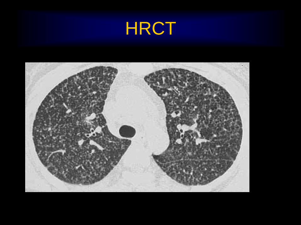
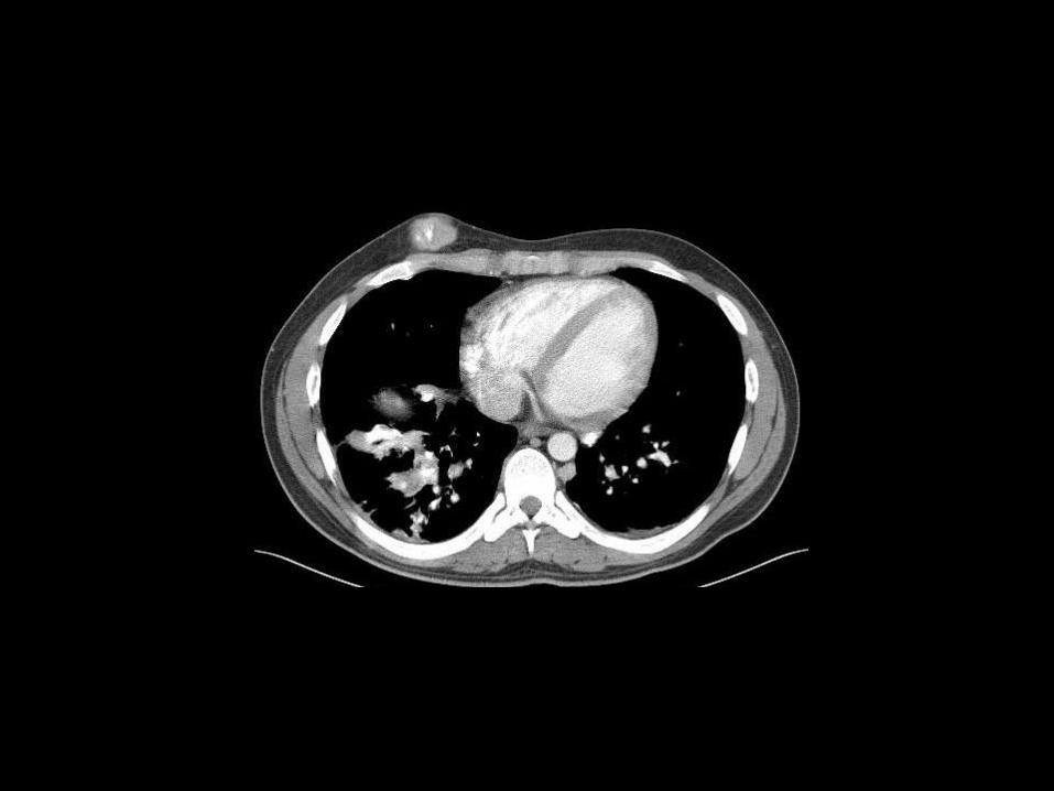
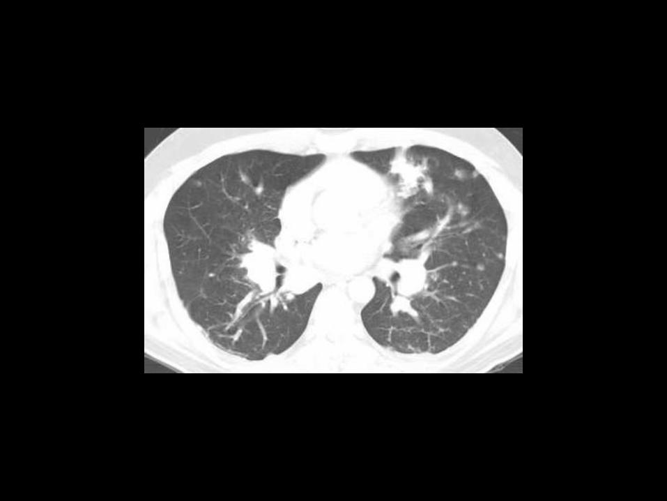
HRCT


Mediastinal Window

Diagnosis
• Primary lung cancer (adenocarcinoma) in
RLL with miliary metastasis and pericardial
seeding
Case Two
Chest Radiology

History
• Age / Sex 21 /M
• Chief complaint Palpable chest
wall mass and
neuralgia in gaiting for
one month


Diagnosis
• Metastasis of Osteosarcoma

Case Three
Chest Radiology

History
• Age / Sex 32 / M
• Chief complaint Weight loss , fatigue,
and poor oral intake for
two months

Diagnosis
Sarcoidosis

Case Four
Chest Radiology

History
• Age/Sex: 27/M
Chief complaints: Fever


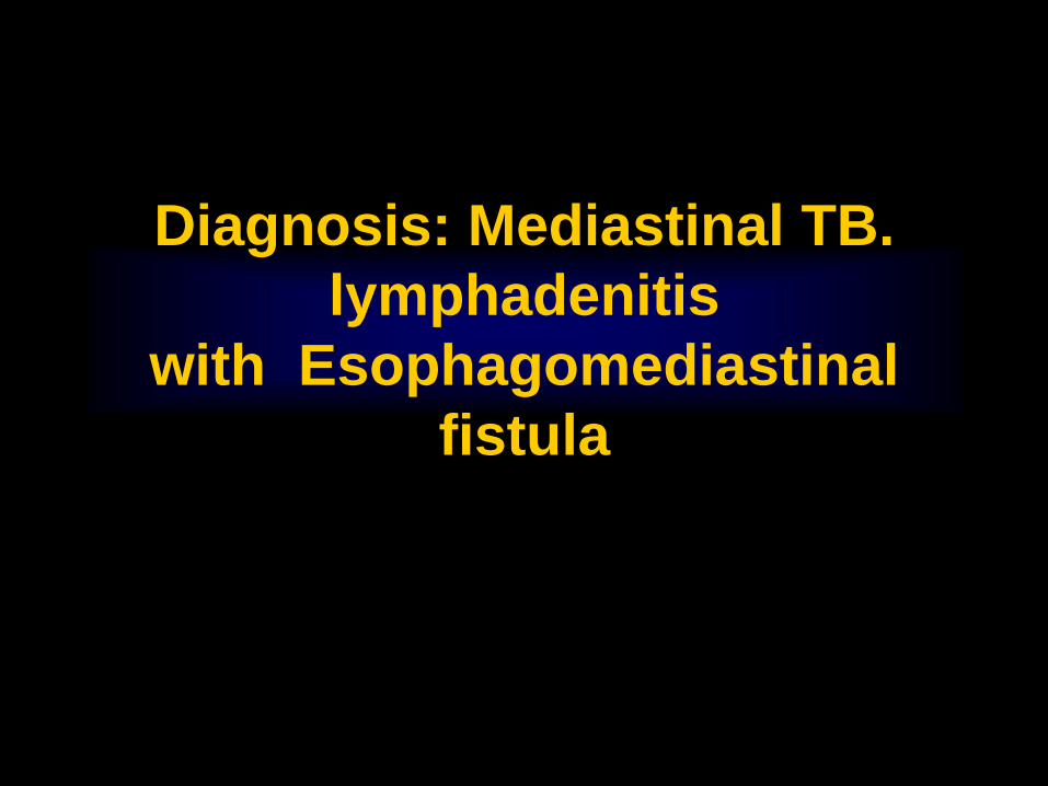
Diagnosis: Mediastinal TB.
lymphadenitis
with Esophagomediastinal
fistula

?

PA

Lateral

COLLAPSE

LLL Collapse

LUL Collapse

RUL COLLAPSE

RML COLLAPSE

RLL COLLAPSE

Consolidation

Lingular
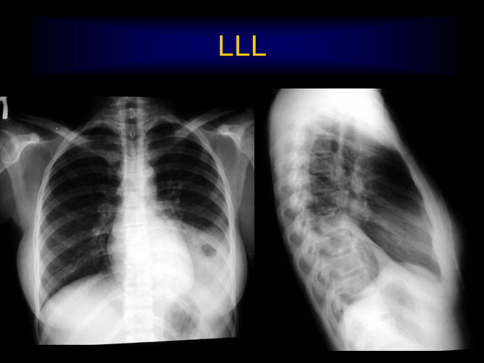
LLL

LUL
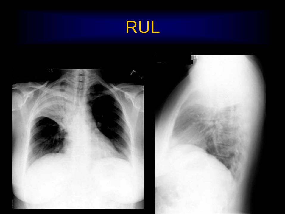
RUL

Tumor
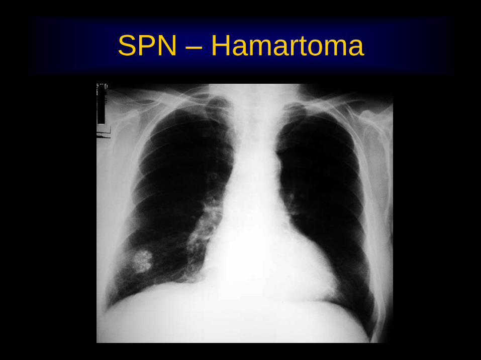
SPN – Hamartoma

CA Thyroid
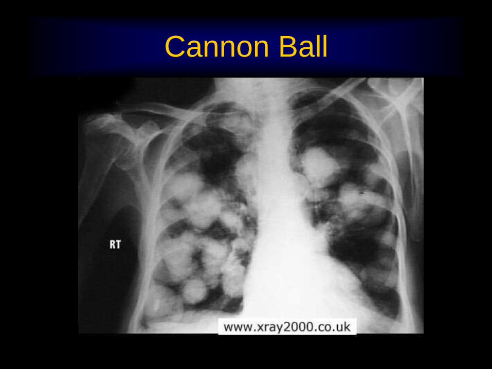
Cannon Ball

Pleural Mets

Absent Clavicle-Metastasis

CA causing RUL Collapse

CA Lung

Pancoast Tumor

Lymph nodes

Lymphoma

Diaphragm
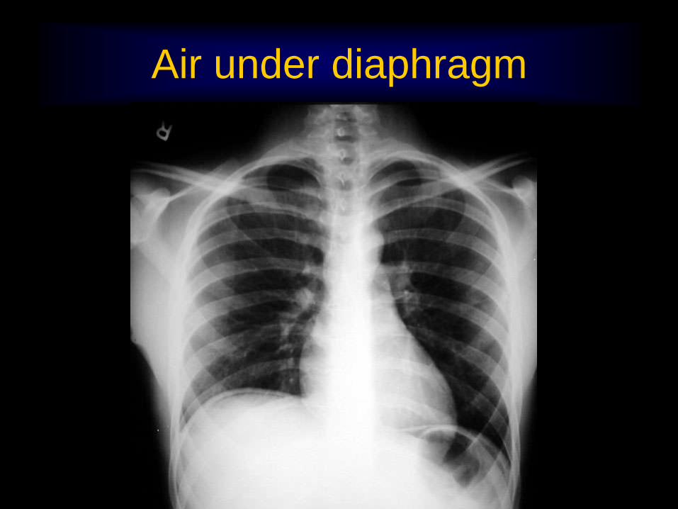
Air under diaphragm

Localized Eventration

Supradiaphragmatic mass
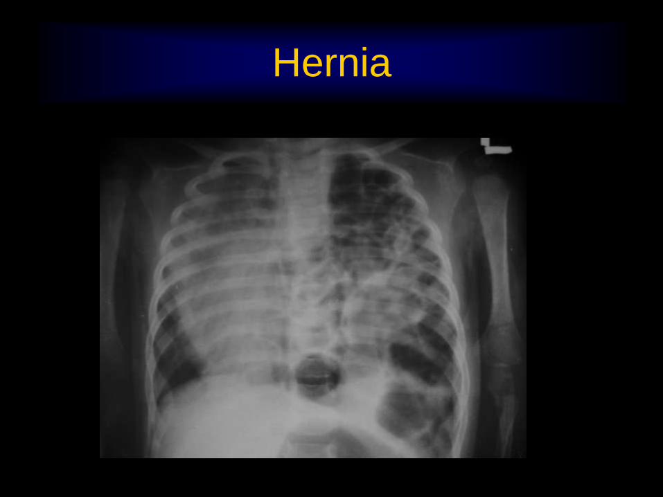
Hernia

Infective

Miliary TB


Tuberculous Cavity with
Aspergilloma


Bronchiectasis

Bronchiectasis

Bronchiectasis

Abscess

Lucent Lung

Agenesis Pulmonary Artery

Breast Resection

Emphysema

Pneumothorax

Pneumo Mediastinum

Miscellaneous

Myositis Ossificans


Achlasia


Kartegner

Goiter
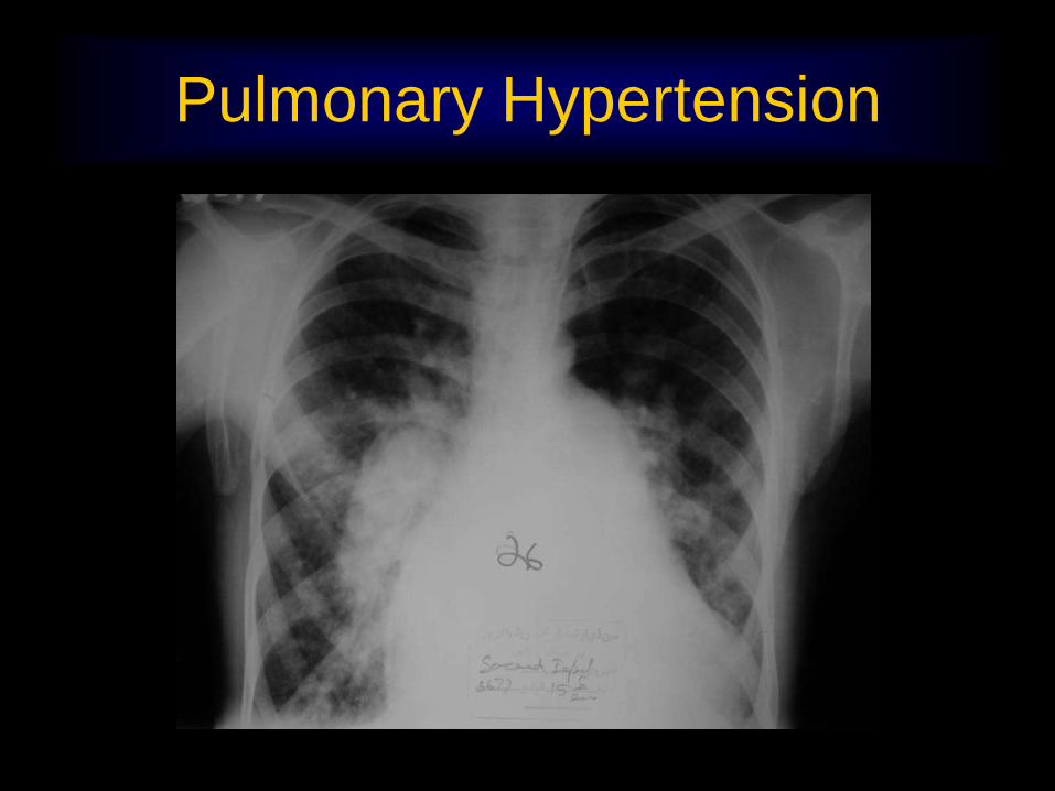
Pulmonary Hypertension
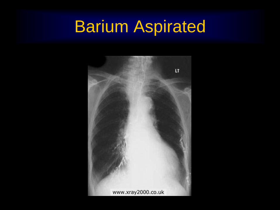
Barium Aspirated
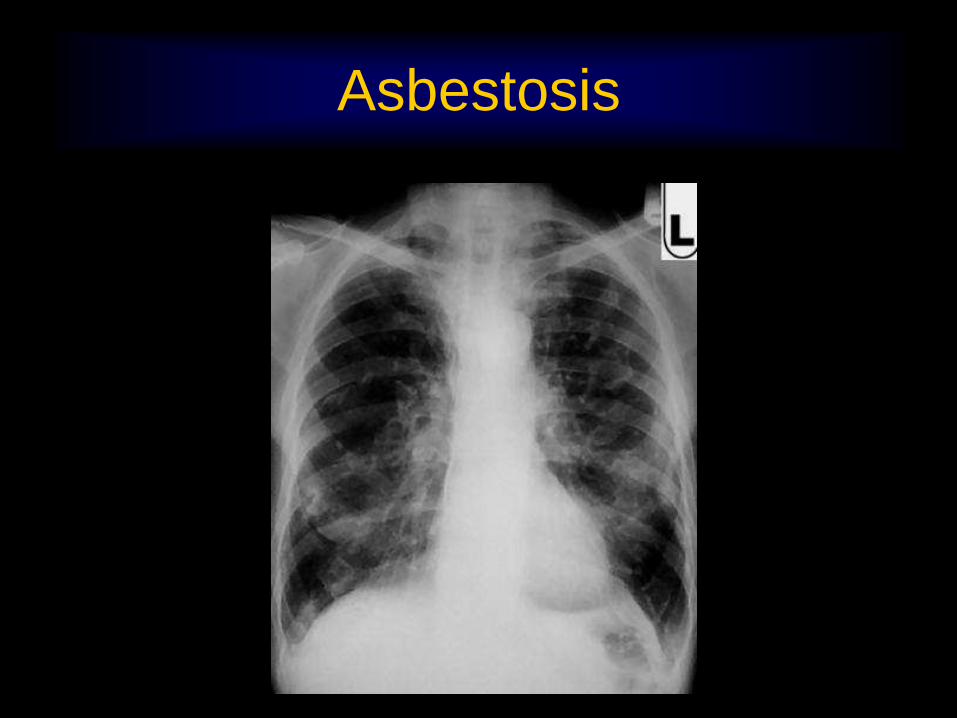
Asbestosis

Reference Sources
• Teaching files of Radiology Department Mayo
Hospital , Lahore
• Teaching Files of Prof MA Jawed Siddiqui
• Website www.xray2000.co.uk
• Website www.Chestxrayatlas.com
• Academic cases of Korean Society of Thoracic
Radiology

THANK YOU

CA Lung

Chondroma
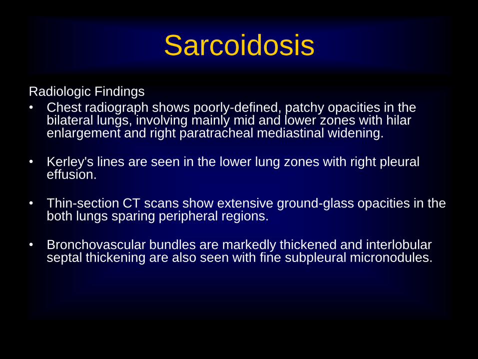
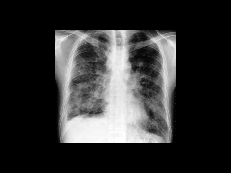
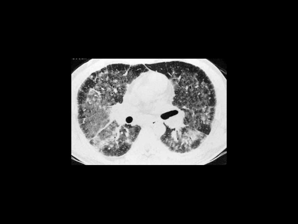
Sarcoidosis
Radiologic Findings
• Chest radiograph shows poorly-defined, patchy opacities in the bilateral lungs, involving mainly mid and lower zones with hilar enlargement and right paratracheal mediastinal widening.
• Kerley's lines are seen in the lower lung zones with right pleural effusion.
• Thin-section CT scans show extensive ground-glass opacities in the both lungs sparing peripheral regions.
• Bronchovascular bundles are markedly thickened and interlobular septal thickening are also seen with fine subpleural micronodules.
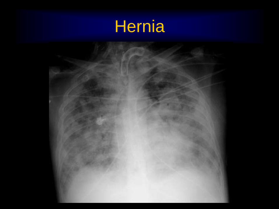
Hernia

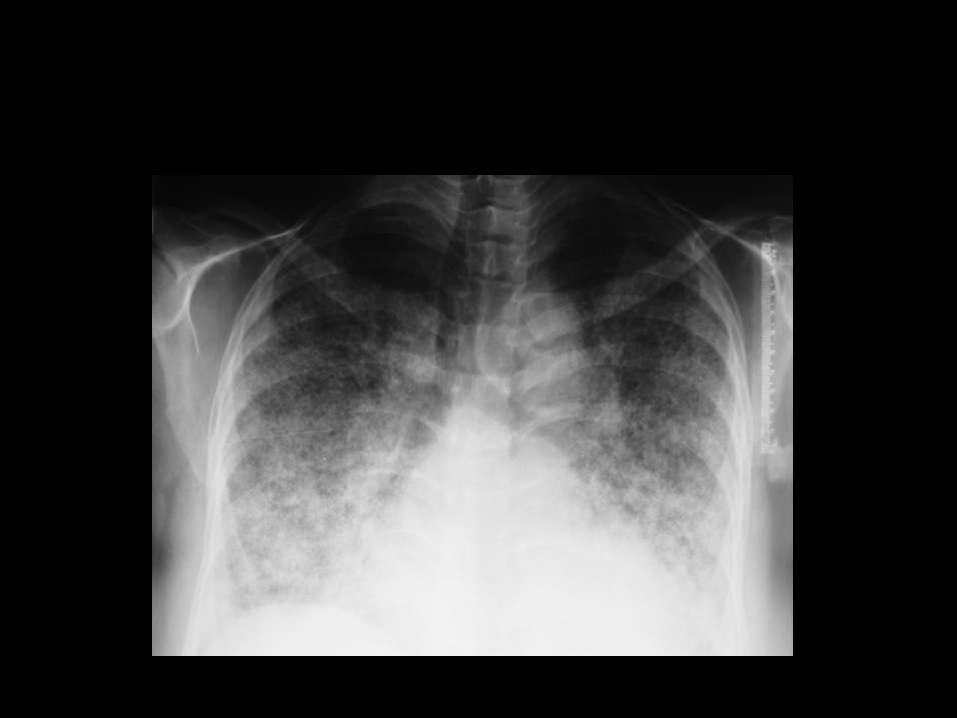
ARDS

Interstitial Lung
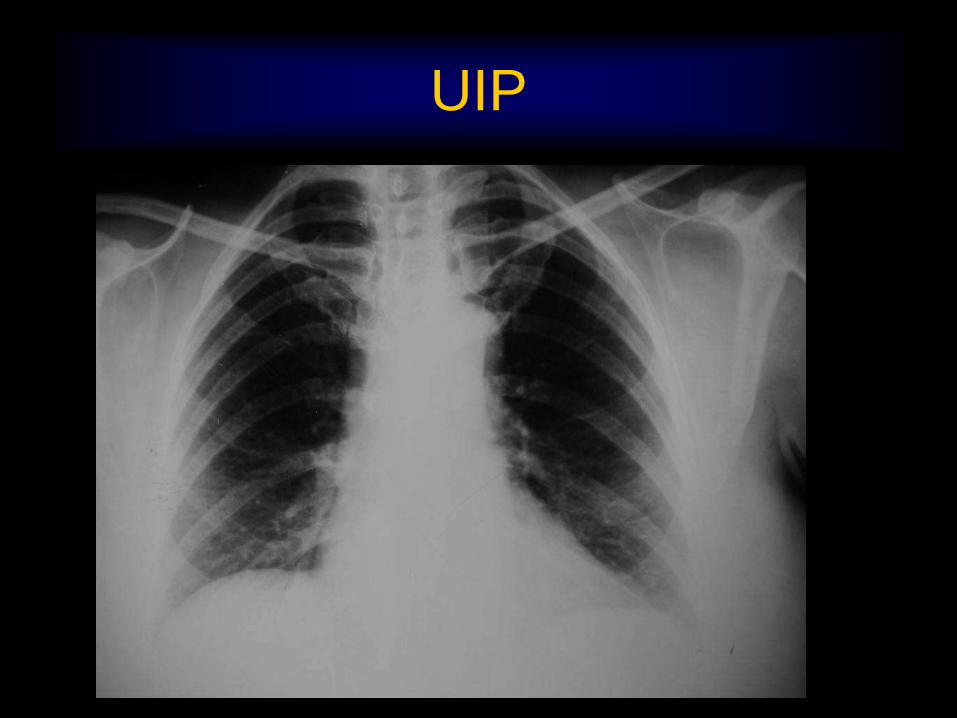
UIP

IPF
ARDS

Peanut obstruction

IPF

IPF

Unfolding Aorta

Alveolar Microlithiasis