characterization of lactic acid bacteria isolated from bukuljac, a homemade goat's milk cheese
TRANSCRIPT

Available online at www.sciencedirect.com
biology 122 (2008) 162–170www.elsevier.com/locate/ijfoodmicro
International Journal of Food Micro
Characterization of lactic acid bacteria isolated from Bukuljac, a homemadegoat's milk cheese
Milica Nikolic ⁎, Amarela Terzic-Vidojevic, Branko Jovcic, Jelena Begovic,Natasa Golic, Ljubisa Topisirovic
Institute of Molecular Genetics and Genetic Engineering, Vojvode Stepe 444a, P.O. Box 23, 11010 Belgrade, Serbia
Received 6 June 2007; received in revised form 13 November 2007; accepted 27 November 2007
Abstract
The Bukuljac cheese is traditionally homemade cheese, produced from heat-treated goat's milk without the addition of any bacterial starterculture. The presence of lactic acid bacteria (LAB) in Bukuljac cheese has been analyzed by using a polyphasic approach includingmicrobiological and molecular methods such as rep-PCR with (GTG)5 primer. Lactobacillus paracasei subsp. paracasei represents a dominantstrain in the microflora of analyzed cheese. Out of 55 Gram-positive and catalase-negative isolates, 48 belonged to L. paracasei subsp.paracasei species. Besides lactobacilli, five Lactococcus lactis subsp. lactis and two Enterococcus faecalis were found. Results of PCR-denaturing gradient gel electrophoresis (DGGE) of DNA extracted directly from the fresh cheese revealed the presence of Leuconostocmesenteroides.
Only lactobacilli showed a high proteolytic activity and hydrolyzed αs1− and β-caseins. They are also producers of diacetyl. In addition, 34 outof 55 isolates, all determined as lactobacilli, showed the ability of auto-aggregation. Among 55 isolates, 50 also exhibited antimicrobial activity.© 2007 Elsevier B.V. All rights reserved.
Keywords: Goat's milk cheese; LAB; DGGE; Rep-PCR; Proteinase; Aggregation; Antimicrobial activity
1. Introduction
The analysis of dominant microorganisms in cheese has beenperformed by bacterial cultivation, followed by characterizationand identification of the microflora by phenotypic, biochemicaland physiological tests (Corroler et al., 1998; Stiles andHolzapfel, 1997). Although conventional culture-dependentmethods have proven to be useful and indispensable tool, theyare regarded as time-consuming and limited in terms of bothdiscriminating ability and accuracy. In the last decade, theprofiling of bacterial populations became more precise with theapplication of molecular methods that enabled direct detectionof DNA and RNA in microbial ecosystems. Moleculartechniques, including 16S ribosomal DNA (rDNA) sequencing,repetitive bacterial DNA elements (rep-PCR) analysis, andother PCR-based techniques, are necessary for the taxonomic
⁎ Corresponding author. Tel.: +381 11 3975960; fax: +381 11 3975808.E-mail address: [email protected] (M. Nikolic).
0168-1605/$ - see front matter © 2007 Elsevier B.V. All rights reserved.doi:10.1016/j.ijfoodmicro.2007.11.075
investigation of complex ecosystems (Amann et al., 1995;Schleifer et al., 1995). In particular, denaturing gradient gelelectrophoresis (DGGE) of 16S rDNA amplicons has beendemonstrated as a suitable tool for the analysis of microbialcommunities allowing the detection of species and changes incommunity structure quickly and economically (Randazzo etal., 2002).
Due to the widely appreciated organoleptic characteristics,the production of goat's milk cheese has attracted growinginterest over recent years. It has been noticed that the goat milkhas more easily digestible fat and protein content than cow milk(Haenlein, 2001). In addition, goat milk has increase content ofvitamin A, thiamine and niacin in comparison to cow's milk(Haenlein, 2001). Characterization and identification of themicroflora from artisanal goat's milk cheeses have beenanalyzed in different countries, like Spain (Fontecha et al.,1990; Requena et al., 1992; Olarte et al., 2000; Sanchez et al.,2005), Greece (Litopoulou-Tzanetaki and Tzanetakis, 1992;Xanthopoulos et al., 2000) and Bulgaria (Tserovska et al.,2000–2002).

163M. Nikolic et al. / International Journal of Food Microbiology 122 (2008) 162–170
The goat's milk cheese (locally called Bukuljac) originatedfrom Serbia. It is a soft white cheese with pleasant flavour madefrom pure heat-treated goat's milk by the addition of rennet andwithout the addition of any starter culture. In general, artisanalcheeses with unique characteristics such as goat's milk cheeseanalyzed in this study are manufactured as a part of historicaland cultural heritage in the Balkan region.
In this study, we performed a polyphasic approach, includingculture-dependent and independent methods for studying thelactic acid bacteria (LAB) from the goat's milk cheese. Sampleswere subjected to traditional microbiological analysis, directDNA extraction, followed by PCR with primers able to detecteubacterial population, and DGGE analysis on the amplifiedproducts. Moreover, strains of LAB were isolated, subsequentlyidentified by molecular methods, and characterized by the useof PCR amplification of repetitive bacterial DNA elements (rep-PCR). The aim of this study was the examination of the wildLAB microflora from a homemade goat's milk cheese andtechnological properties of the isolated strains for the potentialinclusion in a starter culture preparation. According to ourknowledge, this paper reports the first study regarding thephenotypic, genotypic and physiological characterization of themicroflora from goat's milk cheese produced in The WesternBalkan region.
2. Materials and methods
2.1. Cheese manufacture and sampling
Bukuljac cheese was made in a household using traditionalcheese-making procedure, located in Central Serbia, near townof Arandjelovac, at approximate altitude of 500 m. The cheeseBukuljac was made on the only Sana breed goat farm in thisregion. The milk was obtained by mixing refrigerated eveningand fresh morning milk. The milk was heated until boiling andthe temperature of milk was adjusted to approximately 30 °Cprior to addition of the commercial rennet (“Sirela”, Cacak,Serbia) for milk coagulation, without the addition of a starterculture. The formation of a curd took 2 to 3 h. The curd wascut into pieces, approximately 12×6×3 cm weighed 200–350 g and salted by dry salt (10 g kg−1 of cheese). In order toremove whey, the curd was sieved 2 to 3 h without pressure.Afterward, the cheese was left to ripen. The usual ripeningtime was between 1 and 10 days at 10 to 15 °C. Cheesesamples (one sample per batch) from three different batches(300 g in weight per sample), with the same ripening period(5 days) were taken sterile and kept refrigerated at 4 °C untilthe arrival to the laboratory for the analysis. Microbiologicalanalyses of these samples were performed within the following24 to 48 h.
2.2. Microbiological analyses–isolation, characterization andidentification of lactic acid bacteria
For microbiological analysis, 20 g of each cheese samplewas taken from the cheese interior, homogenized with pestlein sterile mortar, and transferred to 180 ml sterile 2% (w/v)
sodium citrate solution. Decimal dilutions of the homogenateswere prepared with sterile 0.85% (w/v) sodium chloride andwere plated on media most suitable for isolation of LAB: a)for presumptive lactobacilli, on MRS agar pH 5.7 (MerckGmbH, Darmstadt, Germany); b) for presumptive lactococcion M17 agar pH 7.2 (Merck GmbH, Darmstadt, Germany).Incubation of inoculated media was performed at 30 °C and45 °C for 72 h in aerobic conditions and in anaerobicconditions (Mannu et al., 2002). For incubation in anaerobicconditions, jars with Anaerocult A (Merck GmbH, Darmstadt,Germany) were used.
Thirty to fifty colonies per sample were randomly taken fromboth M17 and MRS (30 °C and 45 °C) agar platescorresponding to the highest dilution at which growth occurred.Cell morphology of all isolates of LAB was determined bymicroscopy (Olympus U-RFL-T, BX51, GmbH, Hamburg,Germany). After microscopic observations, the colonies weresub-cultured to purity on MRS or M17 medium for rods andcocci, respectively. Gram-positive and catalase-negative iso-lates were frozen at −20 °C and −80 °C in M17 (for cocci) andin MRS (for rods) broth containing 15% of glycerol (v/v)(Mannu et al., 2002).
Overall, 131 pure cultures were isolated and 88 of them wereGram-positive and catalase-negative. According to the mor-phology of bacterial colonies and cells, 55 isolates out of 88were chosen for further analysis and they were identified to thegenus or species level, by phenotypic tests as follows: (a)colony morphology and pigmentation, (b) growth at 15 °C,30 °C and 45 °C in MRS broth for rods and in M17 broth forcocci (the tests were performed three times), (c) salt tolerance:growth with 4%, 6.5% and 8% NaCl in MRS and M17 broth forrods or cocci, respectively (the tests were performed threetimes), (d) production of carbon dioxide from glucose by sub-culturing the isolates in tubes with MRS broth containingDurham's tubes, (e) L-arginin and esculin hydrolysis, (f) onlyfor cocci-test of forming black zone on bile esculin agar(HiMedia, Mumbai, India), (g) citrate-utilization, (h) activity inmilk and test in litmus milk, (i) diacetyl production-only forLAB which coagulated skimmed milk (Kandler and Weiss1986; Mundt, 1986a,b; Prescot et al., 1996).
The bacterial strains used in this study are listed in Table 1.Lactobacillus strains were generally grown in MRS (MerckGmbH, Darmstadt, Germany) at 30 °C or 37 °C, depending onthe strain. Lactococcus lactis and Enterococcus strains weregrown in M17 (Merck GmbH, Darmstadt, Germany) with 10 gl−1 of glucose, at 30 °C and 37 °C, respectively.
2.3. DNA isolation
Total DNA from cheese was extracted as described byRandazzo et al. (2002). Total DNA from pure cultures wasextracted as described by Hopwood et al. (1985). Plasmid DNAfrom the isolates was extracted as described by O' Sullivan andKlaenhammer (1993). Plasmid profiles were observed in 1%agarose gels and visualized in ethidium bromide by CCDcamera Biometra BDR2/5/6 (Bio Doc Analyze GmbH,Göttingen, Germany).

Table 1The list of strains used in this study
Strains Relevantcharacteristic(s)
Source or reference
LactococcusL. lactis subsp. lactisBGSM1-19
Bac+, BacIm⁎ Laboratory collectionBCCM/LMG‡
L. lactis subsp. lactisBGMN1-596
Bac−, Bacs † Gajic et al., 1999
L. lactis subsp. cremoris NS1 Bac−, Bacs Laboratory collectionL. lactis subsp. cremoris Wg2 Prt+ Haandrikman et al., 1991
LactobacillusLact. paracasei subsp.paracasei BGSJ2-8
Bac+, BacIm Lozo et al., 2007
Lact. paracasei BGUB9 Bac+, BacIm Laboratory collectionBCCM/LMG
Lact. paracasei subsp.paracasei BGHN14
Natural isolate Kojic et al., 1991b
Lact. paracasei subsp.paracasei BGBUK2-16
Natural isolate Lozo et al., 2004
Lact. paracasei subsp.paracasei BGBUK2-16 K4
Derivative ofBGBUK2-16
Lozo et al., 2004
EnterococcusEnt. faecium BGGJ8-3 Natural isolate Laboratory collection
BCCM/LMGEnt. faecalis BGZLS60-26a Natural isolate Laboratory collection
BCCM/LMGSalmonella enteritidis Clinical
isolateCollection from the institute“Dr Milan JovanovicBatut”, Belgrade
⁎ Bac+, BacIm=Bacteriocin producer, bacteriocin immune.† Bac−, Bacs=Bacteriocin producer, bacteriocin sensitive.‡ BCCM/LMG=strains determination was done in BCCM/LMG, University ofGent, Gent, Belgium.
164 M. Nikolic et al. / International Journal of Food Microbiology 122 (2008) 162–170
2.4. PCR amplification
All PCR amplifications were performed with the Taq DNApolymerase kit (Fermentas, Vilnius, Latvia). To investigate thedominant bacterial communities by DGGE analysis, PCRproducts were generated with PCR primers U968-GC (5'-CGC CGG GGG CGC GCC CCG GGCGGGGCG GGGGCACGG GGG GAA CGC GAA GAA CCT TAC-3') and L1401(5'-GCG TGT GTA CAA GAC CC-3') to amplify the V6 to V8region of eubacterial 16S rDNA (Randazzo et al., 2002). Thesamples were amplified in GeneAmp PCR System 2700(Applied Biosystems, Foster City, CA, USA) programmed asfollows: initial denaturation of DNA for 5 min at 94 °C, 30cycles of 30 s at 94 °C, 30 s at 55 °C, and 30 s at 72 °C; andextension of incomplete products for 7 min at 72 °C. PCRproducts were quantified by electrophoresis on a 1% (wt/vol)agarose gel containing ethidium bromide, and visualized byCCD camera Biometra BDR2/5/6 (Bio Doc Analyze).
For rep-PCR analysis products were generated with primers(GTG)5 (5'-GTG GTG GTG GTG GTG-3') for all isolates.Samples were amplified in GeneAmp PCR System 2700(Applied Biosystems) programmed as follows: initial denatura-tion of DNA for 7 min at 95 °C, 32 cycles of 1 min at 94 °C,1 min at 40 °C, and 8 min at 65 °C; and extension of incompleteproducts for 16 min at 65 °C. PCR products were separated by
electrophoresis on a 1.5% (wt/vol) agarose gel containing0.5 μg ml−1 ethidium bromide in 1×TAE buffer (2 mol l−1 Trisbase, 1 mol l−1 glacial acetic acid, 0.05 mol l−1 EDTA [pH8.0]), at a constant voltage 60 Vat temperature of 4 °C for 20 h(Versalovic et al., 1994). The electrophoresis was performed inthe horizontal gel electrophoresis system (Bethesda ResearchLaboratories, Gaithersburg, Maryland) and visualized by CCDcamera Biometra BDR2/5/6 (Bio Doc Analyze).
2.5. DGGE analysis of PCR amplicons
DGGE analysis was performed on the DGGE System(DGGE System, C.B.S. Scientific co., Del Mar, CA, USA)essentially as described previously (Randazzo et al., 2002).Samples were added to a 6.5% (wt/vol) polyacrylamide gel(acrylamide–bisacrylamide, 19:1) in 1×TAE buffer (2 mol l−1
Tris base, 1 mol l−1 glacial acetic acid, 0.050 mol l−1 EDTA[pH 8.0]). Optimal separation of the PCR products for thespecies in the cheese samples was achieved with a 40% to 60%urea-formamide denaturant gradient, increasing in the directionof electrophoresis. A 100% denaturant corresponds to 7 mol l−1
urea and 40% (vol/vol) formamide. Electrophoresis wasperformed at a constant voltage of 90 V at temperature of60 °C for 16 h. The DNA bands were visualized by silverstaining according to DNA silver staining kit protocol(PlusOne™ DNA Silver Staining Kit, Amersham Biosciences,Uppsala, Sweden).
Elution of DNA bands from DGGE polyacrylamide gels wasperformed (http://axon.med.harvard.edu/~cepko/protocol/mike/D5.html, last date entered 11/08/07) and DNA fragments weredistained (http://msf.ucdavis.edu/silverstaining_protocols.html,last date entered 11/08/07), amplified by PCR with U968 andL1401 primers and sequenced. Obtained PCR products werepurified by QIAquick PCR Purification KIT (Qiagen, GmbH,Germany), and sequenced by CRIBI-BMR servizio sequenzia-mento DNA (Universita di Padova, Italy). The sequence wasaligned with NCBI database using the standard nucleotide–nucleotide homology search BLAST (http://www.ncbi.nlm.nih.gov/BLAST; RID: 1138633900-27581-131272740575.BLASTQ4).
2.6. Antimicrobial activity assay
For the detection of antimicrobial activity, an agar-welldiffusion assay was used (Kojic et al., 1991a). Soft M17 orMRS agar (0.7%, w/v) containing approximately 105 cells ml−1
of indicator strains were overlaid onto M17 or MRS plates(depending on the indicator strain). Wells were made in the lawnof M17 or MRS soft agar. Aliquots (50 μl) of fresh 12 h growingcultures were poured into the wells. Plates were incubated 24 hat appropriate temperatures for growth of indicator strains. After24 h of incubation, inhibition zones were read. A crystal ofpronase E was placed close to the edge of the well containingthe bacteriocin sample to confirm the proteinaceous nature ofbacteriocin. A clear zone of inhibition around the well but not inthe vicinity of the pronase E crystal was taken as a positivesignal for bacteriocin production. Indicator strains used in this

165M. Nikolic et al. / International Journal of Food Microbiology 122 (2008) 162–170
assay were: L. lactis subsp. lactis BGSM1-19, L. lactis subsp.lactis BGMN1-596, L. lactis subsp. cremoris NS1, Lactoba-cillus paracasei subsp. paracasei BGSJ2-8, Lact. paracaseiBGUB9 and Salmonella enteritidis (Table 1).
Test of H2O2 production, was done as described previously(McGoarty et al., 1992; Rabe and Hillier, 2003).
2.7. Assay of proteolytic activity
Proteolytic activities of the isolates were assayed aspreviously described (Kojic et al., 1991b). Collected freshcells (10 mg approximate density 1010 cells ml−1) wereresuspended in 0.1 mol l−1 ammonium acetate buffer, pH 6.2.The cell suspension was mixed with β-casein (5 mg ml−1in0.1 mol l−1 ammonium acetate buffer, pH 6.2) (Sigma, St.Louis, MO, USA) and incubated for 2 h at 37 °C. Electrophor-esis was carried out on 12.5% polyacrylamide gel. Gels werestained with Coomassie brilliant blue G250 (SERVA, Heidel-berg, Germany) and distained in a mix of methanol (20%) andacetic acid (7%).
3. Results
3.1. Total count of lactic acid bacteria
The microbiota present in the goat's milk cheese wasenumerated by cultivating on MRS or M17 media with theintention to isolate different groups of bacteria. The averageLAB count of all three cheese samples ranged between4.60×103 and 4.13×10⁷cfu ml−1 on MRS and 1.82×10⁷ and8.86×107or M17 media. The initial count of bacteria washigher on M17 media due to a large number of catalase positivebacteria (Table 2).
3.2. Physiological characteristics of LAB isolated fromBukuljac cheese
According to the cell morphology 55 Gram-positive,catalase-negative isolates (designated BGAR) were chosen forfurther analysis. These 55 isolates were divided into three sub-groups. The majority (87.3%) of these isolates was assigned tothe genus Lactobacillus, 9.1% to Lactococcus and 3.6% wereEnterococci.
Among cocci, five isolates (BGAR4, BGAR8, BGAR10,BGAR116, BGAR117) had the typical physiological character-istics for strain L. lactis. subsp. lactis. Two cocci isolates(BGAR124, BGAR131), according to the physiological tests,
Table 2Mean number of total viable bacteria isolated from Bukuljac cheese samplesafter 72 hours cultivation on MRS and M17 plates
Sample Cfu g−1 samplea
MRS M17
1. 4.13×10⁷ 2.09×10⁷
2. 6.74×10⁵ 1.82×10⁷
3. 4.60×10³ 8.86×10⁷
a Average values of three repetitions.
showed characteristics of Enterococcus sp. They also showeddark zones when cultivated on bile esculin agar. Among 48isolated Lactobacillus sp., minor differences in physiologicalcharacteristics were found (Table 3). All isolates curdledskimmed milk within 16 h. Test in litmus milk showed thatall isolates produced acid and changed the colour of theindicator from purple to white. Five isolates belonging to L.lactis subsp. lactis had very good activity in milk and after 6 hof incubation, the pH reached a value 4.8. Majority oflactobacilli (81.25%) were able to produce diacetyl. It isinteresting that 34 of 48 lactobacilli showed auto-aggregationphenotype, seen as clearly visible sand-like particles, formed bycells, which gravited to the bottom of the tubes, leaving a clearsupernatant fluid (Fig. 1).
3.3. DGGE analysis of PCR amplicones
To investigate the diversity of the dominating microbialcommunity in Bukuljac cheese, DNA isolated from cheesesamples was analysed by PCR-DGGE. The 16S rDNA genes ofBGAR isolates were also screened by PCR-DGGE, whichdistinguished three unique types (Fig. 2). DGGE profilerevealed that lactobacilli isolates had high similarity with thestrain Lact. paracasei subsp. paracasei BGHN14 andBGBUK2-16, lactococci isolates showed high similarity withL. lactis subsp. lactis BGSM1-19 and enterococci isolatesshowed high similarity with Enterococcus faecalis BGZLS60-26a (Fig. 2)
In order to further identify the origins of the amplicons,positions of PCR products from each group were compared withthe microbial community DGGE pattern of the Bukuljac cheese.The DGGE profiles of the isolates belonging to Lact. paracaseisubsp. paracasei, L. lactis subsp. lactis and Ent. faecaliscorresponded precisely to the bands A, C, B, respectively,identified in the DGGE pattern of the total DNA extracted fromcheese. One band that did not correspond to any band of theisolates (band D) was cut off from the DGGE gel and after PCRreamplification it was sequenced. Results of the BLASTnucleotide–nucleotide homology search indicated that thesequence from the band corresponds to Leuconostoc mesenter-oides with 98% of identity.
3.4. Identification and diversity of isolates by rep-PCR analysis
The results of the rep-PCR analysis revealed two differentpatterns of GTG-PCR products, which enabled grouping of 48lactobacilli isolates into two different sub-groups. Sub-group Iconsist of 32 isolates and showed high pattern similarity withstrain Lact. paracasei subsp. paracasei BGHN14, while other16 isolates, belonging to sub-group II, showed high similaritywith other reference strain Lact. paracasei subsp. paracaseiBGSJ2-8 (Fig. 3). Comparison of obtained fingerprint profilesof lactococci isolates with L. lactis subsp. cremorisWg2 and L.lactis subsp. lactis BGSM1-19 revealed a higher similarity withL. lactis subsp. lactis BGSM1-19 (Fig. 4). Isolated enterococcishowed higher similarity with Ent. faecalis BGZLS60-26a incomparison to Enterococcus faecium BGGJ8-3 (Fig. 4). Since

Table 3Physiological characteristics and preliminary identification of LAB isolated from Bukuljac cheese
Physiological tests Preliminary identification
Lactobacillus sp. Lactococcus lactissubsp. lactis
Enterococcus sp.
+ − + − + −
Growth at 15 °C 44 4 5 0 2 0Growth at 30 °C 44 0 5 0 2 0Growth at 45 °C 0 48 0 5 2 0Growth in mediumwith 4% NaCl
48 0 5 0 2 0
Growth in mediumwith 6.5% NaCl
36 12 0 5 2 0
Growth in mediumwith 8% NaCl
0 48 0 5 2 0
Arginin hydrolysis 1 46 5 0 2 01 (±)
Esculin hydrolysis 32 1 5 0 2 015 (±)
Using of citrate 26 (±) 22 5 0 1 1Production of CO2
from glucose0 48 0 5 0 2
Production of diacetyl 39 9 0 5 0 2
+═ Number of isolates with positive reaction; − ═ number of isolates with negative reaction; ± ═ weak reaction.
166 M. Nikolic et al. / International Journal of Food Microbiology 122 (2008) 162–170
two enterococcal isolates showed identical rep-PCR profile,DNA of the isolate BGAR124 was amplified with U968 and1401 primers, and PCR product was sequenced. Nucleotidesequence showed that enterococcci isolate belong to Ent.faecalis with 99% of similarity.
3.5. Plasmid profile and proteolytic activities
Plasmid patterns revealed that no difference among all fivelactococci isolates, as well as for the enterococci isolates couldbe observed. According to plasmid profiles, lactobacilli isolatescan be sorted into four groups (data not shown).
Fig. 1. The appearance of overnight cultures of an auto-aggregating isolateBGAR99-1 (Tube 1) and non-aggregating isolate BGAR104 (Tube 2) fromBukuljac cheese.
All lactobacilli were proteolyticaly active. They hydrolyzedboth αS1− and β-casein (Fig. 5). No proteolytic activities inlactococci and enterococci isolates were detected (data notshown).
3.6. Antimicrobial activity assay
According to the test of the ability to produce antimicro-bial substances, all 55 isolates from Bukuljac cheese havebeen analyzed using lactococci and lactobacilli as indicators.
Fig. 2. DGGE of PCR amplicons of the V6 to V8 regions of 16S rDNA of totalDNA isolated from the cheese, DNA isolated from BGAR isolates fromBukuljac cheese and from the referent strains. Lanes: 1. total DNA isolated fromgoat's milk cheese Bukuljac; 2. BGAR75; 3. BGAR76; 4. Lact. paracaseisubsp. paracasei BGHN14; 5. Lact. paracasei subsp. paracasei BGBUK2-16;6. BGAR4; 7. L. lactis subsp. lactis BGSM1-19; 8. BGAR124; 9. Ent. faecalisBGZLS60-26a. The position of bands in DGGE profile of total DNA (indicatedby the letters) are discussed in the text.
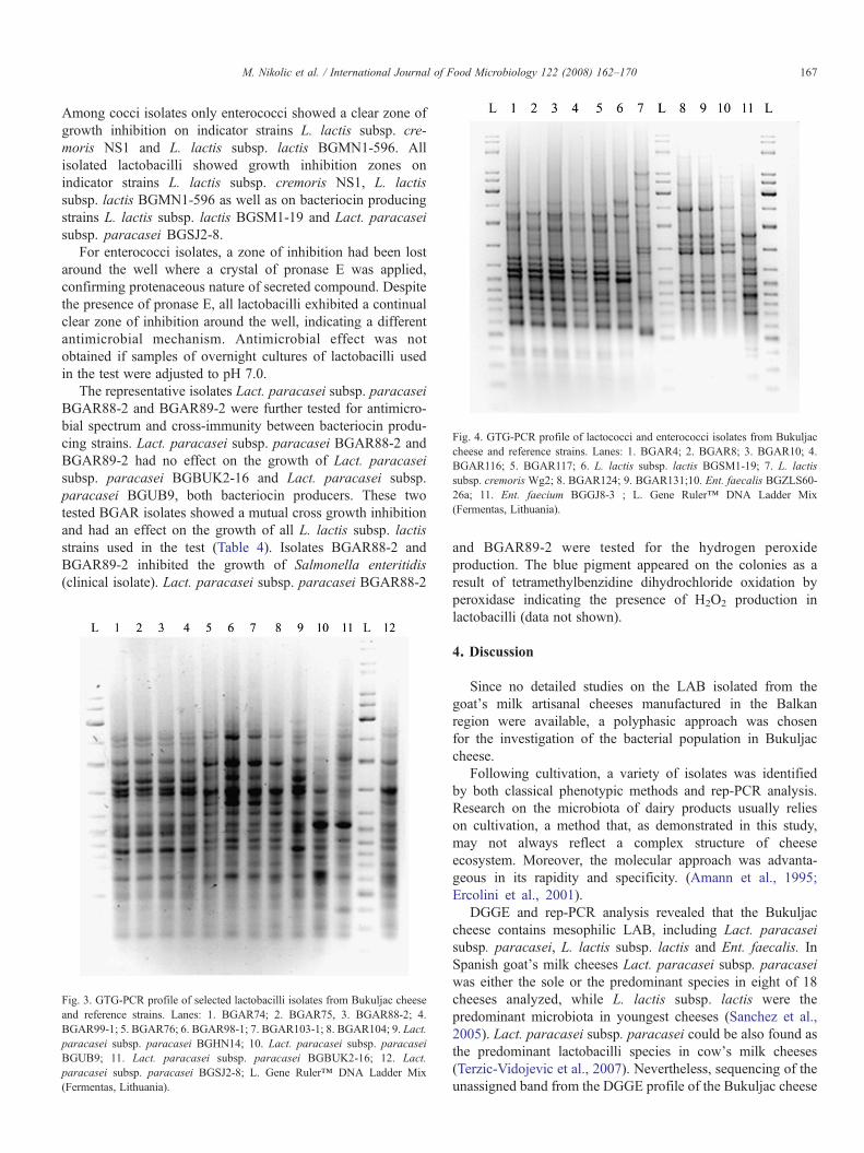
Fig. 4. GTG-PCR profile of lactococci and enterococci isolates from Bukuljaccheese and reference strains. Lanes: 1. BGAR4; 2. BGAR8; 3. BGAR10; 4.BGAR116; 5. BGAR117; 6. L. lactis subsp. lactis BGSM1-19; 7. L. lactissubsp. cremoris Wg2; 8. BGAR124; 9. BGAR131;10. Ent. faecalis BGZLS60-26a; 11. Ent. faecium BGGJ8-3 ; L. Gene Ruler™ DNA Ladder Mix(Fermentas, Lithuania).
167M. Nikolic et al. / International Journal of Food Microbiology 122 (2008) 162–170
Among cocci isolates only enterococci showed a clear zone ofgrowth inhibition on indicator strains L. lactis subsp. cre-moris NS1 and L. lactis subsp. lactis BGMN1-596. Allisolated lactobacilli showed growth inhibition zones onindicator strains L. lactis subsp. cremoris NS1, L. lactissubsp. lactis BGMN1-596 as well as on bacteriocin producingstrains L. lactis subsp. lactis BGSM1-19 and Lact. paracaseisubsp. paracasei BGSJ2-8.
For enterococci isolates, a zone of inhibition had been lostaround the well where a crystal of pronase E was applied,confirming protenaceous nature of secreted compound. Despitethe presence of pronase E, all lactobacilli exhibited a continualclear zone of inhibition around the well, indicating a differentantimicrobial mechanism. Antimicrobial effect was notobtained if samples of overnight cultures of lactobacilli usedin the test were adjusted to pH 7.0.
The representative isolates Lact. paracasei subsp. paracaseiBGAR88-2 and BGAR89-2 were further tested for antimicro-bial spectrum and cross-immunity between bacteriocin produ-cing strains. Lact. paracasei subsp. paracasei BGAR88-2 andBGAR89-2 had no effect on the growth of Lact. paracaseisubsp. paracasei BGBUK2-16 and Lact. paracasei subsp.paracasei BGUB9, both bacteriocin producers. These twotested BGAR isolates showed a mutual cross growth inhibitionand had an effect on the growth of all L. lactis subsp. lactisstrains used in the test (Table 4). Isolates BGAR88-2 andBGAR89-2 inhibited the growth of Salmonella enteritidis(clinical isolate). Lact. paracasei subsp. paracasei BGAR88-2
Fig. 3. GTG-PCR profile of selected lactobacilli isolates from Bukuljac cheeseand reference strains. Lanes: 1. BGAR74; 2. BGAR75, 3. BGAR88-2; 4.BGAR99-1; 5. BGAR76; 6. BGAR98-1; 7. BGAR103-1; 8. BGAR104; 9. Lact.paracasei subsp. paracasei BGHN14; 10. Lact. paracasei subsp. paracaseiBGUB9; 11. Lact. paracasei subsp. paracasei BGBUK2-16; 12. Lact.paracasei subsp. paracasei BGSJ2-8; L. Gene Ruler™ DNA Ladder Mix(Fermentas, Lithuania).
and BGAR89-2 were tested for the hydrogen peroxideproduction. The blue pigment appeared on the colonies as aresult of tetramethylbenzidine dihydrochloride oxidation byperoxidase indicating the presence of H2O2 production inlactobacilli (data not shown).
4. Discussion
Since no detailed studies on the LAB isolated from thegoat's milk artisanal cheeses manufactured in the Balkanregion were available, a polyphasic approach was chosenfor the investigation of the bacterial population in Bukuljaccheese.
Following cultivation, a variety of isolates was identifiedby both classical phenotypic methods and rep-PCR analysis.Research on the microbiota of dairy products usually relieson cultivation, a method that, as demonstrated in this study,may not always reflect a complex structure of cheeseecosystem. Moreover, the molecular approach was advanta-geous in its rapidity and specificity. (Amann et al., 1995;Ercolini et al., 2001).
DGGE and rep-PCR analysis revealed that the Bukuljaccheese contains mesophilic LAB, including Lact. paracaseisubsp. paracasei, L. lactis subsp. lactis and Ent. faecalis. InSpanish goat's milk cheeses Lact. paracasei subsp. paracaseiwas either the sole or the predominant species in eight of 18cheeses analyzed, while L. lactis subsp. lactis were thepredominant microbiota in youngest cheeses (Sanchez et al.,2005). Lact. paracasei subsp. paracasei could be also found asthe predominant lactobacilli species in cow's milk cheeses(Terzic-Vidojevic et al., 2007). Nevertheless, sequencing of theunassigned band from the DGGE profile of the Bukuljac cheese

Fig. 5. Casein hydrolyses of lactobacilli isolates from Bukuljac cheese. Lanes: 1. BGAR81-2; 2. BGAR82; 3. BGAR83-1; 4. BGAR85-2; 5. BGAR87-1; 6. BGAR87-2; 7. BGAR88-1; 8. BGAR88-2; 9. BGAR89-1; 10. BGAR89-2; 11. BGAR91; 12. BGAR92-2; 13. BGAR93-1; 14. BGAR93-2; αS1-casein and β-casein as substratesfor hydrolyses.
168 M. Nikolic et al. / International Journal of Food Microbiology 122 (2008) 162–170
revealed the presence of Leuc. mesenteroides, a species that wasnot discovered with classical methodology of isolation. It isnoteworthy that more than one band for a species in a DGGEprofile does not necessarily mean that different strains of thatspecies are present, since some species harbour more then onecopy of the rDNA operon, which are heterogeneous in theirsequence (Ercolini, 2004).
Overall, results of rep-PCR and results of plasmid profilesallowed us to classify L. paracasei subsp. paracasei isolatesinto four groups. Additionally, more subgroups in Lact. para-casei were identified during the research on the composition ofnon-starter LAB in Cheddar cheese (Fitzsimons et al., 2001) orin the Swiss cheese (Demarigny et al., 1996).
Moreover, while lactococci and enterococci have not shownproteolytic activity, lactobacilli isolates degraded αS1− and β-casein. In addition, only lactobacilli isolates showed diacetylproduction. Therefore, Lact. paracasei subsp. paracasei can beconsidered as both, the dominant and metabolically mostimportant bacteria in this artisan cheese. Furthermore, Lact.paracasei subsp. paracasei is likely to be involved in theflavour and aroma development of the cheese and is welladapted to the particular environmental conditions of cheeseripening (Albenzio et al., 2001). Lactobacillus strains isolatedfrom Bukuljac cheese, displayed a broad-spectrum of anti-microbial activity. Lact. paracasei subsp. paracasei BGAR88-2 and BGAR89-2 had an effect on the growth of Lact.paracasei subsp. paracasei BGSJ2-8, a producer of the
Table 4The cross-immunity between LAB isolates from Bukuljac cheese and different antim
Testedstrains
Indicator strains
EnterococcusfaecalisBGAR124
Lact. paracaseisubsp. paracaseiBGAR88-2
Lact. paracaseisubsp. paracaseiBGAR89-2
L. lactissubsp. lactisBGSM1-19
LsB
BGAR124 ▪ – – – –BGAR88-2 – ▪ 2 t 2 t –BGAR89-2 – 4 t ▪ 2 t –BGSM1-19 – – – ▪ –BGBUK2-16 – 3⁎ 3⁎ 2⁎ ▪BGUB9 1⁎ 3⁎ 4⁎ – 2BGSJ2-8 2⁎ 4⁎ 4⁎ 2⁎ t 3
−═ Without a zone; NA ═ not analyzed; ⁎ ═ an inhibition zone sensitive to pronainhibited Salmonella enteritidis growth and were tested for the hydrogen peroxide p
bacteriocin SJ (Lozo et al., 2007). BGAR88-2 and BGAR89-2 inhibited, among other, the growth of Salmonella enteritidis.This is an important data since Salmonella enteritidis isassociated with food borne diseases (Notermans and Hoogen-boom-Verdegaal, 1992). Results showed that antimicrobialcompound produced by lactobacilli isolates was hydrogenperoxide. This is an antimicrobial substance, with strongoxidizing effect on the bacterial cell surface proteins and lipids(Kamau et al., 1990).
In this study, we identified 34 (61.82%) isolates with theaggregation phenotype, an important probiotic characteristic.This ability of LAB is a mechanism that could increase theirsurvival and colonization of the gastrointestinal tract, prevent-ing bacterial elimination by peristalsis and providing acompetitive advantage in the ecosystem (Johansson et al.,1993; Crociani et al., 1995; Alander et al., 1997). In addition,aggregation may favour exchange of genetic material allowingthe lactobacilli to acquire new phenotypic characteristics(Reniero et al., 1992). Hence, the strains of lactobacilli fromBukuljac cheese will be further characterized as potentialprobiotics.
Obtained results indicated that strains Lact. paracasei subsp.paracasei BGAR88-2 and BGAR89-2 are proteolyticaly active,diacetyl producers and showed antimicrobial activity. Theydisplayed promising properties suggesting they might be goodcandidate strains for the inclusion in starter culture formanufacturing of goat's milk cheese, together with L. lactis
icrobial producers
act. paracaseiubsp. paracaseiGBUK2−16
Lact.paracaseiBGUB9
Lact. paracaseisubsp. paracaseiBGSJ2-8
L. lactissubsp. lactisBGMN1-596
L. lactis subsp.cremoris NS1
– – 1⁎ 1⁎
– 3 t 2 t 2 t– 3 t 2 t 2 t– – 6⁎ 2⁎
– NA NA NA⁎ t ▪ 3 t NA NA⁎ t NA ▪ 2 t 2⁎ t
se E (given in mm); t ═ turbid zone (given in mm) of inhibition. Isolates thatroduction are indicated in bold letters.

169M. Nikolic et al. / International Journal of Food Microbiology 122 (2008) 162–170
strains, which showed good activity in milk. Moreover, twoenterococcal isolates from Bukuljac cheese, Ent. faecalisBGAR124 and BGAR131 have shown antimicrobial activity,since it is well known that the enterocins have antimicrobialeffect on Listeria innocua and L. monocytogenes (Abriouel etal., 2006).
Acknowledgements
This work was funded by the Ministry of Science andEnvironmental Protection, of The Republic of Serbia, grantNo. 143036.
References
Abriouel, H., Ben Omar, N., Lucas, R., Martinez-Canamero, M., Galvez, A.,2006. Bacteriocin production, plasmid content and plasmid location ofenterocin P structural gene in enterococci isolated from food sources. Lettersin Applied Microbiology 42, 331–337.
Alander, M., Korpela, R., Saxelin, M., Vilpponen-Salmera, T., Mattila-Sandholm, T., Von Wright, A., 1997. Recovery of Lactobacillus rhamnosusGG from human colonic biopsies. Letters in Applied Microbiology 24,361–364.
Albenzio, M., Corbo, M.R., Rehman, S.U., Fox, P.F., De Angelis, M., Corsetti,A., Sevi, A., Gobbetti, M., 2001. Microbiological and biochemicalcharacteristics of Canestrato Pugliese cheese made from raw milk,pasteurized milk, or by heating the curd in hot way. International Journalof Food Microbiology 67, 35–48.
Amann, R.I., Ludwig, W., Schleifer, K.H., 1995. Phylogenetic identification andin situ detection of individual microbial cells without cultivation.Microbiological Reviews 59, 143–169.
Corroler, D., Mangin, I., Desmasures, N., Gueguen, M., 1998. An ecologicalstudy of lactococci isolated from raw milk in the camembert cheeseregistered designation of origin area. Applied and Environmental Micro-biology 64, 4729–4735.
Crociani, J., Grill, J.P., Huppert, M., Ballongue, J., 1995. Adhesion of differentbifidobacteria strains to human enterocyte-like Caco-2 cells and comparisonwith in vivo study. Letters in Applied Microbiology 21, 146–148.
Demarigny, Y., Beuvier, E., Dasen, A., Duboz, G., 1996. Influence of raw milkmicroflora on the characteristics of Swiss-type cheese. Evolution ofmicroflora during ripening and characterisation of facultatively hetero-fermentative lactobacilli. Le Lait 76, 371–387.
Ercolini, D., Moschetti, G., Blaiotta, G., Coppola, S., 2001. The potential of apolyphasic PCR-DGGE approach in evaluating microbial diversity ofNatural Whey Cultures for water-buffalo Mozzarella cheese production: biasof “culture dependent” and “culture independent” approaches. Systematicand Applied Microbiology 24, 610–617.
Ercolini, D., 2004. PCR-DGGE fingerprinting: novel strategies fordetection of microbes in food. Journal of Microbiological Methods56, 297–314.
Fitzsimons, N.A., Cogan, T.M., Condon, S., Beresford, T., 2001. Spartial andtemporal distribution of non-starter lactic acid bacteria in cheddar cheese.Journal of Applied Microbiology 90, 600–608.
Fontecha, J., Pelaez, C., Juarez, M., Requena, T., Gomez, C., 1990. Biochemicaland microbiological characteristics of artisanal hard goat's milk cheese.Journal of Dairy Science 73, 1150–1157.
Gajic, O., Kojic, M., Banina, A., Topisirovic, L., 1999. Characterization ofnatural isolate Lactococcus lactis subsp. lactis BGMN1-5, a strainproducing two bacteriocins, cell wall-associated proteinase and showingclumping phenotype. Archives of Biological Sciences 51, 69–78.
Haandrikman, A.J., Meesters, R., Laan, H., Konings, W.N., Kok, J., Venema,G., 1991. Processing of the lactococcal extracellular serine proteinase.Applied and Environmental Microbiology 57, 1899–1904.
Haenlein, G.F.W., 2001. Past, present, and future perspectives of small ruminantdairy research. Journal of Dairy Science 84, 2097–2115.
Hopwood, D.A., Bibb, J.M., Chater, K.F., Kieser, T., Bruton, C.J., Kieser, H.M.,Lydiate, K.M., Smith, C.P., Ward, J.M., Schrempf, H., 1985. Geneticmanipulation of Streptomyces, A Laboratory Manual. The John InnesFoundation, Norwich, UK.
Johansson, M.L., Molin, B., Jeppsson, B., Nobaek, B., Ahrne, B., Bengmark, S.,1993. Administration of different Lactobacillus strains in fermented oatmealsoup: in vivo colonization of human intestinal mucosa and effects on theindigenous flora. Applied and Environmental Microbiology 59, 15–20.
Kamau, D.N., Doores, S., Pruitt, K.M., 1990. Enhanced thermal destruction ofListeria monocytogenes and Staphylococcus aureus by the lactoperoxidasesystem. Applied and Environmental Microbiology 56, 2711–2716.
Kandler, O., Weiss, N., 1986. Genus Lactobacillus Beijerinck 1901. In:Sneath, P.H.A., Mair, N.S., Sharpe, M.E., Holt, J.G. (Eds.), Bergey'sManual of Systematic Bacteriology, vol. 2. Williams and Wilkins,Baltimore, pp. 1209–1243.
Kojic, M., Svircevic, J., Banina, A., Topisirovic, L., 1991a. Bacteriocin-producing strain of Lactococcus lactis subsp. diacetilactis S50. Applied andEnvironmental Microbiology 57, 1835–1837.
Kojic, M., Fira, D., Banina, A., Topisirovic, L., 1991b. Characterization of thecell wall-bound proteinase of Lactobacillus casei HN14. Applied andEnvironmental Microbiology 57, 1753–1757.
Litopoulou-Tzanetaki, E., Tzanetakis, N., 1992. Microbiological study ofwhite-brined cheese made from raw goat milk. Food Microbiology 9,13–19.
Lozo, J., Vukasinovic, M., Strahinic, I., Topisirovic, L., 2004. Characterizationand antimicrobial activity of bacteriocin 217 produced by natural isolateLactobacillus paracasei subsp. paracasei BGBUK2-16. Journal of FoodProtection 67, 2727–2734.
Lozo, J., Jovcic, B., Kojic, M., Dalgalarrondo, Chobert, J., Haertle, T.,Topisirovic, L., 2007. Molecular characterization of a novel bacteriocin andan unusually large aggregation factor of Lactobacillus paracasei subsp.paracasei BGSJ2-8, a natural isolate from home-made cheese. Currentmicrobiology 55, 266–271.
Mannu, L., Riu, G., Comunian, R., Frozzi, M.C., Scintu, M.F., 2002. Apreliminary study of lactic acid bacteria in whey starter culture and industrialPecorino Sardo ewes' milk cheese: PCR-identification and evaluation duringripening. International Dairy Journal 12, 17–26.
McGoarty, J.A., Tomeczek, L., Pond, D.G., Reid, G., Bruce, A.W., 1992.Hydrogen peroxide production by Lactobacillus species: correlation withsusceptibility to the spermicidal compound nonoxynol-9. The Journal ofInfectious Diseases 165, 1142–1144.
Mundt, J.O., 1986a. Lactic acid streptococci. In: Sneath, P.H.A., Mair, N.S.,Sharpe, M.E., Holt, J.G. (Eds.), Bergey's Manual of Systematic Bacteriol-ogy, vol. 2. Williams and Wilkins, Baltimore, pp. 1064–1071.
Mundt, J.O., 1986b. Enterococci. In: Butler, J.P. (Ed.), Bergey's Manual ofSystematic Bacteriology. Williams and Wilkins, Baltimore, pp. 1063–1065.
Notermans, S., Hoogenboom-Verdegaal, A., 1992. Existing and emergingfoodborne diseases. International Journal of Food Microbiology 15,197–205.
Olarte, C., Sanz, S., Gonzales-Fandos, E., Torre, P., 2000. The effect of acommercial starter culture addition on the ripening of an artisanal goat'smilk cheese (Cameros cheese). Journal of Applied Microbiology 88,421–429.
O' Sullivan, D.J., Klaenhammer, T.R., 1993. Rapid mini-prep isolation of high-quality plasmid DNA from Lactococcus and Lactobacillus ssp. Applied andEnvironmental Microbiology 59, 2730–2733.
Prescot, L.M., Harley, J.P., Klein, D.A., 1996. Microbiology, 3th ed. Wm. C.Brown Publishers, London, pp. 685–688.
Rabe, L.K., Hillier, S.L., 2003. Optimization of media for detection of hydrogenperoxide production by Lactobacillus species. Journal of Clinical Micro-biology 41, 3260–3264.
Randazzo, C.L., Torriani, S., Akkermans, A.D.L., de Vos, W.M., Vaughan, E.E.,2002. Diversity, dynamics, and activity of bacterial communities duringproduction of an artisanal sicilian cheese as evaluated by 16s rRNA analysis.Applied and Environmental Microbiology 68, 1882–1892.
Reniero, R., Cocconcelli, P., Bottazzi, V., Morelli, L., 1992. High frequency ofconjugation in Lactobacillus mediated by an aggregation-promoting factor.Journal of General Microbiology 138, 763–768.

170 M. Nikolic et al. / International Journal of Food Microbiology 122 (2008) 162–170
Requena, T., De la Fuente, M.A., Fernandez de Palencia, P., Juarez, M., Pelaez,C., 1992. Evaluation of a specific starter for the production of semi-hardgoat's milk cheese. Lait 72, 437–448.
Sanchez, I., Sesena, S., Poveda, J.M., Cabezas, L., Palop, L., 2005.Phenotypic and genotypic characterization of lactobacilli isolated fromSpanish goat cheeses. International Journal of Food Microbiology 102,355–362.
Schleifer, K.H., Ehrmann, M., Beimfohr, C., Brockmann, E., Ludwig, W.,Amann, R., 1995. Application of molecular methods for the classificationand identification of lactic acid bacteria. International Dairy Journal 5,1081–1094.
Stiles, M.E., Holzapfel, W.H., 1997. Lactic acid bacteria of foods andtheir current taxonomy. International Journal of Food Microbiology 36,1–29.
Terzic-Vidojevic et al., 2007, A., Vukasinovic, M., Veljovic, K., Ostojic, M.,Topisirovic, L., 2007. Characterization of microflora in homemade semi-hard white Zlatar cheese. International Journal of Food Microbiology 114,36–42.
Tserovska, L., Stefanova, S., Yordanova, T., 2000–2002. Identification of lacticacid bacteria isolated from Katyk, goat's milk and cheese. Journal of CultureCollection 3, 48–52.
Versalovic, J., Schneider, M., De Bruijn, F.J., Lupski, J.R., 1994. Genomicfingerprinting of bacteria using repetitive sequence-based polymerase chainreaction. Methods in molecular and cellular biology 5, 25–40.
Xanthopoulos, V., Polychroniadou, A., Litopoulou-Tzanetaki, E., Tzanetakis,N., 2000. Characteristics of Anevato cheese made from raw or heat-treatedgoat milk inoculated with a lactic starter. Lebensmittel-Wissenschaft undTechnologie 33, 483–488.