characterization of camptothecin-induced genomic...
TRANSCRIPT
Abstract. Acquisition of resistance to topoisomerase I(TOP1)-targeting camptothecin (CPT) derivatives is a majorclinical problem. Little is known about the underlyingchromosomal and genomic mechanisms. We characterizedthe CPT-K5 cell line expressing mutant CPT-resistant TOP1and its parental T-cell derived acute lymphoblastic leukemiaCPT-sensitive RPMI-8402 cell line by karyotyping andmolecular genetic methods, including subtractive oligo-based array comparative genomic hybridization (soaCGH)analysis. Karyotyping revealed that CPT-K5 cells hadacquired additional structural aberrations and a reducedmodal chromosomal number compared to RPMI-8402.soaCGH analysis identified vast copy number alterationsand >200 unbalanced DNA breakpoints distributed unevenlyacross the chromosomal complement in CPT-K5. In addition,the short tandem repeat alleles were found to be highlydifferent between CPT-K5 and its parental cell line. Weidentified copy number alterations affecting genes importantfor maintaining genome integrity and reducing CPT-inducedDNA damage. We show for the first time that short tandemrepeats are targets for TOP1 cleavage, that can bedifferentially stimulated by CPT.
The camptothecin (CPT) derivatives, topotecan andirinotecan (SN-38 being the active metabolite), belong to animportant group of drugs used for the treatment of manytypes of malignancies in advanced stages, includingcolorectal, ovarian, small cell lung cancer and acuteleukemia (1-5). However, acquired resistance to these drugsgreatly compromises their efficacy in clinical use (6).
The CPTs are highly selective inhibitors of the nuclearenzyme DNA topoisomerase I (TOP1) [reviewed in (7)].TOP1 is an important monomeric enzyme that is required torelease the torsional tension of DNA introduced during theDNA replication and transcription processes in normal cells[reviewed in (8)]. Moreover, it has been shown that, inaddition to its DNA-relaxing activity, TOP1 also hasindependent kinase activity important for splicing of pre-mRNA (9). The enzyme exerts its relaxing activity bytransiently cleaving and rejoining one strand of the DNAduplex. The CPTs are non-competitive, reversible TOP1inhibitors that cause accumulation of TOP1-cleavagecomplexes (TOP1-cc) in the genome by transientlypreventing the DNA rejoining step of catalysis. The trappedTOP1-cc are converted into one-ended DNA double-strandbreaks (DSBs) when they collide with the DNA replicationor transcription machinery. It is generally accepted that theformation and stabilization of TOP1-cc and their conversioninto one-ended DSBs is a prerequisite for the cytotoxicactivity of CPTs (10).
Although many players have been implicated in thecellular response to CPT, little is known about the pathwaysdownstream of the trapped TOP1-cc that ultimately lead torepair of DNA damage or cell death. Impaired DSB repairmakes cells hypersensitive to CPT and unless the DSBs arerepaired they are lethal to the cell. DNA repair may lead tochromosomal or genomic alterations depending on theinvolved pathways. Several CPT-resistant cell lines havebeen derived (11-22), but only few of these have beencharacterized at the chromosomal or genomic level (21, 23).
91
This article is freely accessible online.
*Current address: Department of Biotechnology, National Instituteof Pharmaceutical Education & Research (NIPER), Hajipur,Vaishali, India.
Correspondence to: Dr. Eigil Kjeldsen, Cancer Cytogenetics Section,HemoDiagnostic Laboratory, Aarhus University Hospital, Tage-Hansens Gade 2, Ent. 4A, DK-8000 Aarhus C, Denmark. E-mail:[email protected]
Key Words: CPT-K5, camptothecin resistance, karyotype, aCGH,genomic instability, short tandem repeat.
CANCER GENOMICS & PROTEOMICS 15: 91-114 (2018)doi:10.21873/cgp.20068
Characterization of Camptothecin-induced Genomic Changes inthe Camptothecin-resistant T-ALL-derived Cell Line CPT-K5
EIGIL KJELDSEN1, CHRISTINE J.F. NIELSEN2, AMIT ROY2*, CINZIA TESAURO2, ANN-KATRINE JAKOBSEN3, MAGNUS STOUGAARD3 and BIRGITTA R. KNUDSEN2
1Cancer Cytogenetics Section, HemoDiagnostic Laboratory, and 3Department of Pathology, Aarhus University Hospital, Aarhus, Denmark;
2Department of Molecular Biology and Genetics, C.F. Møllers Allé, Aarhus University, Aarhus, Denmark

In the present study we aimed to characterize thechromosomal complement and global copy numberaberrations generated by acquired CPT resistance in theCPT-K5 cell line to gain insight into which regions of thegenome may be affected in acquisition of CPT resistance.Furthermore, we examined whether short tandem repeat(STR) sequences, normally used for paternity testing andcell line authentication, are targets for TOP1 DNA cleavage,and whether these DNA cleavage sites can be selectivelystimulated by CPT.
Materials and MethodsCell lines and cell proliferation analysis. Andoh and co-workersestablished the CPT-resistant cell line, CPT-K5, from its parentalsensitive counterpart human T-cell derived acute lymphoblastic (T-ALL) cell line RPMI-8402 (wild-type) by exposure to stepwiseincreasing concentrations of the semi-synthetic water-soluble CPT-derivative, irinotecan (11). For this study, cells stored in liquidnitrogen were thawed and cultured briefly at an initial density of1×106 cells/ml in RPMI 1640 medium supplemented with 10% fetalcalf serum as described elsewhere (24). Growth inhibition of thecells by CPT was determined by counting the number of cells inculture using a hematology analyzer (Sysmex KX-21N; SysmexNordic Aps, Copenhagen, Denmark).
Nuclear extracts, western blotting, and measurement of tyrosyl-DNAphosphodiesterase 1 (TDP1) and TOP1 activity. Preparation ofnuclear extracts from RPMI-8402 and CPT-K5 cells, TOP1-specificwestern blotting and TOP1 activity measurements by rolling-circleenhanced enzyme activity detection (REEAD) was performed asdescribed previously (25), while TDP1 activity measurement wasperformed as described in (26).
Expression and purification of recombinant human TOP1.Recombinant human TOP1 was expressed in yeast and purified tohomogeneity, as described previously (27). The enzyme preparationwas tested for purity by sodium dodecyl sulfate (SDS)polyacrylamide gel electrophoresis followed by Coomassie stainingand the concentration estimated by comparison to a bovine serumalbumin (BSA) standard before use.
G-Banding and fluorescence in situ hybridization (FISH).Chromosomal preparations and G-banding were performed accordingto standard protocols as described in (28). Karyotypes and FISHresults were reported according to ISCN, 2013 (29). Analysis of 10G-banded metaphases was carried out using a Laborlux microscope(Leica, Germany) equipped with a grey-level charge coupled device-camera and Ikaros karyotyping software implemented on a DellOptiplex GX1 (MetaSystems, Heidelberg, Germany). Locus-specificFISH analysis of interphase nuclei and metaphase spreads was carriedout with the following directly fluorescent labeled probe setsaccording to the manufacturers’ instructions: SIL-TAL1 FISH DNAprobes and TOP1/CEN20 (DAKO, Glostrup, Denmark); BlueFISHBacterial Artificial Chromosomes (BAC) probes (BlueGnome,Cambridge, UK): RP11-133H19 fluorescence labeled with red(11p15.4, pos. 8,555,486-8,732,332 bp located telomeric to the LIMdomain only 1 (LMO1) gene pos. 8,202,433-8,241,982) and RP11-298I3 (14q11.2, pos. 22,435,253-22,627,198 located centromeric to
the Tcrd T cell receptor delta chain (TCRD) gene pos. 21,994,085-22,002,042); and custom-made BAC-based probes (EmpireGenomics, Buffalo NY, USA): RP11-368G2 (4q22.1, pos.89,137,575-89,319,451), and RP11-213O13 (14q32.11, pos.89,466,687-89,648,208) were used for validation of array-basedcomparative genomic hybridization (aCGH) results. Interphase nucleiand metaphases were counterstained with 4’,6-diamidino-2-phenylindole (DAPI). The genomic position of the listed BAC probeswas according to National Center for Biotechnology Information(NCBI) build 36.1 (hg18) (http://www.ncbi.nlm.nih.gov).
Spectral karyotyping (SKY). Pretreatment, denaturing, andhybridization of microscopy slides with chromosome suspensionapplied were carried out as previously described (30) according tothe manufacturer’s instructions (Applied Spectral Imaging™,Migdal HaEmek, Israel). Ten fully analyzed mitoses in the SKYanalysis were performed for both cell lines. Numerical andstructural aberrations were counted using a normal female asreference. Karyotypes were described according to InternationalSystem for Human Cytogenetic Nomenclature (ISCN) (29)
aCGH analysis. High-resolution aCGH analysis was performedusing the 180k oligo-based Cancer CytoChip (BlueGnome) asdescribed elsewhere (31). In brief, high molecular weight DNA wasisolated from RPMI-8402 and CPT-K5 cells using Gentra Pure Genekit (Qiagen AB, Sollentuna, Sweden) according to themanufacturer’s instructions. Pooled female DNA (Promega, BiotechAB, Nacka, Sweden) was used as reference DNA to determine theDNA copy number status of the female RPMI-8402 cells andderivative CPT-K5 cells. In addition, subtractive aCGH analysis ofCPT-K5 was performed with isolated DNA from the parental cellline RPMI-8402 as reference DNA. The analysis was carried outaccording to the manufacturer’s instructions using 0.5 μg of DNA.After hybridization, washing and drying, the oligo arrays werescanned at 2.5 μm with a GenePix 4400A microarray scanner(Molecular Devices, Sunnyvale, CA, USA). Initial analysis andnormalization was achieved with BlueFuseMulti v2.6 (BlueGnome).For analysis and visualization, log2 probe signal values wereimported into Nexus Copy Number software v6.1 (BioDiscovery,Hawthorne, CA, USA), and segmented using RANK segmentationalgorithm with a minimum of five probes/segment. Gains and losseswere defined by log2 ratios 0.16 and −0.16, respectively, while 0.5and −1.0 defined high copy gains and large losses, respectively. Thereference genome was NCBI build 36.1 (hg18). Bioinformaticsanalysis was performed by querying the University of California,Santa Cruz database (http://genome.ucsc.edu).
Genomic break point analysis. Break points were defined with highprecision as locations where a change in copy number occurred incalled regions of amplifications and deletions as determined by theNexus software algorithm. To quantify genomic breakage, log2 ratiodifferences larger than 0.3 were used to discriminate putative DNAbreakage points. We identified those breakage points at the edgesof segments of copy number gains and losses as well as points ofabrupt copy number changes called within larger aberrations. Theprecision of this type of measurement is determined by theresolution of the array. The smallest aberration we were able todetect with confidence on the 180k platform was 100 kb in length.In addition, the abnormality needed to be present in at least 10-20%of the cells in order to be detected.
CANCER GENOMICS & PROTEOMICS 15: 91-114 (2018)
92

Immunophenotyping by flow cytometry. Immunophenotyping ofexponentially growing RPMI-8402 and CPT-K5 cells were carriedout as described in (32).
STR profiling. STR profiling was performed at the Department ofForensic Medicine, University of Copenhagen, Copenhagen,Denmark, as part of their routine analyses using the AmpFISTR®Identifiler PCR Amplification kit (Applied Biosystems, ThermoFisher Scientific, Waltham, MA, USA). The STR profiling wasrepeated twice with identical results on genomic DNA purified fromtwo independent cultures from both cell lines.
Generation of STR TOP1 cleavage substrates. The flankingsequences in the STR TOP1 cleavage substrates were the same inthe three substrates (marked below in bold) and the STR repeatlength (marked with underline) were from the parental CPT-sensitive 8402 cell line.
Each of the STR TOP1 cleavage substrates were synthesized assingle-stranded oligonucleotides (DNA technology, Aarhus,Denmark).
I) D7S820 locus (8 repeats of GATA): 50-base top strand (5’->3’): ACGTCACGGGATAGATAGATAGATAGATAGATAGATAGATACGGTTCAGC; 50-base bottom strand (5’->3’):GCTGAACCGTATCTATCTATCTATCTATCTATCTATCTATCCCGTGACGT.
II) D5S818 locus (12 repeats of AGAT): 60-base top strand (5’->3’): ACGTCACGGAGATAGATAGATAGATAGATAGATAGATAGATAGATAGATAGATAGATCGGTTCAGC; 60-base bottomstrand (5’->3’): GCTGAACCGATCTATCTATCTATCTATCTATCTATCTATCTATCTATCTATCTATCTCCGTGACGT.
III) Thyroid peroxidase (TPOX) locus (8 repeats of AATG): 50-base top strand (5’->3’): ACGTCACGGAATGAATGAATGAATGAATGAATGAATGAATGCGGTTCAGC; 50-base bottomstrand (5’->3’): GCTGAACCGCATTCATTCATTCATTCATTCATT CATTCATTCCGTGACGT.
To prepare the STR substrates for TOP1 cleavage reaction, 50pmol of each top strand was separately hot-labeled with gamma-ATP essentially as described before (33). The reaction was stoppedby ethanol precipitation using NaCl and after 80% EtOH wash, anddrying of the pellet, it was resuspended in 10 μl 1x TOP1-cleavagebuffer (5 mM MgCl2, 5 mM CaCl2, and 10 mM Tris-HCl, pH 7.5).Then 50 pmol of the corresponding bottom strand was added, themixture was left at 90˚C for 2 min and allowed to anneal with therespective top strand by slowly cooling down to room temperature.
TOP1 STR cleavage reactions and detection. The TOP1 cleavagereaction was performed as described (33). Briefly, approximately200 copies of the annealed oligonucleotides were used for cleavagereaction with 300 units of human TOP1 in the presence of 10 μMCPT or dimethylsulfoxide, which was used as a solvent for CPT.The reaction was carried out at 37˚C for 30 min. and stopped by theaddition of 0.2% SDS followed by EtOH precipitation. Afterprecipitation and 80% EtOH washing and drying of the pellet, it wasresuspended in 10 μl trypsin mix and incubated at 37˚C for 30 minas described previously (33). Then 10 μl of 2x Seq-buffer (100%formamide, 1 mM EDTA, pH 8.0, 0.1% bromophenol blue (BPB),and 0.1% xylene cyanol) (Sigma-Aldrich, Brøndby, Denmark) wasadded to each reaction. Approximately 16 counts per second wasadded to each lane of a 14% denaturating polyacrylamide gel andrun until BPB left the gel. The gel was transferred to Whatman
3MM filter paper (Sigma-Aldrich Chemie GmbH), dried andexposed in a phosphor imager (PMI Imager; Bio-Rad, Life Science,Bio-RAD laboratories, Copenhagen, Denmark).
Results and Discussion
Cellular characteristics of CPT-K5 and its CPT-resistantCPT-K5 cell line. To obtain a global genomic insight into thedevelopment of acquired CPT resistance and to identifyputative candidate genes involved in this process, we tookadvantage of the CPT-K5 cell line, which exhibits highlystable CPT resistance following prolonged CPT-selectivegrowth of its parental RPMI-8402 cell line (11, 24, 34).
For the purposes of the present study, we wanted toconfirm the cellular and biochemical characteristics of thesecell lines using current methods. Firstly, immunophenotypingof the parental cell line RPMI-8402 confirmed that it isindeed a T-ALL derived cell line (Table I). The CPT-K5 cellline has not previously been characterized byimmunophenotyping but this analysis confirmed that CPT-K5 retains the predominant T-ALL immunophenotype (TableI). Secondly, at the genetic level it was confirmed that theRPMI-8402 harbors two T-ALL specific chromosomalaberrations: i) the common SIL deletion at chromosomeregion 1p32; and ii) the rare t(11;14)(p15;q11)/LMO1-TCRD.Both aberrations were present in two copies (Figure 1). CPT-K5 has retained one of the chromosome 1 that harbors the1p32 deletion, while the t(11;14) was completely lost (Figure1). Thirdly, in a standard cell survival assay the CPT-resistantand the CPT-sensitive phenotypes of the CPT-K5 and RPMI-
Kjeldsen et al: Camptothecin-induced Genomic Changes in CPT-K5 Cells
93
Table I. Immunophenotype of RPMI-8402 and CPT-K5.
Antigen RPMI-8402a RPMI-8402 CPT-K5 (DMSZ)
CD2 + + +CD3 - - -CD4 - n.d.c n.dCD5 + + +CD6 + n.d. n.d.CD7 + + +CD8 - - -CD13 - - -CD19 - - -CD33 n.i.b - -CD34 + + -CD45 n.i. + +CD71 n.i. + +CD235a n.i. - -TCR−α/β - n.d. n.d.TCR-γ/δ - n.d. n.d.
aInformation from DMSZ web-site, http://www.dsmz.de; bn.i., noinformation; cn.d., not done.

8402 cell lines, respectively, were confirmed. Asdemonstrated in Figure 2A, cell proliferation wassignificantly reduced in the presence of 1 μM CPT in theCPT-sensitive parental cell line RPMI-8402, while the CPT-resistant derivative cell line, CPT-K5, did not exhibit anysignificant change in growth rate. In the absence of CPT, thecellular growth rate of the CPT-K5 was lower as comparedto its parental cell line, which seems to be a common featureof CPT-resistant cell lines (22). Fourthly, we compared theTOP1 activity expressed in the two cell lines by westernblotting and REEAD assay (35, 36). In line with previousstudies, we found a reduced intra-cellular level of TOP1protein in CPT-K5 cells relative to RPMI-8402 cells (Figure2B). The cellular TOP1 activity was measured using theREEAD assay, which allows quantification of TOP1cleavage-ligation activity in crude extracts from only a fewhuman cells (schematically illustrated in Figure 2C) (35, 36).With the REEAD assay, we found a 2- to 3-fold reduction incellular TOP1 enzymatic activity (Figure 2D). Moreover, thepreviously observed CPT-resistant phenotype of purifiedTOP1 expressed in CPT-K5 cells (11, 24) was confirmed by
measuring the effect of CPT on TOP1 activity present incrude extracts from the CPT-K5 and its parental cell linesusing the REEAD assay (Figure 2E).
Taken together, our present findings are in agreement withprevious studies by Andoh et al. (11) and Kjeldsen et al.(24), and establish that the CPT-K5 cell line is a T-ALLderived cell line with unique characteristics.
Chromosomal analysis of CPT-K5 and RPMI-8402 cells. TheCPT-K5 cell line has not previously been characterized bykaryotyping, whereas its parental cell line RPMI-8402 had apartial or incomplete karyotype available as describedelsewhere (37-40) and by DSMZ (Braunschweig, Germany;http://www.dsmz.de). In agreement with these earlier reports,we found that the RPMI-8402 cell line is hypo-tetraploidwith a modal chromosomal number of 90 (range=76-92)(Figure 3A and B; Table II). In the present study, wecompleted the karyotype by defining the three previouslyassigned marker chromosomes to be: del(X)(q13.2q22.1),del(2)(q11.2q23.7) and der(15)t(14;15)(q32.2;p11).
The karyotype of the CPT-K5 cell line is also hypo-tetraploid but with a modal chromosome number of 80(range=71-81) (Figure 3C and D; Table II). The mostprominent differences between CPT-K5 and its parental RPMI-8402 are: i) a reduced modal chromosomal number from 90 to80; ii) loss of five structurally aberrant chromosomes: del(Xq),del(2q), dup(4q), del(6q), and t(11;14); and iii) gain of 13 newstructurally aberrant chromosomes: der(1)t(1;10), der(2)t(2;6),der(4)t(4;17), der(6)t(6;16), der(7)t(7;8), der(8)t(8;9),der(9)t(4;9), der(10)t(10;20), der(12)t(4;12), der(16)t(3;16),der(19)t(3;19), der(20)t(9;20), and der(22)t(3;22;3). Despitethese major karyotypic differences between the two cell linesaffecting almost all chromosomes, chromosomes 5, 18, and 21were cytogenetically unaffected.
Although several human cell lines resistant to CPT or itsderivatives have been developed (Table III) only the CPT-K5 cell line has so far been studied by karyotyping analysisas described above. This is intriguing from a biological pointof view since TOP1 exerts its main activity in DNAmetabolism by cleavage and rejoining, and that the latter isinhibited by CPT causing DNA strand breaks, whichultimately may induce chromosomal aberrations andgenomic instability (23, 41). Spectral karyotyping of thedoxorubicin-resistant cell line, NCI-H69AR, which is cross-resistant to several topoisomerase II inhibitors, revealedsubstantial structural chromosomal differences comparedwith its parental NCI-H69 cell line (42). Similarchromosomal findings were obtained for mitoxantrone-resistant SF295 cells (43). Together these data suggest thatacquisition of resistance to drugs inhibiting DNA cleavage-rejoining/metabolizing topoisomerases is accompanied bymajor karyotypic alterations, although the molecularmechanisms behind such alterations remain obscure.
CANCER GENOMICS & PROTEOMICS 15: 91-114 (2018)
94
Figure 1. Fluorescence in situ hybridization (FISH) analysis of RPMI-8402 cell line using the SIL-TAL1 split-apart probe set showing anucleus with 2F2G pattern indicating deletion of the SCL/TAL1-Interrupring locus (STIL) gene in two alleles and a normal fusion in theother two alleles. FISH analysis using of RPMI-8402 cell line using theLMO1-TCRD single fusion probe showing a nucleus with 2F2R2Gpattern indicating fusion of the LIM domain only 1 (LMO1) and theTcrd T cell receptor delta chain (TCRD) genes in two alleles and nofusion in the other two alleles. FISH analysis of CPT-K5 cell line usingthe TAL1-STIL split-apart probe showing a nucleus with 2F1G patternindicating deletion of the STIL gene in one allele and a normal fusionin the other two alleles. FISH analysis using of CPT-K5 cell line usingthe LMO1-TCRD single fusion probe showing a nucleus with 3R3Gpattern indicating no fusion of the genes.

Genomic profiling reveals greatly increased number ofgenomic DNA breakpoints in CPT-K5 cells. To furthercharacterize the observed chromosomal differences betweenRPMI-8402 and CPT-K5 cells, we examined copy numberchanges between the cell lines by oligo-based aCGH analysisusing female DNA pooled from normal individuals asreference (Figure 4A). For RPMI-8402 cells, the observedcopy number aberrations were in agreement with thepublished SNP Array Based LOH and Copy Number
Analysis (Welcome Sanger Trust Institute, http://www.sanger.ac.uk/cgi-bin/genetics/CGP/cghviewer). Theoligo-based aCGH analysis of CPT-K5 cells confirmed themajor differences as revealed by karyotyping (Figure 4B).
For a detailed comparison, however, the approach ofcomparing oaCGH analyses with DNA from normalindividuals serving as a reference is imprecise. SubtractiveCGH analysis is a direct method for determining genomicchanges between drug-resistant cell lines where DNA from
Kjeldsen et al: Camptothecin-induced Genomic Changes in CPT-K5 Cells
95
Figure 2. A: Effects of camptothecin (CPT) on cell growth of parental RPMI-8402 and CPT-K5 cells. Growth of parental cells in the absence (l)and presence of 1 μM CPT (l). Growth of CPT-K5 in the absence (s) and presence of 1 μM CPT (s). B: Western blot of nuclear extracts from106 cells of RPMI-8402 and CPT-K5 using mouse antibody against human topoisomerase 1 (hTOP1). C: Schematic representation of the rolling-circle enhanced enzyme activity detection (REEAD) assay. A dumbbell-shaped DNA substrate with a preferred TOP1 cleavage sequence in thedouble-stranded stem region and an identifier- or primer annealing sequence (denoted “i” or “p”) in each of the loops is converted to a closedcircle upon cleavage and ligation by TOP1. The generated circle is then hybridized to a surface anchored primer with a sequence matching the“p” region and subjected to rolling circle amplification by added phi-polymerase. The resulting tandem repeat rolling circle product is visualizedat the single molecule level by hybridization of fluorescent labeled detection probes matching the “i” region. D: REEAD measurement of hTOP1in nuclear extracts generated from 106 cells and assayed either undiluted (1x) or 10, 100, 1000, or 10,000 times diluted as indicated. E: The CPTsensitivity of hTOP1 in extracts from RPMI-8402 (blue bars) and CPT-K5 (red bars) was analyzed by measuring the activity using the REEADassay in the presence of increasing concentrations of CPT as indicated. The bar chart shows the mean values from three independent experimentsand the error bars indicate standard deviations.

CANCER GENOMICS & PROTEOMICS 15: 91-114 (2018)
96
Figure 3. G-Banding (A, C) and spectral karyotyping (B,D) of the RPMI-8402 (A,B) and CPT-K5 (C,D) cell lines showing representative karyotypes.

the parental cell line serves as reference DNA (44, 45). Thesubtractive oligo-based aCGH analytical approach, where theRPMI-8402 DNA served as a reference, revealed that CPT-K5 cells had acquired 165 copy number alterations (Figure4C, Table IV).
Calculating the mere number of copy number alterationsunderestimates the number of putative DNA breakage points
(46). We assessed those DNA breakage points at the edgesof segments of DNA copy number gains and losses, as wellas at points of abrupt DNA copy number changes calledwithin larger aberrations as exemplified in Figure 4D. Bythis analysis, we found that CPT-K5 cells had acquired atotal of 236 unbalanced breakpoints that were unevenlydistributed across each chromosome (Figure 4D). The
Kjeldsen et al: Camptothecin-induced Genomic Changes in CPT-K5 Cells
97
Table II. Karyotypes of the camptothecin (CPT)-resistant cell line CPT-K5 and its parental CPT-sensitive RPMI-8402 cell line.
Cell line Karyotype
RPMI-8402 90<4n>,XXX,del(X)(q13.2q22.1), del(2)(q11.2q37.3), +3, dup(4)(q13.1q25)x2, -5, del(6)(q13q21)x2, -7, t(11;14) (p15;q11)x2, -13, -13, -15, der(15)t(14;15)(q32.2;p11), -16, -18, der(20)t(1;20)(p36.1;q11.21), der(22)t(20;22) (q11.21;q13.33) and a minor sideline with an additional der(1)t(1;8)(p3?5;q11)CPT-K5 80<4n>,XX,-X,-X,der(1)t(1;10)(p10;q11)del(1)(q25),-2,t(2;6)(q37::p12.3p11.2),-3,+4,der(4)t(4;17)(q?11;q25), del(4)(q11), -5, der(6)t(6;16)(p21;q11), der(7)t(7;8)(q36;q13), der(8)t(8;9)(q12;q33), i(9)(q10),der(9)t(4;9), -10, der(10)t(10;20)(p15;?), -11,-11, t(4;12)(q22q21::p13),-14,-15,der(16)t(3;16)(q21;q11),-17,-18, der(19)t(3;19)(p21;p13), der(20)t(9;20), -21, der(22)t(3;22;3) and a minor sideline with del(6)(q14q21)
Table III. Human cell lines developed after selection of resistance to camptothecin (CPT) or its derivatives.
Resistant Parental Parental Selection TOP1 point Karyotype Referencecell line cancer agent mutation available (Ref)
CPT-K5 RPMI-8402 T-ALL CPT-11 Asp533Gly P: Yes (see Table II) (11, 34) R: Yes, present study (see Table II)PC-7/CPT PC-7 Non-small cell lung cancer CPT-11 Thr729Ala P: No (12, 13) R: NoA549/CPT A549 Lung cancer CPT None P: Yes, (97) (14) R: No, but CGH analysis (44)St-4/CPT St-4 Gastric cancer CPT No, but ex3 P: No (14) to ex9 deletion R: No, but CGH analysis (44)HT-29/CPT HT-29 Colon cancer CPT No, but ex3 P: No (14) to ex9 deletion R: No, but CGH analysis, (44)U937/CR U937 Myeloid leukemia 9-NC Phe361Ser P: Yes (98) (15) R: NoCEM/C1 and CCRF-CEM T-ALL CPT* Asn722Ser P: Yes (99) (16)CEM/C2 R: NoDU-145/RC0.1 DU145 Prostate cancer 9-NC Arg364His P: Yes (100, 101) (17, 18)DU-145/RC1 R: NoU937/RERC U937 Myeloid leukemia 9-NC Asp533Asn P: Yes (19) R: NoCPT30 HONE-1 Nasopharyngeal carcinoma CPT Glu418Lys P: No (20) R: NoMMRU/CR1 and MMRU Melanoma CPT-11 None P: No (21)MMRU/CR2 R: No, but BAC-based aCGH analysis (21)SF295/hCPT50 and SF295 Glioblastoma hCPT and None P: Yes (43) (55)SF295/BN50 BN80915 R: NoHT-29/SN-38 HT-29 Colon cancer SN38 None P: No (22)HCT-116/SN-38 HCT-116 Colon cancer Arg364Lys R: No and Gly721Arg
P: Parental; R: resistant; T-ALL: T-cell lymphoblastic leukemia, 9-NC: 9-nitro-20(S)camptothecin; hCPT: homocamptothecin; BN80915: hCPTderivative; SN-38:7-ethyl-10-hydroxy-20(S)-camptothecin. *Resistance stable for up to 6 months.

CANCER GENOMICS & PROTEOMICS 15: 91-114 (2018)
98
Figure 4. Genomic profiling using 180k oligo-based array comparative genomic hybridization (aCGH) analysis. A: Whole-genome view of RPMI-8402 against normal female DNA pool. B: Whole-genome view of CPT-K5 against normal female DNA pool. C: Subtractive aCGH analysis whereRPMI-8402 served as reference. To the right of the individual ideograms, microarray profiles of copy number gains and losses are depicted. Gainis indicated by blue and loss is indicated by red as an overlay on the ideogram. The log2 ratios for each chromosome are −2.5, 0, and +2.5 asillustrated for chromosome 1. D: Subtractive aCGH-based breakpoint analysis. Left panel: Magnified view of copy number profile of a region onchromosome 6 (pos. 42,500,000-58,000,000 bp) in which asterisks indicate determined putative DNA breakpoints. Focusing on the called regionsof loss (red) and gain (blue) indicates two breakpoints, while taking copy number changes within the called regions into account, eight putativeDNA break points can be determined. Right panel. Diagram showing the number of putative DNA break points determined on each chromosome.

Kjeldsen et al: Camptothecin-induced Genomic Changes in CPT-K5 Cells
99
Table IV. Results from subtractive oligo array-based comparative genomic hybridization analysis between CPT-K5 and RPMI-8402 (hg18) celllines.
Chromosomal region Copy number aberration Length (bp) Cytoband No. of probes
chr1:0-4,617,760 Loss 4617761 p36.33 - p36.32 347chr1:4,617,760-6,731,626 Homozygous copy loss 2113867 p36.32 - p36.31 135chr1:6,731,626-23,383,050 Loss 16651425 p36.31 - p36.12 1028chr1:29,939,359-40,135,057 Loss 10195699 p35.3 - p34.2 667chr1:40,232,695-47,470,334 Loss 7237640 p34.2 - p33 518chr1:47,555,023-76,314,989 Loss 28759967 p33 - p31.1 1702chr1:76,687,243-121,052,423 Loss 44365181 p31.1 - p11.2 2547chr1:147,259,828-148,058,607 Loss 798780 q21.1 - q21.2 30chr1:164,645,796-171,310,019 Gain 6664224 q24.1 - q25.1 417chr1:171,310,019-172,363,398 High copy gain 1053380 q25.1 62chr1:172,363,398-172,978,604 Gain 615207 q25.1 37chr1:172,978,604-247,249,719 Loss 74271116 q25.1 - q44 4372chr2:82,105-2,351,466 Loss 2269362 p25.3 139chr2:17,082,892-19,555,672 Loss 2472781 p24.2 - p24.1 129chr2:47,871,668-47,913,372 Loss 41705 p16.3 26chr2:96,685,923-98,987,015 Gain 2301093 q11.2 122chr2:98,987,015-99,310,234 High copy gain 323220 q11.2 21chr2:99,310,234-115,209,148 Gain 15898915 q11.2 - q14.1 891chr2:115,209,148-115,389,211 High copy gain 180064 q14.1 11chr2:115,389,211-124,331,966 Gain 8942756 q14.1 - q14.3 473chr2:124,331,966-125,952,676 High copy gain 1620711 q14.3 89chr2:147,977,155-148,340,965 Homozygous copy loss 363811 q22.3 16chr2:160,098,019-160,395,597 Loss 297579 q24.2 19chr2:200,685,674-201,878,147 Loss 1192474 q33.1 102chr2:211,746,930-232,604,377 Gain 20857448 q34 - q37.1 1322chr2:232,604,377-232,810,269 High copy gain 205893 q37.1 13chr2:232,810,269-242,118,949 Gain 9308681 q37.1 - q37.3 591chr3:4,499,856-4,714,317 Loss 214462 p26.2 13chr3:15,760,147-30,673,114 Gain 14912968 p24.3 - p24.1 847chr3:32,894,032-36,997,480 Gain 4103449 p22.3 - p22.2 218chr3:46,704,902-75,717,747 Loss 29012846 p21.31 - p12.3 1868chr3:96,119,738-131,498,439 Loss 35378702 q11.2 - q21.3 2074chr3:133,661,863-144,156,885 Loss 10495023 q22.1 - q23 698chr3:154,366,004-199,501,827 Loss 45135824 q25.2 - q29 2836chr4:30,928,350-48,818,171 Gain 17889822 p15.1 - p11 1029chr4:48,818,171-49,324,512 Loss 506342 p11 13chr4:52,379,976-54,001,926 Gain 1621951 q11 - q12 115chr4:54,001,926-55,601,951 Loss 1600026 q12 141chr4:57,756,781-58,477,891 Loss 721111 q12 36chr4:59,282,223-84,667,611 Loss 25385389 q13.1 - q21.23 1379chr4:84,667,611-86,334,752 Gain 1667142 q21.23 91chr4:86,334,752-90,572,723 High copy gain 4237972 q21.23 - q22.1 291chr4:90,572,723-92,216,636 Gain 1643914 q22.1 94chr4:92,216,636-116,617,534 Loss 24400899 q22.1 - q26 1401chr4:150,282,711-156,335,243 High copy gain 6052533 q31.23 - q32.1 408chr4:174,686,909-176,738,183 Gain 2051275 q34.1 - q34.2 107chr4:180,929,325-183,265,266 High copy gain 2335942 q34.3 - q35.1 115chr4:189,587,206-191,040,830 Loss 1453625 q35.2 84chr5:72,172,204-76,597,348 Loss 4425145 q13.2 - q14.1 254chr5:85,672,364-86,090,775 High copy gain 418412 q14.3 22chr5:130,374,979-142,235,475 Loss 11860497 q23.3 - q31.3 763chr5:172,636,434-180,857,866 Homozygous copy loss 8221433 q35.2 - q35.3 452chr6:0-44,324,035 Loss 44324036 p25.3 - p21.1 2863chr6:44,324,035-44,330,142 Gain 6108 p21.1 17chr6:44,330,142-46,756,760 Loss 2426619 p21.1 - p12.3 147chr6:46,756,760-49,038,560 High copy gain 2281801 p12.3 127chr6:49,038,560-55,583,943 Gain 6545384 p12.3 - p12.1 369
Table IV. Continued

CANCER GENOMICS & PROTEOMICS 15: 91-114 (2018)
100
Table IV. Continued
Chromosomal region Copy number aberration Length (bp) Cytoband No. of probes
chr6:55,583,943-57,048,799 High copy gain 1464857 p12.1 91chr6:58,733,711-58,827,870 Loss 94160 p11.1 5chr6:74,906,801-107,152,528 High copy gain 32245728 q13 - q21 1733chr6:130,196,488-133,067,603 Gain 2871116 q22.33 - q23.2 153chr6:135,489,105-135,956,121 Loss 467017 q23.3 44chr6:137,243,750-137,499,193 Gain 255444 q23.3 15chr7:0-1,121,607 High copy gain 1121608 p22.3 53chr7:1,121,607-7,035,288 Gain 5913682 p22.3 - p22.1 446chr7:7,035,288-16,220,801 High copy gain 9185514 p22.1 - p21.1 531chr7:16,220,801-17,144,093 Gain 923293 p21.1 53chr7:17,144,093-17,274,506 High copy gain 130414 p21.1 7chr7:17,274,506-57,931,426 Gain 40656921 p21.1 - p11.1 2454chr7:61,060,634-73,060,264 Gain 11999631 q11.1 - q11.23 641chr7:75,463,022-91,853,628 Gain 16390607 q11.23 - q21.2 1061chr7:92,385,908-98,849,814 Gain 6463907 q21.2 - q22.1 392chr7:102,194,682-108,995,676 Gain 6800995 q22.1 - q31.1 404chr7:108,995,676-109,629,306 High copy gain 633631 q31.1 38chr7:110,609,962-111,191,239 High copy gain 581278 q31.1 34chr7:138,278,951-140,733,077 Loss 2454127 q34 155chr7:141,028,260-141,716,184 Loss 687925 q34 43chr7:141,716,184-142,001,388 Homozygous copy loss 285205 q34 34chr7:142,001,388-158,821,424 Loss 16820037 q34 - q36.3 1033chr8:6,939,167-8,140,313 Loss 1201147 p23.1 42chr8:51,018,695-51,876,043 Gain 857349 q11.22 50chr8:52,319,845-56,579,297 Loss 4259453 q11.22 - q12.1 313chr8:75,594,717-75,816,663 Loss 221947 q21.11 13chr8:100,169,590-100,592,848 Loss 423259 q22.2 26chr8:111,840,142-112,654,585 Gain 814444 q23.2 - q23.3 39chr9:0-21,791,953 Loss 21791954 p24.3 - p21.3 1323chr9:22,331,315-47,059,042 Loss 24727728 p21.3 - p11.1 1193chr9:101,646,358-102,093,247 Loss 446890 q31.1 35chr9:133,061,462-133,365,057 High copy gain 303596 q34.13 36chr10:0-15,888,483 Loss 15888484 p15.3 - p13 952chr10:16,915,490-36,318,413 Loss 19402924 p13 - p11.21 1177chr10:36,593,683-37,866,631 Loss 1272949 p11.21 64chr10:41,927,663-42,119,299 Gain 191637 q11.1 - q11.21 9chr10:46,207,943-48,328,997 Gain 2121055 q11.22 81chr10:49,279,264-51,016,781 Gain 1737518 q11.22 - q11.23 95chr10:51,592,423-71,893,858 Gain 20301436 q11.23 - q22.1 1183chr10:77,740,437-79,305,369 Gain 1564933 q22.3 91chr10:89,615,986-89,851,240 Loss 235255 q23.31 25chr10:89,851,240-104,145,580 Gain 14294341 q23.31 - q24.32 889chr10:106,426,059-114,163,946 Gain 7737888 q25.1 - q25.2 445chr10:114,163,946-124,316,045 Loss 10152100 q25.2 - q26.13 598chr10:124,382,777-135,374,737 Loss 10991961 q26.13 - q26.3 652chr11:0-3,759,910 Loss 3759911 p15.5 - p15.4 395chr11:3,759,910-4,116,151 Homozygous copy loss 356242 p15.4 25chr11:4,116,151-51,407,929 Loss 47291779 p15.4 - p11.11 2735chr11:54,691,084-56,019,679 Loss 1328596 q11 79chr11:56,348,485-111,109,293 Loss 54760809 q11 - q23.1 3569chr11:111,109,293-111,379,863 Gain 270571 q23.1 15chr11:111,379,863-134,452,384 Loss 23072522 q23.1 - q25 1626chr12:10,914,787-11,201,643 Loss 286857 p13.2 17chr12:11,201,643-11,831,298 High copy gain 629656 p13.2 39chr12:33,409,429-35,400,000 Loss 1990572 p11.1 - q11 57chr12:100,808,565-101,022,195 Gain 213631 q23.2 13chr13:17,928,209-34,651,641 High copy gain 16723433 q11 - q13.2 1132chr13:34,651,641-34,923,584 Gain 271944 q13.2 - q13.3 17
Table IV. Continued

highest number of breakpoints were observed onchromosomes 1 and 4, whereas chromosomes 18 and 21 hadthe lowest; in fact, there were no unbalanced breakpoints onchromosome 21 and only two on chromosome 18. The long
arm of chromosome 4 had a complex pattern of alternatingcopy number changes (Figure 4C), which is compatible withchromothripsis (47). Chromothripsis has been invoked toexplain clusters of gross chromosomal rearrangements in a
Kjeldsen et al: Camptothecin-induced Genomic Changes in CPT-K5 Cells
101
Table IV. Continued
Chromosomal region Copy number aberration Length (bp) Cytoband No. of probes
chr13:34,923,584-49,660,630 High copy gain 14737047 q13.3 - q14.3 927chr13:49,660,630-49,890,816 Gain 230187 q14.3 14chr13:49,890,816-114,142,980 High copy gain 64252165 q14.3 - q34 3468chr14:19,242,599-19,473,021 Loss 230423 q11.2 13chr14:19,758,344-21,385,999 Loss 1627656 q11.2 127chr14:21,385,999-21,678,310 Gain 292312 q11.2 34chr14:21,678,310-22,073,646 Homozygous copy loss 395337 q11.2 43chr14:22,073,646-54,510,912 Loss 32437267 q11.2 - q22.3 1849chr14:61,276,314-88,287,948 High copy gain 27011635 q23.2 - q31.3 1613chr14:88,287,948-90,535,004 Gain 2247057 q31.3 - q32.12 132chr14:98,445,434-105,601,857 High copy gain 7156424 q32.2 - q32.33 526chr14:105,700,924-106,368,585 High copy gain 667662 q32.33 40chr15:20,145,866-30,712,202 Loss 10566337 q11.2 - q13.3 545chr15:63,566,907-63,826,238 Loss 259332 q22.31 16chr16:0-1,998,049 Gain 1998050 p13.3 122chr16:2,074,844-23,832,350 Gain 21757507 p13.3 - p12.1 1434chr16:23,832,350-24,085,795 Loss 253446 p12.1 15chr16:24,085,795-29,582,504 Gain 5496710 p12.1 - p11.2 305chr16:29,582,504-33,746,468 Loss 4163965 p11.2 227chr16:33,746,468-35,006,622 Gain 1260155 p11.2 - p11.1 58chr16:44,945,309-55,455,741 Gain 10510433 q11.2 - q13 647chr16:56,876,211-76,791,921 Gain 19915711 q21 - q23.1 1270chr16:77,529,554-88,827,254 Gain 11297701 q23.1 - q24.3 718chr17:4,964,674-5,005,652 Loss 40979 p13.2 53chr17:6,030,004-6,459,217 Loss 429214 p13.2 25chr17:20,079,654-20,634,224 Loss 554571 p11.2 28chr17:22,200,000-70,815,509 Loss 48615510 q11.1 - q25.1 3395chr17:74,616,873-75,422,920 Loss 806048 q25.3 39chr18:19,868,443-20,151,388 Loss 282946 q11.2 17chr19:10,645,176-10,984,732 Loss 339557 p13.2 46chr19:47,876,805-48,456,771 Loss 579967 q13.31 27chr19:51,972,368-55,041,795 Loss 3069428 q13.32 - q13.33 206chr19:55,041,795-59,364,356 Gain 4322562 q13.33 - q13.42 362chr19:59,364,356-59,743,803 High copy gain 379448 q13.42 23chr19:59,743,803-63,811,651 Gain 4067849 q13.42 - q13.43 242chr20:7,042,589-8,048,700 High copy gain 1006112 p12.3 56chr20:27,100,000-28,266,142 Gain 1166143 q11.1 10chr20:29,297,076-39,079,714 Loss 9782639 q11.21 - q12 714chr20:39,079,714-39,209,853 Homozygous copy loss 130140 q12 19chr20:39,209,853-62,435,964 Loss 23226112 q12 - q13.33 1760chr22:14,433,500-18,072,209 Gain 3638710 q11.1 - q11.21 238chr22:18,529,725-28,719,750 Gain 10190026 q11.21 - q12.2 730chr22:28,719,750-29,265,348 Loss 545599 q12.2 33chr22:29,265,348-40,253,100 Gain 10987753 q12.2 - q13.2 846chr22:44,546,688-44,803,212 High copy gain 256525 q13.31 16chrX:0-59,500,000 Loss 59500001 p22.33 - q11.1 3431chrX:61,697,828-95,798,355 Loss 34100528 q11.1 - q21.33 1923chrX:95,798,355-96,306,484 Homozygous copy loss 508130 q21.33 33chrX:96,306,484-101,646,251 Loss 5339768 q21.33 - q22.1 288chrX:101,646,251-154,913,754 Homozygous copy loss 53267504 q22.1 - q28 3033
wide variety of tumor types (48-52) but has not previouslybeen described in a CPT-resistant cell line.
The detailed karyotype findings and the highly significantnumber of increased DNA break points generated in CPT-K5cells not only indicates that the prolonged CPT inhibition ofTOP1 at sublethal cellular doses is associated with theacquisition of major chromosomal and genomic reorganizationboth in terms of numerical and structural aberrations, but alsothat some chromosomes are unaffected. Absence of genomicor chromosomal aberrations suggests that these chromosomesor chromosomal regions might be lacking CPT-involvedtargets, and that these chromosomes or regions may containgenes of importance so only cells without aberrations affectingthese genes may survive or that they have been repaired withgreat accuracy. This could be due to the essential functions ofgenes in those regions. The acquisition of major chromosomalreorganizations may at some point have resulted from DSBs,which in turn may have resulted from formation of TOP1-ccthat were repaired erroneously, giving rise to copy numberalterations, perhaps providing selective growth advantages.
Genomic copy number changes in CPT-K5 cells. Tospecifically address gene dosage changes that might beinvolved in generating highly stable CPT resistance, weclosely examined different chromosomal locations harboringgenes that are known to be involved in maintaining cellularand genomic homeostasis (53). Resistance of cancer cells toCPT is multifactorial, and five major mechanisms mayaccount for cellular CPT resistance (6, 53-55): i) reducedTOP1 activity, either by reduced specific activity or areduced cellular amount e.g. due to reduced gene dosage; ii)TOP1 mutations that render the enzyme drug-resistant; iii)reduced intracellular active drug content by decreased druginflux or increased drug efflux; iv) resistance to apoptosis;and v) efficient repair of TOP1-cc by the cell.
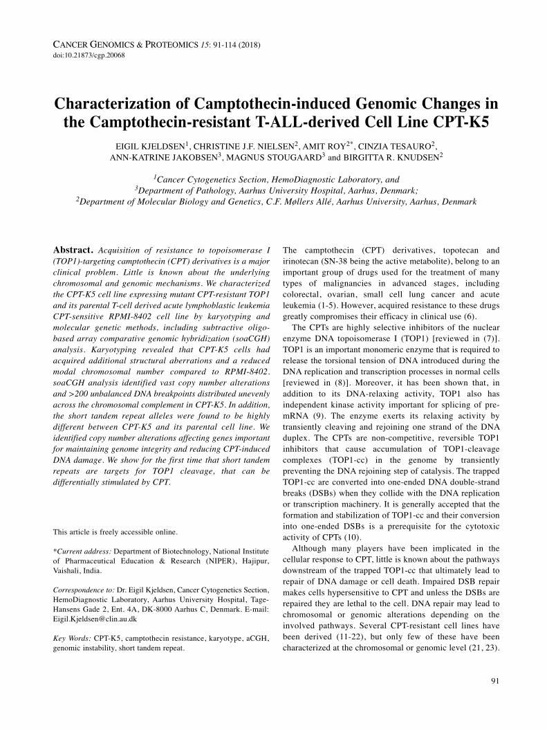
A first line of defense for a cell against CPT-induced damageis down-regulation of its TOP1 activity, thereby reducing TOP1-cc (55, 56). Our subtractive oligo-based aCGH analysis revealedthat the gene dosage of the TOP1 locus was diminished by afactor of approximately 1.7, which was confirmed by FISH(Figure 5). The loss of TOP1 gene copy numbers correlatedwith a reduced level of cellular TOP1 protein in CPT-K5 cells(Figure 2). Other studies have shown that a copy numberchange of the TOP1 gene correlates with the cellular amount ofenzyme and seems to be a common mechanism contributing toCPT resistance (56). By CGH analysis of the CPT-resistant celllines HT-29/CPT, A549/CPT and st-4/CPT, a reduced DNAcopy number of TOP1 was shown together with a reducedrelative expression of TOP1 (44). In our subtractive oligo-basedaCGH analysis, we further screened for copy number changesof other type I topoisomerases (TOP1MT, TOP3A and TOP3B)and type II topoisomerases (TOP2A, TOP2B and TOPOVIA(also known as S. cerevisiae, homolog of, SPO11). We observed
that TOP2A and SPO11 exhibited large losses, and loci ofTOP3B and TOP2B were gained, while TOP3A and TOP1MTdisplayed no copy number change between the CPT-K5 andRPMI-8402 cells (Table IV).
Another line of defense for a cell to resist the effects ofCPT is through the acquisition of single nucleotide mutationsof TOP1 thereby preventing the action of CPT on TOP1-cc[reviewed in (57)] (Table III). We previously showed thatCPT-K5 harbors mutated TOP1 containing the Asp533Glymutation (34). These types of mutations, however, cannot berevealed by oligo-based aCGH analysis.
Reduced intracellular CPT concentration is a third importantway to reduce the formation of devastating TOP1-cc. One ofthe major mechanisms responsible for multidrug resistance incancer chemotherapy is overexpression of some members ofthe ATP-binding cassette (ABC) transporter superfamily,including ABCB1, ABCC1 and ABCG2, resulting in decreasedintracellular levels of drugs [reviewed in (58)]. In mammaliancells, expression of ABC transporters such as Pgp (ABCB1)and ABCG2 confers resistance to CPT and its derivatives (59-61). Gene amplification is a major contributor to increased
CANCER GENOMICS & PROTEOMICS 15: 91-114 (2018)
102
→
Figure 5. Fluorescence in situ hybridization (FISH) validation of copynumber changes at specific genomic regions important for thecamptothecin resistance in CPT-K5 cells. Left hand-side panel:Genomic profiles of chromosome 20 (upper panel), chromosome 4(middle panel) and chromosome 14 (lower panel) together with theirrespective ideograms beneath. Magnified genomic profile views ofcorresponding altered chromosomal regions are given belowchromosome 20, chromosome 4, and chromosome 14 indicating deletedregion at 20q12 containing topoisomerase 1 (TOP1) gene, highlyamplified region at 4q22.1 containing ATP-binding cassette sub-familyG member 2 (ABCG2) gene and amplified region at 14q31.3q32.11containing the tyrosyl-DNA phosphodiesterase 1 (TDP1) gene,respectively. Blue and red bars around the ideograms indicate regionsof gains and losses, respectively. Regions with high gains and losses areindicated by double bars in their respective color. A yellow bar in thecopy number profile of chromosome 14 indicates the region of gain inthe CR1-CR2/MMRU cell lines (see Table III). Right hand-side panel:FISH analyses confirm the genomic array comparative genomichybridization (aCGH) findings in the RPMI-8402 and CPT-K5 cell lineson interphase nuclei with hybridization signals using the followingprobe sets as indicated by red and green marks on the respectiveideograms in the left hand-side panel: i) Bacterial artificialchromosome (BAC)-based probe containing the TOP1 gene (red) andcentromere 20 probe (green); ii) BAC-based probe RP11-368G2 (green)containing the ABCG2 gene together with centromere 4 probe (red) –to the right metaphase spreads represented by partial karyograms ofchromosomes 4 and 12; and iii) BAC-based probe RP11-213O13 (red)containing the TDP1 gene together with subtelomeric 14-qter probe(green) - to the right metaphase spreads represented by partialkaryograms of derivative chromosomes 11 (der(11)t(11;14)) and 15(der(15)t(14;15)) for RPMI-8402 and partial karyograms of derivativechromosomes 5 (der(5)t(5;14)(q35;q32)) and 14 for CPT-K5.

Kjeldsen et al: Camptothecin-induced Genomic Changes in CPT-K5 Cells
103

CANCER GENOMICS & PROTEOMICS 15: 91-114 (2018)
104
Table V. Copy number findings of key genes involved in DNA repair pathways and cell-cycle arrest in CPT-K5 cells relative to RPMI-8402 cells.
HGNC-approved gene symbol Function Cytoband Pos. (hg18) Copy number
Proteasome-dependent TOP1 degradation pathway/phosphodiesterase pathway for TOP1-cc repair of single-ended DSBPARP1 DSB recognition, XRCC1 and TDP1 recruitment 1q42.12 224,615,015-224,662,424 LossTDP1 Hydrolyze the tyrosyl-DNA link in Top1-DNA complex 14q32.11 89,491,999-89,580,861 High gainXRCC1 DNA binding 19q13.31 48,739,304-48,771,570 ncPTEN Phosphatase, negative regulator of PI3K/AKT pathway 10q23.31 89,613,175-89,718,512 LossPNKP End processing 19q13.33 55,056,273-55,062,630 GainAPEX1 or APE1 Apurinic/apyrimidinic endodeoxyribonuclease 1 14q11.2 19,993,130-19,995,766 LossPOLB DNA polymerase beta 8p11.21 42,315,187-42,348,470 ncPRKDC 8q11.21 48,848,222-49,035,296 nc
Proteasome-independent pathway/endonuclease pathway for TOP1-cc repair of single-ended DSBERCC1 Interacts with ERCC4 to form endonuclease 19q13.32 50,608,532-50,618,642 ncERCC4 or XPF 3’-Flap endonuclease subunit 16p13.12 13,921,515-13,953,706 GainRPA1 Replication protein A1 subunit 17p13.3 1,680,023-1,749,598 ncRPA2 Replication protein A2 subunit 1p35.3 28,090,636-28,223,823 ncRPA3 Replication protein A3 subunit 7p21.3 7,643,100-7,724,763 High gainPOLD1 Polymerase delta catalytic subunit A 19q13.33 55,579,405-55,613,083 GainPOLD2 Polymerase delta catalytic subunit B 7p13 44,120,811-44,129,672 GainPOLD3 Polymerase delta catalytic subunit D 11q13.4 73,981,277-74,031,413 LossPOLD4 Polymerase delta catalytic subunit A 11q13.1 66,875,595-66,877,593 LossFEN1 Flap structure-specific endonuclease 1 11q12.2 61,316,726-61,321,286 LossLIG1 DNA ligase 1 19q13.32 53,130,515-53,365,372 LossSLX4 Structure-specific endonuclease subunit 16p13.3 3,571,185-3,601,608 GainHR & NHEJATM Binds to MRN complex, phosphorylates CHK2 11q22.3 107,598,769-107,745,036 LossH2AFX Activated by ATM, recruit further DNA repair components 11q23.3 118,469,795-118,471,387 LossMRE11 Endonuclease, 3’-5’ exonuclease, MRN complex 11q21 93,790,115-93,866,688 LossNBN or NBS1 Adaptor, check point roles, MRN complex 8q21.3 91,014,740-91,060,075 ncPARP1 DSB recognition, MRN complex recruitment 1q42.12 224,615,015-224,662,424 LossRAD50 ATPase/Scaffold, MRN complex 5q31.1 131,920,529-132,007,494 LossHRBARD1 BRCA1-associated ring domain 2q35 215,301,520-215,382,673 GainBLM or RECQL3 Bloom syndrome, helicase 15q26.1 89,061,583-89,159,690 GainBRCA1 Mediator/adaptor, ubiquitin ligase 17q21.31 38,449,840-38,530,994 LossBRCA2 Recombination mediator, binds to RAD51 13q13.1 31,787,617-31,871,809 High gainRBBP8 or CtIP Endonuclease; binds to BRCA1 18q11.2 18,767,293-18,860,447 ncDNA2 DNA replication helicase 2 10q21.3 69,843,827-69,901,885 GainEXO1 5’-3’ Exonuclease 1q43 240,078,158-240,119,671 LossLIG1 ATP-dependent DNA ligase 19q13.32 53,310,515-53,365,372 LossPALB2 Partner and localizer of BRCA2 16p12.1 23,521,984-23,560,179 GainPOLD See above for POLD1-4 - - -POLE Catalytic subunit DNA polymerase epsilon 12q24.33 131,710,421-131,774,018 ncRAD51 Recombinase, homology pairing 15q15.1 38,774,651-38,811,648 ncRAD52 Mediator of repair 12p13.33 904,877-912,503 ncATRX or RAD54 Recombinase, D-loop formation Xq21.1 76,647,012-76,928,375 LossRPA See above for RPA1-3 - - -Classic NHEJAPTX End processing 9p13.3 32,962,608-32,991,626 LossAPLF End processing 2p14 68,548,246-68,660,798 ncLIG4 DNA ligase 4, complexes with XRCC4 13q33.3 107,657,793-107,665,883 High gainNHEJ1 or XLF Scaffold protein 2q35 219,648,290-219,733,831 GainPNKP End processing 19q13.33 55,056,273-55,062,630 GainPRKDC or DNA-PKcs DSB-responsive PIKK family protein kinase 8q11.21 48,848,222-49,035,296 ncPOLL or DNA-Pol μ End processing, gap filling 10q24.32 103,328,629-103,337,963 GainPOLM or DNA-Pol μ End processing, gap filling 7p13 44,078,372-44,088,607 GainXRCC4 Scaffold protein 5q14.2 82,409,073-82,685,335 ncXRCC5 or Ku80 dsDNA end binding, resection inhibition 2q35 216,682,377-216,779,248 GainXRCC6 or Ku70 dsDNA end binding, resection inhibition 22q13.2 40,347,241-40,389,998 nc
Table V. Continued

expression of ABCB1 and ABCG2 in colorectal and breastcancer cells making them resistant towards the CPT-analogSN-38 and mitoxantrone (62, 63). Remarkably, we found thatin the CPT-K5 cell line, the genes ABCB1 and ABCC1 arelocated in regions of copy number gain at 7q21.12 and16p13.11, respectively, while ABCG2 is in the region of highestcopy gain at 4q22.1 (Figure 5, Table IV). The CPT-resistantcell lines HT-29/CPT and A549/CPT had amplified ABCtransporter family genes (44). These observations indicate thatcopy numbers gains of ABC transporters are of importance indecreased intracellular CPT accumulation in CPT-K5 cells. Ithas been shown that CPT-K5 is multidrug resistant e.g. it isalso resistant to topoisomerase II inhibitors (64). Thisobservation is in line with the findings that there are manydrugs that are substrates for the ABCG2 transporter, includingtopoisomerase II inhibitors (e.g. mitoxantrone, etoposide,daunorubicin, and doxorubicin), CPT analogs (e.g. topotecan,and irinotecan), tyrosine kinase inhibitors, antimetabolites, andmany other drug types [reviewed in (58)]. Interestingly, aglioblastoma cell line, SF295, which became mitoxantrone-resistant by exposure to step-wise increasing concentrations ofthe drug had a resulting ABCG2 amplification by formingdouble minutes, which were later reintegrated into the genomeat multiple chromosomal regions during subsequent selectionsteps (43). In the CPT-K5 cell line, we found that the ABCG2gene in addition to its location at 4q22.1 is highly amplified ata single chromosomal site on der(12)t(4;12) (Figure 5),suggesting a similar mechanism of amplification to thatobserved in the SF295 cell line. Taken together, these findingssupport the idea that ABCG2 is one of the most important
transporter genes in relation to CPT and its derivatives, whichis in agreement with previous suggestions (65-67).
Alteration of apoptotic pathways is a fourth line ofdefense in influencing resistance to CPT. The biochemicalpathways of apoptosis include pro-apoptotic and anti-apoptotic proteins, which affect the cellular response to CPTas reviewed elsewhere (7, 68). A delicate balance betweenpro- and anti-apoptotic pathways has been proposed toprovide alternative means of providing cancer cells with aselective advantage promoting chemoresistance (69).Apoptosis involves a complex network of many proteins ofwhich the proteins encoded by tumour protein p53 (TP53),inhibitor of apoptosis, X-linked (XIAP) and baculoviral IAPrepeat-containing protein 5 (BIRC5) play important roles inregulating apoptosis (70). TP53 plays an important role insensitivity to CPT and apoptosis induced by CPT. Whenleukemia cells are deficient in p53 function, the cells arehypersensitive to CPT, whereas cells with normalfunctioning p53 are resistant to CPT (71, 72). In our study,we found that the copy number of TP53 was unchanged(Tables IV and V), which is in agreement with previousfindings that normal TP53 is important for resistance to CPTand its derivatives. The anti-apoptotic genes XIAP andBIRC5 possess a wide range of biological activities thatpromote cancer cell survival and proliferation to overcomeendogenous and exogenous insults [reviewed in (68)]. Wefound CPT-K5 cells had loss of the XIAP gene and thatBIRC5 had no copy number change (Table IV).
A fifth line of defense against CPT is efficient repair of CPT-induced DNA damage by the cell (53). Although the
Kjeldsen et al: Camptothecin-induced Genomic Changes in CPT-K5 Cells
105
Table V. Continued
HGNC-approved gene symbol Function Cytoband Pos. (hg18) Copy number
Alternative NHEJRBBP8 or CtIP Nuclease; binds to BRCA1 18q11.2 18,767,293-18,860,447 ncPOLB Gap filling 8p11.21 42,315,187-42,348,470 ncXRCC1 DNA binding 19q13.31 48,739,304-48,771,570 ncLIG3 DNA ligase 3, complexes with XRCC1 17q12 30,331,651-30,356,201 LossWRN DSB repair mediator, Helicase 3’-5 exonuclease 8p12 31,010,320-51,150,819 ncMismatch repairMLH1 Forms a heterodimer with PSM2 3p22.2 37,009,983-37,067,341 ncMLH3 Forms heterodimer with MLH1 14q24.3 74,550,220-74,587,988 High gainMSH2 Forms heterodimer with MSH6 2p16.3 47,483,767-47,563,864 ncMSH3 Forms heterodimer with MSH3 5q14.1 79,986,050-80,208,390 ncMSH6 Forms heterodimer with MSH2 2p16.3 47,863,725-47,887,596 LossPMS1 Forms heterodimer with MLH1 2q32.2 190,357,056-190,450,600 ncPMS2 Forms heterodimer with MLH1 7p22.1 5,979,396-6,015,263 GainCell-cycle arrestCHK1 Phosphorylated by ATR, G2/M cell-cycle arrest 11q24.2 125,001,854-125,030,850 LossCHK2 Phosphorylated by ATM, G1 cell-cycle arrest 22q12.1 27,413,731-27,460,715 GainTP53 G1 and G2 Cell cycle arrest 17p13.1 7,512,445-7,531,588 nc
DSB: Double=strand break HR: homologous recombination; NHEJ: non-homologous end-joining; nc: no change.

mechanisms implicated in repair of TOP1-cc have been widelystudied they still remain largely elusive (73-78). The repair ofthis rather unique form of DNA damage involves differentpathways (Figure 6) that cooperate to remove the TOP1-cc andrestore intact DNA, thus allowing the resumption ofphysiological cellular functions including replication andtranscription. An important step in the repair of TOP1-cc isdegradation of DNA-bound TOP1 (76). Furthermore, it hasbeen shown that cells that rapidly degrade TOP1 are resistantto CPT, while cells that fail to degrade TOP1 are sensitive toCPT (75, 79). TOP1 covalently stalled on DNA is degraded viaa ubiquitin-26S proteasome pathway into a small peptide witha tyrosyl residue attached to the 3’-end of the nicked DNA (75,76). A recent finding suggests that phoaphatase and tensinhomolog (PTEN) availability may determine the fate of cellsharboring CPT-induced DNA damage by regulating activity ofDNA-dependent protein kinase (PRKDC) kinase, whichphosphorylates TOP1 on serine 10 (79). It was found that
PTEN deletion ensures higher TOP1 serine 10 phosphorylationleading to rapid TOP1 degradation and CPT resistance.PRKDC-dependent phosphorylation of TOP1 in complex withDNA seems to be a prerequisite for ubiquitinylation, which inturn is required for proteasomal degradation (79).
Another important aspect of the repair of CPT-inducedDNA damage is the fate of stalled or collapsed replication ortranscription fork complexes which are generated by CPT(80). Colliding replication forks on the leading strand forDNA synthesis or transcription on the template strandconvert CPT-stabilized TOP1-cc into devastating DSBs ifunrepaired. Break-induced replication (BIR) is a homologousrecombination pathway which was recently identified asserving for repair of collapsed or broken replication forks[reviewed in (81, 82)]. Another more recent BIR-relatedpathway called micro-homology-mediated BIR has beenproposed to explain copy number alterations, complexchromosomal rearrangements and microsatellite expansion
CANCER GENOMICS & PROTEOMICS 15: 91-114 (2018)
106
Figure 6. Schematic overview of mechanisms implicated in repair of camptothecin (CPT)-induced DNA damage. BER: Base excision repair; BIR:breakage-induced replication; CPT: camptothecin or its derivatives; DSB: double-strand break; HR: homologous recombination; MMBIR: micro-homology-mediated BIR; MRN: meiotic recombination 11 (MRE11)/DNA repair protein RAD50(RAD50)/nibrin (NBS1) complex; NER: nucleotideexcision repair; NHEJ: non-homologous end-joining; NHR: non-homologous recombination; PTEN(−) indicates phosphatase and tensin homologdeficiency; SSB: single-strand DNA break. Open red circle indicates fork collision and repair mechanisms; Y-P: 3’-phosphotyrosyl.

associated with cancer and various other diseases in humans(81-83). It is beyond the scope of this study to describe theseprocesses in more detail, as excellent reviews exist (81, 82).
A summary of the genes involved in the repair processesdescribed above is given in Table V, which also providesinformation about copy number changes affecting these genesin the CPT-K5 cell line. Interestingly, important genesinvolved in the repair of TOP1-induced DNA damage hadcopy number changes. Firstly, the PTEN gene, an upstreameffector of the proteasomal pathway, had a copy number loss.Secondly, a 50% increase in copy number of the TDP1 gene,which was confirmed by FISH analysis (Figure 5), and a 1.42-fold higher relative activity of TDP1 (Figure 7) wereidentified. Both of these observations are in agreement withthe recent findings by Ando et al. (79) suggesting that PTENdeletion ensures higher TOP1 serine 10 phosphorylation byPRKDC, favoring rapid TOP1 degradation and CPTresistance. It might be interesting in future studies todetermine PTEN expression or the level of phosphorylatedserine 10 in TOP1 in the CPT-K5 cell line and investigatewhether TOP1 is more degraded in response to CPT in CPT-K5 cells. A previous BAC-based aCGH analysis on theirinotecan-resistant cell lines CR1 and CR2, derived fromparental melanoma MMRU cell lines, showed a gain of theregion 14q23.2 to q31.1 (21). The amplified region in that
study is within the region gained at 14q23.2-q32.12 observedin CPT-K5 cells (Table IV; Figure 5) but amplification in theCR1 and CR2 cells did not include the TDP1 gene. A majordifference between the CPT-K5 and the CR1/CR2 cell lines isthat the latter needs CPT to be present for the cells to conservetheir CPT resistance as opposed to CPT-K5 cells, which arestably CPT-resistant in the absence of CPT. This differencemay influence the cellular dynamics of e.g. DNA repair.
The DNA mismatch repair (MMR) system also plays animportant role in maintaining genomic stability (84). The maingenes involved in MMR include, E. coli homolog of MutL(MLH1), MLH2, E. coli homolog of MutS (MSH2), MSH3,MSH6, postmeiotic segregation increased, S. cerevisiae 1(PMS1), and PMS2. Defects in any of these genes result inmicrosatellite instability (85) and repeat expansions (86). Itwas shown in one study that MSH2 deficiency results in ahigher sensitivity to CPT, while loss of MLH1 results in CPTresistance (87). In the CPT-K5 cell line, we observed a gainof MLH3 and PMS2, and a 3’-partial loss of MSH6, whilethere were no copy number changes in the other MMR genes(Table V). Although only few studies exist on MMR and CPTsensitivity, gain of MLH3 located at 14q24.3 might be arecurrent rearrangement in CPT-resistant cells as it was alsoobserved in CPT-resistant MMRU cells (21).
A slower growth rate has been reported in several CPT-resistant cell lines (55, 11, 22), although this does not seemto be a major mechanism of resistance to TOP1 inhibitors(55). It may, however, be a reminiscence of altered propertiesas a result of obtaining CPT resistance because cell-cyclecheckpoint arrest is important to allow time for repair and toprevent progression of replication. CPT-induced DNAdamage activates S. pombe homolog of checkpoint 1 (CHK1)and CHK2 checkpoint kinases (88). The two independentCPT-resistant colorectal cancer cell lines derived from HT-29 and HCT-116 had an approximately 30% increaseddoubling time (22). Increased basal levels of H2A histonefamily, member X (γ-H2AX) and activated CHK2accompanied these findings, without notable changes in thelevel of CHK1. Consistent with these findings, the CPT-K5cell line has an increased generation doubling time(approximately 100% increase) (11) and amplification of theCHK2 gene together with a relative loss of CHK1 (Table V).
STR profiling of RPMI-8402 and CPT-K5. Because of themajor cytogenetic and genomic differences between RPMI-8402 and CPT-K5 cells, we wanted to examine whether STRsdiffer between the two cell lines. STRs are microsatelliteswith repetitive sequences characterized by a variable numberof repeated short sequence elements of 2-6 bp in length as aunit (e.g. di-, tri-, and tetra-sequences). They are highlypolymorphic between individuals and are especially used inforensic studies for human identification, and more recentlyfor authentication of human cell lines (89, 90).
Kjeldsen et al: Camptothecin-induced Genomic Changes in CPT-K5 Cells
107
Figure 7. Bar chart showing the mean of the results obtained whenmeasuring relative tyrosyl-DNA phosphodiesterase 1 (TDP1) activity inwhole-cell extracts from 1.0×10⁶ and 0.5×10⁶ RPMI-8402 (black bars)and CPT-K5 (grey bars) cells, as indicated in the figure, in threeindependent repetitions. Relative TDP1 activity in CPT-K5 cells wasincreased by a factor of 1.42 (p<0.001; unpaired t-test). The error barsindicate standard deviation.

The AmpFISTR® Identifiler PCR Amplification tests STRalleles (tetra-nucleotide repeats) at 15 loci that are located ondistinct chromosomes, behave according to known principlesof population genetics and contain low mutation rates[reviewed in (91)]. The results from the STR profiling analysisof RPMI-8402 and CPT-K5 cells are summarized in Table VI.
A comparison of the STR results of the two cell lines showsthat it is only the TPOX locus which has a complete match,with the homozygous STR allele bearing the eight repeat unitspresent in both cell lines. For the remaining 14 STR loci, therewas either a partial match (D2S1338, D3S1358, D5S818,CSF1PO, vWA, D13S317, D16S539, D19S433, D21S11) or
CANCER GENOMICS & PROTEOMICS 15: 91-114 (2018)
108
Table VI. Short tandem repeat (STR) profiling of RPMI-8402 and CPT-K5 cells using the AmpFISTR® Identifiler PCR amplification kit.
STR locus Chromosomal STR allelesa Match Repeat unitb location RPMI-8402 CPT-K5
TPOX* 2p25.3 8 8 M (AATG)6-14D2S1338 2q35 19 23 m (TGCC)6(TTCC)11 23 24 26 (27)D3S1358 3p21.31 14 14 m TCTA (TCTG)2-3(TCTA)10-15 16 15 16FGA 4q28 23 21 (TTTC)3TTTTTTCT(CTTT)7CTCC(TTCC)2 22 24 D5S818* 5q23.2 12 11 m (AGAT)7-15 13 12 13CSF1PO 5q33.1 11 9 m (AGAT)5-16 12 (10) 11 12D7S820* 7q21.11 8 9 None (GATA)6-14 13 12 14D8S1179 8q24.13 11 13 None (TCTA)7-12 14TH01 11p15.5 9 6 None (AATG)3-12 9.3 7vWA 12p13.31 14 (17) m TCTA TCTG TCTA (TCTG)4 (TCTA)3 20 18 20 22D13S317 13q31.1 12 10 m (TATC)7-15 12D16S539 16q24.1 10 10 m (GATA)5-15 11 14D18S51 18q21.33 16 14 None (AGAA)8-27 20 17 18D19S433 19q12 12 14 m (AAGG)(AAAG)(AAGG)(TAGG)(AAGG)7-12 (13) 15 14D21S11 21q21.1 30 28 m (TCTA)4(TCTG)6(TCTA)3TA(TCTA)3TCA (TCTA)2TCCATA(TCTA)6 30 33.2 34.2
M: Complete match, m:partial match, None: no match of genotypes. *Denotes STR loci analyzed by topoisomerase 1 (TOP1) DNA cleavage assay;aNumbers in brackets indicate a minor peak; bSubscripts indicate number of repeat units. A more detailed allele listing of these STR loci is availableat www.cstl.nist.gov/biotech/strbase/fr_fact.htm.

no match (FGA, D7S820, D8S1179, TH01, D18S51) of theSTR alleles between the two cell lines. The aberrant STR locidid not correlate with the observed differences intranslocations or breakpoints as identified by karyotyping orgenomic profiling. The total number of STR alleles increasedfrom 26 in the RPMI-8402 cells to 42 in the CPT-K5 cell line(p<0.01), which is in line with the observed number ofchanges by karyotyping and by breakpoints identified byoligo-based aCGH analysis. These findings are in agreementwith the fact that CPT induces genomic instability.
STRs can be cleaved in vitro by TOP1 and DNA cleavagecan be stimulated by CPT. The most obvious conclusionfrom the STR data is that either the two cell lines are notrelated or alternatively that differences may be the result ofCPT-induced DNA damage and repair.
To test whether STR alleles are targets for CPT-inducedDNA damage, we performed a number of classical in vitroTOP1 DNA cleavage experiments in the presence and absenceof the drug (Figure 8). We found that the substrate containingthe sequence of the D7S820 locus was a stronger TOP1 DNAcleavage site than the substrate containing the sequence for theD5S818 locus in the absence of CPT. Addition of CPT to theTOP1 cleavage reaction resulted in very strong stimulation ofDNA cleavage of the D7S820 STR substrate, whereas onlyvery weak stimulation for the D5S818 STR substrate wasobserved. In contrast, the TPOX STR substrate was not cleavedby TOP1 and neither was cleavage stimulated by CPT.
Correlating these findings with the observed STR allelicdifferences between the two cell lines, it should be noted thatthe TPOX STR was the only allele that showed a completematch in the STR profiling and that we found only one allelein both cell lines. This finding indicates that because theTPOX STR does not contain a TOP1 cleavage site it is stablein the presence of CPT. Furthermore, the D7S820 locus,which did not exhibit any match in the STR profiling,contained the strongest TOP1 DNA cleavage site. This sitewas also the site with the strongest cleavage stimulation byCPT of the three loci sequences tested. Taken together, ourfindings indicate that the genomic stability of the tested STRalleles depends upon their ability to be cleaved by TOP1 andhow strongly CPT can enhance this cleavage. Thisinterpretation is in agreement with the studies ontranscription-induced trinucleotide STR instability (92-94).Another way STR can become unstable or mutate is byreplication strand slippage or misalignment (95). Replicationfork collapse or arrest is one source of DNA strand slippageand as CPT greatly enhances the frequency of replicationfork arrests, it is not unlikely that it will affect the STRs asexemplified by the tetra-nucleotides we examined. Normallythe rate of slippage is highest in dinucleotide STRs andlowest in tetra-nucleotides but how CPT affects the rate ofslippage remains unknown (96).
Kjeldsen et al: Camptothecin-induced Genomic Changes in CPT-K5 Cells
109
Figure 8. Topoisomerase 1 (TOP1) DNA cleavage assay using shorttandem repeat (STR) sequences as DNA substrate. The top panel showsthe construction of the double-stranded oligonucleotide substrates. Thegrey box indicate the double-stranded STR alleles representing nomatch (None) D7S820, a partial match (m) D5S818, and a completematch (M) for thyroxin peroxidase (TPOX) as observed in the STRprofiling of DNA from RPMI-8402 and CPT-K5 cell lines. Theseoligonucleotides were constructed so that the sequence of each of thethree selected RPMI-8402 STR alleles were flanked by a commonsequence identical in the three different substrates. Each construct was5’-radiolabeled and subjected to human topoisomerase 1 (hTOP1) DNAcleavage with purified recombinant hTOP1 in the absence and presenceof CPT before the products were separated by 12% polyacrylamide gelelectrophoresis. Lanes 1, 4 and 7: incubation without enzyme and CPT;lanes 2, 5 and 8: incubation with hTOP1 in the absence of CPT; lanes3, 6 and 9: incubation with hTOP1 and 10 μM CPT. Uncleavedsubstrates are marked (S) and *denotes hTOP1 DNA cleavage sites.
In conclusion, it is highly likely that cells subjected tocontinuous sublethal concentrations of CPT acquireincreased STR instability, but further studies focusing onCPT and STR are warranted.
Conclusion
A major clinical problem is that initially responding patientswith cancer treated with CPT or its derivatives graduallyacquire resistance. We used the stably and highly CPT-resistant cell line CPT-K5 to obtain global genomic insightinto acquired CPT resistance by characterizing the cell line,and its parental, using 24-color karyotyping and subtractiveoligo-based aCGH analysis.
Although many human cell lines resistant to CPT or itsderivatives have been developed, only very few of thesehave been examined at the chromosomal or genomic level.This is intriguing because CPT is a specific inhibitor ofTOP1, which has a major role in DNA metabolism, such asin transcription, replication, recombination and repair.
By 24-color karyotyping, we showed that CPT-K5 is hypo-tetraploid with a modal chromosomal number reduced to 80,and had loss or gain of 18 structural aberrant chromosomescompared with its parental RPMI-8402. Subtractive oligo-based aCGH analysis identified 165 copy number alterationsand 236 unbalanced DNA breakpoints unevenly distributedacross the chromosomal complement in CPT-K5 cells.Furthermore, we found that STRs highly differ between CPT-K5 and its parental cell line, and we show for the first time thatSTRs are TOP1 targets that can be differentially stimulated byCPT. These findings are in agreement with repair pathways thatefficiently repaired CPT-induced DNA damage at the expenseof conserved genome integrity providing substantialchromosomal and genomic alterations that may have beenbeneficial for survival and cellular proliferation. However,further studies are needed to establish whether STR alterationsactually contribute to cellular tolerance to CPT.
We suggest that acquired resistance to CPT in the CPT-K5cell line is a multifactorial process. Four major mechanismsseem to be involved in addition to its acquired previouslydescribed TOP1 mutation. These are: i) reduced gene dosageof TOP1 resulting in decreased cellular amount and activity ofthe enzyme; ii) high amplification of ABCG2, which maycontribute to reduced intracellular concentration of active drugby decreased drug influx/increased drug efflux; iii) no copynumber changes in TP53 and BIRC5 genes, which maycontribute to resistance to apoptosis; and iv) high amplificationof TDP1 accompanied by increased TDP1 activity and reducedgene dosage of PTEN, which were recently shown to be ofgreat importance for repair of CPT-induced DNA damage.
CPT-resistant cell lines have variable degrees of resistance toCPT and variable dependency on the presence of CPT to retaintheir resistance to it. These observations may relate to
mechanisms implicated in their resistance. Only a few CPT-resistant cell lines have been examined at the genomic level,and clearly, more CPT-resistant cell lines need to be examinedwith up-to-date genomic methods in order to enhance theunderstanding of their resistance mechanisms. Furthermore, itshould be kept in mind that CPT-resistant cell lines aredeveloped by exposure to longitudinal increments of sublethaldrug doses, and often prior to their selection, had a mutagenicstep, which may provide the cells with potential initial genomicaberrations amenable to selective advantage. This is different tothe clinical situation in that patients becoming resistant totreatment with CPT-derived drugs did not have an initialmutagenic step and most often no increment in drug dose.
Nevertheless, identification of recurrent regions implicatedin the acquisition of CPT resistance might provide the firstclues to identifying important regions of the genome, andconsequently genes, which could serve as potentialtherapeutic targets for intervention against development ofCPT resistance, or for its reversal.
Conflicts of Interest
The Authors declare no conflicts of interest in regard to this study.
Acknowledgements
The biotechnologists Bente Madsen and Pia Kristensen are greatlythanked for excellent technical assistance. Technician NorikoHansen is thanked for purification of human TOP1. Dr. ChristopherVeigaard is greatly thanked for providing cell growth characteristics.The Danish Cancer Society supported the study.
References
1 Kantarjian HM, Beran M, Ellis A, Zwelling L, O’Brien S,Cazenave L, Koller C, Rios MB, Plunkett W and Keating MJ:Phase I study of Topotecan, a new topoisomerase I inhibitor, inpatients with refractory or relapsed acute leukemia. Blood 81:1146-1151, 1993.
2 Smith DH, Adams JR, Johnston SR, Gordon A, Drummond MFand Bennett CL: A comparative economic analysis of pegylatedliposomal doxorubicin versus topotecan in ovarian cancer in theUSA and the UK. Ann Oncol 13: 1590-1597, 2002.
3 Tsavaris N, Kosmas C, Skopelitis H, Papadoniou N, Polyzos A,Zografos G, Adoniou E, Gryniatsos J, Felekouras E, ZacharakisM, Sigala F, Bacoyiannis C, Papastratis G and Papalambros E:Sequential administration of 5-fluorouracil (5FU)/leucovorin(LV) followed by irinotecan (CPT-11) at relapse versus CPT-11followed by 5-FU/LV in advanced colorectal carcinoma. Aphase III randomized study. Chemotherapy 53: 282-291, 2007.
4 Riemsma R, Simons JP, Bashir Z, Gooch CL and Kleijnen J:Systematic Review of topotecan (Hycamtin) in relapsed smallcell lung cancer. BMC Cancer 10: 436, 2010.
5 Douillard JY, Cunningham D, Roth AD, Navarro M, James RD,Karasek P, Jandik P, Iveson T, Carmichael J, Alakl M, Gruia G,Awad L and Rougier P: Irinotecan combined with fluorouracil
CANCER GENOMICS & PROTEOMICS 15: 91-114 (2018)
110

compared with fluorouracil alone as first-line treatment formetastatic colorectal cancer: a multicentre randomised trial.Lancet 355: 1041-1047, 2000.
6 Beretta GL, Gatti L, Perego P and Zaffaroni N: Camptothecinresistance in cancer: insights into the molecular mechanisms ofa DNA-damaging drug. Curr Med Chem 20: 1541-1565, 2013.
7 Pommier Y: Topoisomerase I inhibitors: camptothecins andbeyond. Nat Rev Cancer 6: 789-802, 2006.
8 Wang JC: Cellular roles of DNA topoisomerases: a molecularperspective. Nat Rev Mol Cell Biol 3: 430-440, 2002.
9 Rossi F, Labourier E, Forne T, Divita G, Derancourt J, Riou JF,Antoine E, Cathala G, Brunel C and Tazi J: Specificphosphorylation of SR proteins by mammalian DNAtopoisomerase I. Nature 381: 80-82, 1996.
10 Pfister TD, Reinhold WC, Agama K, Gupta S, Khin SA,Kinders RJ, Parchment RE, Tomaszewski JE, Doroshow JH andPommier Y: Topoisomerase I levels in the NCI-60 cancer cellline panel determined by validated ELISA and microarrayanalysis and correlation with indenoisoquinoline sensitivity.Mol Cancer Ther 8: 1878-1884, 2009.
11 Andoh T, Ishii K, Suzuki Y, Ikegami Y, Kusunoki Y, TakemotoY and Okada K: Characterization of a mammalian mutant witha camptothecin-resistant DNA topoisomerase I. Proc Natl AcadSci USA 84: 5565-5569, 1987.
12 Kanzawa F, Sugimoto Y, Minato K, Kasahara K, Bungo M,Nakagawa K, Fujiwara Y, Liu LF and Saijo N: Establishmentof a camptothecin analogue (CPT-11)-resistant cell line ofhuman non-small cell lung cancer: characterization andmechanism of resistance. Cancer Res 50: 5919-5924, 1990.
13 Kubota N, Kanzawa F, Nishio K, Takeda Y, Ohmori T, FujiwaraY, Terashima Y and Saijo N: Detection of topoisomerase I genepoint mutation in CPT-11 resistant lung cancer cell line.Biochem Biophys Res Commun 188: 571-577, 1992.
14 Sugimoto Y, Tsukahara S, Oh-hara T, Isoe T and Tsuruo T:Decreased expression of DNA topoisomerase I in camptothecin-resistant tumor cell lines as determined by a monoclonalantibody. Cancer Res 50: 6925-6930, 1990.
15 Rubin E, Pantazis P, Bharti A, Toppmeyer D, Giovanella B andKufe D: Identification of a mutant human topoisomerase I withintact catalytic activity and resistance to 9-nitro-camptothecin.J Biol Chem 269: 2433-2439, 1994.
16 Fujimori A, Harker WG, Kohlhagen G, Hoki Y and PommierY: Mutation at the catalytic site of topoisomerase I in CEM/C2,a human leukemia cell line resistant to camptothecin. CancerRes 55: 1339-1346, 1995.
17 Chatterjee D, Wyche JH and Pantazis P: Induction of apoptosisin malignant and camptothecin-resistant human cells. Ann N YAcad Sci 803: 143-156, 1996.
18 Urasaki Y, Laco GS, Pourquier P, Takebayashi Y, Kohlhagen G,Gioffre C, Zhang H, Chatterjee D, Pantazis P and Pommier Y:Characterization of a novel topoisomerase I mutation from acamptothecin-resistant human prostate cancer cell line. CancerRes 61: 1964-1969, 2001.
19 Saleem A, Ibrahim N, Patel M, Li XG, Gupta E, Mendoza J,Pantazis P and Rubin EH: Mechanisms of resistance in a humancell line exposed to sequential topoisomerase poisoning. CancerRes 57: 5100-5106, 1997.
20 Chang JY, Liu JF, Juang SH, Liu TW and Chen LT: Novelmutation of topoisomerase I in rendering cells resistant tocamptothecin. Cancer Res 62: 3716-3721, 2002.
21 Gao K, Lockwood WW, Li J, Lam W and Li G: Genomicanalyses identify gene candidates for acquired irinotecanresistance in melanoma cells. Int J Oncol 32: 1343-1349, 2008.
22 Petitprez A, Poindessous V, Ouaret D, Regairaz M, Bastian G,Guerin E, Escargueil AE and Larsen AK: Acquired irinotecanresistance is accompanied by stable modifications of cell cycledynamics independent of MSI status. Int J Oncol 42: 1644-1653, 2013.
23 Tuduri S, Crabbe L, Conti C, Tourriere H, Holtgreve-Grez H,Jauch A, Pantesco V, De Vos J, Thomas A, Theillet C, PommierY, Tazi J, Coquelle A and Pasero P: Topoisomerase I suppressesgenomic instability by preventing interference betweenreplication and transcription. Nat Cell Biol 11: 1315-1324, 2009.
24 Kjeldsen E, Bonven BJ andoh T, Ishii K, Okada K, Bolund Land Westergaard O: Characterization of a camptothecin-resistant human DNA topoisomerase I. J Biol Chem 263: 3912-3916, 1988.
25 Roy A, Tesauro C, Frohlich R, Hede MS, Nielsen MJ, KjeldsenE, Bonven B, Stougaard M, Gromova I and Knudsen BR:Decreased camptothecin sensitivity of the stem-cell-likefraction of Caco2 cells correlates with an alteredphosphorylation pattern of topoisomerase I. PloS One 9:e99628, 2014.
26 Jensen PW, Falconi M, Kristoffersen EL, Simonsen AT,Cifuentes JB, Marcussen LB, Frohlich R, Vagner J, HarmsenC, Juul S, Ho YP, Withers MA, Lupski JR, Koch J, Desideri A,Knudsen BR and Stougaard M: Real-time detection of TDP1activity using a fluorophore-quencher coupled DNA-biosensor.Biosens Bioelectron 48: 230-237, 2013.
27 Lisby M, Krogh BO, Boege F, Westergaard O and Knudsen BR:Camptothecins inhibit the utilization of hydrogen peroxide inthe ligation step of topoisomerase I catalysis. Biochemistry 37:10815-10827, 1998.
28 Veigaard C and Kjeldsen E: Exploring the genome-widerelation between copy number status and microRNAexpression. Genomics 104: 271-278, 2014.
29 ISCN: An International System for Human CytogeneticNomenclature (2013). Shaffer LG, McGowan-Jordan andSchmid M (Eds.) Basel: S. Karger and Cytogenetic andGenome Research, 2013.
30 Kerndrup GB and Kjeldsen E: Acute leukemia cytogenetics: anevaluation of combining G-band karyotyping with multi-colorspectral karyotyping. Cancer Genet Cytogenet 124: 7-11, 2001.
31 Kjeldsen E and Roug AS: A novel unbalanced de novotranslocation der(5)t(4;5)(q26;q21.1) in adult T-cell precursorlymphoblastic leukemia. Mol Cytogenet 5: 21, 2012.
32 Braylan RC, Orfao A, Borowitz MJ and Davis BH: Optimalnumber of reagents required to evaluate hematolymphoidneoplasias: results of an international consensus meeting.Cytometry 46: 23-27, 2001.
33 Christiansen K, Svejstrup AB andersen AH and Westergaard O:Eukaryotic topoisomerase I-mediated cleavage requires bipartiteDNA interaction. Cleavage of DNA substrates containing strandinterruptions implicates a role for topoisomerase I inillegitimate recombination. J Biol Chem 268: 9690-9701, 1993.
34 Tamura H, Kohchi C, Yamada R, Ikeda T, Koiwai O, PattersonE, Keene JD, Okada K, Kjeldsen E, Nishikawa K and AndohT: Molecular cloning of a cDNA of a camptothecin-resistanthuman DNA topoisomerase I and identification of mutationsites. Nucleic Acids Res 19: 69-75, 1991.
Kjeldsen et al: Camptothecin-induced Genomic Changes in CPT-K5 Cells
111

35 Stougaard M, Lohmann JS, Mancino A, Celik S, Andersen FF,Koch J and Knudsen BR: Single-molecule detection of humantopoisomerase I cleavage-ligation activity. ACS Nano 3: 223-233, 2009.
36 Andersen FF, Stougaard M, Jorgensen HL, Bendsen S, Juul S,Hald K andersen AH, Koch J and Knudsen BR: Multiplexeddetection of site specific recombinase and DNA topoisomeraseactivities at the single molecule level. ACS Nano 3: 4043-4054,2009.
37 Huang CC, Hou Y, Woods LK, Moore GE and Minowada J:Cytogenetic study of human lymphoid T-cell lines derived fromlymphocytic leukemia. J Natl Cancer Inst 53: 655-660, 1974.
38 Hayata I, Oshimura M, Minowada J and Sandberg AA:Chromosomal banding of cultured T and B lymphocytes. InVitro 11: 361-368, 1975.
39 Le Beau MM, McKeithan TW, Shima EA, Goldman-Leikin RE,Chan SJ, Bell GI, Rowley JD and Diaz MO: T-cell receptoralpha-chain gene is split in a human T-cell leukemia cell linewith a t(11;14)(p15;q11). Proc Natl Acad Sci USA 83: 9744-9748, 1986.
40 McGuire EA, Hockett RD, Pollock KM, Bartholdi MF, O’BrienSJ and Korsmeyer SJ: The t(11;14)(p15;q11) in a T-cell acutelymphoblastic leukemia cell line activates multiple transcripts,including Ttg-1, a gene encoding a potential zinc finger protein.Mol Cell Biol 9: 2124-2132, 1989.
41 Kjeldsen E, Tordrup D, Hubner GM, Knudsen BR andAndersen FF: Topoisomerase I deficiency results inchromosomal alterations in cervical cancer cells. AnticancerRes 30: 3257-3265, 2010.
42 Salido M, Arriola E, Carracedo A, Canadas I, Rovira A, EspinetB, Rojo F, Arumi M, Serrano S, Albanell J and Sole F:Cytogenetic characterization of NCI-H69 and NCI-H69ARsmall cell lung cancer cell lines by spectral karyotyping. CancerGenet Cytogenet 191: 97-101, 2009.
43 Rao VK, Wangsa D, Robey RW, Huff L, Honjo Y, Hung J,Knutsen T, Ried T and Bates SE: Characterization of ABCG2gene amplification manifesting as extrachromosomal DNA inmitoxantrone-selected SF295 human glioblastoma cells. CancerGenet Cytogenet 160: 126-133, 2005.
44 Yasui K, Mihara S, Zhao C, Okamoto H, Saito-Ohara F, TomidaA, Funato T, Yokomizo A, Naito S, Imoto I, Tsuruo T andInazawa J: Alteration in copy numbers of genes as a mechanismfor acquired drug resistance. Cancer Res 64: 1403-1410, 2004.
45 Veigaard C and Kjeldsen E: microRNA global expressionanalysis and genomic profiling of the camptothecin-resistant T-ALL derived cell line CPT-K5. RNA Dis 1: 12, 2014.
46 Przybytkowski E, Lenkiewicz E, Barrett MT, Klein K, NabaviS, Greenwood CM and Basik M: Chromosome-breakagegenomic instability and chromothripsis in breast cancer. BMCGenomics 15: 579, 2014.
47 Jones MJ and Jallepalli PV: Chromothripsis: chromosomes incrisis. Dev Cell 23: 908-917, 2012.
48 Kloosterman WP, Hoogstraat M, Paling O, Tavakoli-Yaraki M,Renkens I, Vermaat JS, van Roosmalen MJ, van Lieshout S,Nijman IJ, Roessingh W, van ‘t Slot R, van de Belt J, GuryevV, Koudijs M, Voest E and Cuppen E: Chromothripsis is acommon mechanism driving genomic rearrangements in primaryand metastatic colorectal cancer. Genome Biol 12: R103, 2011.
49 Lapuk AV, Wu C, Wyatt AW, McPherson A, McConeghy BJ,Brahmbhatt S, Mo F, Zoubeidi A anderson S, Bell RH, Haegert
A, Shukin R, Wang Y, Fazli L, Hurtado-Coll A, Jones EC, HachF, Hormozdiari F, Hajirasouliha I, Boutros PC, Bristow RG,Zhao Y, Marra MA, Fanjul A, Maher CA, Chinnaiyan AM,Rubin MA, Beltran H, Sahinalp SC, Gleave ME, Volik SV andCollins CC: From sequence to molecular pathology and amechanism driving the neuroendocrine phenotype in prostatecancer. J Pathol 227: 286-297, 2012.
50 Magrangeas F, Avet-Loiseau H, Munshi NC and Minvielle S:Chromothripsis identifies a rare and aggressive entity amongnewly diagnosed multiple myeloma patients. Blood 118: 675-678, 2011.
51 Molenaar JJ, Koster J, Zwijnenburg DA, van Sluis P, ValentijnLJ, van der Ploeg I, Hamdi M, van Nes J, Westerman BA, vanArkel J, Ebus ME, Haneveld F, Lakeman A, Schild L, MolenaarP, Stroeken P, van Noesel MM, Ora I, Santo EE, Caron HN,Westerhout EM and Versteeg R: Sequencing of neuroblastomaidentifies chromothripsis and defects in neuritogenesis genes.Nature 483: 589-593, 2012.
52 Rausch T, Jones DT, Zapatka M, Stutz AM, Zichner T,Weischenfeldt J, Jager N, Remke M, Shih D, Northcott PA,Pfaff E, Tica J, Wang Q, Massimi L, Witt H, Bender S, PleierS, Cin H, Hawkins C, Beck C, von Deimling A, Hans V, BrorsB, Eils R, Scheurlen W, Blake J, Benes V, Kulozik AE, Witt O,Martin D, Zhang C, Porat R, Merino DM, Wasserman J, JabadoN, Fontebasso A, Bullinger L, Rucker FG, Dohner K, DohnerH, Koster J, Molenaar JJ, Versteeg R, Kool M, Tabori U,Malkin D, Korshunov A, Taylor MD, Lichter P, Pfister SM andKorbel JO: Genome sequencing of pediatric medulloblastomalinks catastrophic DNA rearrangements with TP53 mutations.Cell 148: 59-71, 2012.
53 Rasheed ZA and Rubin EH: Mechanisms of resistance totopoisomerase I-targeting drugs. Oncogene 22: 7296-7304,2003.
54 Rubin EH, Li TK, Duann P and Liu LF: Cellular resistance totopoisomerase poisons. Cancer Treat and Res 87: 243-260, 1996.
55 Liao Z, Robey RW, Guirouilh-Barbat J, To KK, Polgar O, BatesSE and Pommier Y: Reduced expression of DNA topoisomeraseI in SF295 human glioblastoma cells selected for resistance tohomocamptothecin and diflomotecan. Mol Pharmacol 73: 490-497, 2008.
56 Pommier Y, Pourquier P, Urasaki Y, Wu J and Laco GS:Topoisomerase I inhibitors: selectivity and cellular resistance.Drug Resist Updat 2: 307-318, 1999.
57 Chrencik JE, Staker BL, Burgin AB, Pourquier P, Pommier Y,Stewart L and Redinbo MR: Mechanisms of camptothecinresistance by human topoisomerase I mutations. J Mol Biol339: 773-784, 2004.
58 Mo W and Zhang JT: Human ABCG2: structure, function andits role in multidrug resistance. Int J Biochem Mol Biol 3: 1-27, 2012.
59 Hoki Y, Fujimori A and Pommier Y: Differential cytotoxicity ofclinically important camptothecin derivatives in P-glycoprotein-overexpressing cell lines. Cancer Chemother Pharmacol 40:433-438, 1997.
60 Brangi M, Litman T, Ciotti M, Nishiyama K, Kohlhagen G,Takimoto C, Robey R, Pommier Y, Fojo T and Bates SE:Camptothecin resistance: role of the ATP-binding cassette(ABC), mitoxantrone-resistance half-transporter (MXR) andpotential for glucuronidation in MXR-expressing cells. CancerRes 59: 5938-5946, 1999.
CANCER GENOMICS & PROTEOMICS 15: 91-114 (2018)
112
61 Rajendra R, Gounder MK, Saleem A, Schellens JH, Ross DD,Bates SE, Sinko P and Rubin EH: Differential effects of thebreast cancer resistance protein on the cellular accumulationand cytotoxicity of 9-aminocamptothecin and 9-nitrocamptothecin. Cancer Res 63: 3228-3233, 2003.
62 Ross DD, Yang W, Abruzzo LV, Dalton WS, Schneider E, LageH, Dietel M, Greenberger L, Cole SP and Doyle LA: Atypicalmultidrug resistance: breast cancer resistance protein messengerRNA expression in mitoxantrone-selected cell lines. J NatlCancer Inst 91: 429-433, 1999.
63 Candeil L, Gourdier I, Peyron D, Vezzio N, Copois V, BibeauF, Orsetti B, Scheffer GL, Ychou M, Khan QA, Pommier Y,Pau B, Martineau P and Del Rio M: ABCG2 overexpression incolon cancer cells resistant to SN38 and in irinotecan-treatedmetastases. Int J Cancer 109: 848-854, 2004.
64 Su Y, Lee SH and Sinko PJ: Inhibition of efflux transporterABCG2/BCRP does not restore mitoxantrone sensitivity inirinotecan-selected human leukemia CPT-K5 cells: evidence formultifactorial multidrug resistance. Eur J Pharm Sci 29: 102-110, 2006.
65 Bates SE, Medina-Perez WY, Kohlhagen G, Antony S, NadjemT, Robey RW and Pommier Y: ABCG2 mediates differentialresistance to SN-38 (7-ethyl-10-hydroxycamptothecin) andhomocamptothecins. J Pharmacol Exp Ther 310: 836-842, 2004.
66 Maliepaard M, van Gastelen MA, de Jong LA, Pluim D, vanWaardenburg RC, Ruevekamp-Helmers MC, Floot BG andSchellens JH: Overexpression of the BCRP/MXR/ABCP genein a topotecan-selected ovarian tumor cell line. Cancer Res 59:4559-4563, 1999.
67 Kawabata S, Oka M, Shiozawa K, Tsukamoto K, Nakatomi K,Soda H, Fukuda M, Ikegami Y, Sugahara K, Yamada Y, KamihiraS, Doyle LA, Ross DD and Kohno S: Breast cancer resistanceprotein directly confers SN-38 resistance of lung cancer cells.Biochem Biophys Res Commun 280: 1216-1223, 2001.
68 Tomicic MT and Kaina B: Topoisomerase degradation, DSBrepair, p53 and IAPs in cancer cell resistance to camptothecin-like topoisomerase I inhibitors. Biochim Biophys Acta 1835:11-27, 2013.
69 Nieves-Neira W and Pommier Y: Apoptotic response tocamptothecin and 7-hydroxystaurosporine (UCN-01) in the 8human breast cancer cell lines of the NCI Anticancer DrugScreen: multifactorial relationships with topoisomerase I,protein kinase C, Bcl-2, p53, MDM-2 and caspase pathways.Int J Cancer 82: 396-404, 1999.
70 Roos WP and Kaina B: DNA damage-induced cell death byapoptosis. Trends Mol Med 12: 440-450, 2006.
71 Dubrez L, Goldwasser F, Genne P, Pommier Y and Solary E:The role of cell cycle regulation and apoptosis triggering indetermining the sensitivity of leukemic cells to topoisomeraseI and II inhibitors. Leukemia 9: 1013-1024, 1995.
72 Solary E, Dubrez L, Eymin B, Bertrand R and Pommier Y:Apoptosis of human leukemic cells induced by topoisomeraseI and II inhibitors. Bull Cancer 83: 205-212, 1996.
73 Beretta GL, Perego P and Zunino F: Targeting topoisomeraseI: molecular mechanisms and cellular determinants of responseto topoisomerase I inhibitors. Expert Opin Ther Targets 12:1243-1256, 2008.
74 Pommier Y, Huang SY, Gao R, Das BB, Murai J and MarchandC: Tyrosyl-DNA-phosphodiesterases (TDP1 and TDP2). DNARepair (Amst) 19: 114-129, 2014.
75 Desai SD, Liu LF, Vazquez-Abad D and D’Arpa P: Ubiquitin-dependent destruction of topoisomerase I is stimulated by theantitumor drug camptothecin. J Biol Chem 272: 24159-24164,1997.
76 Takahata C, Masuda Y, Takedachi A, Tanaka K, Iwai S andKuraoka I: Repair synthesis step involving ERCC1-XPFparticipates in DNA repair of the Top1-DNA damage complex.Carcinogenesis 36: 841-851, 2015.
77 Takashima H, Boerkoel CF, John J, Saifi GM, Salih MA,Armstrong D, Mao Y, Quiocho FA, Roa BB, Nakagawa M,Stockton DW and Lupski JR: Mutation of TDP1, encoding atopoisomerase I-dependent DNA damage repair enzyme, inspinocerebellar ataxia with axonal neuropathy. Nat Genet 32:267-272, 2002.
78 Das BB, Huang SY, Murai J, Rehman I, Ame JC, Sengupta S,Das SK, Majumdar P, Zhang H, Biard D, Majumder HK,Schreiber V and Pommier Y: PARP1-TDP1 coupling for therepair of topoisomerase I-induced DNA damage. Nucleic AcidsRes 42: 4435-4449, 2014.
79 Ando K, Shah AK, Sachdev V, Kleinstiver BP, Taylor-Parker J,Welch MM, Hu Y, Salgia R, White FM, Parvin JD, Ozonoff A,Rameh LE, Joung JK and Bharti AK: Camptothecin resistanceis determined by the regulation of topoisomerase I degradationmediated by ubiquitin proteasome pathway. Oncotarget 8:43733-43751, 2017.
80 Yamauchi T, Yoshida A and Ueda T: Camptothecin inducesDNA strand breaks and is cytotoxic in stimulated normallymphocytes. Oncol Rep 25: 347-352, 2011.
81 Sakofsky CJ, Ayyar S and Malkova A: Break-inducedreplication and genome stability. Biomolecules 2: 483-504,2012.
82 Leffak M: Break-induced replication links microsatelliteexpansion to complex genome rearrangements. Bioessays39(8): 2017. doi: 10.1002/bies.201700025.
83 Hastings PJ, Ira G and Lupski JR: A microhomology-mediated break-induced replication model for the origin ofhuman copy number variation. PLoS Genet 5: e1000327,2009.
84 Reyes GX, Schmidt TT, Kolodner RD and Hombauer H: Newinsights into the mechanism of DNA mismatch repair.Chromosoma 124: 443-462, 2015.
85 Li GM: Mechanisms and functions of DNA mismatch repair.Cell Res 18: 85-98, 2008.
86 Gomes-Pereira M, Fortune MT, Ingram L, McAbney JP andMonckton DG: Pms2 is a genetic enhancer of trinucleotideCAG.CTG repeat somatic mosaicism: implications for themechanism of triplet repeat expansion. Hum Mol Genet 13:1815-1825, 2004.
87 Pichierri P, Franchitto A, Piergentili R, Colussi C and Palitti F:Hypersensitivity to camptothecin in MSH2 deficient cells iscorrelated with a role for MSH2 protein in recombinationalrepair. Carcinogenesis 22: 1781-1787, 2001.
88 Xiao Z, Chen Z, Gunasekera AH, Sowin TJ, Rosenberg SH,Fesik S and Zhang H: Chk1 mediates S and G2 arrests throughCdc25A degradation in response to DNA-damaging agents. JBiol Chem 278: 21767-21773, 2003.
89 Nims RW, Sykes G, Cottrill K, Ikonomi P and Elmore E: Shorttandem repeat profiling: part of an overall strategy for reducingthe frequency of cell misidentification. In Vitro Cell Dev BiolAnim 46: 811-819, 2010.
Kjeldsen et al: Camptothecin-induced Genomic Changes in CPT-K5 Cells
113
90 Kerrigan L and Nims RW: Authentication of human cell-basedproducts: the role of a new consensus standard. Regen Med 6:255-260, 2011.
91 Butler JM: Genetics and genomics of core short tandem repeatloci used in human identity testing. J Forensic Sci 51: 253-265,2006.
92 Hubert L, Jr., Lin Y, Dion V and Wilson JH: Topoisomerase 1and single-strand break repair modulate transcription-inducedCAG repeat contraction in human cells. Mol Cell Biol 31:3105-3112, 2011.
93 Lin Y and Wilson JH: Diverse effects of individual mismatchrepair components on transcription-induced CAG repeatinstability in human cells. DNA Repair (Amst) 8: 878-885,2009.
94 Lin Y and Wilson JH: Transcription-induced CAG repeatcontraction in human cells is mediated in part by transcription-coupled nucleotide excision repair. Mol Cell Biol 27: 6209-6217, 2007.
95 Kim JC and Mirkin SM: The balancing act of DNA repeatexpansions. Curr Opin Genet Dev 23: 280-288, 2013.
96 Yang Z, Lau R, Marcadier JL, Chitayat D and Pearson CE:Replication inhibitors modulate instability of an expandedtrinucleotide repeat at the myotonic dystrophy type 1 diseaselocus in human cells. Am J Hum Genet 73: 1092-1105, 2003.
97 Park SY, Choi HC, Chun YH, Kim H and Park SH:Characterization of chromosomal aberrations in lung cancer celllines by cross-species color banding. Cancer Genet Cytogenet124: 62-70, 2001.
98 Shipley JM, Sheppard DM and Sheer D: Karyotypic analysisof the human monoblastic cell line U937. Cancer GenetCytogenet 30: 277-284, 1988.
99 Pittman SM, Kavallaris M and Stewart BW: Karyotypicanalysis of CCRF-CEM and drug-resistant cell lines with stableand unstable ploidy. Cancer Genet Cytogenet 66: 54-62, 1993.
100 Bernardino J, Bourgeois CA, Muleris M, Dutrillaux AM,Malfoy B and Dutrillaux B: Characterization of chromosomechanges in two human prostatic carcinoma cell lines (PC-3 andDU145) using chromosome painting and comparative genomichybridization. Cancer Genet Cytogenet 96: 123-128, 1997.
101 Beheshti B, Park PC, Sweet JM, Trachtenberg J, Jewett MA andSquire JA: Evidence of chromosomal instability in prostatecancer determined by spectral karyotyping (SKY) andinterphase fish analysis. Neoplasia 3: 62-69, 2001.
Received November 17, 2017Revised December 12, 2017
Accepted December 13, 2017
CANCER GENOMICS & PROTEOMICS 15: 91-114 (2018)
114