characterisation of plasmodium knowlesi from … of plasmodium...abstract (plasmodium knowlesi is a...
TRANSCRIPT

rl
.'
.
1ý1"'
CHARACTERISATION OF Plasmodium knowlesi FROM MONKEY AND HUMAN SAMPLES IN KAPIT DIVISION, SARAWAK
Siti Khatijah Binti Zakaria
Master of Science 2011

Pusat Khidmat iVlaktuuiat Akacleniik U1vIVERSITI MALAYSIA SARAWAK
CHARACTERISATION OF Plasmodium knowlesi FROM MONKEY AND HUMAN SAMPLES IN KAPIT DIVISION, SARAWAK
P. KHIDMAT MAKLUMAT AKADEMIK
i; iii IIII1Ii' TiIIIIIIIII 1000246488
t
SITI KHATIJAH BINTI ZAKARIA
A thesis submitted in fulfillment of the requirement for the Degree of
Master of Science (Molecular Parasitology)
Faculty of Medicine and Health Sciences UNIVERSITI MALAYSIA SARAWAK
2011

ACKNOWLEDGEMENTS
I am very grateful to my supervisors, Professor Dr. Balbir Singh and Professor Dr. Janet Cox-
Singh for their assistance and support throughout the whole process from collecting the data to
writing this thesis. I am highly indebted to them for believing in me to conduct this study under
their guidance.
My utmost thanks to David Lee for his valuable advice and patience for his guidance regarding
lab experiments, data collecting and also phylogenetic analyses. Also, I would like to extend my
appreciation to friends and lab mates, Angela Sinner, Lau Hui Chong, Paul Divis, Roynston
Albert, Sunita Sara-Gill Shamsul and Wilson Tan who have directly and indirectly played a
major role towards helping me to complete the study.
To my parents, Zakaria bin Abdullah and Salihah binti Sejili, I couldn't have done this without
their love and support. Also, to my husband, Mohammad Haizar Khan bin Haidar Khan, thank
you for always being there for me when I needed you.
This study was supported by a grant from the Wellcome Trust and a Student Fellowship from the
Malaysian Ministry of Science, Technology and Innovation.
It

ABSTRACT
(Plasmodium knowlesi is a malaria parasite naturally found in long-tailed and pig-tailed
macaques. Recent studies in the Kapit Division of Sarawak showed that there was a large focus
_. t of naturally acquired P. knowlesi infections in humans. Studies on macaques in the Kapit
Division have shown that they harbour knowlesi malaria parasites and other types of monkey
malarias. It is important to study the mode of transmission of P. knowlesi in order to establish
whether human infections of P. knowlesi are zoonotic or acquired from other humans so that
preventive measures against the disease can be taken) The overall aim of this study was to
identify and characterise the P. knowlesi circumsporozoite protein (csp) genes in macaque and
human hosts from the Kapit Division in order to provide data that would help to determine
whether knowlesi malaria is a zoonosis or human-to-human transmission is occurring. The csp
genes, isolated from 31 humans and 16 macaques, were successfully amplified, cloned and
sequenced. Based on full length P. knowlesi csp gene sequences, 61 different genotypes were
identified out of a total of 82 gene sequences obtained. Thirty-seven different genotypes were A
found only in macaques, 20 different genotypes were found in humans and 3 different genotypes
were found in both human and macaques. Most of the macaques (10 out of 16) and only a
minority of humans (3 out of 31) contained 2 or more P. knowlesi csp genotypes. This probably
reflects lower intensity of P. knowlesi transmission among humans compared with macaques.
However, it is possible that the lower number of csp genotypes detected per infection in humans
compared with macaques could be due to only certain P. knowlesi genotypes being able to
survive in humans. The phylogenetic inferences, based on the neighbour joining and Bayesian
methods, showed that the csp gene sequences of the non-repeat region isolated from both
humans and macaques were P. knowlesi. In addition, there were no csp gene lineages associated
U1

exclusively with either host. Analysis of the segregating nucleotide sites in the non-repeat region
of the P. knowlesi csp gene revealed 39 polymorphic sites, of which 27 were due to non-
synonymous mutations and 12 were due to synonymous mutations. A majority (24) of these 39
mutations occurred at the 3' end of the csp gene. Out of 61 P. knowlesi csp gene sequences
obtained, 13 were found to have the KPKQP motif for Region I instead of the typical KLKQP
motif. Meanwhile, for Region Il-plus, 21 of the P. knowlesi csp sequences have the RIRRK
motif while 40 csp sequences have the RIRRR motif. However, none of the different Region I or
Region II sequences was associated exclusively with either human or monkey isolates. The
tandem repeat region of P. knowlesi was very polymorphic as 5 types of repeat motifs were
identified; each consists of 7,9,10,11 or 12 amino acids, compared to P. falciparum and P.
vivax which have only two types of repeat motifs. The predominant consensus family throughout
human and monkey isolates were nano amino acids repeat motifs, with 15 different variants,
whereas the octo amino acid repeat motif had the least number of members, with only 2 variants.
The overall nucleotide diversity of the P. knowlesi csp gene (7t= 1.7 x 10⁻²) was found to be
significantly higher than previously observed in a study done on P. falciparum csp genes (n= 1.0
x 10,22). The P. knowlesi csp gene sequences from macaques exhibited similar nucleotide
diversity (n= 1.8 x 10⁻²) as compared to those from humans (7t= 1.6 x 10⁻²). The nucleotide
diversity of the 3' region (7t- 2.3 x 10⁻²) of the csp of P. knowlesi was also higher than that of the
5' region (it= 9.1 x 10⁻³), similar to that reported in a study on the csp gene of P. falciparum.
Previous epidemiological data indicates that there is no clustering of knowlesi malaria cases in
longhouses in the Kapit Division and that P. knowlesi infections predominantly occur in adults,
who mainly work as farmers and loggers. This implies that the transmission occurs away from
the vicinity of the longhouse. Furthermore, previous entomological studies showed that the
iv

vectors for P. knowlesi in Kapit, An. leucosphyrus, are forest-dwelling mosquitoes that prefer to
feed outdoors after dusk and they feed on both humans and macaques. Therefore, the molecular
data from this study together with the previous epidemiological and entomological data supports
the notion that P. knowlesi is primarily a zoonosis. However, it is possible that deforestation and
an increase in the number of humans in these areas may alter the transmission dynamics of P.
knowlesi in the future, and could lead to P. knowlesi switching to humans as the preferred host.
V

ABSTRAK
Plasmodium knowlesi merupakan parasit malaria yang menjangkiti kera dan beruk secara
semulajadi. Kajian terdahulu di Bahagian Kapit, Sarawak menunjukkan bahawa terdapat
tumpuan kepada jangkitan P. knowlesi secara semulajadi kepada manusia. Kajian ke atas monyet
di Bahagian Kapit menunjukkan bahawa mereka mempunyai parasit malaria knowlesi dan juga
malaria monyet yang lain. Adalah penting untuk mengkaji mod jangkitan P. knowlesi untuk
menentukan sama ada jangkitan P. knowlesi pada manusia adalah secara zoonosis ataupun
jangkitan daripada manusia lain supaya langkah berjaga-jaga dapat diambil. Tujuan kajian ini
adalah untuk mengenalpasti dan mencirikan gen protin circumsporozoite (csp) P. knowlesi dalam
monyet dan manusia daripada Bahagian Kapit bagi mengumpul data yang mungkin membantu
untuk menentukan sama ada knowlesi malaria adalah zoonosis ataupun jangkitan manusia ke
manusia berlaku. Gen csp daripada 31 manusia serta 16 monyet berjaya diamplifikasi, diklon dan
dijujuk. Berdasarkan keseluruhan jujukan gen csp P. knowlesi, 61 genotip berjaya dikenalpasti
daripada 82 jujukan gen yang diperolehi. Tiga puluh tujuh genotip bebeza dijumpai hanya pada
monyet, 20 genotip berbeza hanya dijumpai pada manusia manakala 3 genotip berbeza dijumpai
pada manusia dan monyet. Kebanyakan monyet (10 daripada 16) dan sebilangan manusia (3
daripada 31) didapati mempunyai 2 atau lebih genotip csp P. knowlesi. Ini kemungkinan
menunjukkan jangkitan P. knowlesi di kalangan manusia adalah kurang berbanding dengan
jangkitan pada monyet. Walau bagaimanapun, bilangan genotip csp yang rendah per jangkitan di
dalam manusia adalah berkemungkinan disebabkan hanya sesetengah genotip P. knowlesi sahaja
yang mampu bertahan di dalam badan manusia. Inferens filogenetik berdasarkan kaedah-kaedah
nei hý bour_joiniU dan Bayesian, menunjukkan bahawa jujukan gen csp kawasan tidak berulang
yang diperolehi daripada manusia dan monyet adalah P. knowlesi. Selain itu, didapati bahawa
vi

tiada garis keturunan antara gen csp kedua-dua hos. Analisis tapak nukleotida di kawasan tidak
berulang mendapati terdapat 39 tapak polimorfik yang mana 27 adalah mutasi tidak sinonim dan
12 tapak adalah disebabkan oleh mutasi sinonim. Majoriti iaitu 24 daripada 39 mutasi ini
didapati berlaku di kawasan 3' gen csp. Darpada 61 jujukan gen csp P. knowlesi. 13 didapati
mempunyai motif KPKQP untuk Region I sebaliknya, merupakan motif tipikal iaitu KLKQP.
Manakala untuk Region-11 plus, 21 daripada jujukan gen csp P. knowlesi didapati mempunyai
motif RIRRK sementara 40 jujukan lagi mempunyai motif RIRRR. Namun begitu, jujukan untuk
Region I dan Region 11-plus tidak dapat dikaitkan secara ekslusif sama ada dengan isolat
manusia mahupun monyet. Kawasan ulangan tandem P. knowlesi didapati tersangat polimorfik
kerana terdapat 5 jenis motif ulangan dikenalpasti yang terdiri daripada 7,9,10,11 atau 12 asid
amino bagi setiap jenis motif, berbanding dengan P. falciparum dan P. vivax yang mempunyai 2
jenis motif ulangan. Konsensus famili yang utama di kalangan manusia dan monyet adalah motif
asid amino ulangan nano, dengan 15 varian dan motif asid amino ulangan okto yang mempunyai
bilangan ahli yang paling sedikit dan mempunyai 2 varian. Keseluruhan kepelbagaian genetik
gen csp P. knowlesi (n= 1.7 x 10⁻²) didapati adalah lebih tinggi berbanding yang telah dilaporkan
dalam kajian ke atas gen csp P. falciparum (n= 1.0 x 10⁻²). Jujukan gen csp P. knowlesi daripada
monyet (n= 1.8 x 10⁻²) didapati mempunyai kepelbagaian genetik yang lebih kurang sama
dengan manusia (7t= 1.6 x 10⁻²). Manakala kepelbagaian genetik kawasan 3' (n= 2.3 x 10⁻²) gen
csp P. knowlesi didapati adalah lebih tinggi jika dibandingkan dengan kepelbagaian pada
kawasan 5' (zn= 9.1 x 10⁻³), sepertimana yang pernah dilaporkan di dalam satu kajian ke atas gen
csp P. falciparum. Data entolomogi terdahulu menunjukkan bahawa tiada pengkelompokan kes-
kes malaria di rumah-rumah panjang di Bahagian Kapit dan kebanyakkan jangkitan P. knowlesi
berlaku pada orang dewasa yang bekerja sebagai petani dan pembalak. Ini menunjukkan bahawa
vii

jangkitan penyakit berlaku di luar kawasan rumah panjang. Selain itu, kajian entomologi
sebelum ini juga menunjukkan bahawa vektor bagi P. knowlesi di Kapit adalah An.
Leucosphyrus, iaitu nyamuk hutan yang akan mendapatkan bekalan makanan di luar selepas
waktu senja dan nyamuk tersebut menghisap darah manusia dan monyet. Oleh itu, data -a
molecular daripada kajian ini dan juga kajian epidemiologi serta entomologi terdahulu
menyokong anggapan bahawa P. knowlesi merupakan zoonosis. Walau bagaimanapun,
kemungkinan juga penebangan hutan yang berleluasan dan juga pertambahan manusia di
kawasan tersebut akan menyebabkan dinamik jangkitan P. knowlesi berubah di masa hadapan,
dan berkemungkinan P. knowlesi bertukar dengan menjadikan manusia sebagai hos semulajadi
yang baru.
viii

Pusat Khidmat Makiumat Akademik UNIVERSITI MALAYSIA SARAWAK
TABLE OF CONTENTS
ACKNOWLEDGEMENTS ABSTRACT ABSTRAK TABLE OF CONTENTS LIST OF TABLES LIST OF FIGURES ABBREVIATIONS
Chapter One: Introduction and literature review 1.1 General introduction to malaria 1.2 Life cycle of malaria parasites 1.3 Epidemiology of Plasmodium species causing malaria in humans
1.3.1 Plasmodiumfalciparum 1.3.2 Plasmodium vivax 1.3.3 Plasmodium malariae 1.3.4 Plasmodium ovale 1.3.5 Plasmodium knowlesi
1.4 Malaria incidence in Malaysia 1.5 Molecular characterisation of malaria parasite
1.5.1 Merozoite surface protein-1 (MSP-1) gene 1.5.2 Merozoite surface protein-2 (MSP-2) gene 1.5.3 Apical membrane antigen-1 (AMA-1) gene 1.5.4 Circumsporozoite protein (csp) gene
1.6 Objectives of the study
Chapter Two: Materials and methods 2.1 Extraction and screening for Plasmodium species
2.1.1 Study area and trapping of wild macaques 2.1.2 Blood samples
2.1.2.1 Human samples 2.1.2.2 Macaque samples
2.1.3 Preparation of Plasmodiumfalciparum 2.1.3.1 Retrieval of parasite culture 2.1.3.2 Daily maintenance of a culture 2.1.3.3 Synchronizing a culture 2.1.3.4 Preparation of blood spots on filter paper for control
2.1.3.4.1 Five fold serial dilution of parasite culture 2.1.3.4.2 Preparation of blood spots
2.1.4 DNA extraction 2.1.4.1 Blood spots on filter paper 2.1.4.2 Whole blood
2.1.5 Detection of malaria parasites by nested PCR assays 2.1.5.2 Primers used for nested PCR assays 2.1.5.3 Nest 1 amplification and PCR parameters
ii
vi ix
xi xiii xiv
1 2 5 5 6 6 7 7 11 15 15 17 18 18 24
26 26 26 26 26 28 28 28 30 31 32 32 32 33 33 34 35 35 36
ix

2.1.5.4 Nest 2 amplification and PCR parameters 2.1.5.5 Preparation of agarose gel 2.1.5.6 Electrophoresis and Gel Imaging
2.2 Sequencing of circumsporozoite protein (csp) gene 2.2.1 PCR amplification of the csp gene and parameters 2.2.2 Cloning of PCR products 2.2.3 Analyzing Transformants
2.2.3.1 Httman samples 2.2.3.2 Monkey samples
2.2.4 Preparation of glycerol stocks 2.2.5 Extraction of plasmid DNA 2.2.6 EcoR I digestion of plasmid DNA 2.2.7 Sequencing of csp genes 2.2.8 Sequence Data analysis 2.2.9 Phylogenetic analyses 2.2.10 Genetic analysis
Chapter Three: Results 3.0 Detection of malaria parasites by PCR assays
3.0.1 Human samples 3.0.2 Macaque samples
3.1 P. knowlesi csp genes 3.1.1 Amplification, cloning and sequencing of the csp gene 3.1.2 Phylogenetic analysis 3.1.3 Polymorphisms of the P. knowlesi csp gene 3.1.4 Polymorphisms within the Region I, Region 11-plus and central
tandem repeat region 3.1.5 Nucleotide Diversity
38 39 39 42 42 43 44 44 45 47 47 49 49 51 52 54
55 55 55 55 58 58 66 70 74
Chapter Four: Discussion and Conclusion 82
References 94
Appendixes A: Repeat Motifs 115
X
LIST OF TABLES
Table 1.1 Distribution of malaria cases in Sarawak by state (Sarawak Health 14 Department, 2008)
Table 1.2 Distribution of malaria species in Sarawak (Sarawak Health 16 Department, 2008)
Table 1.3 Region r of the csp of Plasmodium species 22
Table 1.4 Region 11-plus of the csp of Plasmodium species and human 23 trombospondin
Table 2.1 Blood samples collected from patients with P. knowlesi 27
Table 2.2 Blood samples collected from long-tailed (Macaca fascicularis) 29 and pig-tailed (Macaca nemestrina) macaques of the Kapit Division, Sarawak.
Table 2.3 Primer sequence used in the amplifications of the SSUrRNA 37 genes of Plasmodium.
Table 2.4 PCR Cycling Parameters for each primer pair 40
Table 2.5 Oligonucleotide sequences based on the Plasmodium csp gene 46
Table 2.6 Oligonucleotides sequence used in the sequencing of csp genes of 50 P. knowlesi
Table 2.7 Sequences of the csp gene of various Plasmodium species used in 53 the phylogenetic analyses.
Table 3.1 Summary of PCR assay for the identification of malaria parasites 56 from human whole blood samples.
Table 3.2 Summary of PCR assay for the identification of malaria parasites 57 from macaque whole blood sample.
Table 3.3 Summary of number of colony- PCR, approximate insert sizes and 60 codes for selected complete sequences of Plasmodium knowlesi csp clones.
Table 3.4 (a) The number and types of csp genotypes obtained from blood 64 samples
Table 3.4 (b) The number of genotypes obtained from monkey isolates 65
Table 3.5 Polymorphism of N-terminal (195 bp) and C-terminal (258 bp) of 72 P. knowlesi csp gene obtained from monkey and human samples in Kapit Division. Nucleotide positions are numbered vertically above the polymorphic sites. Dots indicate identical nucleotide
XI
residues, `n' indicates non-synonymous and `s' indicates synonymous mutations. The highlighted isolates with dark background are from monkey samples.
Table 3.6 The comparisons of the amino acid sequences of Region I, Region 75 11 and Region II-plus. Dots indicate identical and underlined amino acid indicates substituted residue.
Table 3.7 Polymorphisms of the repeat regions of P. knowlesi csp gene 78 obtained from monkey and human samples in Kapit Division according to number of amino acids per motifs. Dots indicate identical amino acid residues when compared to one of the motifs within the same type.
Table 3.8 Genetic diversity for 82 P. knowlesi csp gene sequences analysed 81 using DnaSp version 4.5
Table 4.1 Composition of the P. knowlesi csp gene repeat region obtained 88 from monkey and human samples in Kapit Division according to number of amino acid (a. a) per motif
Table 4.2 Compostion of the csp gene repeat region of Plasmodium species. 89 (Galinski et al., 1987)
XU

LIST OF FIGURES
Figure 1.1 Life cycle of malaria parasite (Source: Centre for Disease 3 Control, CDC)
Figure 1.2 Annual malaria incidence from the year 1961 until 2006 13 (Ministry of Health, 2006)
Figure 1.3 Schematic diagram of the circumsporozite protein (csp) gene of 20 Plasmodium species.
Figure 3.1 The sizes of the P. knowlesi csp gene in the colonies randomly 59 chosen for colony-PCR (derived from isolate KH54). Molecular size markers in base pairs (bp) are in lane M
Figure 3.2 Histogram showing numbers of human and macaque individuals 67 with different numbers of full length csp genotypes detected per infection.
Figure 3.3 Phylogenetic tree based on the non-repeat region of the csp 68 genes of Plasmodium sp. produced by the neighbour joining
method. Figures on the branches are bootstrap percentages based on 1,000 replicates and only figures above 70% are shown. Clones underlined represent DNA sequences that are 100% identical for the complete csp gene.
Figure 3.4 Phylogenetic tree based on the non-repeat region of the csp 69
genes of Plasmodium sp. produced by the Bayesian method. Figures on the branches are bootstrap percentages based on 1,000 replicates and only figures above 70% are shown. Clones
underlined are those that are 100% identical for the complete csp gene.
Figure 3.5 The P. knowlesi Glade of the phylogenetic tree based on the 71
non-repeat region of the csp genes of Plasmodium sp. produced by the neighbour joining method. Figures on the branches are bootstrap percentages based on 1,000 replicates and only figures
above 70% are shown. Clones underlined are those that are 100% identical for the complete csp gene.
Xlll

ABBREVIATIONS
bp
csp
cytb
DNA
dNTP
EDTA
G positive
LB agar
LB broth
M fascicularis
M
MgCI2
MGW
mI
MOH
N
NA
NaCl
NaOAc
NJ
NRBC
PCR
RBC
rpm
S
base pair
circumsporozoite protein gene
cytochrome b gene
deoxyribonucleic acid
deoxynuleotide triphosphate
ethylenediaminatetraacetic acid
only when examined with Plasmodium SSU
Luria Bertani agar
Luria Bertani broth
Macacafascicularis or long-tailed macaque
Molar
magnesium chloride
molecular grade water
millilitre
Ministry of Health
non-synonymous
not available
sodium chloride
sodium acetate
neighbour joining
uninfected red blood cell
polymerase chain reaction
red blood cell
revolutions per minute
synonymous
rRNA genus specific primers
XIV

sp. species
SSHD Sarawak State Heath Department
TBE Tris-borate EDTA
µl microlitre
UV ultra-siolet
V voltage
WBC white blood cell
WHO World Heath Organisation
xv

Chapter One
Introduction and literature review
1.1 General introduction to malaria
Malaria is one of mankind's oldest enemies and is widely distributed in the tropical and
subtropical regions. Despite advances in knowledge, malaria continues to remain as the
world's most pervasive infection, affecting people in at least 91 different countries, causing
around 500 million cases every year and one to three million deaths (Guinovart et. al, 2006;
White and Breman, 1998). Although malaria is largely under control in Asia, it remains a
major health problem in many parts of sub-Saharan Africa, mostly among poorest without
access to health facilities. Moreover, the disease has shown resistance to antimalarial drugs
and also the vector has become resistant to some of insecticides that were used to control it
(Lewison and Srivastava, 2008).
Malaria is commonly known as the `poor man's disease' due to problems in controlling the
disease arising from inadequate health infrastructures and poor socio-economic conditions in
poverty stricken malaria-endemic countries. Malaria not only impedes economic development
by causing premature deaths in children, but also though lost and diminished productivity,
absenteeism, huge medical costs, population growth and country's savings and investments
(Muturi et al., 2008). Efforts to control the disease with better understanding of the parasite
and appropriate use of knowledge have been initiated by introducing the Roll Back Malaria
Global Partnership, Multilateral Initiative for Malaria of the WHO and the Millennium
Development Goals (Nabarro, 1998; WHO, 2005).
1

Malaria, a disease caused by protozoan parasites of the genus Plasmodium and it is
transmitted from one host to another by the bite of infected female Anopheline mosquitoes.
As of 2004, five Plasmodium species are known to be infectious to humans under natural
conditions: Plasmodiumfalciparum, P. vivar, P. malariae, P. ovale and P. knowlesi (Singh et
al., 2004).
1.2 Life cycle of malaria parasites
Malaria parasites exhibit a complex life cycle, with asexual multiplication occurring in the red
blood cells (RBC) of the vertebrate hosts and sexual and asexual multiplication in the
invertebrate hosts (Garnham, 1977; Knell, 1991).
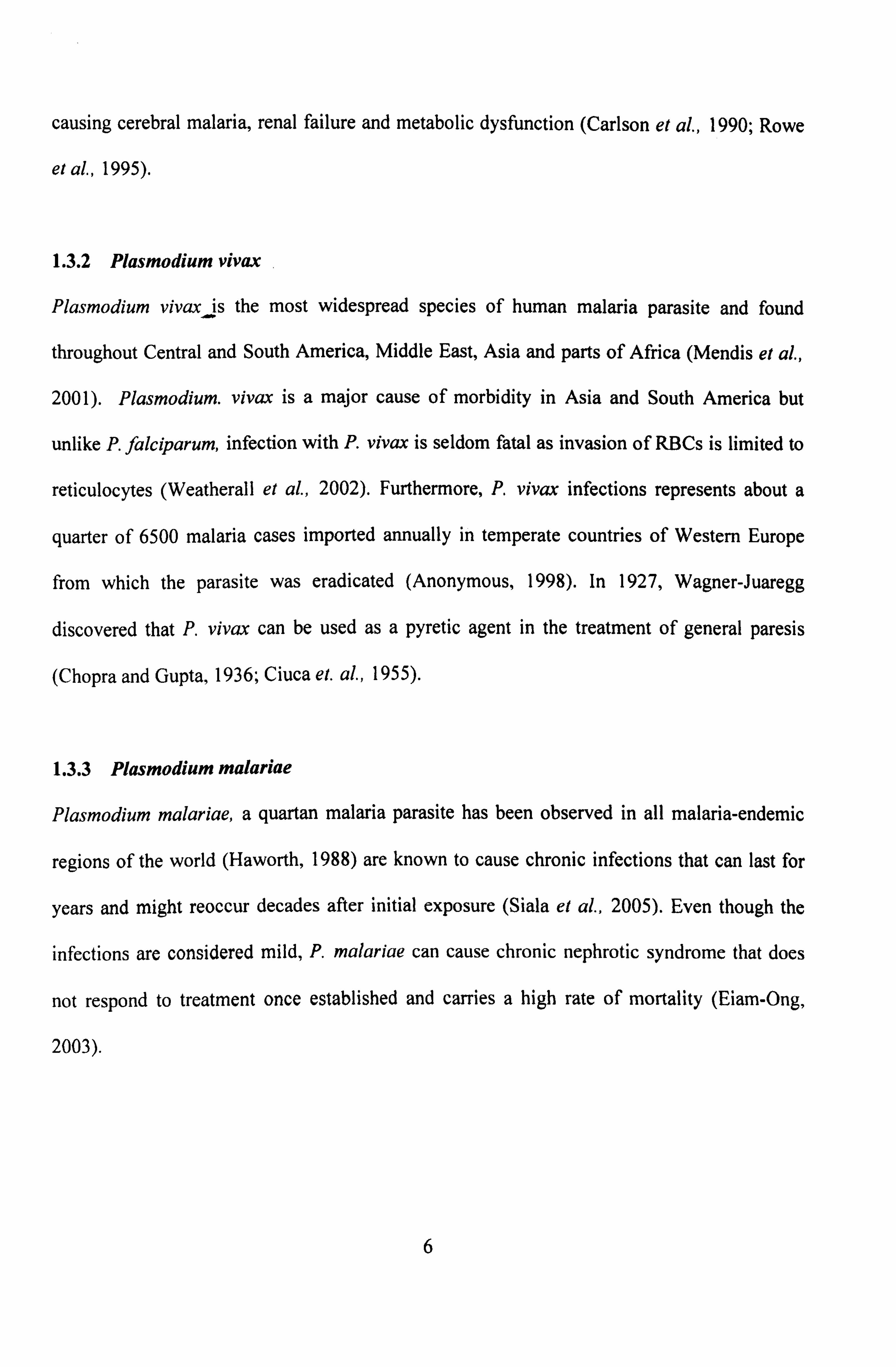
In the human host, the asexual stage of malaria life cycle begins with the exoerythrocytic
phase. When a Plasmodium infected female mosquito feeds on a human, sporozoites in the
saliva of the mosquito are injected into the bloodstream (Figure 1.1). Within 30-60 minutes,
the sporozoites then travel to the liver, where they pass through Kuppfer cells and invade
hepatocytes. The Kuppfer cells destroy most of the sporozoites in the bloodstream with only a
fraction manages to avoid the destruction by entering the hepatocyte. Then, the sporozoite
undergoes asexual replication known as pre-erythrocytic schizogony within the hepatocytes
over a period of approximately 4 weeks where the sporozoites develop into schizonts. In P.
vivax and P. ovale infections, schizonts can remain dormant as hypnozoites for weeks and
even years before causing any clinical relapse (Suh el al., 2004).
For P. , falciparum, the incubation period in the liver is 18 days, 14 days for P. ovale, 26 days
for short term incubation period and 48 weeks for long term incubation period for P. vivax, 28
2

Human Liver Stages
liver tell .ý r',
Infette0
r"ý :. ý" "". } Iner Ceq
. 7_ý "ý
`P °r r:
ý
.ý
Li1.
1 j
"+ Human Blood Slags
---`
®aaknete
ý r+' MKtogamele enlerkq
'ý. °ý°ýý ° ýº ExlepeUated rncropsmeaocyle
A= Infective Stage
A- Diagnostic Stags
. d_ --A4:. ý~"ý"rýr 'ý- ý' Gemawcyiea
Figure 1.1 Life cycle of malaria parasite (Source: Centre for Disease Control, CDC)
13 EwO+ryüvorytie Cycle
ý 1171RIOYHII!
lropholoAs (rwv suge)
U-3 EryUwOý, 'ytiC Cyd.
,.. ._ý . ý''". 'ý!
-- .... w tbkwwi, m a di eb ý ý_ ý. I
RuDWrrdW** ý
fthisOM ýýý*" ý 00 3chýzont e
cän, eoocycs8©
0
r. 1l4 i . "/ý
3

days for P. malariae and P. knowlesi has the shortest incubation period with 7 days (Nishiura
et al., 2007; Collins et al., 2005; Tuteja et al., 2007; Garnham, 1966; Wiser,
http: //www. tulane. edu/-wiser/malaria/). Each liver schizont contains tens of thousands of
merozoites with each able to invade an erythrocyte in the bloodstream upon release from the
liver (erythrocytic schizogony). Disease begins only once the asexual parasite multiplies
within the erythrocyte (Weatherall, 2002).
Once the merozoite enters the erythrocyte, it develops into an early trophozoite or often
referred as ring form due to its morphology. The maturation of the trophozoite and asexual
multiplication into a schizont is accompanied by an active metabolism including ingestion of
host cytoplasm and proteolysis of hemoglobin into amino acids (Garnham, 1977). The
schizont then replicates to produce 16-32 daughter merozoites inside the erythrocyte. Once
mature, it will subsequently rupture, thus releasing merozoites into the bloodstream to invade
new erythrocytes and reinitiate the erythrocytic cycle. When erythrocytes rupture, parasite
proteins and metabolites are released, resulting in clinical symptoms such as fever and chills
(Knell, 1991). Malaria parasites exhibit similar life cycle with only minor variations in terms
of length of time required to complete the erythrocytic cycle depending on the species. P.
malariae has the longest period which is 72 hours (quartan periodicity), P. falciparum, P.
vivax and P. ovale are 48 hours (tertian periodicity), whereas P. knowlesi only requires 24
hours (quotidian periodicity) to complete the erythrocytic cycle (Coatney et al., 1971).
In the blood, as an alternative to the asexual replicative cycle, some of the merozoites
differentiate into non-multiplying sexual forms known as macrogametocytes (female) and
microgametocytes (male) inside the erythrocyte without rupturing them. The gametocytes are
4

Pusat Khidmat Makiumat Akadeuºik UNIVERSITI MALAYSIA SARAWAK
crucial for the next part of the life cycle as the erythrocytes that contain gametocytes are
ingested by a feeding Anopheles mosquito during blood a meal. Ingestion of the gametocytes
induces gametogenesis and escape from the host erythrocyte. During gametogenesis, the
macrogametocytes form macrogametes while microgametocytes exflagellate and develops
into eight haploid motilgmicrogametes. The highly motile microgamete will seek out and fuse
with a macrogamete (sexual multiplication) resulting in a diploid zygote. The zygote then
develops into an ookinete that penetrates the extracellular space between the midgut epithelial
cells. The ookinete later matures into an oocyst and undergoes multiple rounds of asexual
multiplication, called sporogony which culminates in the production of sporozoites. Upon
maturation, the oocyst ruptures, consequently releasing the sporozoites into haemocoel of the
mosquito. The sporozoites migrate to the salivary gland of the mosquito, ready to initiate
another life cycle into another vertebrate host during a blood meal.
1.3 Epidemiology of Plasmodium species causing malaria in humans
1.3.1 Plasmodiumfalciparum
Plasmodium falciparum is the most virulent malaria parasite. The high multiplication rate of
the blood stages and the adhesive properties of the infected RBCs contribute to virulence
(MacPherson et al., 1985). P. falciparum modifies the surface of the RBCs for adherence of
both asexual parasites and gametocytes to the endotheliums and asexual parasites within
placenta. Thus, only ring forms of P. falciparum are found circulating within the blood vessels
(Baruch, 1999; Newbold et al., 1999; Chen el al., 2000). The adherence protects the parasites
from destruction as the non-adherent mature parasitized RBCs are rapidly cleared within the
spleen (Langreth and Peterson, 1985). The sequestration of P. falciparum-infected RBCs in
the peripheral circulation of vital organs, consequently resulting in dysfunction of the organs
5
causing cerebral malaria, renal failure and metabolic dysfunction (Carlson et at, 1990; Rowe
et al., 1995).
1.3.2 Plasmodium vivax
Plasmodium vivax is the most widespread species of human malaria parasite and found
throughout Central and South America, Middle East, Asia and parts of Africa (Mendis et al.,
2001). Plasmodium. vivax is a major cause of morbidity in Asia and South America but
unlike P. falciparum, infection with P. vivax is seldom fatal as invasion of RBCs is limited to
reticulocytes (Weatherall et al., 2002). Furthermore, P. vivax infections represents about a
quarter of 6500 malaria cases imported annually in temperate countries of Western Europe
from which the parasite was eradicated (Anonymous, 1998). In 1927, Wagner-Juaregg
discovered that P. vivax can be used as a pyretic agent in the treatment of general paresis
(Chopra and Gupta, 1936; Ciuca et. al., 1955).
1.3.3 Plasmodium malariae
Plasmodium malariae, a quartan malaria parasite has been observed in all malaria-endemic
regions of the world (Haworth, 1988) are known to cause chronic infections that can last for
years and might reoccur decades after initial exposure (Siala el al., 2005). Even though the
infections are considered mild, P. malariae can cause chronic nephrotic syndrome that does
not respond to treatment once established and carries a high rate of mortality (Eiam-Ong,
2003).
6

1.3.4 Plasmodium ovale
Plasmodium ovale has limited distribution as compared to P. malariae with endemic
transmission being limited to areas of tropical Africa, New Guinea, Middle East, Indian
subcontinent and different parts of Southeast Asia (Baird et al., 1990; Kawamoto et al., 1999;
Win et al., 2002). Humans are the only natural hosts for P. ovale (Collins et al., 2005). And
until now, only two strains of P. ovale are known; the Donaldson and Liberian strain. The
Donaldson strain was used in malaria therapy for the treatment of patients with neurosyphilis
(Jeffery, 1954; Jeffery and Young, 1954; Jeffery et al., 1954). Compared to patients infected
with P. falciparum or P. vivax, the parasite counts are usually low (Christophers, 1934).
Previous infection with P. ovale does not prevent reinfection but results in reduced levels of
parasitaemia and fever whereas previous infection with P. falciparum, P. vivax or P. malariae
does not prevent P. ovale infection but the frequency and the intensity of the fever and
parasite counts are reduced (Collins et al., 2005). Plasmodium, ovale is a relapsing infection
and can be generated from latent parasites in the liver. The relapse occurs as early as 17 days
after treatment of the primary attack to as late as 255 days (Chin and Coatney, 1971).
1.3.5 Plasmodium knowlesi
Plasmodium knowlesi was first discovered by Napier and Campbell in 1931 in the blood of a
long-tailed macaque (Macaca fascicularis) imported from Singapore, when they were
working in India (Gamham, 1966; Coatney et at, 1971). When Campbell drew the macaque's
blood and inoculated it into three rhesus macaques (M. mulatta), the macaques developed a
fulminating infection (Sinton and Mulligan, 1932). The following year, Knowles and Das
Gupta injected blood from the original long-tailed macaque containing the parasites into
rhesus and long tailed macaques. They found that the parasite caused lethal infection in rhesus
7

macaques although it only caused chronic infections in long-tailed macaque (Knowles and
Das Gupta, 1932). They also found that the parasite is infectious to humans when the two
human patients developed malaria with a 24-hour fever peak when injected with blood from
the infected long-tailed macaques. P. knowlesi was later used as a pyretic agent to treat
patients with neuro yphilis (Chopra & Das Gupta, 1936).
The natural hosts for P. knowlesi are the long-tailed macaques, pig-tailed macaques (M
nemestrina) and silver leaf macaques (Trachypithecus cristatus). These macaques are
naturally found widespread in Asia, particularly throughout the islands of South East Asia and
also mainland Asia. The macaques were found to be infected with P. knowlesi in Peninsular
Malaysia (Fong et al., 1971) and Thailand (Jongwutiwes et al., 2004). Research done on these
early macaques noted that P. knowlesi and P. malariae was morphologically similar
(Garnham, 1966).
The first case of a human with naturally acquired P. knowlesi was reported in 1965 by Chin et
al. They described an American surveyor who became ill upon return to the United States of
America from working in Pahang, Malaysia. Initially, his infection was diagnosed as P.
falciparum, due to only ring forms being observed during examination by microscopy. The
following day, `band forms' trophozoites were observed and the diagnosis was changed to P.
malariae. His blood was inoculated into human volunteers at Georgia State Penitentiary,
Atlanta and the human volunteers developed malaria with 24-hour fever peak which was
unexpected as P. malariae patients would produce a 72-hour fever peak (Coatney et al.,
1971). Blood was injected into rhesus macaques and later found dead, confirming that the
infection was indeed P. knowlesi (Chin et al., 1965). Following this, there was a second report
8