chapter one - princeton university press home pageassets.press.princeton.edu/chapters/s7313.pdf ·...
TRANSCRIPT
Chapter One
THE STRUCTURE OF BONE TISSUE
THROUGHOUT this book I shall be suggesting that the struc-ture of bone tissue, and of whole bones, makes sense only if itsfunction, particularly its mechanical function, is known or
guessed. (As Rik Huiskes of Eindhoven is fond of saying [2000]: “Ifbone is the answer, then what is the question?”) However, in this firstchapter I shall deal only with the structure of bone, leaving almost alldiscussion of function until later. Of course, the mechanical propertiesof bone and bones are determined by their structure, and we cannotbegin to understand the function without having a good idea of thestructure. Much of the subject matter will be familiar to some readers,but not all to everyone. Indeed, some readers may be coming to bonefor the first time, from say materials science, so I shall start with a singleparagraph overview of bone structure.
Bone of present-day mammals and birds is a stiff skeletal materialmade principally of the fibrous protein collagen, impregnated with amineral closely resembling calcium phosphate. Bone also contains wa-ter, which is very important mechanically. Bone is produced inside thebody and is usually covered with cells throughout life, though in fishscales, for instance, the external lining of cells may be rubbed off. Mostbone not only is covered by cells but has living cells and blood vesselswithin it. Bone, being hard, cannot swell or shrink; all changes in shapemust take place at surfaces. Most bones are hollow and contain hema-topoietic or fatty marrow. Marrow probably has little mechanical sig-nificance. Tendons and ligaments insert into the bone substance, and theends of bones are often covered by a thin layer of cartilage for lubrica-tion. Some tissues, such as antler and dentin, are not called bone but areactually bone, or extremely like it. Horn, such as is found in cattle, is acompletely different material, usually unmineralized, though the horncore, which supports the horn, is made of bone.
To start straight off talking about the structure of bone begs the ques-tion. It is not really at all clear what bone is. Consideration of a present-day mammal or bird would allow a clear distinction to be made, be-cause bone is the only structure that is essentially collagen mineralizedwith calcium phosphate and containing cell bodies, though in antlerbone the cells are all dead by the time the antler comes to be used.Dentin is collagen mineralized with calcium phosphate but it does not

4 C H A P T E R 1 . T H E S T R U C T U R E O F B O N E T I S S U E
contain cell bodies, only tubular extensions of cells. The other signifi-cant tissue mineralized with calcium phosphate (as opposed to calciumcarbonate) is enamel, and this is very different in that it has virtually nocells or cell processes or, indeed, much organic matrix. However, assoon as one looks outside the mammals and birds the situation becomesmuch more complex. Bone is found only in vertebrates. Many teleostfish have bone without bone cells, and the range of structures seen inscales of different fish species forms an almost complete spectrum fromwhat is obviously “typical” bone or “‘typical” dentin to what is obvi-ously “typical” enamel. This situation is often found in biology, sincenature is concerned not with categorization, but with producing effec-tive results. (The great evolutionary biologist John Maynard Smith likessaying that “all biology is false,” meaning, of course, that there are veryfew absolutes in biology.)
For the purposes of this book, the fact that there are so many differ-ent types of scales does not matter greatly because virtually nothing isknown about the mechanical properties of scales. I shall be concernedalmost entirely with the mechanics of typical bone, as found in mam-mals, but I shall devote some space to tissues such as dentin. Antler,which is dead when functioning, will figure prominently. A good ac-count of the variation of structure in vertebrates is found in Francillon-Vieillot et al. (1990), which, though not an enthralling read, is veryclear, comprehensive, and well illustrated.
Even “typical” bone is such a complex structure that there is no levelof organization at which one can truly be said to be looking at bone assuch. I shall start at the lowest level and work up to a brief descriptionof the variety of shapes one sees in whole bones.
1.1 Bone at the Molecular Level
At the lowest level bone can be considered to be a composite materialconsisting of a fibrous protein, collagen, stiffened by an extremely densefilling and surrounding of calcium phosphate crystals. There are otherconstituents, notably water, some ill-understood proteins and polysac-charides, and, in many types of bone, living cells and blood vessels. Theamount of water present in bone is an important determinant of itsmechanical behavior, and I shall say more about it in chapter 4.
A word about terminology. In this book I use the word matrix tomean the water and the soft organic material, mostly collagen, in whichthe mineral crystals are deposited. This accords with what materialsscientists would consider to be the matrix (though some might considerthe mineral to be the matrix). However, bone biologists, who are fo-

1 . 1 B O N E A T T H E M O L E C U L A R L E V E L 5
cused almost entirely on the cells of bone, use the word matrix to meanthe bone tissue itself, that is, the water, the organic material, and themineral. There is no way round this possible source of confusion; onesimply has to be aware of it.
Collagen is a structural protein found in probably all metazoan ani-mal phyla. It is the most abundant protein found in animals, but only inthe vertebrates does it undergo a wholehearted transformation into amineralized skeletal structure, although some soft corals have traveledsome way along the road. A classified bibliography of more than 3400references to collagen, comprehensive up to that time, is given in Kadler(1994).
Unmineralized collagen is also found in the vertebrates, and in manyinvertebrates, in skin, tendon, ligament, blood vessel walls, cartilage,basement membrane, and in connective tissue generally, in those cir-cumstances where the material is required to be flexible but not veryextensible. Collagen makes up more than half the protein in the humanbody (Miller 1984). Collagen from different sites often has differentamino acid compositions; in the mid 1990s 19 types of collagen wereknown throughout the animal kingdom, and the known number in-creases relentlessly (Prockop and Kivirikko 1995). The collagens ofskin, tendon, dentin, and bone share the same type of composition, andare called type 1 collagen. The protein molecule tropocollagen, whichaggregates to form the microfibrils of collagen, consists of three poly-peptides of the same length—two have the same amino acid composi-tion, one a different one. These form on ribosomes, are connected bymeans of disulfide cysteine links, and leave the cell. Outside the cell theends of the joined polypeptides are snipped off, the lost part containingthe disulfide bonds. The three chains are by now held together by hy-drogen bonds in a characteristic left-handed triple helix.
The primary structure of the polypeptides in the tropocollagen mole-cule is unusual, great stretches of it being repeats of glycine–X–Y, withX often being proline and Y sometimes hydroxyproline. The iminoacids proline and hydroxyproline are unlike amino acids in that thenitrogen atom is included in the side chain as part of a five-memberedring. The effect of this is to reduce the amount of rotation possiblebetween units of the polypeptide. It also prevents α-helix formation andlimits hydrogen-bond formation. These constraints result in a rather in-flexible polypeptide, 300 nm long (Olsen and Ninomiya 1993).
The tropocollagen molecules line up in files and bond, not with mole-cules in the same file, but with molecules in neighboring files, to formmicrofibrils. The tropocollagen molecules alongside each other are stag-gered by about one-fourth of their length. There is a gap between thehead of one molecule and the tail of the next in the file, the hole region,

6 C H A P T E R 1 . T H E S T R U C T U R E O F B O N E T I S S U E
and, because many tropocollagen molecules are stacked side by side,these gaps and other features of the molecules produce a characteristicperiodicity, 67 nm long. The whole microfibril becomes stabilized byintermolecular cross-links. Microfibrils aggregate to form fibrils. Al-though the longitudinal arrangement of the tropocollagen molecules inthe microfibrils is fairly well understood, the way in which the micro-fibrils themselves are arranged laterally to form fibrils is much less wellunderstood. A clear introduction to the subject is provided by Prockopand Fertala (1998). Hulmes et al. (1995) produce evidence that the fi-brils are arranged in concentric rings. Wess et al. (1998a,b) produce arather different model that they claim will explain the way in whichmineral is able to pack in bone. This is a difficult subject, with themajority view changing often as experimentation becomes ever moresophisticated. It could well be that when a stable view is formed theresults will be useful in helping to model the mechanical behavior ofbone, but at the moment this is not really the case.
Collagen comprises about 85 to 90% of the protein in bone. Theproteins that are not collagen are called, negatively, noncollagenousproteins (NCPs). The literature on them is vast and expanding rapidly(Ganss et al. 1999; Gerstenfield 1999; Gorski 1998; Nanci 1999). SomeNCPs are restricted to bone, and some are also found in other places inthe body. Some of these proteins almost certainly have a role in theinitiation and control of mineralization or reconstruction, and somemay have a role in binding the collagen and mineral together (Roach1994). However, we are almost completely in the dark at the momentabout any quantitative effect NCPs may have on the mechanical proper-ties of bone.
Impregnating and surrounding the collagen is the bone mineral,which is some variety of calcium phosphate. The precise nature of themineral of bone, both its chemistry and its morphology, is still a matterof some dispute. The problem is that the mineral in bone comes in verysmall crystals that have a very high surface-area-to-volume ratio. Thesize of the crystal is such that in one dimension it is only about 10atomic layers thick (Lowenstam and Weiner 1989). This makes it reac-tive, and so most preparative techniques used for investigating it, suchas, drying under vacuum for electron microscopy, may cause alterationsfrom the living state. There is agreement that some of the bone mineralis the version of calcium phosphate called hydroxyapatite, whose unitcell (the smallest part of a crystal that is repeated uniformly throughouta crystal) contains Ca10(PO4)6(OH)2. The crystals are impure. In partic-ular, there is about 4–6% of carbonate replacing the phosphate groups,making the mineral more truly a carbonate apatite (dahllite). This car-bonate substitution takes place more near edges of the bone, close to

1 . 1 B O N E A T T H E M O L E C U L A R L E V E L 7
vascular and marrow spaces and tends to reduce the crystallinity of thecrystals (Ou-Yang et al. 2001). Various other substitutions may takeplace (Boyde and Jones 1998; McConnell 1962).
At the moment, we are ignorant of the mechanical properties of themineral itself, and all modeling, such as that of Wagner and Weiner(1992) and Sasaki et al. (1991), which I discuss in section 3.7, makesuse of somewhat insecurely based (though not necessarily far wrong!)estimates. The mineral is certainly stiff, but its strength, in such smallblocks, is unknown.
The positioning of the mineral relative to the collagen fibrils, as wellas its shape, is becoming clearer, though there is still controversy. Thereis some argument as to whether the crystalline mineral, which can beseen in electron micrographs, is needle-shaped or plate-shaped. Ascenziet al. (1978) claimed that the mineralization process starts off withsmall granules, about 4.5 nm across, which coalesce or grow into nee-dles about 40 nm long. However, the observations of Landis and his co-workers make it almost certain that in mineralized tendon (Landis et al.1993) and in embryonic chick bone (Landis et al. 1996) the crystals areplatelet-shaped. They have used the technique of taking multiple viewsof bone using high-voltage electron microscopy to produce a tomo-graphic image. This method shows very clearly the three-dimensionalshape of the crystals and to some extent their spatial relationship to thecollagen (fig. 1.1). These visualizations show that the crystals’ thicknessis rather unvarying at about 4–6 nm, their width is about 30–45 nm,and their length is typically 100 nm. Later, these mineral platelets seemto fuse sideways, and lengthways, producing at times sword-shapedblades that are quite long and broad. However, they do not seem togrow in the depth direction, remaining about 5 nm deep. Erts et al.(1994), using scanning probe microscopy, found similar values for tur-key tendon.
Reports of the visualization of the crystals directly overwhelminglysupports this view that the crystals in all bone examined are platelet-shaped. Weiner and Price (1986) examined the size of bone mineralcrystals, extracting them from the bone by a gentle procedure, and pro-posed values of about 50 � 20 � 2 nm. Kim et al. (1995) report platelet-shaped crystals from tissues of a taxonomically satisfyingly variedgroup of species: chickens, bovines, mice, and herring. The averagelength and breadth, in nanometers, for the four species are given inTable 1.1. Kim et al. did not measure the thickness, but suggested it wasabout 2 nm. Ziv and Weiner (1994) suggest that most estimates of thesize of crystals are underestimates, because the plates are so fragile, andthat crystals may be often hundreds of nanometers long in untreatedbone.

8 C H A P T E R 1 . T H E S T R U C T U R E O F B O N E T I S S U E
FIG. 1.1 Diagram of mineralizing turkey leg tendon according to the microtomographicinvestigations of Landis. Mineralization is proceeding from the top down. The crystalsare platelet-shaped, and are initially registered in line with the hole region of the fibrils(�67 nm apart). This initial relationship between the platelets and the collagen fibrils isshown in the right-hand part of the diagram. Toward the top of the diagram some plate-lets are shown as having fused longitudinally. (From Landis, W. J., Hodgens, K. J.,Arena, J., Song, M. J., and McEwen, B. F. 1996. Structural relations between collagenand mineral in bone as determined by high voltage electron microscopic tomography.Microscopy research and technique 33:192–202. Reprinted by permission of Wiley–Liss,Inc., a subsidiary of John Wiley & Sons, Inc.)
Fratzl et al. (1992) have produced indirect evidence, using small-angleX-ray scattering, that the crystals in ossified tendon are indeed platelet-shaped but that in ordinary compact bone they are more likely to beneedle-shaped. On the other hand, Wachtel and Weiner (1994) showthat the small-angle X-ray scattering picture from crystals from rat boneis very similar to that from mineralizing turkey tendon, and suggest thatit is probably reasonably safe to generalize about the crystal morphol-ogy from mineralizing turkey tendon.

1 . 1 B O N E A T T H E M O L E C U L A R L E V E L 9
Table 1.1Length and Breadth of Mineral Crystals (in nm) in the Bone of Four Species
Species Length Breadth
Chicken 23.3 12.2Bovine 27.3 15.8Mouse 21.2 12.0Herring 37.3 15.4
Source. From Kim et al. (1995).
More contentious is where the mineral is in relation to the collagenfibrils. For years, following a suggestion by Hodge and Petruska (1963),it was thought that the mineral is initially deposited in the holes be-tween the heads and the tails of the tropocollagen molecules (the gapzones). This results in the initial mineralization having a 67-nm peri-odicity (Berthet-Colominas et al. 1979). Many studies seemed to con-firm this, but nearly all were carried out on mineralizing turkey legtendon, which, although very convenient to study because the collagenfibrils are so well arranged, is not typical bone, particularly in relationto the arrangement of the crystals (Wenk and Heidelbach 1999). It isprobable that in some way the particular conformation of the collagenmolecule allows it to act as a nucleation site, permitting the precipita-tion of lumps of mineral that, without the presence of the energeticallyfavorable sites, could not come out of solution. There is some evidencethat the mineral deposits preferentially in parts of the fibril that are highin hydrophilic residues (Maitland and Arsenault 1991). Later, the min-eral is deposited all over the collagen fibrils, and also within them.Weiner and Traub (1986) have published stereopairs of mineralizingturkey leg tendon, showing how the crystals lie within the fibers. Landiset al. (1993, 1996) show similar pictures (fig. 1.1), and point out thatthe individual platelets seem to remain separated by a space in the depthdirection of about 5 nm. This would, of course, allow collagen micro-fibrils to exist between the platelets. Jager and Fratzl (2000) suggest,though with no observations to back the suggestion up, that the crystalsmay be arranged circumferentially round the center of the fibril. Thiswould accord with the radial fibril model of Hulmes et al. (1995).
All these observations relate to early stages of mineralization, andit is much less obvious what happens when mineralization has proceed-ed to its full extent. There must be considerable derangement of theinitially very uniform collagen structure. It is strange that some quitemodern works (e.g., An [2000], table 3.1) still hold to the idea thatthe only place that the mineral resides in mature bone is in the gap

10 C H A P T E R 1 . T H E S T R U C T U R E O F B O N E T I S S U E
region between the ends of adjoining collagen molecules. This idea isquite wrong.
Much work has been done on the mineralizing turkey leg tendon,because, although it is called tendon, it is actually proper bone andmineralizes in a particularly regular way, so that there is a close rela-tionship along the tendon between distance and the progression of min-eralization. This makes it very convenient for studies of mineralization.However, most bone mineralizes on surfaces, rather than at the end oftendons. On surfaces what happens is that a matrix of collagen, plus afew other organic components, is laid down first. This organic materialis called osteoid. Mineral is deposited in the collagen, initially, it seems,in the gap zones, but then, unlike the situation in tendon, all along thelength of the collagen fibrils. The plate like mineral crystals as theygrow tend to form quite large lumps, and the individual mineral crystalstend to be oriented in the direction of the collagen fibrils. Within thecollagen fibrils the precipitation is not random; one of the long axes ofthe mineral plates is always fairly well aligned with the collagen fibrils.Also, later, mineral is deposited between the fibrils, in the amorphousand rather tenuous ground substance. The relative amount depositedwithin and between the collagen is quite hotly argued even now, withsome people proposing that more is between than within (Bonar et al.1985; Lees et al. 1990; Pidaparti et al. 1996). Furthermore, it is likely,certainly in mineralized tendon and possibly ordinary bone, that themineral outside the fibrils may have a different orientation from thatwithin the fibrils (Lees et al. 1994; Pidaparti et al. 1996). In some fishbone, however, the vast majority of the mineral is within the fibrils, andnot between them (Lee and Glimcher 1991). Knowing the truth of thesematters is important for understanding why bone behaves mechanicallyas it does.
Added to our ignorance of the disposition of the mineral in bone isour ignorance of how, and the extent to which, the collagen and themineral are bound together. The relationship between those mineralcrystals that are inside the collagen fibrils and the collagen is extremelyintimate, and such short-range forces as van der Waals forces may wellbe important. Also, ionic bonding probably occurs, which I discuss insection 3.7.
I am conscious, in reviewing the last few pages, of how often I havesaid that we are ignorant of the true situation. I suspect that much ofour ignorance will be removed in the next decade. It will then becomepossible to try to understand in detail how collagen/mineral interactionsdetermine the mechanical properties of bone at the molecular level, inthe same way that metallurgists have a good idea of how, for instance,steel behaves at this level. However, bone has several levels of structural

1 . 2 T H E C E L L S O F B O N E 11
hierarchy above the molecular level, and these all have important effectson the mechanics of bone, as we shall see.
1.2 The Cells of Bone
Bone is permeated by and lined by various kinds of specialized cells,which will be introduced later. I here list them and briefly describe theirproperties.
Bone-lining cells cover all surfaces of bones, including the bloodchannels, forming a thin continuous sheet that controls the movementof ions between the body and the bone (Miller et al. 1988). The layer ofcells on the outside of the bone is called the periosteum, although thisword is often used to include the strong collagenous sheet covering theouter surface. The layer of cells on the inside of the bone is called theendosteum. The bone-lining cells, which are often considered to be qui-escent osteoblasts, are derived, via complex series of changes, from os-teoprogenitor cells. These stages are described by Lian and Stein (1996).
Osteoblasts derive from bone-lining cells and are responsible for theformation of bone. They initially lay down the collagenous matrix, os-teoid, in which mineral is later deposited, and they probably also have arole in its mineralization.
Osteocytes are the cells in the body of the bone. In cancellous bonethe density of osteocytes varies from about 90,000 mm�3 in rats toabout 30,000 mm�3 in cows. In general, the larger the animal the lowerthe density of osteocytes (Mullender et al. 1996). They derive from os-teoblasts. They are imprisoned in the hard bone tissue and connect withneighboring osteocytes and with bone-lining cells by means of processesthat are housed in little channels (canaliculi), of about 0.2–.03 µm di-ameter (Cooper et al. 1966). The actual connections with neighboringcells are by means of gap junctions that allow small molecules througheasily.
Osteoclasts are bone-destroying cells. They are large, multinucleatedcells derived from precursor cells circulating in the blood. In time-lapsephotography they give the appearance of being extremely aggressive,clamping themselves to the bone’s surface and leaving a space under-neath a ruffled border that is very mobile and beneath which the bonecan be seen dissolving. Debris, both organic and mineral, are packedinto little vesicles and pass through the cell body of the osteoclast andare dumped into the space above (Nesbitt and Morton 1997; Salo et al.1997). When osteoclasts have done their job they disappear and pre-sumably die. (A colleague of mine, who initially studied osteoclasts, saidthat for some strange reason most people who studied them were as

12 C H A P T E R 1 . T H E S T R U C T U R E O F B O N E T I S S U E
aggressive as their subjects. My colleague eventually gave up, and turnedto studying benign osteoblasts!)
1.3 Woven and Lamellar Bone
Above the level of the collagen fibril and its associated mineral, mam-malian bone exists in two usually fairly distinct forms: woven bone andlamellar bone. Parallel-fibered bone is intermediate.
Woven bone is usually laid down very quickly, more than 4 µm a dayand often much more, most characteristically in the fetus and in thecallus that is produced during fracture repair. The collagen in wovenbone is variable, the fibrils being 0.1–3 µm or so in diameter and ori-ented almost randomly, so it is difficult to make out any preferred direc-tion over distances greater than about a millimeter (Boyde 1980; Boydeand Jones 1998). The mineralization process involves roughly sphericalcenters, impregnating both the collagen and ground substance at thesame time, in which the crystals seem to be randomly arranged. Asthese mineralization centers spread they abut and often leave mineral-free spaces (Boyde 1980). As a result, woven bone, though highly min-eralized, is often quite porous at the micron level. As in most bone,woven bone contains cells (osteocytes) and blood vessels. Rather fre-quently, the spaces surrounding the osteocytes are extensive and differin this way from those in lamellar bone. There are of the order of 60canaliculi per osteocyte (Boyde 1972), though no doubt this numbervaries greatly between osteocytes and between species. “Woven” bone isa misnomer, because there are very few examples of weaving, that is,true interlacing, in biology. (It would be a trick almost impossible tobring off, though, surprisingly, the enamel of some rodents has some-thing almost as good—structures arranged in three orthogonal direc-tions, see Section 6.3.)
Lamellar bone is more precisely arranged, and is laid down muchmore slowly than woven bone, less than 1 µm a day (Boyde 1980). Thecollagen fibrils and their associated mineral are arranged in sheets(lamellae), which often appear to alternate in thickness. The final degreeof mineralization of lamellar bone is less than that of woven bone. Theclassical view is that the fibrils lie within the plane of the lamella, rarelypassing from one to the next and that the fibrils tend to be oriented inone direction within the lamella. Indeed, some workers suggest that thecollagen fibrils in a particular lamella are all oriented in the same direc-tion (Ascenzi et al. 1978). However, this is probably not the case; inmany lamellae the fibrils are in small domains about 30–100 µm across.Within a domain the fibril orientation is constant, but it changes, within

1 . 3 W O V E N A N D L A M E L L A R B O N E 13
one lamella, from one domain to the next (Boyde and Hobdell 1969;Frasca et al. 1977). The collagen fibrils in lamellar bone form branchingbundles, 2–3 µm in diameter (Boyde 1980), thicker than in most wovenbone. The osteocyte lacunae in lamellar bone are oblate spheroids, theequatorial diameters being about five times longer than the polar axis.The shorter axis of each lacuna is oriented parallel to the direction ofthe thickness of the lamella.
The division between one lamella and the next looks abrupt underthe light microscope, particularly under polarized light. However, scan-ning electron microscope pictures show a much messier situation. Fre-quently, lamellae seem to come in alternating thicknesses. There is arelatively thick one, about 5 µm thick, whose mineral crystals areroughly arranged all in one direction, over small distances at least, at anangle both to the long axis of the bone and to the circumferential exten-sion of the lamella itself The thin lamella is about 1 µm thick, and hasmineral plates oriented in the plane of the circumferential extension ofthe lamellae, with their smallest dimension normal to the long axis ofthe bone. This kind of lamellar organization is held to occur in mice byWeiner et al. (1991), and from my observations of fracture surfaces ofbone of a large number of species I think this arrangement is wide-spread. If the mineral is arranged like this, presumably the collagen fi-brils also lie in the same direction. As we shall see in the next chapter,such changes in direction have mechanical consequences. There is dis-agreement at the moment as to whether there is a difference in the den-sity of mineral in the thick and thin lamellae. Marotti (1993) argues,giving evidence from the appearance of the collagen fibrils, that thethicker lamellae are looser and have more mineral, the thinner lamellaeare somewhat more collagen-rich, and there is not a great deal of differ-ence in the collagen orientation between the different layers. Yamomotoet al. (2000) argue convicingly that, at least in human dentin, the ap-pearance found by Marotti is an artifact and that the differently appearinglamellae are only different in respect of their orientation. The uncer-tainty in the literature may be partially because some people concen-trate on the mineral and some on the collagen.
Giraud-Guille (1988) produces strong evidence that in some lamellaethe collagen fibrils, and presumably, therefore, their associated mineralcrystals, are arranged in a “twisted plywood” or helicoidal structure. Ina helicoidal structure there are many layers, and within a layer the fibersall point in the same direction. There is a change of angle between thelayers, usually quite small, and this is constant in size and sense. Theresult is a multilayer composite with no preferred orientation. Ziv et al.(1996), Weiner et al. (1997), and Weiner and Wagner (1998) producefurther evidence that such continuous transitions between, and even

14 C H A P T E R 1 . T H E S T R U C T U R E O F B O N E T I S S U E
through, apparently discontinuous lamellae are common. The model ofWeiner and his co-workers suggests that collagen fibrils, only 80 nm orso in diameter, are the basic unit. These may lie parallel to each otherthrough the thickness of the lamella, or be at an angle, usually about30�, to their neighbors. Helicoidal arrangements of fibers are found fre-quently in biological tissues, and Neville (1993) has written a wholebook about them. They occur in all sorts of structures, such as insectcuticle and plant cell walls, and are a prime example of the ability oforganisms to produce complex structures by self-assembly, that is tosay, without cells being involved directly in the placing of the succeedinglayers. Helicoidal structures give rise to a confusing artifact if cut andexamined at an angle to the plane of the sheets: The structure appearsto consist of a series of arcs (Kingsmill et al. 1998, fig. 5f), but, in fact,there are no arcs.
Parallel-fibered bone (described by Ascenzi et al. [1967] and by En-low [1969]) is structurally intermediate between woven bone and lamel-lar bone. It is quite highly calcified, but the collagen fiber bundles aremuch more parallel than those in woven bone.
1.4 Fibrolamellar and Haversian Bone
In mammals there are, at higher levels of structure, four main types ofbone. Woven bone can extend uniformly for many millimeters in alldirections. Such a large block is found only in very young bone (ofrather large mammals) and in large fracture calluses. Lamellar bonemay also occupy quite large volumes. Usually, in mammals it does so incircumferential lamellae, initially wrapped around the outside or theinner cavity of bones. There are blood channels in such bone, but theydo not much disturb the general arrangement of the lamellae.
Lamellar bone also exists in a quite separate form: Haversian sys-tems, or secondary osteons. The British and other Europeans are in-clined to use “Haversian systems,” whereas Americans prefer “second-ary osteons.” It does not matter which is used, except that it is criticalto distinguish primary osteons, which I shall describe in a moment,from secondary osteons. Haversian systems form like this: many bone-destroying cells, called osteoclasts, move forward in a concerted attackon the bone tissue. They form a so-called cutting cone, shaped, as Mar-tin and Burr (1989) say in an excellent account of the process, like “halfan eggshell which is about 200 µm in diameter 300 µm long.” Osteo-clasts are not derived from cells that occur locally, but instead comefrom cells circulating in the blood. As the cutting cone advances itleaves a cylindrical cavity of diameter about 200 µm behind. Almost as

1 . 4 F I B R O L A M E L L A R A N D H AV E R S I A N B O N E 15
FIG. 1.2 Diagram of a forming secondary osteon (Haversian system) in lon-gitudinal and cross-sectional views. The system is extending toward the left.The times give, very roughly, the time course of the process in humans. At 5days the osteoclasts are still widening the cavity in the bone. At 3 weeks thecavity it at its widest. By 6 weeks the cavity is half filled in by osteoblasts,and by 10 weeks or so the process is completed, although it will take a longtime for the bone to become completely mineralized. In the cross sectionsthe central cavity is shown black.
soon as the cavity forms, it begins to fill in (Parfitt 1994). The walls ofthe cavity are made smooth, and bone is deposited on the internal sur-face in concentric lamellae (fig. 1.2). The end result is arranged like aleek Allium porrum, with usually clearly distinguishable cylindricallayers, except that there is a central cavity in the Haversian system,which contains one, or sometimes two, blood vessels, and nerves (Mar-otti and Zallone 1980). In humans the whole process from initiation ofthe osteoclastic activity to the completion of the filling in takes about 2–4 months.
The Haversian system is the classic result of the process of remodel-ing. There are two ways in which new bone may appear. In modelingthe gross shape of the bone may be altered; that is, bone may be addedto the periosteal or endosteal surfaces or it may be taken away fromthese surfaces. In remodeling all surfaces of the bone may be affected,including the internal body of the bone, by the formation of Haversiansystems. In remodeling the bone involved is usually a small individual

16 C H A P T E R 1 . T H E S T R U C T U R E O F B O N E T I S S U E
packet called a basic nulticellular unit (BMU), and typically the amountof bone remaining after the remodeling cycle is little changed; new bonehas more or less replaced old bone. This distinction will be seen to beimportant in chapter 11.
There is a type of secondary osteon that is different from the typedescribed above; this is called the drifting osteon. It is found in youngeranimals, in general. After the cavity is formed by the cutting cone, it isnot filled in at once; instead, one side continues to be eroded by osteo-clasts, while the opposite side is filled in by osteoblasts. As a result thecylinder of the osteon drifts sideways through the tissue, and in so do-ing erodes and replaces a great deal of preexisting bony tissue (Roblingand Stout 1999). Nothing is known about the mechanical consequencesof the presence of such secondary osteons.
There is an outer sheath to the Haversian system, called the cementsheath (or line, because it is usually viewed in cross section). This isformed when the cutting cone stops its erosional activity and just beforenew lamellar bone is laid down on the raw surface so formed. Thecomposition of the cement sheath is still controversial. Some, for in-stance, Frasca (1981), propose that it is more highly mineralized thansurrounding bone; others, for instance, Schaffler et al. (1987), proposethat it is less highly mineralized. There is agreement that there is verylittle collagen in it. Schaffler et al. produce evidence that it has moresulfur and less calcium and phosphorus than the neighboring bonelamellae, and is therefore probably less mineralized than them. It maybe that the cement line is simply a very thin layer of osteoid that re-mains when the process of bone erosion is replaced by bone deposition(Martin and Burr 1989). Zhou et al. (1994) suggest that there areglobular accretions (of unknown composition) on the ends of the de-graded collagen fibrils that have partially eroded away, and that theseaccretions act as a bridge between the old degraded collagen fibrils, andthe newly deposited ones. The cement line is also the site of a concen-tration of osteopontin, a bone protein that probably has some role inbone remodeling (Gerstenfield 1999; McKee and Nanci 1996; Terai etal. 1999).
It is a pity that this matter is not settled, because it is very importantto know, for the purpose of understanding bone mechanics, whethercement sheaths are likely to be more or less compliant, and more or lessstrong, than the surrounding lamellae. Some canaliculi cross the cementsheath, so cells outside do have some metabolic connection with theblood vessel in the middle of the Haversian system. This is shown infigure 1.3 in which the blood vessels, osteocytes, and canaliculi are im-pregnated with resin and stand proud when the outer few microns of aspecimen of bone are etched away. The density of canaliculi in bone is

1 . 4 F I B R O L A M E L L A R A N D H AV E R S I A N B O N E 17
FIG. 1.3 Scanning electron micrograph of a resin-impregnated etched piece of humancortical bone. The resin stands proud of the bone. Two vascular channels can be seen (topcenter, bottom right). The lamellation of the bone is made obvious by the differentialerosion produced by the etching acid. Osteocyte lacunae have radiating canaliculi, whichcan be seen passing over places where the lamellae obviously belong to two generations ofosteogenesis, and which therefore have a cement sheath between them (upper left). How-ever, the density of canaliculi crossing the sheath is generally less than the density nearby(bottom left, top right). The resorption and redeposition associated with the bottom rightvascular channel starts at the bottom left of the picture. Width of image �500 �m. (Froma Christmas card kindly sent by Dr. Peter Atkinson.)
high, averaging about 5 canaliculi per hundred square microns in theplane normal to the predominant direction of the canaliculi (Marotti etal. 1995). The canaliculi account for a porosity of about 1.5% (Frost1960). The cell biology of osteoclasis, the process of destruction of bonetissue, was reviewed by Zaidi et al. (1993).
A fourth characteristic type of mammalian bone is known as plexi-form, or laminar, or fibrolamellar bone (Enlow and Brown 1956, 1957,1958; Currey 1960; de Ricqles 1977; Francillon-Vieillot et al. 1990;Stover et al. 1992). It is found particularly in large mammals, whosebones have to grow in diameter rather quickly. Lamellar bone cannot belaid down as fast as woven bone (as I said above, lamellar bone is laiddown at a rate of less than 1 µm a day, while woven bone is laid downat more than 4 µm and, indeed, often much more, a day). If a bone hasto grow in diameter faster than lamellar bone can be laid down, wovenbone must be laid down instead. For reasons that will be discussed in
18 C H A P T E R 1 . T H E S T R U C T U R E O F B O N E T I S S U E
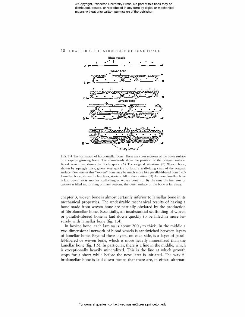
FIG. 1.4 The formation of fibrolamellar bone. These are cross sections of the outer surfaceof a rapidly growing bone. The arrowheads show the position of the original surface.Blood vessels are shown by black spots. (A) The original situation. (B) Woven bone,shown by squiggly lines, grows very quickly to form a scaffolding clear of the originalsurface. (Sometimes this “woven” bone may be much more like parallel-fibered bone.) (C)Lamellar bone, shown by fine lines, starts to fill in the cavities. (D) As more lamellar boneis laid down, so is another scaffolding of woven bone. (E) By the time the first row ofcavities is filled in, forming primary osteons, the outer surface of the bone is far away.
chapter 3, woven bone is almost certainly inferior to lamellar bone in itsmechanical properties. The undesirable mechanical results of having abone made from woven bone are partially obviated by the productionof fibrolamellar bone. Essentially, an insubstantial scaffolding of wovenor parallel-fibered bone is laid down quickly to be filled in more lei-surely with lamellar bone (fig. 1.4).
In bovine bone, each lamina is about 200 µm thick. In the middle atwo-dimensional network of blood vessels is sandwiched between layersof lamellar bone. Beyond these layers, on each side, is a layer of paral-lel-fibered or woven bone, which is more heavily mineralized than thelamellar bone (fig. 1.5). In particular, there is a line in the middle, whichis exceptionally heavily mineralized. This is the line at which growthstops for a short while before the next later is initiated. The way fi-brolamellar bone is laid down means that there are, in effect, alternat-

1 . 4 F I B R O L A M E L L A R A N D H AV E R S I A N B O N E 19
FIG. 1.5 Block diagram of fibrolamellar bone. Conventions concerning his-tological type as in Figure 1.4 Two-dimensional networks of blood chan-nels, sheathed by lamellar bone, alternate with layers of woven bone.
ing layers of parallel-fibered or woven bone and lamellar bone tissueextending, quite often, for many millimeters, or even centimeters, in theradial direction. Fibrolamellar bone can be laid down vey rapidly. Forinstance Castanet et al. (2000) report that the femur of a young emuDromaius novaehollandiae can be added up to 80 µm per day on thesubperiosteal surface.
This description is of particularly neatly arranged laminar bone. Fre-quently the blood channels are more irregularly disposed or do notform a network, and the laminar arrangement gives way to one inwhich the blood vessels anastomose in three dimensions, and each issurrounded by more or less concentric layers of lamellar bone. Thisproduces an appearance somewhat like that of Haversian systems, andthe structures around the blood vessels are called primary osteons.However, there is a most important difference between primary osteonsand secondary osteons, or Haversian systems: Haversian systems aresecondary, that is, they replace bone that has existed previously. Thereare differences that enable one to distinguish Haversian systems fromprimary osteons histologically. In particular, secondary osteons are sur-rounded by a cement sheath, whereas primary osteons are not. Also,secondary osteons appear to drill through the preexisting bone, withoutregard to its structure, whereas the lamellae round primary osteons

20 C H A P T E R 1 . T H E S T R U C T U R E O F B O N E T I S S U E
merge smoothly with the surrounding bone. The distinction between thetwo types of osteon is not mere semantic hairsplitting, because differ-ences between primary osteonal bone and Haversian bone correlatewith differences in mechanical behavior, as we shall see. Haversian boneis weaker than primary bone. The distinction is frequently not made,and is a grave source of muddled thinking.
The kind of primary bone laid down depends on the rate of accretion.Castanet et al. (1996) related the changes in the histology of the bone ofthe mallard duck Anas platyrhyncos to the rate of accretion, which theydetermined by periodic labeling with a fluorescent dye. Different bonesgrow at different rates, and they were able to see the different kinds ofhistology being laid down at the same time, so what they found was notan aging or maturation effect. The humerus grew fastest and, initially,at seven weeks of age, had a rate of accretion of about 25 µm a day; thebone was completely fibrolamellar. As the rate of accretion diminished,the fibrolamellar bone gave way to anastomizing primary osteons. Fi-nally, when the accretion rate was only about 1 µm a day, the bloodvessels were sparse, all parallel to the long axis of the bone, and thebone consisted of circumferential lamellae. The phalanges, on the otherhand, which had a much lower accretion rate right from the start, nevershowed any sign of fibrolamellar histology.
Stover et al. (1992) show that in bone that grows very fast in theradial direction, such as in the young foal, the outer sheet of wovenbone may initially be connected to the rest of the bone by bony struts sosparsely, if at all, as to be effectively lying lie free in the periosteum. Theouter sheet becomes connected to the rest of the bone only when miner-alization is well underway. This process, which they call “saltatory pri-mary osteonal bone formation,” allows growth to be even more rapidthan the process I described above. Indeed, Sue Stover tells me that,very occasionally, two layers of free-floating bone can be seen. However,it is obviously a somewhat risky process, because a blow to the peri-osteum would cause the relative positions of the outer sheet and themain bone to become deranged.
1.5 Primary and Secondary Bone
Primary bone is replaced by secondary bone in two ways: The bone canbe eroded away at its surface, and then new bone can be laid down, orelse Haversian systems can be formed. Enlow (1963, 1969) gives veryclear descriptions of these processes. It is often quite difficult to tellwhen the former has happened, and the effects, if any, of such replace-ment on mechanical properties are uncertain. The adaptive reason (if it
1 . 6 C O M P A C T A N D C A N C E L L O U S B O N E 21
is adaptive) for the formation of Haversian systems, which have a some-what deleterious effect on mechanical properties, is obscure. The com-mon explanation half-heartedly held by much of the bone communityfor a long time was that Haversian systems form when the bone mineralhas, from time to time, to be released into the blood system for pur-poses of mineral homeostasis (Hancox 1972). Nowadays, mechanicalexplanations are more fashionable. There are various problems associ-ated with such explanations that need not concern us yet, because wecan accept Haversian remodeling as a fact and explore its mechanicalconsequences. I shall say much more, although rather inconclusively,about the function of Haversian remodeling in chapter 11.
The formation of Haversian systems tends to lead to the productionof more Haversian systems. Each Haversian system is bounded by itscement sheath, and the passage of canaliculi across these cement sheathsis variable in amount (fig. 1.3), and is often rather sparse (Curtis et al.1985). When it is poor, blood vessels will be separated from some oftheir catchment area, and osteocytes outside this area may find it diffi-cult to obtain nutrients and are more likely to die (Currey 1960,1964b). It is probably for this reason that the formation of a few Haver-sian systems in a region is often followed by the formation of manymore in the immediate vicinity. Haversian systems often occur in clus-ters more often than would be expected by chance (Bell et al. 2000).Eventually, a region of bone may be completely occupied by Haversiansystems and by luckless interstitial lamellae, little bits of bone that areseparated by cement sheaths from all blood vessels, and so tend to bedead. However, death of bone cells by no means always leads to theformation of new bone to replace the old.
Human bone is like that of many primates and carnivores in thatprimary fibrolamellar bone is laid down initially, but this bone type issoon replaced by Haversian bone. However, this is not the case in manyother mammalian groups. In most bovids (cattle) and cervids (deer), forexample, the long bones keep their primary, fibrolamellar structure allthrough life, with only small regions, usually under the insertion ofstrong muscles, becoming Haversian. Many smaller mammals show noremodeling at all (Enlow and Brown 1958), the bone being fibrolamel-lar or, often, mainly composed of circumferential lamellae.
1.6 Compact and Cancellous Bone
At the next higher order of structure there is the mechanically impor-tant distinction between compact and cancellous bone. Compact bone issolid, with the only spaces in it being for osteocytes, canaliculi, blood

22 C H A P T E R 1 . T H E S T R U C T U R E O F B O N E T I S S U E
vessels, and erosion cavities. In cancellous bone there are large spaces.The difference between the two types of bone is visible to the naked eye.The material making up cancellous bone of adults is usually primarylamellar bone or fragments of Haversian bone. In young mammals itmay be made of woven or parallel-fibered bone.
The structure of cancellous bone varies in three ways: in its fine-scalestructure, in its large-scale structure, and in its porosity. At the lowestlevel, cancellous bone is usually made of lamellar, not woven bone.However, the lamellae usually do not usually run precisely parallel withthe external surfaces of the trabecular struts and so they come out tothe surface, rather like rocky strata coming to the surface of the earth,at odd angles. Singh (1978) has a convenient description of cancellousbone morphology at the next level. The simplest kind of cancellousbone consists of randomly oriented cylindrical struts, about 0.1 mm indiameter, each extending for about 1 mm before making a connectionwith one or more other struts (fig. 1.6), usually roughly at right angles.In a variation of this pattern the cylindrical struts are replaced by littleplates. The amount of variation ranges from cancellous bone in whichthere is just the occasional plate among the struts to cancellous bone inwhich there is just the occasional strut among the plates. In other can-cellous bone the plates may be considerably longer, up to several milli-meters. When this happens there is a higher level of anisotropy: theselonger plates are not randomly oriented but are preferentially aligned inone direction. The final form of such cancellous bone is shown in figure1.6C, where there are parallel sheets of bone with fine struts joiningthem. Another type of cancellous bone consists almost wholly of sheets,forming long tubular cavities that interconnect by means of fenestrae inthe walls. Gibson (1985) produced a somewhat idealized classification,which, though not grounded so firmly in the messy reality of life, isnevertheless convenient for mechanical modeling. This is discussed inChapter 5. These different versions of cancellous bone as classified bySingh are found in characteristically different places. The type made ofcylindrical struts, with no preferred orientation, is usually found deep inbones, well away from any loaded surface, while the more orientedtypes, made of many sheets, are found just underneath loaded surfaces,particularly where the pattern of stress is reasonably constant. If thetrabeculae are more than about 300 µm thick, they often contain bloodvessels, usually within secondary osteons (Lozupone and Favia 1990).
The porosity of cancellous bone is the proportion of the total volumethat is not occupied by bone tissue. Usually it is filled with marrow, butin birds there may be gas. The porosity varies from being effectivelycomplete, where there is only the occasional tentative strut sticking intothe marrow cavity, down to about 50%. If the porosity is less thanabout 50%, then cancellous bone becomes difficult to distinguish from

1 . 6 C O M P A C T A N D C A N C E L L O U S B O N E 23
FIG. 1.6 Drawings of cancellous bone, seen by SEM. In each, the hatched parts are at thelevel of the top of the section. (A) Middle of the human sternum. Rather fine, nearlyrandom network of mainly cylindrical struts. Width of picture 3.4 mm. (B) Humangreater trochanter. Many of the elements are plates. Width of picture 3.4 mm. (C) Humanfemoral neck. The longitudinal plates are very obvious. There are many plates and strutslying orthogonal to them. Width of picture 8 mm (note smaller scale). ([A] Derived fromWhitehouse [1975]; [B, C] derived from Whitehouse and Dyson [1974].)
compact bone with many holes in it. However, the change from com-pact to cancellous bone is usually clear and takes place over a smalldistance, and bone with a porosity of between 50 and 15% is uncom-mon. The mechanical reasons for this are discussed in chapter 5.
Bone grows by accretion on preexisting surfaces. Long bones havecancellous bone at their ends for reasons discussed in section 7.6. Aslong bones grow in length, cross sections that start off near the ends(and as the bone grows, move relatively closer to the middle of thelength of the bone) usually undergo a reduction in diameter. This isbecause the ends (the epiphyses) are wider than the middle (the dia-physis) and although the diaphysis is slowly growing in diameter, themetaphysis (the part of bone just underneath the epiphysis) is usuallyreducing in diameter. The geometry of the situation is such that, quiteoften, compact bone has to be formed in a region where cancellousbone already exists. Here the old cancellous bone is not replaced; newbone is merely wrapped around the trabeculae, producing an extremelyconfused structure, with no obvious grain, called compact coarse-

24 C H A P T E R 1 . T H E S T R U C T U R E O F B O N E T I S S U E
cancellous bone. The effect of bone growth on bone histology and manyother aspects of bone growth and structure (but not fine structure) arevery clearly discussed by Enlow (1963, 1975).
1.7 A Summary of Mammalian Bone Structure• Tropocollagen molecules (wrapped in a triple helix) lined up in
files, and bonding side to side to form:• Microfibrils, which aggregate to form:• Fibrils, which are impregnated by and surrounded by the mineral
hydroxyapatite or, somewhat more accurately, dahllite.These fibrils appear in three different forms:
Woven bone Parallel-fibered bone Lamellar boneFibrils 0.1–3µm indiameter, arrangedfairly randomly
Intermediate Fibrils 2–3 µm in di-ameter, arranged insheets (lamellae) 2–6µm in thickness
Bone has bone cells, enclosed in lacunae:
Roughly isodiametric�20 µm in diameter
Oblate spheroids, 5:1ratio of major andminor axes. Majoraxes 20 µm
Canaliculi, about 0.2–0.3 µm in diameter, are channels containing cellprocesses that connect the cells with each other and with the nearestblood channel. Each osteocyte has about 60 canaliculi.
The bone is organized, at the next higher level, in four different ways:
Lamellarbone
Wovenbone
Fibrolamellarbone
Secondary osteons(Haversian systems)
Often foundin large lumpsin reptiles.Found as cir-cumferentiallamellae inmammals andbirds.Primary andsecondary
Found inlarge lumpsin young ani-mals and infracturecallus.Primary
Alternatingsheets of la-mellar andwoven bone/parallel-fiberedbone, with2-dimensionalnets of bloodvessels. Ca.200 µm be-tween bloodvessel nets.
Cylinders of lamel-lar bone, solid ex-cept for a tube inthe middle forblood vessels.Ca. 200 µm indiameter.Secondary
Primary
(continued on next page)
1 . 8 N O N M A M M A L I A N B O N E 25
The bone is further organized into two different types of bone:
Compact bone Cancellous boneSolid, only porosity is for cana-liculi, osteocyte lacunae, bloodchannels and erosion cavities.
Porosity easily visible to the na-ked eye. Rods and plates of bone,multiply connected, never formingclosed cells.
1.8 Nonmammalian Bone
The bone I have discussed so far is mammalian bone. Rather little isknown about the mechanical properties of nonmammalian bone, but Ishall say something about its structure because of its interesting sim-ilarities and dissimilarities with mammalian bone. Many years ago En-low and Brown wrote a useful summary of fossil and recent bone of allthe vertebrates (1956, 1957, 1958). De Ricqles produced a massive sur-vey in ten parts of the histology of tetrapod bone, mainly that of rep-tiles. These papers are all cited in a comprehensive bibliography of 618references, chiefly on bone histology, in de Ricqles (1977). A (some-what) shorter account is in Francillon-Vieillot et al. (1990).
The bone tissue of birds is like that of mammals, although at thenaked eye level there are important differences in the proportions ofwall thickness to overall diameter, which I shall discuss in section 7.3.2.However, the lamellae are usually less well developed than in mam-malian bone, and the canaliculi have a much more wandering course(Rensberger and Watabe 2000). Some reptile bone is like mammalianbone; dinosaurs, in particular, often had well-developed fibrolamellarbone, Haversian systems, and a particularly rich blood supply (Currey1962a). The bone of pterodactyls (pterosaurs) is more like that of birds(de Ricqles et al. 2000). However, in many reptiles the bone is poorlyvascularized and, indeed, is often avascular, although it does containliving bone cells. This poor vascularization is presumably possible be-cause of the low metabolic rate of many reptiles. A characteristic ofreptiles is the lamellar–zonal structure. This is bone principally made ofparallel-fibered or true lamellar bone. It has poor vascularization and isparticularly characterized by zones where growth comes to a halt thenstarts again. This pattern is characteristic of ectotherms, which oftenstop growing in the winter, and leads to lively debate among paleon-tologists trying to determine the physiology of extinct groups from theirbone histology. The modern amphibia tend to have a rather simple,often avascular, bone structure. However, the earlier amphibia, such asthe Embolomeri, which were quite large, show ill-developed lamellar–zonal or fibrolamellar bone, and also Haversian systems.
In the lower (less derived, in modern biology-speak) teleosts and in
26 C H A P T E R 1 . T H E S T R U C T U R E O F B O N E T I S S U E
lungfish there are bone cells, although there is a tendency for bone to bereplaced by cartilage in these groups. However, the bone of most mod-ern bony fish—the advanced teleosts—has no bone cells (Moss 1961b).This is a remarkable fact whose significance, physiological or mechani-cal, is obscure. The acellularity is brought about in different ways indifferent groups. In some the bone cells form in the ordinary way fromosteoblasts, are incorporated into the bone, and then die, the lacunaethey leave being filled up with mineral (Moss 1961a). In other groupsthe osteoblasts avoid being incorporated in the bone at all (Ekanayakeand Hall 1988). Another remarkable feature of the bones of these fish isthe way in which they hardly remodel. Fish bones appear to spreadfrom centers of ossification in almost straight lines. (Cod skulls arecheap. Boil one for a while till all the flesh drops off, and you will seethe striking difference between “ordinary” bones and the cod’s skullbones. The bones themselves are extremely graceful.) In many speciesosteoclasts have never been observed, though they may be induced todevelop by playing physiological tricks on the body chemistry(Glowacki et al. 1986), and sometimes rather peculiar osteoclasts arefound naturally (Witten and Villwock 1997). If bones do not remodel,there must be considerable constraints on their functional adaptation.
Fish bone, despite being acellular, and in general not remodeling, can,as always in biology, produce splendid exceptions. The rostrum of theswordfish, which is made of true bone and probably has some hydro-dynamic function, is acellular. Parts of it are intensely remodeled, show-ing dense Haversian tissue and sometimes little groups of secondaryosteons surrounding a large blood vessel (Poplin et al. 1976). Thelamellar structure characteristic of secondary osteons is not very welldeveloped, and the whole tissue is completely devoid of osteocytes. Thisstrange tissue’s adaptations are a mystery.
Very little is known about the mechanical properties of fish bone tissue orof whole bones. One reason for this ignorance is interesting: it is very difficultto obtain specimens of teleost bone that are not pervaded with large, elon-gated cavities. These cavities, of course, make it difficult to prepare useful testspecimens. The anatomy and physiology of teleost bone is a neglected sub-ject. Since most vertebrate species are teleost fish, this is a shame.
Mineralized tissues in the vertebrates have been evolving for a longtime; 450-million-year-old fossils of what are probably vertebrate tis-sues have been discovered in the late Cambrian (Young et al. 1996). Themineralized tissue of “lower” vertebrates, including extinct groups, isdiscussed by Ørvig (1967) and Francillon-Vieillot et al. (1990). Theconsiderable range of histological structures seen in the nonmammalianvertebrates is a challenge, because so little is known of their mechanicalproperties. Undoubtedly, when fully investigated they will turn out tohave instructive similarities to, and differences from, mammalian bone.