chapter 7 knee and thigh. 9/2/2015copyright 2006 2 knee anatomy largest joint of the body...
TRANSCRIPT

Chapter 7
Knee and Thigh

04/19/23 copyright 2006 www.brainybetty.com
2
KneeAnatomy
• Largest Joint of the body
• Structurally weak– Weakness due
to unstable boney structure.

04/19/23 copyright 2006 www.brainybetty.com
3
KneeAnatomy
Consider the femur the longest & strongest bone of the body.
Sits on the much smaller tibia; which is the main weight bearing bone!

04/19/23 copyright 2006 www.brainybetty.com
4
Knee Femur / Tibia
Condyles – 2 slightly convex surfaces on the distal end of the femur• Condyles articulate with the slightly concave
surfaces of the tibia– This is stable– However once the knee bends (like in walking &
running) stability of these surfaces decrease.

04/19/23 copyright 2006 www.brainybetty.com
5
Knee Femur / Tibia

04/19/23 copyright 2006 www.brainybetty.com
6
Knee Femur / Tibia
These two bones slide back & forth on each other.• Even in non-athletic activities
Add to the issue the lack of rotation of the joint

04/19/23 copyright 2006 www.brainybetty.com
7
KneeFibula
• Non-Weight bearing bone• Serves as the attachment for the Lateral
collateral ligament (LCL) & the Bicep Femoris Muscle

04/19/23 copyright 2006 www.brainybetty.com
8
KneePatella “knee cap”
Incased in the powerful patellar tendon• Moves up & down in front of the knee in the
space between the 2 condyles of the femur
04/19/23 copyright 2006 www.brainybetty.com
9
Ligaments & Muscles
Instability of boney structures is compensated by strong ligaments and even stronger
muscles

04/19/23 copyright 2006 www.brainybetty.com
10
Ligaments & Muscles
4 Ligaments that stabilize the knee.1. Medial Collateral Ligament2. Lateral Collateral Ligament3. Anterior Cruciate Ligament4. Posterior Cruciate Ligament

04/19/23 copyright 2006 www.brainybetty.com
11
Ligaments & Muscles
MCLRemember ligaments hold bones to bones. MCL – Helps secure the femur to the tibia• Also connects to medial meniscus
– Creates issue with evaluation– Not as strong as the MCL

04/19/23 copyright 2006 www.brainybetty.com
12
Ligaments & Muscles
LCLCordlike and does not attach to the meniscus• Assists in valgus / varus movement of the
knee.

04/19/23 copyright 2006 www.brainybetty.com
13
Ligaments & Muscles
ACL / PCL• Form an X in the middle of the knee• Controls movement Posterior & Anterior
movement of femur on the tibia.

04/19/23 copyright 2006 www.brainybetty.com
14
Ligaments & Muscles
More than any other joint, the depends on good muscle support.
• There are 12 muscles that support the anatomical structures of the knee
– Most of this support comes from the large muscles of the quad and lower leg.

04/19/23 copyright 2006 www.brainybetty.com
15
Ligaments & Muscles
Quadriceps:• Anterior
– Rectus Femoris– Vastis medialialis– Vastus intermedialis
• Quads extends when straightenedHamstrings• Posterior
– Semitendinosis– Semimembranosus– Biceps femoris
Controls rotary movements and flexes the knee.

04/19/23 copyright 2006 www.brainybetty.com
16
Ligaments & Muscles
Other “less” popular muscles:• Sartorius• Gracilis• Popliteus• Gastronemius• Plantaris• Tensor Fascia Latae / Iliotibial Band (IT Band)

04/19/23 copyright 2006 www.brainybetty.com
17
Menisci
Medial & Lateral Meniscus:• 2 Tough, fibrous cartilages• Rest on top of the Tibia

04/19/23 copyright 2006 www.brainybetty.com
18
Menisci
Function:• Form a cushion for Femoral Condyles• Shock absorption• Adds to Joint Stability• Helps to smooth the gliding & rotating
movements of femur and tibia
04/19/23 copyright 2006 www.brainybetty.com
19
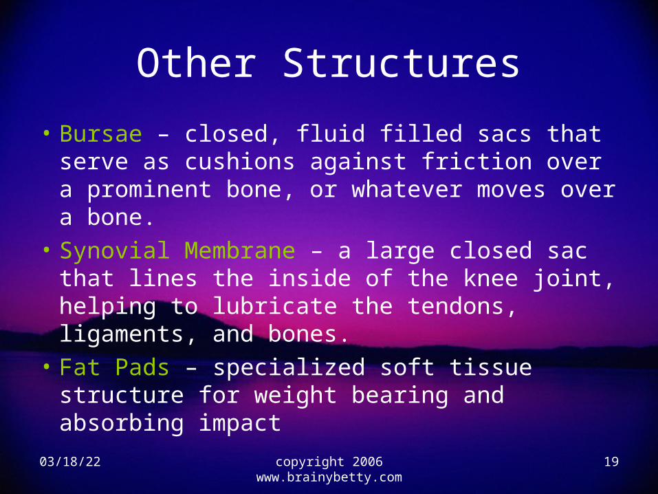
Other Structures
• Bursae – closed, fluid filled sacs that serve as cushions against friction over a prominent bone, or whatever moves over a bone.
• Synovial Membrane – a large closed sac that lines the inside of the knee joint, helping to lubricate the tendons, ligaments, and bones.
• Fat Pads – specialized soft tissue structure for weight bearing and absorbing impact

04/19/23 copyright 2006 www.brainybetty.com
20
NervesDermatome – the sensory distribution of a
nerve root; produces feeling in a certain anatomical area.
Myotome – the motor distribution of a group of muscles innervated by a single nerve root; it produces movement of the anatomical structures.

Range Of MotionFlexion – Decreasing angle between the Femur and the TibiaExtension – Increasing the angle between the Femur and the TibiaTibial Internal Rotation – Rotation of the Tibia toward the midline of the bodyTibial External Rotation - Rotation of the Tibia away from the midline of the body
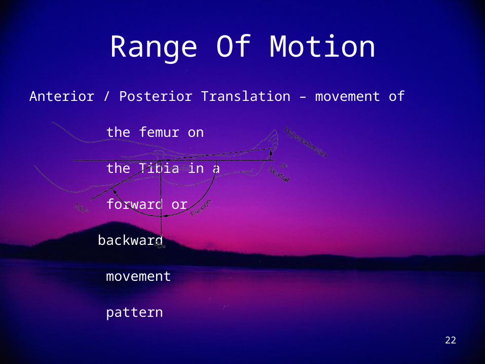
Range Of Motion
Anterior / Posterior Translation – movement of the femur on the Tibia in a forward or backward movement pattern
22

Evaluation• Proper evaluation is used to determine
seriousness of the injuryHistoryMechanism of injury, location of pain, sensations experienced,
and previous history• This will guide you through the rest of the evaluation
ObservationCompare the uninjured to the injured, look for: deformation,
swelling, discoloration, scars, other signs of trauma or abnormality.
23

Evaluation
PalpationAgain; compare right to leftCheck for:• Neurological Trauma• Circulation (nail bed return / pulse)• Anatomical structures• Potential Fractures
24

Evaluation
Special Test• Start with strength evaluation
Valgus / Varus Test – used to evaluate medial and lateral ligament stabilityAnterior / Posterior Drawer – Assesses stability of the ACL & PCL
Lachman – Used to evaluate ACL
Apley’s Compression – Evaluates the integrity of the menisci
25

Refer When….• Gross deformity• Significant Pain• Increase Swelling• Circulation or neurological impairment• Joint instability• Suspect a fx or dislocation• Dislocated patella• Abnormal sensations such as clicking, popping,
grating, or weakness• Locked knee or excessive / limited motion• Any doubt
26

Common InjuriesDue to the complexity of the knee it is
frequently injured• It is possible to severely injure the knee & not get a lot of
swelling.• And get very little pain
Ligament Sprains: can be cased by multidirectional forces and are compounded when the athlete's foot is stationary (planted)• Most common is a direct blow to the lateral aspect of the
knee injuring medial structures 27

Common InjuriesKnee Ligaments are usually injured by one of the
following methods:1.Compression (Direct Blow)2.Torsion (Fixed foot, twist body part or body)3.Shearing (Forced applied to the opposite side
of the joint)Sports that use cleats have an increase chance
of injury.28

Common InjuriesAll Ligamentous Sprains are classified the same.
1st Degree Sprain – one or more supporting ligaments & surrounding tissue stretched.2nd Degree Sprain – A portion of 1 or more ligaments is torn.3rd Degree Sprain – 1 or more ligaments is torn.
29

Common InjuriesPatellar Tendinitis
• Excessive stress placed on the patellar tendon cause inflammation above or below the patella
• Athlete complains of pain “when they first get up” and/or after activity. May have swelling.– Treat with Cold/Heat/Ultrasound/Rest– Rehab Strengthen Quads / Hamstrings
30

Common InjuriesChondromalacia Patellae
• A degenerative condition that results in the irritation and softening of the cartilage on the posterior aspect of the patella.– Running, jumping, kneeling, and climbing stairs
will elicit pain– Causes muscle weakness or imbalance, body
structure• Treatment: Ice before & after activity
– Surgery?• Rehab: Again work on Strengthening
31

Common InjuriesFemale Athletes Knee
(Anterior Superior Iliac Spine – ASIS)• Patellar problem may be more prevalent in
female athletes b/c of structural differences in the pelvic girdle.
• Wider pelvis creates a sharper angle where the femur attaches to the pelvis.
32

Common InjuriesFemale: Q Angle – an imaginary line from the
ASIS to the medial edge of patella.
copyright 2006 www.brainybetty.com
33

Common Injuries• A sharper Q Angle changes the line of pull of
the quads and may cause the patella to be pulled laterally, with muscle contraction.
• Changes in mechanics can increase conditions like:– Chondromalacia– Patellar dislocation/subluxation– ACL?
• Key to treatment is pevention!– Strengthen medial aspects of quads (VMO) 34

Common InjuriesOsgood-Slatters
• Common to adolescents due to rapid growth during “growth spurts”
• Characterized by swelling & point tenderness below 1 or both knees.– Can be caused by partial separation of the patellar
tendon from tibial tubercle– Inflammation of the tibial tubercle
35

Common InjuriesWhatever the cause it is
aggravated by activity, relieved by rest
In cases of long duration the front of the knee appears enlarged and a bony prominence can be felt.
Although the condition is usually disappears after adolescence the boney prominence remains.
36

Common InjuriesOther Musculoskeletal Disorders / Conditions
• Muscle strains• Bursitis• Dislocation (knee, patella)• Iliotibial Band Friction Syndrome• Meniscal Tear• Myositis Ossificans• Osteochondritis Dissecans (OCD)• Popliteal Cyst
37