chapter 4 platelet activation by the apob/e receptor ... 4 platelet activation by the apob/e...
TRANSCRIPT
75
Chapter 4
Chapter 4 Platelet activation by the apoB/E
receptor-binding domain of LDL
Ingrid A.M. Relou 1, 3, Gertie Gorter 1, Herman J.M.
van Rijn 2, Jan-Willem N. Akkerman 1,3
1 Laboratory for Thrombosis and Haemostasis,
Department of Haematology, University Medical
Center Utrecht, Utrecht, The Netherlands2 Department of Clinical Chemistry, University Medical
Center Utrecht, Utrecht, The Netherlands3 Institute for Biomembranes, Utrecht University,
Utrecht, The Netherlands.
Thrombosis and Haemostasis, 2002, 87 (5): 880-887
Platelet activation by apoB100
76
Summary
Low Density Lipoprotein (LDL) increases the sensitivity of human platelets for agonists
by activating p38MAPK. Antibody 4G3 disturbs apoB100 binding to the classical apoB/
E receptor and inhibits LDL-induced p38MAPK activation, whereas an antibody against
a distal domain on apoB100 has no effect. Peptide RLTRKRGLKLA mimics the
binding domain of apoB100 called the B-site and activates platelet p38MAPK. Activa-
tion by B-site peptide is dose-dependent, transient and followed by desensitization,
in accordance with receptor-mediated signalling. A scrambled peptide and a partially
homologous peptide RKLRKRLLRDA mimicking the apoB/E receptor binding site of
apoE in High Density Lipoprotein (HDL) also activate p38MAPK albeit 40% weaker,
but an uncharged peptide lacks p38MAPK activating capacity. LDL and B-site peptide
bind to the same binding sites and initiate similar signaling to p38MAPK and cytosolic
phospholipase A2. Thus, LDL and to a lesser extent HDL activate platelets via
specific domains in the protein moiety that recognize receptors of the LDL receptor
family.

77
Chapter 4
Introduction
The Low Density Lipoprotein (LDL) particle contains one molecule of apolipoprotein
B100 (apoB100) that enwraps LDL like a belt, with the carboxyl tail crossing
apoB100 close to amino acid 3500 1. The positively charged amino acids arginine
and lysine on apoB100 are essential for recognition of the classical LDL receptor
on nucleated cells, the apoB/E receptor 2-4. ApoB100 contains several positively
charged segments of which residues 3359-3369 are critical for receptor recognition5. This domain is called the B-site and consists of the amino acids RLTRKRGLKLA.
There is a high similarity with the receptor-binding domain of apoE, present on
HDL, which consists of RKLRKRLLRDA 6. The interaction of the carboxyl tail of
apoB100 with residue 3500 allows the B-site to interact with the receptor 7. This
interaction is critical since an R3500Q mutation found in patients with familial
defective apoB100 results in loss of receptor binding 8.
Native LDL activates endothelial cells 9 and increases the sensitivity of blood platelets
to agonist stimulation 10-14. How LDL interacts with platelets is still unclear. Patients
with an apoB/E receptor defect have platelets that respond normally to LDL.
Antibodies against the apoB/E receptor interfere with LDL binding to lympho-cytes
and fibroblasts but leave LDL binding to platelets unaffected. These observations
suggest that the LDL receptor on platelets differs from the classical apoB/E receptor
present on nucleated cells 15. A recent study identified LRP8, a member of the LDL
receptor family, on platelets and showed that apoE changes platelet functions by
binding to this receptor 16. By analogy, the same or a closely related family member
might function in LDL binding to platelets.
A first step in platelet activation by LDL is the activation of the enzyme p38MAPK,
which is activated within 10 seconds (at 1 g apoB100/L) after addition of LDL and at
concentrations as low as 0.1 g apoB100/L (within 10 minutes) 17. P38MAPK is a member
of the family of proline directed serine/threonine kinases, which is activated by the
simultaneous phosphorylation of Thr180 and Tyr182 18. LDL-induced p38MAPK activation
is insensitive to many inhibitors of signal transduction suggesting that it is an early
step in the activation cascade initiated by LDL. An upstream inhibitor of LDL-
induced p38MAPK activation is cAMP. An important downstream effect is the
phosphorylation and activation of cytosolic phospholipase A2 (cPLA
2) resulting in
formation of thromboxane A2 and enhanced platelet functions 17.
There is little insight in the activating properties of LDL. LDL consists of free and
esterified cholesterol, triglycerides and phospholipids, which are all components
that might change cell composition and behaviour. 19 Furthermore, LDL is easily
oxidized which leads to the generation of lysophosphatidic acid, which is an inducer
of platelet shape change 20. On the other hand, modification of lysine residues on
apoB100 impairs LDL binding to platelets 21 and abolishes LDL induced phosphor-
ylation of p38MAPK 17, suggesting that the signaling properties of LDL are located in
apoB100.
Platelet activation by apoB100
78
Here we report that an activating property of LDL resides in the B-site of apoB100.
By applying antibodies against apoB100 domains and peptides that mimic the B-
site we find activation of p38MAPK with characteristics that point to receptor medi-
ated platelet activation. A weaker activation is found by a peptide mimetic for the
receptor recognition site of apoE, a constituent of High Density Lipoprotein (HDL)
suggesting that the platelet LDL receptor shares many characteristics with the
classical apoB/E receptor on other cells.
Materials and Methods
Antibodies and Reagents
BAPTA-AM was from Calbiochem Corporation (La Jolla, CA, USA) and SK&F was obtained from Biomol
(Sanvertech, Boechout, Belgium). Dibutyryl cyclic AMP (dbcAMP) was from Sigma (St. Louis, MO, USA)
and iloprost from Schering (Berlin, FRG). PP1 was obtained from Alexis Biochemicals (San Diego, CA,
USA). MoAb directed against cPLA2 (4-4B-3C) was from Santa Cruz Biotechnology (Santa Cruz, CA,
USA). Renaissance chemiluminescence Western-blot reagent was from NEN-Dupont, Boston, MA, USA.
Polyclonal antibodies against p38MAPK and dual phosphorylated p38MAPK (phosphoplus p38MAPK) and
horseradish peroxidase labelled anti-rabbit IgG were from New England Biolabs (Beverly, USA).
Non-fat dry milk was obtained from Nutricia (Zoetermeer, the Netherlands). The fibrinogen- derived peptide
HHLGGAKQAGDV (γ400-411
) was kindly provided by the department of Biochemistry, Utrecht University.
SDZ-GPI-562 (GPI-562) was a kind gift of Dr. H-G. Zerwes (Novartis Pharmaceuticals, Basel, Switzerland)22.
The anti-human apoB100 monoclonal antibodies (moAb) 4G3 and 2D8 were obtained from the University
of Ottowa Heart Institute (Ontario, Canada). The production of these antibodies was described before 23.
Peptides
Peptides were synthesized by standard solid-phase peptide synthesis and purified by C18 reverse-phase
chromatography (HPLC, Genosphere biotechnologies, Paris, France). The purity of the peptides was
>99% as determined by HPLC and the molecular weights were verified by matrix-assisted laser desorption
mass spectrometry by the manufacturer.
The peptide RLTRKRGLKLA (Mw = 1311 Da), designated B-site peptide, represents the apoB receptor-
binding domain; SLTSASGLALA (Mw = 990 Da) is a related peptide in which charged amino acids
have been replaced by uncharged amino acids and was named uncharged peptide. Peptide
TKLRALRKLRG is a scrambled peptide with the same composition as B-site peptide. Peptide
RKLRKRLLRDA (Mw = 1426 Da) mimics the receptor binding site of apoE and was named apoE
peptide.
Lipoprotein Isolation
Lipoproteins were isolated as described previously 10. In short, fresh, non-frozen plasma from 4 healthy
subjects each containing less than 100 mg lipoprotein(a)/L was pooled and LDL (density range 1.019-
1.063 kg/L), HDL (density range 1.063-1.21 kg/L), HDL2 (density range 1.063-1.125 kg/L) and HDL
3
(density range 1.125-1.21 kg/L) were isolated by sequential flotation in a Beckman L-70 ultracentrifuge 24.
Centrifugations (20 hr, 175000 g, 10°C) were carried out in the presence of NaN3 and EDTA.
The LDL preparations contained only minimal amounts TBARS (0.20 ± 0.07 nmol/mg apoB100), lipid
peroxides (6.7 ± 1.9 nmol/mg apoB100) and contaminating plasma proteins. The concentrations of these
oxidation markers were below or within reported values for native LDL 10. Lp(a) concentrations (Apotech,
Organon Technika, Rockville, U.S.A) were below 14 mg/L.
79
Chapter 4
Lipoproteins were stored at 4°C under nitrogen for not longer than 14 days and before each experiment
dialyzed overnight against 104 volumes 150 mmol/L NaCl. ApoB100, apoAI and lipoprotein(a)
concentrations were measured using the Behring Nephelometer 100. The concentration of LDL was
expressed as g apoB100 protein/L and the concentration of HDL as g apoAI protein/L.
Platelet Isolation
Freshly drawn venous blood from healthy volunteers was collected with informed consent into 0.1 vol.
130 mmol/L trisodium citrate. The donors claimed not to have taken any medication during two weeks
prior to blood collection.
Platelet-rich plasma (PRP) was prepared by centrifugation (200 g for 15 min at 22°C). Gel-filtered platelets
(GFP) were isolated as described before 17. GFP were adjusted to a final count of 2 * 1011 platelets/L and
incubated with peptides, LDL or HDL at 37°C without stirring.
Binding of 125I-LDL to human platelets
LDL was labelled with 125I according to the method by Mc Farlane 25, modified by Bilheimer 26. GFP in
Tyrode’s solution were incubated with 125I-LDL (60 min, 22°C), and aliquots of 100 µL were layered on top
of 100 µL 25 % (w/v) sucrose in Tyrode solution in microsedimentation tubes (Sarstedt, Nümbrecht,
Germany).
Cells were separated from medium by centrifugation (12000 g, 2 min, 22°C) and both fractions were
counted in a Cobra gamma-counter (Packard, Downers Grove, Il., U.S.A.). Aspecific binding (about 10%
of total binding) was determined by addition of a 30-fold excess unlabeled LDL and substracted from total
binding to obtain specific binding data.
Measurement of p38MAPK and cPLA2
The phosphorylation of p38MAPK and cPLA2 was measured as described elsewhere 17. In short, GFP were
incubated at 37°C with peptides or LDL as indicated. Samples of 100 µL were fixed in 1% formaldehyde
(15 min, 4°C), centrifuged (9000 g, 30 sec) and resuspended in 40 µL Laemmli sample buffer. Samples
were heated prior to SDS-polyacrylamide gel electrophoresis (12%). Proteins were electrophoretically
transferred (1hour, 100 volts) to a nitrocellulose membrane using a mini-protean system (Biorad, Richmond,
CA, USA). The blots were blocked in 5% non-fat dry milk, 0.1% Tween 20 in phosphate buffered saline (1
hr, 4°C) and incubated with the phosphoplus p38MAPK (Thr180/Tyr182) or p38MAPK antibody (1:2000 in 1%
non-fat dry milk, 0.1% Tween in PBS, 16 hr, 4°C). Both antibodies are raised against residues 171-
186 of human p38MAPK. After washing, the membranes were incubated with horseradish peroxidase labelled
anti-rabbit (1:2000, 1 hour, 4°C) and p38MAPK was visualized using the enhanced chemiluminescence
reaction. For semi-quantitative determination of the amount of dual phosphorylated or total p38MAPK, the
density of the bands was analysed using ImageQuant software (Molecular Dynamics).
For the measurement of cPLA2 phosphorylation, samples were withdrawn and collected in Laemmlli
sample buffer (0.001% bromophenol blue, 5% β-mercaptoethanol, 100% glycerol, 2% (w/v) SDS, 62.5
mmol/L Tris pH 6.8). Measurement of cPLA2 was based on the mobility shift on SDS-PAGE that
accompanies phosphorylation of the protein.27 The running buffer for electrophoresis of cPLA2 was
pH 8.3. cPLA2 was detected using the moAb 4-4B-3C.
Immune complexes were detected by enhanced chemiluminescence.
Statistics
Data are expressed as means ± SD with number of observations n, and were analyzed by ANOVA.
Differences were considered significant at P < 0.05.
Platelet activation by apoB100
80
Results
Identification of the activating domain in apoB100
In an attempt to identify which part in LDL induced platelet activation via p38MAPK,
antibodies were sought that interfere with LDL binding to the classical apoB/E
receptor. Anti-human apoB100 monoclonal antibody 4G3 is directed against an
amino terminal part of apoB100 close to the apoB receptor-binding site (amino
acids 2980-3084) and called B-site. This antibody inhibits LDL-binding to the apoB/
E receptor on human fibroblasts. Antibody 2D8 is directed against a domain dis-
tant from the apoB/E receptor-binding site of human fibroblasts (aa 1438-1481)
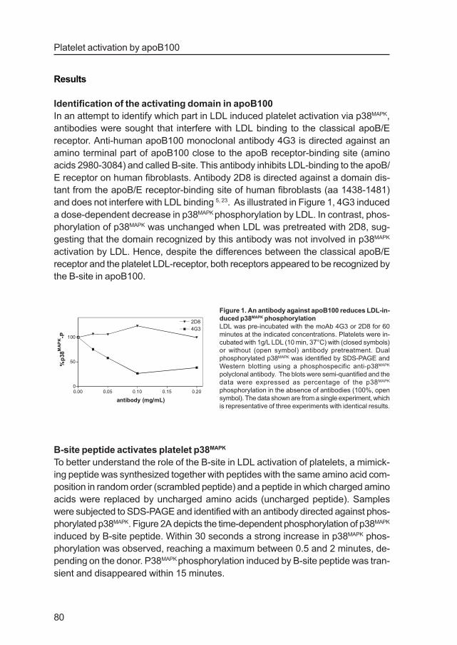
and does not interfere with LDL binding 5, 23. As illustrated in Figure 1, 4G3 induced
a dose-dependent decrease in p38MAPK phosphorylation by LDL. In contrast, phos-
phorylation of p38MAPK was unchanged when LDL was pretreated with 2D8, sug-
gesting that the domain recognized by this antibody was not involved in p38MAPK
activation by LDL. Hence, despite the differences between the classical apoB/E
receptor and the platelet LDL-receptor, both receptors appeared to be recognized by
the B-site in apoB100.
B-site peptide activates platelet p38MAPK
To better understand the role of the B-site in LDL activation of platelets, a mimick-
ing peptide was synthesized together with peptides with the same amino acid com-
position in random order (scrambled peptide) and a peptide in which charged amino
acids were replaced by uncharged amino acids (uncharged peptide). Samples
were subjected to SDS-PAGE and identified with an antibody directed against phos-
phorylated p38MAPK. Figure 2A depicts the time-dependent phosphorylation of p38MAPK
induced by B-site peptide. Within 30 seconds a strong increase in p38MAPK phos-
phorylation was observed, reaching a maximum between 0.5 and 2 minutes, de-
pending on the donor. P38MAPK phosphorylation induced by B-site peptide was tran-
sient and disappeared within 15 minutes.
0.00 0.05 0.10 0.15 0.200
50
100
4G3
2D8
antibody (mg/mL)
%p
38M
AP
K-P
Figure 1. An antibody against apoB100 reduces LDL-in-
duced p38MAPK phosphorylation
LDL was pre-incubated with the moAb 4G3 or 2D8 for 60
minutes at the indicated concentrations. Platelets were in-
cubated with 1g/L LDL (10 min, 37°C) with (closed symbols)
or without (open symbol) antibody pretreatment. Dual
phosphorylated p38MAPK was identified by SDS-PAGE and
Western blotting using a phosphospecific anti-p38MAPK
polyclonal antibody. The blots were semi-quantified and the
data were expressed as percentage of the p38MAPK
phosphorylation in the absence of antibodies (100%, open
symbol). The data shown are from a single experiment, which
is representative of three experiments with identical results.

81
Chapter 4
A B
C D
E
p38 -PMAPK
p38 MAPK
0 1 5 100.5 2 20 30 time (min)10
LDLB-site peptide
0 1 5 100.5 2 20 30 10
p38 -PMAPK
time (min)0 5 100.5 2 30 0.5
B-site peptideunchargedB-site peptide
0 10 20 300
50
100
LDL
B-site peptide
time (min)
%p
38M
AP
K-P
0 100 2000
50
100
150
300 500
B-site peptide (µmol/L)
%p
38M
AP
K-P
0 5 10 15 200
50
100 B-site peptide
scrambled peptide
time (min)
%p
38M
AP
K-P
Figure 2. B-site peptide induces phosphorylation of p38MAPK
(A), Platelets were incubated with B-site peptide (100 µmol/L, 37°C) for the indicated time periods. Samples were
drawn and centrifuged (30 sec, 9000 g, 22°C) and resuspended in sample buffer. One part of the samples was applied
to gel and dual phosphorylated p38MAPK was identified with a phosphospecific anti-p38MAPK polyclonal antibody (upper
panel). Another part was applied to gel and total p38MAPK was detected using an antibody against p38MAPK as a control
for equal lane loading (lower panel). Shown are representative blots of five experiments with identical results. (B),
Platelets were incubated with the indicated concentrations of B-site peptide (1 min, 37°C). Dual phosphorylated
p38MAPK was identified and the data were expressed as percentage of the p38MAPK phosphorylation in the presence of
100 µmol/L B-site peptide. Data are expressed as means ± SD, n=4. (C), Platelets were incubated with B-site peptide
(100 µmol/L, 37°C) or LDL (1 g/L, 37°C) for the indicated time periods. Dual phosphorylated p38MAPK was identified and
the data were expressed as percentage of the p38MAPK phosphorylation in the presence of 10 min LDL. Data are
expressed as means ± SD, n=5. (D), Platelets were incubated with B-site peptide or scrambled peptide (100 µmol/L,
37°C) for the indicated time periods. Dual phosphorylated p38MAPK was identified and the data were expressed as
percentage of the p38MAPK phosphorylation observed after incubation with 1 min B-site peptide. Data are expressed as
means ± SD, n=4. (E), Platelets were incubated with B-site peptide or uncharged peptide (100 µmol/L, 37°C) for the
indicated time periods. Dual phosphorylated p38MAPK was identified as described in Figure 2A. Shown is a representa-
tive blot of three experiments with identical results.
Platelet activation by apoB100
82
In the lower panel the same samples were analysed with an antibody directed against
total p38MAPK, showing that similar amounts of p38MAPK were present in all samples.
An optimal effect was observed at 100 µmol/L B-site peptide (Figure 2B). At this
concentration B-site peptide induced 93.6 ± 4.1% (n=5, P>0.5) of the p38MAPK
phosphorylation induced by 1 g/L LDL. However, activation by the peptide lasted
shorter than activation by LDL, indicating that B-site peptide mimicked only part of
the activating properties of native LDL (Figure 2C). The scrambled B-site peptide
had retained part of the activating properties of intact B-site peptide (Figure 2D).
There was a slight and transient activation which reached about 60% of the increase
in phosphorylated p38MAPK triggered by intact B-site peptide. This indicates that part
of the activating properties of B-site peptide are caused by positively charged amino
acids. Indeed, when all charged amino acids were replaced by neutral amino acids
by means of arginine-serine and lysine-alanine replacements, the activating proper-
ties were lost (Figure 2E).
Together these data illustrate that B-site peptide is a rapid and strong activator of
p38MAPK in platelets.
Comparison between B-site peptide and LDL
The observation that B-site peptide mimicked at least part of the p38MAPK activating
properties of LDL suggested that both induced signal generation by binding to a
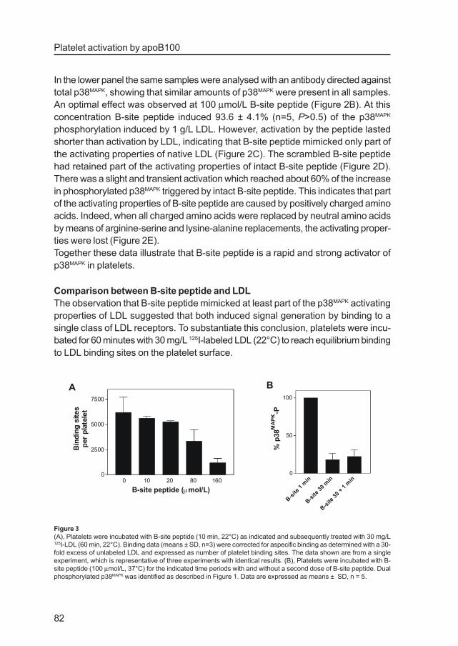
single class of LDL receptors. To substantiate this conclusion, platelets were incu-
bated for 60 minutes with 30 mg/L 125I-labeled LDL (22°C) to reach equilibrium binding
to LDL binding sites on the platelet surface.
0 10 20 80 160 0
2500
5000
7500
B-site peptide (µmol/L)
Bin
din
gsi
tes
per
pla
tele
t
B-site
1m
in
B-site
30m
in
B-site
30+
1m
in0
50
100
%p
38M
AP
K-P
A B
Figure 3
(A), Platelets were incubated with B-site peptide (10 min, 22°C) as indicated and subsequently treated with 30 mg/L125I-LDL (60 min, 22°C). Binding data (means ± SD, n=3) were corrected for aspecific binding as determined with a 30-
fold excess of unlabeled LDL and expressed as number of platelet binding sites. The data shown are from a single
experiment, which is representative of three experiments with identical results. (B), Platelets were incubated with B-
site peptide (100 µmol/L, 37°C) for the indicated time periods with and without a second dose of B-site peptide. Dual
phosphorylated p38MAPK was identified as described in Figure 1. Data are expressed as means ± SD, n = 5.
83
Chapter 4
Prior addition of B-site peptide (10-160 µmol/L) led to a dose-related displacement
of bound 125I-labeled LDL (Figure 3A). This accords with competition for a single
class of binding sites. A common property of most receptors is that they become
unresponsive after prolonged ligand occupation. Indeed, a 30 minutes incubation
with B-site peptide led to downregulation of p38MAPK activation (Figures 2C and 3B).
A second addition of 100µmol/L B-site peptide failed to trigger a second p38MAPK
phosphorylation response, suggesting that the B-site recognition mechanism had
become desensitized. Together these findings support the concept that LDL and B-
site peptide bind to the same receptors thereby starting signal transduction to
p38MAPK.
To further compare the signaling properties of B-site peptide and LDL, the sensitiv-
ity of p38MAPK phosphorylation for agents that interfere with platelet activating
pathways was investigated.
p38 -PMAPK
PP1 ( M)µ10 10 1020 20 20
LDL B-site peptide
00
C
ilopro
st
dbcA
MP
BAPTA-A
MSK&F
0
50
100
%p
38M
AP
K-P
A B
C
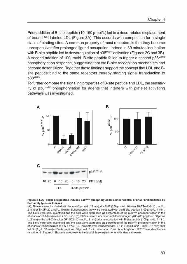
Figure 4. LDL- and B-site peptide-induced p38MAPK phosphorylation is under control of cAMP and mediated by
Src family tyrosine kinases
(A), Platelets were incubated with iloprost (2 µmol/L, 15 min), dbcAMP (250 µmol/L, 10 min), BAPTA-AM (10 µmol/L,
2 min) or SK&F (20 µmol/L, 10 min). Subsequently, they were incubated with the B-site peptide (100 µmol/L, 1 min).
The blots were semi-quantified and the data were expressed as percentage of the p38MAPK phosphorylation in the
absence of inhibitors (means ± SD, n=3). (B), Platelets were incubated with the fibrinogen γ400-411 peptide (100 µmol/
L, 2 min) or the αIIbβ3 blocker GPI-562 (10 nmol/L, 1 min) prior to incubation with B-site peptide (100 µmol/L, 1 min).
The blots were semi-quantified and the data were expressed as percentage of the p38MAPK phosphorylation in the
absence of inhibitors (means ± SD, n=3). (C), Platelets were incubated with PP1 (10 µmol/L or 20 µmol/L, 15 min) prior
to LDL (1 g/L, 10 min) or B-site peptide (100 µmol/L, 1 min) incubation. Dual phosphorylated p38MAPK was identified as
described in Figure 1. Shown is a representative blot of three experiments with identical results.

Platelet activation by apoB100
84
We have shown earlier that LDL-induced p38MAPK was inhibited by agents that raise
cAMP whereas the Ca2+-scavenger BAPTA-AM and the Ca2+- entry blocker SK&F
had no effect 17. B-site induced p38MAPK phosphorylation showed the same
characteristics. There was a strong inhibition by the stable PGI2- analogue iloprost
and the cell- permeable cAMP analogue dibutyryl cAMP but no effect of treatment
with BAPTA-AM and SK&F (Figure 4A). Another feature of LDL- induced p38MAPK
phosphorylation is lack of involvement of integrin αIIbβ3. B-site peptide-induced
p38MAPK activation also showed no requirement of integrin αIIbβ3 as blockers of
ligand binding to the integrin, fibrinogen γ-chain derived dodecapeptide (γ400-411
) and
GPI-562 showed no effect (Figure 4B).
The role of Src family tyrosine kinases in B-site and LDL-induced p38MAPK activation
was investigated with the specific inhibitor PP1. At two concentrations (10 µmol/L
and 20 µmol/L) the inhibitor markedly reduced B-site and LDL-induced p38MAPK
phosphorylation, illustrating the involvement of Src family tyrosine kinases (Figure
4C).
One of the downstream effects of LDL-induced p38MAPK phosphorylation is the
phosphorylation of cPLA2, which is a first step in the generation of thromboxane
A2. Also B-site peptide (100 µmol/L) induced a transient phosphorylation of cPLA
2
but the effect was considerably faster than observed with LDL 27 such in accordance
with the faster phosphorylation of p38MAPK induced by the peptide (Figure 5). B-site
peptide phosphorylation of cPLA2 was blocked completely by the p38MAPK inhibitor
SB203580 confirming the role of p38MAPK in the control of cPLA2. These data indi-
cate that B-site peptide initiates the same type of signal transduction in platelets as
LDL.
P38MAPK phosphorylation by the receptor-binding site of apoE
The apoB/E receptor equally recognizes the apoB-100 part of LDL as the apoE part
of HDL and a high degree of similarity between the receptor recognition domains of
both apolipoproteins can be expected. Indeed, also HDL induced p38MAPK phospho-
rylation but the response was weaker than observed with LDL (Figure 6A).
1 5 100.5 2 20 30
B-site peptide
1 5 100.5 2 20 30 30+1
SB203580
SB203580 + B-site
control
cPLA -P2
cPLA 2
Figure 5. B-site peptide phosphorylates cPLA2
Platelets were incubated with B-site peptide (100 µmol/L) for
the indicated time periods. Platelets were incubated with
SB203580 (10 µmol/L, 15 min) prior to B-site stimulation where
indicated. cPLA2 was identified by western blotting with a moAb
against cPLA2. Shown is a representative blot of four
experiments with identical results.

85
Chapter 4
Subtypes of HDL, HDL2 and HDL
3, which are described respectively as anti-
aggregatory and proaggregatory 28 induce p38MAPK phosphorylation with HDL2 acting
as a weaker agonist (Figure 6B). Residues 142-152 of apoE show a high degree of
homology with the B-site of apoB100. A peptide mimicking this domain called apoE
peptide (RKLRKRLLRDA) also induced phosphorylation of p38MAPK (Figure 6C). The
phosphorylation was fast and transient and also accompanied by loss of respon-
siveness to a second addition of apoE peptide. These properties closely resemble
those observed with B-site peptide.
Discussion
This report shows that at least part of the platelet activating properties of LDL are
located in apoB100. This agrees with earlier studies in which carbamylation of
apoB100 abolished LDL-induced p38MAPK phosphorylation reflecting a crucial role
of the protein moiety in platelet activation by LDL 17. Antibodies against different
domains of apoB100 reveal a central role for the B-site in induction of p38MAPK
phosphorylation. This domain is crucial for LDL binding to the apoB/E receptor on
fibroblasts and apparently also functions in LDL binding to the LDL receptor on
platelets. This is of interest since observations in patients with an apoB/E receptor
defect as well as in vitro studies with antibodies indicate that platelets lack the
classical apoB/E receptor. Possibly, platelets contain a closely related LDL receptor
family member with some properties in common with the apoB/E receptor.
p38 -PMAPK
1 5 100.5 2 20 30
ApoE peptide
30+1
0
p38 -PMAPK
0.5 0.51 1
LDL HDL
0.10.10 lipoprotein (g/L)
0.0 0.5 1.0 1.5 2.00
50
100
150
LDL
HDL2
HDL3
lipoprotein (g/L)
%p
38M
AP
K-P
A B
C
Figure 6. The receptor-binding domain of apoE phosphorylates p38MAPK
(A), Platelets were incubated with LDL or HDL with the indicated concentrations at 37°C for 10 min. Samples were
applied to gel and dual phosphorylated p38MAPK was identified with a phosphospecific anti-p38MAPK pAb. A representa-
tive blot of three experiments with identical result is shown. (B), Platelets were incubated with LDL, HDL2 or HDL
3 with
the indicated concentrations at 37°C for 10 min. The blots were semi-quantified and the data were expressed as
percentage of the p38MAPK phosphorylation after treatment with 1 g/L LDL. C, Platelets were incubated with apoE
peptide (100 µmol/L, 37°C) for the indicated time periods. Samples were applied to gel and dual phosphorylated
p38MAPK was identified with a phosphospecific anti-p38MAPK pAb. Shown is a representative blot of three experiments
with identical result.

Platelet activation by apoB100
86
Earlier studies by Borén et al suggested that the B-site of apoB100 (residues 3359-
3369) was critical site for LDL binding to the apoB/E receptor 29. In contrast, Law
and Scott proposed that multiple sites on apoB100 mediated receptor-binding 30
with the B-site playing a dominant but not an exclusive role. The present study
shows that a B-site mimicking peptide induces signal transduction in platelets.
Phosphorylation of p38MAPK was observed within 30 seconds after addition of B-site
peptide, illustrating rapid binding and activation of platelets. The activation was
maximal at about 100 µmol B-site peptide/L, which is equivalent to 0.13 g protein/
L. At this concentration B-site peptide induced the same amount of p38MAPK phos-
phorylation as 1.0 g LDL protein/L, suggesting that the B-site contains a major part
of the platelet activating properties of LDL. P38MAPK phosphorylation induced by the
peptide was faster and more transient than induced by LDL. The difference might
be caused by different binding affinities of the peptide and LDL. Alternatively, the
B-site might be important for platelet- LDL receptor recognition and initial signal
transduction generation with other components in LDL supporting this process at
later stages of LDL-platelet contact. A B-site peptide with arginine-serine and lysine-
alanine replacements failed to induce p38MAPK activation. A scrambled B-site peptide
has preserved part of the activating properties. This illustrates the importance of
clusters of positively charged amino acids lysine and arginine for platelet activation
by apoB100, such in common which apoB100 binding to the apoB/E receptor 2, 3 .
Also LDL binding to proteoglycans is charge-dependent 31. A minimum of five positive
charges with intervening nonbasic amino acids in a nonapeptide was required for
the binding to proteoglycans 32. Binding affinity depended on number and type of
nonbasic amino acids. In this respect, the B-site and the apoE site investigated in
the present study bind much stronger to proteoglycans than other charged domains
in these apolipoproteins.
The possibility to displace platelet-bound LDL by B-site peptide and the loss of
responsiveness after prolonged incubation with B-site peptide suggests the involve-
ment of a surface receptor that mediates signals to p38MAPK. The desensitization
might be the result of endocytosis of receptor-ligand complexes as has been reported
for ligand occupied apoB/E receptors on fibroblasts 33. However, in studies by Pedreno
et al 125I-LDL binding was reversible and unaffected by changes in temperature
which would argue against receptor internalization 15, 34. In contrast, Mazurov et al
reported a temperature dependent decrease of fluorescent-LDL bound to platelets
and concluded that bound LDL was internalized 35.
B-site peptide and LDL appear to trigger the same signaling pathway to p38MAPK.
P38MAPK phosphorylation by both agonists was inhibited by cAMP, independent of
Ca2+ increases and abolished by PP1, an inhibitor of Src family tyrosine kinases. In
addition, both agonists induced phosphorylation of cPLA2, which is a key step in
enzyme activation leading to formation of thromboxane A2. Although B-site peptide
and LDL induced the same degree of p38MAPK activation during initial contact with
the platelet, the peptide failed to sensitize collagen-induced secretion of dense
87
Chapter 4
granule contents, such in contrast to LDL (data not shown). Thus, the more prolonged
activation observed with LDL might initiate a stronger stimulation of the cPLA2
pathway leading to more support of platelet functions 12, 14, 28, 36-38.
Since the B-site of apoB100 shows a high degree of homology with the receptor-
binding domain of apoE 39, one might expect a similar activation by a peptide that
mimics this part of apoE. Indeed, apoE peptide induced a similar p38MAPK phospho-
rylation as B-site peptide but the activation was much weaker. A 30 minutes incubation
followed by a second peptide addition revealed a similar desensitization as observed
with B-site peptide. These findings are in concert with the rapid down-regulation of
binding sites for radiolabeled HDL3 on platelets as well as desensitization of the
signaling pathway 40, 41. In this respect apoE peptide mimicked the effect of HDL
which was a weaker activator than LDL when concentrations were based on apoA-
I and apoB100 content respectively. Applying molar concentrations and an apoA-I/
apoE ratio of about 30, the same concentration range resulted in a 2-3 fold lower
HDL-apoE concentration compared with LDL-apoB100. Also on this basis, HDL was
a weaker activator of p38MAPK than LDL. HDL3 induced a stronger p38MAPK activation
compared to HDL2 in concert with the pro-aggregatory effect of HDL
3 on platelets 28.
However, HDL2 is the apoE-rich fraction of HDL whereas HDL
3 is the apoE-poor
fraction which argues against an unique role of apoE for p38MAPK activation. The
observations suggest that in HDL-induced p38MAPK phosphorylation in addition to
apoE other lipoprotein-related components contribute to platelet sensitization.
Although previous observations suggest that the platelet LDL receptor is different
from the classical apoB/E receptor, the present data reveal many similarities. Both
receptors recognize apoB100 and apoE and the crucial binding domains are the
same. Furthermore, positively charged amino acids are essential for receptor
binding. These characteristics make it likely that platelets contain a member of the
LDL receptor family. A recent study shows that platelets contain LDL receptor-
related protein 8 (formerly termed apoER2). So far, LRP8 has been exclusively
demonstrated in human brain and placenta and its presence in platelets is
unexpected 42. Binding of apoE to this receptor inhibited platelet aggregation which
is in sharp contrast to the activating effect of apoE peptide on p38MAPK phosphory-
lation 16. The cytoplasmic tail of LRP8 contains several motifs with potential for
cellular signaling. Unfortunately, it is not known whether the extracellular part contains
an apoB100 binding site which would make it a candidate receptor for the effects
presented in the present study.
In conclusion, the results of this study indicate that LDL -and to a lesser extent
HDL- induce platelet activation via specific domains in the protein moiety. The
receptors that bind these domains share many properties with classical apoB/E
receptors which may become desensitized after prolonged contact with these
lipoproteins. This might reflect an important protection mechanism by which circulating
platelets remain dormant despite the continuous contact with LDL and HDL.

Platelet activation by apoB100
88
Acknowledgements
We are grateful to Dr. H. A. M. Voorbij for fruitful discussions. JWNA is supported by
the Netherlands Thrombosis Foundation.

89
Chapter 4
References
1. Chatterton JE, Phillips ML, Curtiss LK, Milne R, Fruchart JC, Schumaker VN. Immunoelectron
microscopy of low density lipoproteins yields a ribbon and bow model for the conformation of
apolipoprotein B on the lipoprotein surface. J Lipid Res. 1995; 36: 2027-2037
2. Mahley RW, Innerarity TL, Pitas RE, Weisgraber KH, Brown JH, Gross E. Inhibition of lipopro-
tein binding to cell surface receptors of fibroblasts following selective modification of arginyl
residues in arginine-rich and B apoproteins. J Biol Chem. 1977; 252: 7279-7287
3. Weisgraber KH, Innerrarity TL, Mahley RW. Role of the lysine residues of plasma lipoproteins
in high-affinity binding to cell surface receptors on human fibroblasts. J Biol Chem. 1978; 253:
9053-9062
4. Brown MS, Goldstein JL. Regulation of the activity of the low density lipoprotein receptor in
human fibroblasts. Cell. 1975; 6: 307-316
5. Boren J, Lee I, Zhu W, Arnold K, Taylor S, Innerarity TL. Identification of the low density
lipoprotein receptor-binding site in apolipoprotein B100 and the modulation of its binding activity
by the carboxyl terminus in familial defective apo-B100. J Clin Invest. 1998; 101: 1084-1093
6. Mahley RW. Apolipoprotein E: cholesterol transport protein with expanding role in cell biology.
Science. 1988; 240: 622-630
7. Boren J, Lee I, Zhu W, Arnold K, Taylor S, Innerarity TL. Identification of the low density
lipoprotein receptor-binding site in apolipoprotein B100 and the modulation of its binding activity
by the carboxyl terminus in familial defective apo-B100. J Clin Invest. 1998; 101: 1084-1093
8. Innerarity TL, Weisgraber KH, Arnold KS, Mahley RW, Krauss RM, Vega GL, Grundy SM.
Familial defective apolipoprotein B-100: low density lipoproteins with abnormal receptor binding.
Proc Natl Acad Sci U S A. 1987; 84: 6919-6923
9. Allen S, Khan S, Al-Mohanna F, Batten P, Yacoub M. Native low density lipoprotein-induced
calcium transients trigger VCAM-1 and E-selectin expression in cultured human vascular
endothelial cells. J Clin Invest. 1998; 101: 1064-1075
10. Hackeng CM, Huigsloot M, Pladet MW, Nieuwenhuis HK, Rijn HJMv, Akkerman JWN. Low-
density lipoprotein enhances platelet secretion via integrin-aðIIbbð3 -mediated signaling.
Arterioscler Thromb Vasc Biol. 1999; 19: 239-247
11. Hackeng CM, Pladet MW, Akkerman JW, van Rijn HJ. Low density lipoprotein phosphorylates
the focal adhesion-associated kinase p125(FAK) in human platelets independent of integrin
alphaIIb beta3. J Biol Chem. 1999; 274: 384-388
12. Surya II, Gorter G, Mommersteeg M, Akkerman JWN. Enhancement of platelet functions by
low density lipoproteins. Biochim Biophys Acta. 1992; 1165: 19-26
13. Lyman S, Gilmore A, Burridge K, Gidwitz S, White GC, II. Integrin-mediated activation of focal
adhesion kinase is independent of focal adhesion formation or integrin activation - Studies with
activated and inhibitory bð3 cytoplasmic domain mutants. J Biol Chem. 1997; 272: 22538-22547
14. van Willigen G, Gorter G, Akkerman JWN. LDLs increase the exposure of fibrinogen binding
sites on platelets and secretion of dense granules. Arterioscler Thromb. 1994; 14: 41-46
15. Pedreno J, de Castellarnau C, Cullare C, Sanchez J, Gomez Gerique J, Ordonez Llanos J,
Gonzalez Sastre F. LDL binding sites on platelets differ from the “classical” receptor of nucle-
ated cells. Arterioscler Thromb. 1992; 12: 1353-1362
16. Riddell DR, Vinogradov DV, Stannard AK, Chadwick N, Owen JS. Identification and characteri-
zation of LRP8 (apoER2) in human blood platelets. J Lipid Res. 1999; 40: 1925-1930
17. Hackeng CM, Relou IA, Pladet MW, Gorter G, van Rijn HJ, Akkerman JW. Early platelet
activation by low density lipoprotein via p38MAP kinase. Thromb Haemost. 1999; 82: 1749-
1756
18. Whitmarsh AJ, Davis RJ. Transcription factor AP-1 regulation by mitogen-activated protein
kinase signal transduction pathways. J Mol Med. 1996; 74: 589-607
19. Esterbauer H, Gebicki J, Puhl H, Jurgens G. The role of lipid peroxidation and antioxidants in
oxidative modification of LDL. Free Radic Biol Med. 1992; 13: 341-390
Platelet activation by apoB100
90
20. Siess W, Zangl KJ, Essler M, Bauer M, Brandl R, Corrinth C, Bittman R, Tigyi G, Aepfelbacher
M. Lysophosphatidic acid mediates the rapid activation of platelets and endothelial cells by
mildly oxidized low density lipoprotein and accumulates in human atherosclerotic lesions. Proc
Natl Acad Sci U S A. 1999; 96: 6931-6936
21. Aviram M, Brook JG, Lees AM, Lees RS. Low density lipoprotein binding to human platelets: role
of charge and of specific amino acids. Biochem Biophys Res Commun. 1981; 99: 308-318
22. Choudhri TF, Hoh BL, Zerwes HG, Prestigiacomo CJ, Kim SC, Connolly ESJ, Kottirsch G, Pinsky
DJ. Reduced microvascular thrombosis and improved outcome in acute murine stroke by inhib-
iting GP IIb/IIIa receptor-mediated platelet aggregation. J Clin Invest. 1998; 102: 1301-1310
23. Milne RW, Theolis R, Jr., Verdery RB, Marcel YL. Characterization of monoclonal antibodies
against human low density lipoprotein. Arteriosclerosis. 1983; 3: 23-30
24. Havel RJ, Eder HA, Bragdon JH. The distribution and chemical composition of ultracentrifugally
separated lipoproteins in human serum. J Clin Invest. 1955; 34: 1345-1353
25. Kramer RM, Roberts EF, Hyslop PA, Utterback BG, Hui KY, Jakubowski JA . Differential activa-
tion of cytosolic phospholipase A2 (cPLA2) by thrombin and thrombin receptor agonist peptide
in human platelets. Evidence for activation of cPLA2 independent of the mitogen-activated pro-
tein kinases ERK1/2. J Biol Chem. 1995; 270: 14816-14823
26. Jacobs DS: Laboratory test handbook, ed4, Cleveland, U.S., Lexi-Comp inc, 1996, pp 11-11
27. Hackeng CM, Franke B, Relou IA, Gorter G , Bos JL, van Rijn HJ, Akkerman JW. Low-density
lipoprotein activates the small GTPases Rap1 and Ral in human platelets. Biochem J. 2000;
349: 231-238
28. Desai K, Bruckdorfer KR, Hutton RA, Owen JS. Binding of apoE-rich high density lipoprotein
particles by saturable sites on human blood platelets inhibits agonist-induced platelet aggrega-
tion. J Lipid Res. 1989; 30: 831-840
29. Boren J, Lee I, Zhu W, Arnold K, Taylor S, Innerarity TL. Identification of the low density lipopro-
tein receptor-binding site in apolipoprotein B100 and the modulation of its binding activity by the
carboxyl terminus in familial defective apo-B100. J Clin Invest. 1998; 101: 1084-1093
30. Law A, Scott J. A cross-species comparison of the apolipoprotein B domain that binds to the
LDL receptor. J Lipid Res. 1990; 31: 1109-1120
31. Mahley RW, Weisgraber KH, Innerarity TL. Interaction of plasma lipoproteins containing
apolipoproteins B and E with heparin and cell surface receptors. Biochim Biophys Acta. 1979;
575: 81-91
32. Olsson U, Camejo G, Olofsson SO, Bondjers G. Molecular parameters that control the associa-
tion of low density lipoprotein apo B-100 with chondroitin sulphate. Biochim Biophys Acta.
1991; 1097: 37-44
33. Goldstein JL, Basu SK, Brown MS. Receptor-mediated endocytosis of low-density lipoprotein in
cultured cells. Methods Enzymol. 1983; 98:241-60: 241-260
34. Pedreno J, de Castellarnau C, Cullare C, Ortin R, Sanchez JL, Llopart R , Gonzalez Sastre F.
Platelet LDL receptor recognizes with the same apparent affinity both oxidized and native LDL.
Evidence that the receptor-ligand complexes are not internalized. Arterioscler Thromb. 1994;
14: 401-408
35. Mazurov AV, Preobrazhensky SN, Leytin VL, Repin VS, Smirnov VN. Study of low density lipo-
protein interaction with platelets by flow cytofluorimetry. FEBS Lett. 1982; 137: 319-322
36. Aviram M, Sirtori CR, Colli S, Maderna P , Morazzoni G, Tremoli E. Plasma lipoproteins affect
platelet malondialdehyde and thromboxane B2 production. Biochem Med. 1985; 34: 29-36
37. Ardlie NG, Selley ML, Simons LA. Platelet activation by oxidatively modified low density
lipoproteins. Atherosclerosis. 1989; 76: 117-124
38. Riddell DR, Graham A, Owen JS. Apolipoprotein E inhibits platelet aggregation through the L-
arginine:nitric oxide pathway. Implications for vascular disease. J Biol Chem. 1997; 272: 89-95
39. Knott TJ, Rall SCJ, Innerarity TL, Jacobson SF, Urdea MS, Levy-Wilson B, Powell LM, Pease
RJ, Eddy R, Nakai H. Human apolipoprotein B: structure of carboxyl-terminal domains, sites of
gene expression, and chromosomal localization. Science. 1985; 230: 37-43

91
Chapter 4
40. Nazih H, Nazih Sanderson F, Magret V, Caron B, Goudemand J, Fruchart JC, Delbart C.
Protein kinase C-dependent desensitization of HDL3-activated phospholipase C in human
platelets. Arterioscler Thromb. 1994; 14: 1321-1326
41. Pedreno J, Vila M, Masana L. Mechanisms for regulating platelet high density lipoprotein type3
binding sites: evidence that binding sites are downregulated by a protein kinase C-dependent
mechanism. Thromb Res. 1999; 94: 33-44
42. Kim DH, Iijima H, Goto K, Sakai J, Ishii H, Kim HJ, Suzuki H, Kondo H, Saeki S , Yamamoto T.
Human apolipoprotein E receptor 2. A novel lipoprotein receptor of the low density lipoprotein
receptor family predominantly expressed in brain. J Biol Chem. 1996; 271: 8373-8380

Platelet activation by apoB100
92