chapter 22 the lymphatic and immune systems. the lymphatic system basic organization lymph fluid in...
TRANSCRIPT

Chapter 22
The Lymphatic and Immune Systems

The Lymphatic System Basic organization
Lymph fluid in lymph vessels Structures, organs w/ lymph tissue, red
bone marrow Functions
drain interstitial fluid and proteins transport dietary fats protect against invasion
resistance - fight off disease nonspecific resistance - general protection against
disease immunity - specific protection
susceptibility - lack of resistance

Lymphatic Vessels Lymphatic capillaries
Blind ended vessels between cells, larger than blood capillaries
Not in avascular tissues, CNS, splenic pulp, bone marrow

Lymphatic Capillaries Structure/Function regulates fluid flow
Anchoring filaments - from lymphatic endothelium attach to surrounding tissues
Endothelial cells overlap high hydrostatic fluid pressure separates cells, fluid into caps hydrostatic fluid pressure in cap prevents fluid movement out

The Lymphatic System Lymph flow follows
venous circulation Lymph vessels have
same organization as vascular tree
Smaller vessels drain into larger vessels

The Lymphatic System (cont.) Lymph returns
to venous blood through R,L lymphatic ducts
Enter at internal jugular and subclavian veins

The Lymphatic System Lymph ducts
Right lymphatic duct
about ½ inch long drains lymph from
upper right side of body
Thoracic (left) duct main collecting
duct of lymph system
38-45 cm long drains 75% of body begins as a dilation
known as cisterna chyli located anterior to lumbar disk #2

The Lymphatic System Flow of Lymph
More fluid out of blood capillaries by filtration than returns by absorption - Starling’s Law
3Ll day of lymph fluid Proteins from blood returned by lymphatics Lymph flow by muscle pump, respiratory pump,
valves

Lymphatic Tissue - General 1º lymphatic organs - site of B and T
cell production bone marrow - produces B cells, pre-T
cells thymus gland - migrate to thymus gland
2 º lymphatic organs site of most immune responses lymph nodes, spleen – surrounded by
connective tissue capsule lymphatic nodules – not surrounded by
capsule

Thymus Gland Thymus Gland
Between sternum, heart 2 lobes Atrophies w/ age
Structure/Function Outer cortex
pre-T cells migrate to thymus
proliferation Maturation
dendritic cells macrophages
Inner medulla – more of the same

Lymph Node Structure oval, bean shaped
structures along lymph vessels
may be deep or superficial scattered throughout body
concentrated in mammary glands, axillae, groin
filter lymph fluid
Lymph Nodes

Lymph Nodes Connective tissue
capsule w/ trabeculae extending into cortex
Stroma -supportive network of reticular fibers, fibroblasts

Lymph Nodes
Cortex
Medulla
Parenchyma Outer cortex -
lymphatic nodules inner germinal
center site of B-cell proliferation
Site of B-cell maturation and plasma cell formation
Inner medulla - medullary cords of lymphycytes, macrophages, plasma cells

Node fluid flow Enter cortex through
afferent vessels Filter, remove damaged
cells, microorganisms, foreign substances by reticular fibers
macrophages phagocytize some, lymphocytes destroy others
exit medulla from hilus by efferent vessels
Metastasis cancer cells from tumor trapped in lymph nodes
Lymphatic Tissue - Specific

Lymph Nodes Histology

Lymphatic Tissue - Specific Spleen
largest mass of lymphoid tissue in body
Left side of body between stomach/diaphragm
thick fibrous capsule w/ artery, vein, efferent lymph vessel organ function:
immune function removal of worn
out, damaged RBC’s
storage of platelets
production of RBC’s during fetal life

Lymphatic Tissue - Specific Lymphatic nodules
(tonsils) oval-shaped non-
encapsulated groups of lymphatic tissue
scattered in mucous membranes, GI tract, respiratory tract, urinary tract, reproductive tract
protect against invasion of inhaled or ingested foreign substances

Nonspecific Resistance to Disease Summarized in Table 22.1 Surface Barrier – Skin and mucosa Internal Defenses
1) Antimicrobial proteins2) Natural killer cells and Phagocytosis3) Inflammation4) Fever

Nonspecific Resistance to Disease Surface Barrier – Skin and mucosa
Mechanical protection intact epidermis mucous membranes
line body cavities, mucus prevents drying, traps foreign bodies
nose hairs, respiratory tract cilia lacrimal apparatus saliva - dilute microbes, wash them from teeth
surface urine flow, vaginal secretions – flow and pH help
kill microorganisms defecation and vomiting - expel microbes

Nonspecific Resistance to Disease Surface Barrier – Skin and mucosa
Chemical protection Skin
sebum (unsaturated FA’s) forms layer, prevents bacterial growth
perspiration has fatty acids, lo pH lysozyme - enzyme breaks down bacterial walls gastric juice - stomach nearly sterile due to lo pH,
2ish

Nonspecific Resistance to Disease1) Antimicrobial substances produced
against pathogens that penetrate 1º defense
A. Interferons Released from infected cells Stimulate production of antiviral proteins from
neighboring cells
B. Complement system 20 plasma and cell membrane proteins Function to complement (enhance) certain
immune, allergic and inflammatory systems
C. Transferrins – inhabit bacterial growth by binding iron

Nonspecific Resistance to Disease
2) Natural killer (NK) cells and phagocytosis
NK nonspecific killers that respond before immune
system is activated ability to kill virus infected body cells and some
tumor cells by poking holes in cells and stimulating cell death

Nonspecific Resistance to Disease2) Natural killer (NK) cells and phagocytosis
Phagocytosis ingestion of microbes or foreign material by phagocytes 2 kinds of phagocytosis - neutrophils and macrophages steps
chemotaxis adherence ingestion digestion killing

Nonspecific Resistance to Disease3) Inflammation 4 symptoms:
Redness Pain Heat Swelling
3 steps in process1. Vasodilation/increased
vessel permeability2. Phagocyte migration3. Tissue repair

Nonspecific Resistance to Disease Step 1. Vasodilation
and increased vessel permeability Release of factors from
leukocytes stimulate vascular changes
Histamine, kinins, prostaglandins, leukotrienes
Vasodilate, increase permeability, stimulate emigration and chemotaxis
Cause heat, redness and swelling
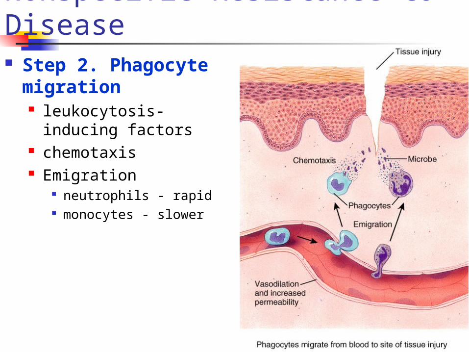
Nonspecific Resistance to Disease Step 2. Phagocyte
migration leukocytosis-inducing
factors chemotaxis Emigration
neutrophils - rapid monocytes - slower

Nonspecific Resistance to Disease Step 3. Tissue
repair Tissue regrowth and
repair of damage Pus
dead phagocytes, damaged tissue
if too numerous for easy removal may form abscess

Nonspecific Resistance to Disease 4) Fever
increase effects of antimicrobial substances inhibits some microbes increase rate of body repair

Nonspecific Resistance to Disease Surface Barrier – Skin and mucosa Internal Defenses
1) Antimicrobial proteins2) Natural killer cells and Phagocytosis3) Inflammation4) Fever

Immunity
Immunity ability of body to defend itself against specific
invaders specificity and memory differentiate this from
non-specific system two types
Humoral (antibody mediated) immunity Cellular (cell mediated) immunity

Antigens Antigen - substances
that provoke immune response
Epitope antigen part that triggers
immune response most antigens have
many epitopes

Antigens (cont.) Chemical nature of
antigens large, complex
molecules - mostly proteins, nucleo-, lipo-, glyco-
smaller substances may be incomplete antigens - hapten
react with antibodies but not cause immune response
poison ivy toxin usually foreign
substances

Antigens (cont.) Antigen receptor diversity
>1 billion different epitopes recognized by body
diversity - genetic recombination, shuffle, reorganize different genes
Major Histocompatibility Complex antigens (MHC)
unique to each individuals cells, help in identifying foreign bodies
2 classes of MHC antigens class I MHC - all body cells but
RBC's class II MHC - only on antigen
presenting cells (APC’s), thymus cells, activated T cells

Pathways of Antigen Processing For immune response to occur, B and T
cells must recognize foreign antigen B cells can recognize, bind to antigens in
ECF, blood, lymph T cells only recognize antigen protein
fragments presented w/ MHC self-antigens - good/bad proteins

Pathways of Antigen Processing During protein digestion and production in cell,
small peptides used to stabilize MHC proteins if peptides from normal body cells, no response if peptides from antigens
T cells recognize them immune response!
preparation of foreign antigen for cell surface known as processing and presenting of antigen
Antigen Presenting Cell's (APC’s) macrophages, activated B cells, dendritic cells process exogenous antigens present them w/ MHC
class II molecules to T cells

Pathways of Antigen Processing Processing of exogenous antigen – produced outside cell
1) phagocytosis/endocytosis 2) digestion3) vesicles fuse, peptide fragments bind to MHC-II’s 4) exocytosis to membrane5) move to lymphatic tissue, present antigen to T-cells

Pathways of Antigen Processing Processing of endogenous antigen –
produced within body cells viral proteins from viral infection of cell produced, incorporated into MHC-I
molecules during normal cell growth put on surface of cell identifies cell as infected, signals that cell
needs help

T and B cell formation
Both B and T cells produced from stem cells in bone marrow
Development of immunocompetence occurs in different sites B cells complete
maturation in Bone marrow
Pre-T cells move to Thymus - complete maturation in thymus

Immunity - General Before mature
cells leave home:
both types acquire surface proteins - antigen receptors
T cells exit as CD4+ or CD8+ cells w/ different functions
CD4+ cells become helper T cells
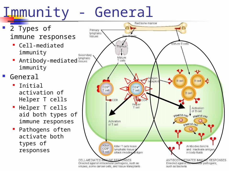
Immunity - General 2 Types of
immune responses
Cell-mediated immunity
Antibody-mediated immunity
General Initial activation of
Helper T cells Helper T cells aid
both types of immune responses
Pathogens often activate both types of responses

Immunity - General1) Cell-mediated
immune (CMI) responses
CD8+ cells change into killer T cells with aid from Helper T cells
directly attack infecting antigen
Effective against: intracellular
pathogens some cancer cells foreign tissue
transplants
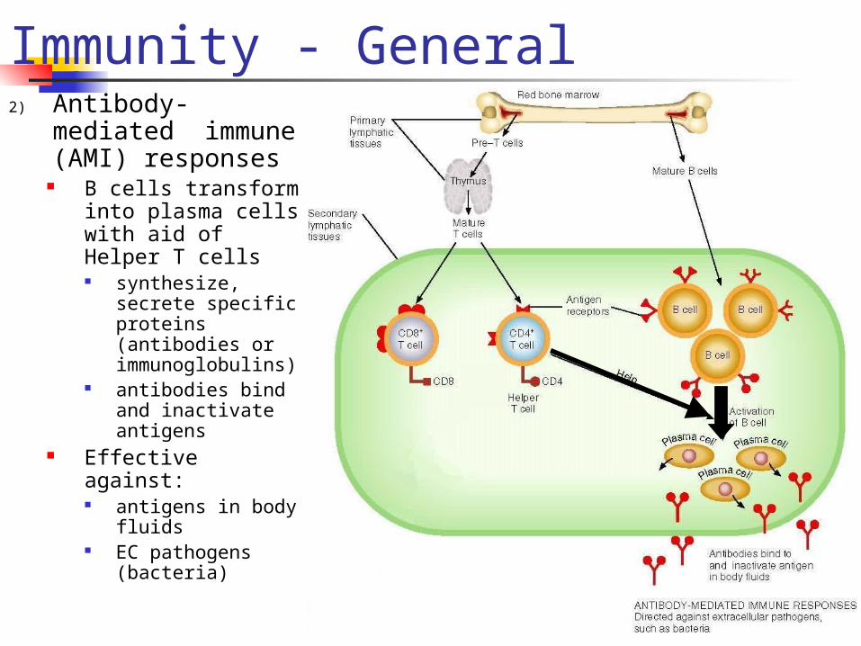
Immunity - General2) Antibody-
mediated immune (AMI) responses
B cells transform into plasma cells with aid of Helper T cells synthesize,
secrete specific proteins (antibodies or immunoglobulins)
antibodies bind and inactivate antigens
Effective against: antigens in body
fluids EC pathogens
(bacteria)

Cell -Mediated Immunity
Basic steps1. Recognition of APC antigen by T cell
receptors (TCR’s) – first signal2. Costimulation for activation3. Proliferation and differentiation 4. Clone effector cells capable of recognizing
initial activator (antigen)5. Elimination of intruder

Cell-Mediated Immunity (cont.) T cell recognition,
proliferation, differentiation
APC presents antigen TC’s recognize, bind
foreign antigen - millions of T cells each specific for 1 antigen
Need co-stimulator 20 known, cytokines,
interleukins, needed for full immune response
prevent false activation?

Cell-Mediated Immunity (cont.) T cell enlarges,
makes clones capable of recognizing antigen

Cell-Mediated Immunity (cont.) Clones
Helper T (TH) cells - CD4+
after co-stimulation helpers secrete co-stimulators
co-stimulates helper T cells, cytotoxic T cells, B cells
Cytotoxic T (TC) cells - CD8+ recognize antigen fragments associated w/ MHC-I molecules,
some tumor cells and tissue transplant cells become cytolytic need stimulators from helper T cells

Cell-Mediated Immunity (cont.) Tc elimination of
invaders1. Migrate from lymph to
infection site2. Recognize, attach to
target antigen/cell3. Kill invaders 4. Detach, seek out
another invader w/ proper antigen

Cell-Mediated Immunity (cont.) More Clones
Suppressor T (TS) cells
produced with other clones
downregulate immune system by producing other cytokines
Memory T (TM) cells recognize original invading antigen second response rapid due to large numbers of TM cells present

Antibody-Mediated Immunity Body contains millions
of B cells located in lymphoid
tissue each responds to
specific antigen Become activated
in presence of foreign antigen unprocessed antigen
weak response need to process
antigen for stronger response

Antibody-Mediated Immunity Unprocessed
antigen taken into cell incorporated into
MHC-II self-antigen activates helper T
cells Costimulation -
production of cytokines
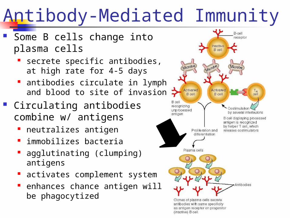
Antibody-Mediated Immunity Some B cells change into
plasma cells secrete specific antibodies, at high
rate for 4-5 days antibodies circulate in lymph and
blood to site of invasion Circulating antibodies combine
w/ antigens neutralizes antigen immobilizes bacteria agglutinating (clumping) antigens activates complement system enhances chance antigen will be
phagocytized

Antibody-Mediated Immunity
Activated B cells some turn into
memory B cells respond more rapidly,
forcefully if antigen appears again

Antibodies Antibodies (Abs)
Globulins (proteins) specific for antigenic determinant of antigen that triggered its production
5 different classes - IgG, IgA, IgM, IgD, IgE

Immunological Memory Long lived
antibodies and lymphocytes arise during activation of antigen-stimulated B, T cells
Immunization possible because memory B cells and memory T cells remain after 1º response w/ secondary exposure system responds more
quickly, forcefully secondary response - antibodies produced during
second exposure have a higher attraction for antigen

Acquired Immunity

Self-recognition and Self-tolerance

The Immune System - Pathologies Immunology and cancer
With development of cancer cell surface proteins (tumor antigens) appear that rarely appear
If body recognizes surface proteins as non-self will destroy them
most effective in eliminating viral tumor cells Hypersensitivities - allergies
first exposure sensitizes to allergen - IgE production next exposure anaphylaxis - massive histamine
release local - surface exposure systemic - body

The Immune System - Pathologies Autoimmune diseases
multiple sclerosis - white matter destruction myasthenia gravis - effects nerve/muscle
communication Graves disease - excessive thyroid hormone
production Type I diabetes - destruction of pancreatic
islet cells glomerulonephritis - impaired renal function rheumatoid arthritis - destruction of the joints

Acquired Immunodeficiency Syndrome Human Immunodeficiency Virus
Enters cells through receptor mediated endocytosis
infects mainly helper T cells uses the CD4 protein on cell surface
A retrovirus carries genetic material as RNA inserts genetic material into host DNA w/
enzyme reverse transcriptase cell makes copies of the virus, releases
them for further infection May be carried unknowingly in cells
for years, being passed on during cloning
Activation will destroy Helper T’s Poor immune response to simple
things

Applied Immunity and HIV Enter the body?
Spread?
Effect of infection?
Cause of death?