chapter 2studentsrepo.um.edu.my/4354/12/chapter_2_nurul… · · 2014-09-25chapter 2 general...
TRANSCRIPT

22
CHAPTER 2
GENERAL CHEMICAL
ASPECTS

23
CHAPTER 2
GENERAL CHEMICAL ASPECTS
2.1 Introduction
The exploration of the Lauraceous plants for alkaloids of medicinal value has
been going on since last decade. Indeed, herbalism and folk medicine, ancient and
modern, have been the source of much useful theraphy, pharmacology and taxonomy.
These chemical compounds also important to economic and nutrition field. Besides
alkaloids, they produced a wide range of non-alkaloidal compounds including
carbohydrate, proteins, lipid, amino acids, terpenes, essential oils, acetogenins,
polyphenols and aromatic compounds.
The alkaloids are one of the most diverst groups of secondary metabolites found
in living organisms and have an array of structure types, biosynthethic pathways, and
pharmacological activities and are of limited distribution in the plant kingdom. Their
amine character produce and alkaline solution in water and hence the origin of their
name-alkaloid27
.
It is generally accepted that secondary metabolites have a role in the survival of
the organism. In plants, these compounds are involved as attractants to ensure
pollination and are found to play an important role in plant interactions with animals
and higher and lower plants. Alkaloids are now generally considered to be part of an
elaborate system of chemical defense in plants and indeed the same seems to be true in
vertebrates, invertebrates and microorganisms28
.

24
2.2 Alkaloids
From ancient times man has utilized alkaloids as medicines, poisons, and
magical potions. Only recently has he gained precise knowledge about the chemical
structures of many of these interesting compounds. The term alkaloid, or ‘’alkali-like’’
was first proposed by the pharmacist, W. Meissner, in 1819. It is usually applied to
basic, nitrogen-containing compounds of plant origin. Two further qualifications are
usually added to this definition, alkaloids have complex molecular structure, and they
manifest significant pharmacological activity. Such compounds occur only in certain
genera and families, rarely being universally distributed in large groups of plants.
Chemical, pharmacological, and botanical properties must all be considered when
classifying a compound as an alkaloid. Examples of well-known alkaloids are morphine
27 (opium poppy), nicotine 28 (tobacco), quinine 29 (cinchona bark), reserpine 30
(rauwolfia) and strychnine 31(strychnos nuxvomica).
It should be emphasized that many widely distributed bases of plant origin, such
as methyl, trimethyl, and other open chain simple alkylamines, the cholines, and the
betaines are not classed as alkaloids. These are designated by some authorities as
‘’biological amines’’ or ‘’protoakaloids’’. Certain authorities also class the
phenylalkylamines among the ‘’biological amines’’. In certain cases, the distinction
drawn between the ‘’biological amines’’ and alkaloids is rather arbitrary. Alkaloids
usually have a rather complex structure with the nitrogen atom involved in a
heterocyclic ring. Interestingly, colchicine 32 is classed as an alkaloid even though it is
not basic, and its nitrogen atom is not incorporated in a heterocyclic ring, because of its
particular pharmacological activity and limited distribution in the plant world29
.

25
Nearly 300 alkaloids had been isolated by 1939 and about 200 of these had at
least reasonable well defined structure. In the first publishing in 1950, more than 1000
alkaloids are noted.
With the development of chromatographic techniques and sophisticated
spectroscopic instrumentations, the number of known alkaloids has risen dramatically.
In 1978 review, nearly 4000, structurally defined alkaloids were reported30
.
27 28
29 30

26
31 32
2.3 Classification of the Alkaloids
Alkaloids are the largest group of natural nitrogen-containing heterocyclic
compounds. When a nitrogen atom is part of a ring, the molecule is refered to as a
heterocyclic compound, meaning that a hetero atom (an atom other than carbon) exists
in the ring31
. Alkaloid can be categorised into three groups based on their biogenetic
pathways, chemical structure, pharmacological action, botanical and biochemical
origin32
. Several examples of common alkaloid ring skeletons are shown in Scheme 2.1
a. The true alkaloids mainly contain nitrogen in their heterocyclic system, derived from a
biogenetic amine and formed by decarboxylation of an amino acid. It can be found in
the plant as salt E.g.: morphine 27 and hygrine 33.
b. The pseudoalkaloids mainly contain characteristic of all true basic alkaloids, but they
are not derived from amino acid. It also known as heterocyclic containing nitrogen and
derived from terpenoids. E g.: actinidine 34 and coniine 35.

27
c. The protoalkaloids mainly contain nitrogen but that not as part of the heterocyclic
system, also basic and like true alkaloids, they are derived from amino acid. E. g.:
serotonine 36 and cathinone 37.
33 34 35
36 37
.

28
Pyrolidine piperidine pyridine
indolizidine quinolizidine pyrrolizidine
Indole quinoline isoquinoline
Purine quinazoline tropane
Scheme 2.1: Examples of Alkaloid Ring Skeletons.

29
2.4 Classification of Isoquinoline Alkaloids
Benzyltetrahydroisoquinolines are intermediate in the metabolism of
isoquinoline alkaloids. They are found by a Manich-type condensation between two
metabolites of phenylalanine. The experiments with labeled precursors and cell culture
showed that the true precursors are dopamine on one hand and 4-
hydroxyphenylacetaldehyde on the other hand. The condensation of these two
molecules leads to (S)-6-demethylcoclaurine, which is subsequently methylated (on the
6-position of the phenol and on the nitrogen atom) before being hydroxylated at C-12
and finally methylated to (S)-reticuline 1433
(Scheme 2.2).
Isoquinoline can be categorised into several classes base on the skeletal of the
structure (Table 2.1).

30
Table 2.1: Categorises of Isoquinoline Alkaloids
Simple isoquinoline Dibenzazonines
Benzylisoquinoline Protoberberines and retroprotoberberines
Isoquinolones Secoberberines
Pavines and isopavines Benzophenantridhines
Bisbenzylisoquinolines Arylisoquinolines
Baluchistanamines Protopines
Cularines Phthlideisoquinolines
Dibenzopyrrocolines Rhoeadines
Proaporphines Emetines
Aporphines Phenethylisoquinolines
Proaporphine-benzylisoquinoline Homoaporphines and homoproaporphines
Dimers 1-Phenylisoquinolines
Aporphine-pavine dimmers N-Benzyltetrahydroisoquinolines
Oxoaporphines 4-Arylisoquinolines
Aristolochic acid and aristolactams Azafluoranthenes and tropolosoquinolines
1, 6-Diazafluoanthenes

31
Scheme 2.2: Biosynthetic origin of the benzyltetrahydroisoquinoline.

32
2.4.1 Simple Isoquinoline
The simple isoquinoline are structurally the simplest of the isoquinoline type and
usually bicyclic. These alkaloids are derived from tetrahydroisoquinoline and they may
be found in the fully aromatic form or in partially reduced form34
. They also may be
defined as those containing only one aromatic nucleus and no other cyclic structure
except a methylenedioxy substituent and most of it has a carbon chain attached to C-1,
often a carbon substituent35
. Simple isoquinoline derivatives can be further sub-divided
into:
a) those not bearing a carbon substituent at C-1 and which are
basic, eg. anhalamine 38
b) those with an amide carbonyl group at C-1 and therefore non-
basic, eg. corydaldine 39 and
c) those with a methyl group at C-1 eg. salsoline 40.
38 39 40

33
2.4.2 Benzylisoquinoline
A B
The benzylisoquinoline types of alkaloids were derived from phenylalanine or
tyrosine and are the parent skeleton of a wide variety of alkaloids belonging to
numerous different ring systems. The benzylisoquinoline alkaloids include both the
benzylisoquinoline bases of type A and the benzyltetrahydroisoqunolines of type B. The
alkaloids with the unmodified benzlisoquinoline skeleton may be divided into two
subgroups:
a) 1,2,3,4-tetrahydrobenzylisoquinolines, e.g. reticuline 14 – of central
importance in the elaboration of other alkaloids; and
b) alkaloids with a carbon substituent at C-2; such as canadaline 41. These may
be regarded as ring – opened berberines.
It was proposed that reticuline 14 acts as a precursor for the C-9, C-10 or the C-
10, C-11 substituted aporphine (scheme 2.3) while N-methylcoclaurine 44 is the
precursor for most of the mono-oxygenated C-10 or non oxygenated ring D (scheme
2.4) and orientaline 1 precursor for the C-9 or C-11 mono-oxygenated ring D aporphine

34
(scheme 2.5). All the reactions occurred involved radicals and in the cases of the latter
two the corresponding alkaloids were formed via proaporphine which is another type of
isoquinoline alkaloid that occur in the nature.
Benzylisoquinoline alkaloids are widely distributed in the family
Anonaceae36-43
, Lauraceae13,20,43-44
, Menispermaceae45-48
, Papaveraceae49-51
,
Fumariaceae52-53
, Ranuculaceae54-55
and Berberidaceae56-57
.
41 44

35
Scheme 2.3: The biogenetic pathway to C-9,10 and C-10,11-disubstituted aporphine.
36
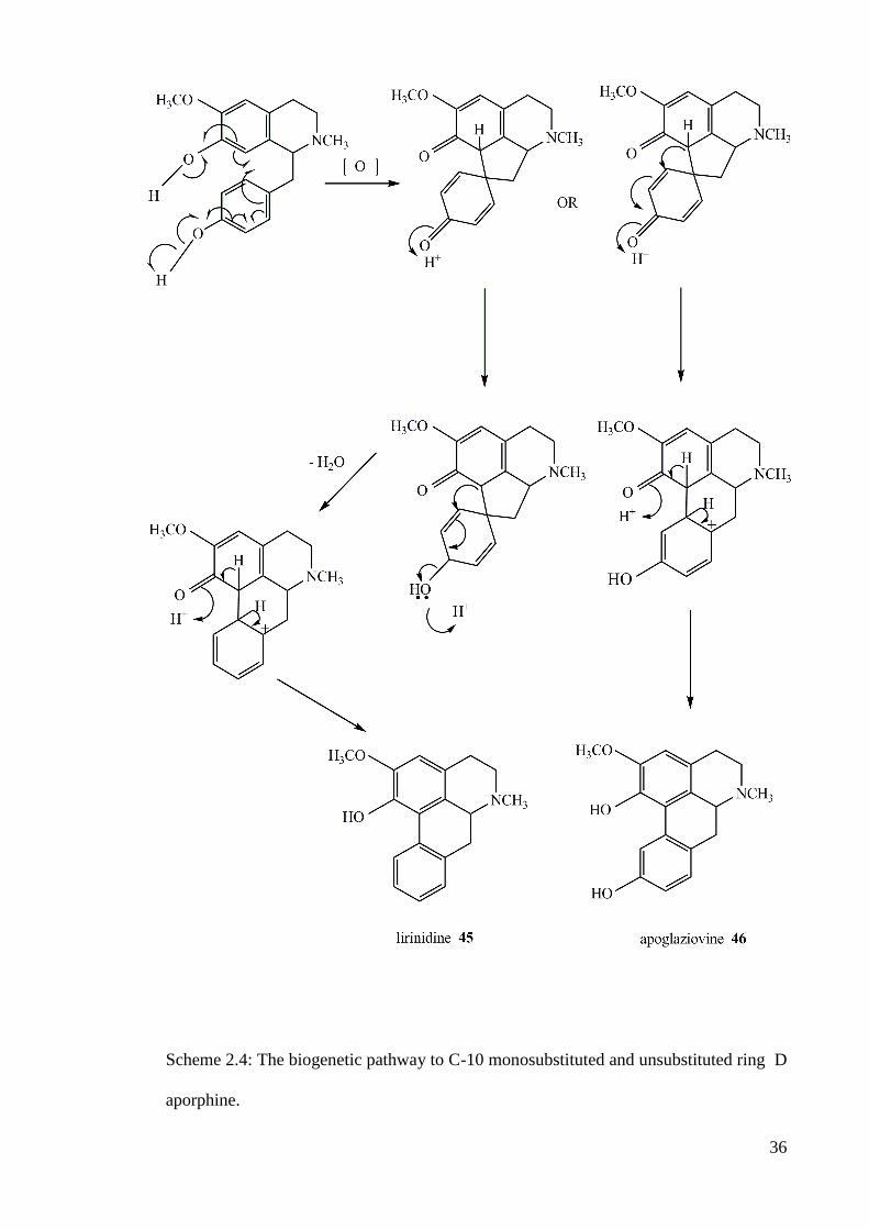
Scheme 2.4: The biogenetic pathway to C-10 monosubstituted and unsubstituted ring D
aporphine.

37
Scheme 2.5: The biogenetic pathway to C-11 and C-9 monosubstituted aporphine.

38
1H NMR
The 1H NMR data of benzylisoquinoline (Table 2.2) shows a number of
interesting features due to the one asymmetric center. The H-1 showed a triplet or
doublet-doublet (J1 = 8-9, J2 = 1.5-3 Hz) with chemical shift (in CDCl3) between δ 3.6-
3.7 while the aliphatic proton signals for H-3, H-4 and H-α, normally appeared as
multiplet at δ 2.5-3.5.
The methylenedioxy resonated at δ 5.6-6.0. Base on the study of
benzylisoquinoline, this group can be attached to C-6,7 and C-3’,4’. When the position
of methylenedioxy attached to C-6 and C-7, the signal showed as a doublet or singlet.
The methoxyl groups of the benzylisoquinoline, generally resonated at δ 3.50-
4.00. Normally, N-methyl groups resonated in the region of δ 2.4-2.6. Table 2.3 showed
the examples chemical shift of 1H NMR for some benzylisoquinolines, i.e.
hydrocotarnine 49, laudanosine 50, laudanine 51,
49 50 R1 = OCH3, R2 = OCH3
51 R1 = OCH3, R2 = H

39
Table 2.2: 1H NMR (in CDCl3, ppm) for some benzylisoquinoline.
Position of H
49
50
51
H-1 3.44 3.64 3.64
H-3 2.60 2.73 and 3.12 2.74 and 3.09
H-4 2.80 2.55 and 2.78 2.57 and 2.74
H-5 6.31 6.50 6.45
H-6 - - -
H-7 - - -
H-8 - 6.02 5.81
H-α - 2.17 and 3.10 2.54 and 3.03
H-2’ - 6.55 6.58
H-3’ - - -
H-4’ - - -
H-5’ - 6.71 6.60
H-6’ - 6.58 6.36
6-OCH3 - 3.77 3.68
7-OCH3 - 3.53 3.37
8-OCH3 3.98 - -
3’-OCH3 - 3.73 -
4’-OCH3
N-CH3
-
2.45
3.78
2.49
3.68
2.40
OCH2O 5.85 - -
13
C NMR
In the 13
C NMR spectra, C-1 normally resonated at δ 52-58, but it resonated at
lower field i.e. δ 60-67 in the presence of N-methyl group. Substituted carbons N-
methyl, methoxyl and methylenedioxy appeared at δ 40-45, δ 54-63 and δ 100-103,
respectively. The quaternary carbon at the position 4a, 8a and 1’ resonated at δ 115-132.
The quaternary carbons with methoxyl and hydroxyl groups appeared at δ 140-152.
Unsubstituted sp2 carbons usually appeared at δ 100-130 and the sp
3 carbons at the

40
position C-α and C-3 resonated at δ 38-40 and δ 45-46, respectively. The chemical shift
of C-4 with N-methyl group in the structure appeared at δ 23-24, but it will appear at δ
28-29 without N-methyl group.
Mass spectrometry
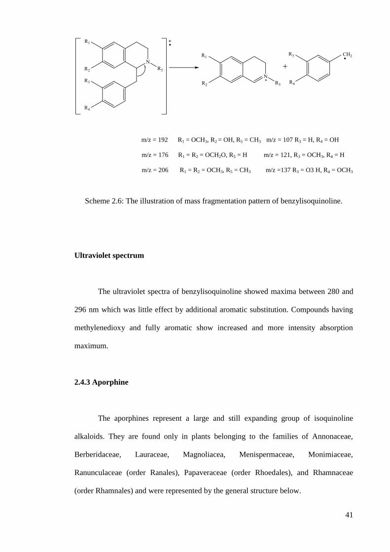
In the mass spectra of benzylisoquinoline, the main cleavage occurs between C-
1 and C-α to form an imine ion. The fragmentation at m/z 192 appeared as a base peak
indicated that the carbons C-6 and C-7 was substituted with methoxyl and hydroxyl
groups, respectively and in the structure beared N-methyl group. If both C-6 and C-7
was substituted with methoxyl groups peak at m/z 206 appeared as a base peak. The
fragmentation at m/z 176 which appeared as a base peak indicated the carbons C-6 and
C-7 attached to methylenedioxy without N-methyl in the structure.
The compounds having a methoxyl and hydroxyl groups in the ring C displayed
peak at m/z 137. Two methoxyl groups attached to C-3’ and C-4’ showed the
fragmentation peak at m/z 151 and a hydroxyl group in the ring C showed peak at m/z
10758
. The illustration of mass fragmentation pattern of benzylisoquinoline is shown in
Scheme 2.6.
41
m/z = 192 R1 = OCH3, R2 = OH, R5 = CH3 m/z = 107 R3 = H, R4 = OH
m/z = 176 R1 = R2 = OCH2O, R5 = H m/z = 121, R3 = OCH3, R4 = H
m/z = 206 R1 = R2 = OCH3, R5 = CH3 m/z =137 R3 = O3 H, R4 = OCH3
Scheme 2.6: The illustration of mass fragmentation pattern of benzylisoquinoline.
Ultraviolet spectrum
The ultraviolet spectra of benzylisoquinoline showed maxima between 280 and
296 nm which was little effect by additional aromatic substitution. Compounds having
methylenedioxy and fully aromatic show increased and more intensity absorption
maximum.
2.4.3 Aporphine
The aporphines represent a large and still expanding group of isoquinoline
alkaloids. They are found only in plants belonging to the families of Annonaceae,
Berberidaceae, Lauraceae, Magnoliacea, Menispermaceae, Monimiaceae,
Ranunculaceae (order Ranales), Papaveraceae (order Rhoedales), and Rhamnaceae
(order Rhamnales) and were represented by the general structure below.

42
The aporphine alkaloids contain a twisted biphenyl system. Its consists with four
rings, A, B, C, D. The whole aporphines alkaloids are based on the 4H-
diben[de,g]quinoline structure or its N-methyl derivative commonly known as the
aporphine nucleus. It can be divided into three groups:
a) The noraporphine, which possess a secondary nitrogen atom, eg:
norlirioferine 52
b) The aporphines which contain N-methyl function, lirioferine 13, N-
methyllauroteanine 53
c) The quaternary aporphines salts, for example N, N-dimethylhernovine 54
52 53 54

43
1H NMR
The 1H NMR spectrum can yield important and valuable information leading to
the structural elucidation of aporphines. The chemical shifts are very dependent on the
position of the protons with respect to the aromatic rings. Several general features have
been observed in the proton shifts of these alkaloids. The following chemical shifts have
been observed.
Methoxyl group
From the 1H NMR spectrum, the aromatic protons appeared at δ 6.3-8.2 and the
methoxyl groups revealed at δ 3.3-3.9. The most deshielded methoxyl groups were
attached at C-2, C-9 and C-10 and the most shielded was C-1, except if C-1 substituted
with methylenedioxy. The methoxyl that attached to C-1 appeared at high field due the
steric effect of ring D.
The most downfield aromatic proton (H-11) was observed between δ 7.5 and 8.2
depending on the adjacent group C-10. The substituted of C-10 will shield the H-11
proton. The C-3 proton was most shielded and typically appeared as a singlet in the
range of δ 6.5-7.5. The presence of methoxyl group at C-11 causes the proton at H-8
and H-9 to had significantly different chemical shift. If a C-11 bears a hydroxyl group,
the chemical shift of H-8 and H-9 were overlapped and no coupling was observed. The
position of C-2 is also substituted when position of C-1 and C-11 are substituted. This
affects the methoxyl groups at C-1 and C-11 would be sterically hindered. As a result,
the methoxyl protons are pushed out of the aromatic plane, which is shielded area. In
addition, ring A and ring D is facing each other. Hence the protons of the methoxyl

44
groups can arrange themselves on top of the adjacent ring, which happens to be a
shielded zone giving a more upfield shift59
.
Methylenedioxy group
The methylenedioxy group shows resonances at range of δ 5.87-6.02. The five
possible location for this group are C-1, 2; C-2, 3; C-9, 10; and C-10, 11. The presence
of C-1, 2 methylenedioxy group is proved by an up field shift of the C-11 proton which
appeared in the range δ 7.47-7.86, and caused the twisted biphenyl system induce
magnetic nonequivalent between the methylene proton, which then appeared as doublets
at δ 5.9 and 6.1.
At position C-9 and C-10, the two protons appear as a singlet whereas at
position C-1 and C-2; C-2 and C-3; C-10 and C-11, they appear as two doublets with
coupling constant of about 1.5 Hz. This inequivalence arises from the torsion caused by
the twisted biphenyl system of ring A and D60. The appearance of the torsional effect on
the methylenedioxy depends on their positions and at position C-9, 10 the effect appears
to be negligible61
.
Aromatic proton
The hydrogens at C-3, C-8 and C-9 of aporphine are located upfield between δ
6.38-7.00 and cannot be easily differentiated from one another while hydrogen at C-11
is found relatively downfield between δ 7.57-8.05. Nevertheless, H-3 normally
resonates at a higher field compared to the other aromatic protons (δ 6.50-6.70) when it
is ortho to a hydroxyl or a methoxyl. This is due to induction effect. On the other hand,

45
H-11 is usually resonates at a lower field with respect to the other protons due to the
deshielding effect imposed by the facing aromatic ring A and hydrogen bonding with
the C-1 substituent in ring A.
N-methyl group and aliphatic protons
The N-methyl group was typically observed at δ 2.4-2.8. The aliphatic protons
of C-4, C-5 and C-7 displayed a complex resonance pattern with absorption in the
region of δ 2.40-4.40 whereas the methyl group resonated in the region of δ 2.50-2.60.
Summary of the 1H NMR data of aporphine given in the table 2.3 below
Table 2.3: 1H NMR data (δ/ppm) of aporphine alkaloids in CDCl3
Position of
substituted
Methoxyl
group
Methylenedioxy Aromatic
proton
N-methyl
group
Aliphatic
group
C-1 3.70-3.55
C-2 4.12-3.75
C-3
C-8 7.00-6.38
C-9 4.12-3.75 7.00-6.38
C-10 4.12-3.75
C-11 3.75-3.65 8.74-8.68
C-1, 2 5.87-6.02
C-2,3 5.87-6.02
C-8, 9 5.87-6.02
C-9, 10 5.87-6.02
C-10, 11 5.87-6.02
N-Me 2.50-4.44
C-4 2.40-4.00
C-5 2.40-4.00

46
13C NMR
The general characteristic of the different type of carbons are mentioned below.
a) Sp2 carbon bearing hydrogen: δ 105-112.
b) Sp2
carbons at position 1a, 1b, 3a, 7a and 11a: δ 119-130.
c) Sp3 carbons at position 4(δ 28-30), 7(δ 35) and 5 and 6a (about δ 42 and 53
for noraporphine) and (about δ 53 and 62 for aporphine).
d) Carbon of the substituents: N-methyl (about δ 43), methoxyl (δ 56-62) and
methylenedioxy (δ 100).
The quaternization of the N-atom causes deshielding of C-5 and C-6a and
shielding of C-1b, C-3a, C-7 and C-7a.
Mass spectrometry
In mass spectrum, the principle fragmentation of the aporphine is the loss of the
hydrogen atom on C-6a. The [M-1]+
peak always serves as the base peak of the
molecule. If the molecule was substituted; [M-15]+ and [M-31]
+ peak will also be
observed due to the expulsion of a methyl, hydroxyl or methoxyl group respectively
(Scheme 2.7). Also if it is a hydroxyl group substituted at the ring D, a [M-17]+ peak
will be observed.
Aporphine compound having the NH or N-CH3 groups will display peaks at [M-
29]+
and [M-43]+ respectively. The fragment lost is methylene imine group (-CH2=NR)
which is expelled via a retro Diels-Alder mechanism (scheme 2.8) (or by the cyclic

47
process in ring B.) The ion formed can further loose another methyl or methoxyl to
produce peaks at [M-74]+, [M-58]
+, [M-60]
+, and [M-44]
62.

48
Scheme 2.7: The principle mass fragmentation of aporphines.

49
R = H or CH3
If R = H, peak observed is [M-29] +
If R = CH3, peak observed is [M-43] +
Scheme 2.8: The mass fragmentation of aporphine with N-methyl or N-H function
group.

50
Ultraviolet spectrum
The positions of the maximum absorptions in the ultraviolet spectra of
aporphines depend mainly upon the location of the substituents. It is derived from the
basic biphenyl system with the added influence of several auxochromes. The
approximate absorption for various substitution patterns are listed as below.
Position of substituents Absorption maximums (nm)
1, 2
234, 273, 312
1, 2, 9 233, 280, 305
1, 2, 10 226, 266, 275, 305
1, 2, 11 220, 265, 272, 300
1, 2, 9, 10
220, 282, 305
1, 2, 10, 11 220, 270, 305
The shape of the curve and the density of the latter two maxima depend on the
substitution in ring D. Furthermore, the monophenolic aporphine position at C-3 and C-
9 display a bathochromic shift at 315 nm and 350 nm in the alkaline environment63-64
.

51
2.4.4 Oxoaporphine
The oxoaporphines consist of carbonyl group at C-7. They are usually colored
such as red, orange and yellow because of their high degree of aromaticity.
Oxoaporphines are widely distributed and the first reported oxoaporphine was
liriodenine 55, which was isolated from Liriodendron tulipfera65-66
. From the species of
Artabotrys unicinatus, a novel 11-oxoaporphine were isolated, namely artacinatine 5665
.
55 56
52
1H NMR
The most characteristic features of oxoaporphine are the existence of highly
deshielded chemical shift values of the aromatic protons and the absence of the aliphatic
proton signals. A characteristic AB system signal at about δ 7.65 and 8.75 with a
coupling constant of 6 Hz which correspond to H-4 and H-5 is referred to a double bond
between C-4 and C-5. The C-3 proton singlet appeared at higher field if C-1 and C-2
were substituted. On contrary, the C-11 proton is usually the most downfield as a result
of the ring current effect of the facing ring A.
The methylenedioxy group observed a singlet with two protons at about δ 6.1
compared to the two doublets in the aporphine. This was due to the planarity of the
oxoaporphine skeleton. Moreover, H-8 resonates at a lower field (δ 8.2-8.6) because of
the neighouring C-7 carbonyl which exerts an inductive effect compared to the H-8 in
aporphine.
The methoxyl group located at C-1 gave the average shifts of about δ 3.55
meanwhile methoxyl groups present at position C-2, C-9 and C-10 reveal peaks at δ
3.80. Nevertheless we found that in the position C-1 and C-11, the methoxyl gave
chemical shift at δ 3.70. In the latter case, the more upfield shift may be due to the
OCH3 bond sticking out of the plane of the benzene rings which a deshielded effect is
expected.

53
13C NMR
The 13
C-NMR spectral data of oxoaporphines showed much closer to those
observed in aporphines. The carbons are all sp2, indicating that it was unsaturated and
fully aromatic. The general characteristic of the dfferent type of carbons are mentioned
below.
a) Sp2
carbon bearing a hydrogen: 105-112 ppm
b) Sp2
carbons at position 1a, 1b, 3a, 7a, and 11a: δ 119-130
c) Sp3
carbons at positions 4(δ 28-30), 7(δ 35) and 5 and 6a (about δ 42
and 53 for noraporphine) and (about δ 53 and 62 for aporphine).
d) Carbon of the substituents: N-methyl (about δ 43), methoxyl (δ 56-
62) and methylenedioxy (δ 100).
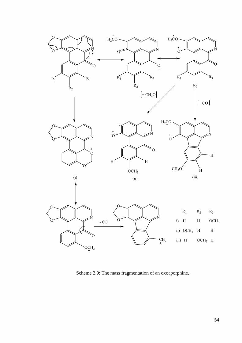
Mass spectrometry
The main fragment ions observed in the mass spectrum of oxoaporphine are
given in scheme 2.9 The important fragmentations are the [M-CO]+, [M-CH2O]
+ and
[M-CHO]+
54
Scheme 2.9: The mass fragmentation of an oxoaporphine.
55
Ultraviolet spectrum
The UV spectral data for the oxoaprphines are quite characteristic for the
skeletal type. These yellowish colored alkaloids posses a highly unsaturated
chromophoric system with extended absorption in the ultraviolet and visible.
For example liriodenine 55 shows three main absorption bands at 245-270, 309
and 413 nm. On acidification, the oxoaporphine exhibit bathochromic shift or the
spectrum is shifted to longer wavelengths with a series of undulating maxima between
325 and 460 nm67
.
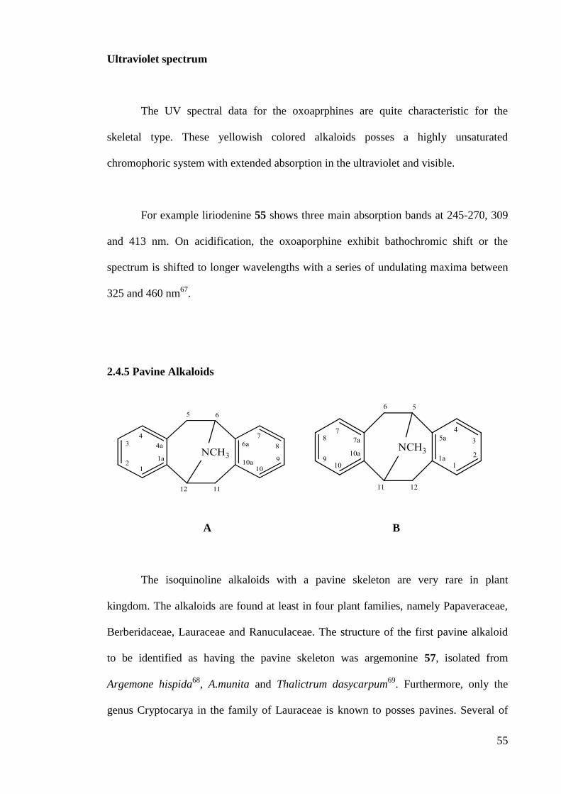
2.4.5 Pavine Alkaloids
A B
The isoquinoline alkaloids with a pavine skeleton are very rare in plant
kingdom. The alkaloids are found at least in four plant families, namely Papaveraceae,
Berberidaceae, Lauraceae and Ranuculaceae. The structure of the first pavine alkaloid
to be identified as having the pavine skeleton was argemonine 57, isolated from
Argemone hispida68
, A.munita and Thalictrum dasycarpum69
. Furthermore, only the
genus Cryptocarya in the family of Lauraceae is known to posses pavines. Several of

56
the pavines are probably derived biogenetically from the tetrahydrobenzylisoquinoline.
Two numbering system have been used for the pavines, represented by expressions A
and B. The characteristic of pavine is due to nitrogen atom must bridge the eight-
membered ring in the cis configuration and only two stereoisomers of the alkaloids are
possible.
1H NMR
In the 1H NMR, the chemical shifts of methoxyl group of the pavine alkaloids
appeared at δ 3.7-3.9. The methylenedioxy group resonated at δ 5.8-5.9 while aromatic
proton will appeared at δ 6.5-6.7. One peak attributed to methyl group attached to
nitrogen was observed at δ 2.4-2.5. The determining factor has been stated to be the
inductive effect of the bridgehead C-N bond, causing deshielding and consequently
downfield shifting of H-1 and H-770
. Chemical shift of pavine alkaloids, (-)
thalimonine-N- oxide 58 and (-) thalimonine 59 are shown in Table 2.4.
57 58
59 60

57
Table 2.4: 1H NMR of some pavine alkaloids
Position of H 58 59
1 6.37, s 6.32, s
5a 3.33, d (17.2) 2.63, s
5b 3.10, d (5.9) 3.20, dd (5.9, 16.5)
6 4.65, d (5.7) 4.03, t
7 6.61, s 6.61, s
10 6.53, s 6.45, s
11a 2.73, d (16.0) 2.56, s
11b 4.24, dd (5.8, 16.00) 3.41, dd (5.9, 16.2)
12 4.54, d (5.7) 4.03, t
2-OMe 3.90, s 3.86, s
8-OMe 3.85, s 3.84, s
9-OMe 3.80, s 3.78, s
3, 4-OCH2O 5.95, dd (1.4, 26.7) 5.88, dd (1.4, 23.2)
N-Me 3.39, s 2.53, s
13C NMR
The 13
C chemical shifts of the symmetrical pavine alkaloids argemonine 57 and
crychine 60 are presented in table 2.5.The different in chemical shifts between the two
alkaloids in large measure reflect the different in substituents on the aromatic rings. A
discrepancy is present between the two alkaloids in the chemical shift of C-6 but the
reason for this is not apparent71
.

58
57 Argemonine R1 = R2 = R3 = R4 = CH3
60 Crychine R1 + R2 = R2 + R3 = CH2
Table 2.5: 13
C NMR of Argemonine and Crychine
Position 57 60
1, 7 109.9 107.1
2, 8 147.3 146.1
3, 9 147.7 146.5
4, 10 111.4 108.7
4a, 10a 123.7 125.6
5, 11 33.3 34.1
6, 12 66.2 56.8
6a, 12a 129.7 131.1
2, 8-OCH3 55.4
3, 9-OCH3 55.8
2, 3-OCH2 100.6
8, 9-OCH2 100.6
N-CH3 40.6 40.8

59
The general shift regions of the different type of the carbons in the 13
C NMR
spectra are summarized as a follows:
a) Sp2
carbon bearing hydrogen: δ 106-112
b) Sp2
carbon at positions 4a, 6a, 10a and 12a: 120-135
c) Sp3 carbon at positions 5 and 11: δ 27-32 and sp
3 at positions 11 and
12: δ 56-70
d) Carbon of the substituent N-methyl: δ 40-41 and δ 75-76 (N-oxide)
e) Carbon methoxyl: δ 55-57 and methylenedioxy: δ 100-101.
Mass spectrometry
In mass spectra, the principle fragmentation of the pavine alkaloids occurred the
molecule lost one phenyl ring to give a basic ion as a base peak. For example, the
isopavine alkaloid amurensinine 61, shows a molecular ion at m/z 339, a strong [M-43]+
ion and a base peak at m/z 18872
. These ions have been proposed as shown in Scheme
2.10.

60
Scheme 2.10: The mass fragmentation of pavine alkaloid.

61
Ultraviolet spectrum
A pavine skeleton may be regarded as two N-methyl-1,2,3,4-
tetrahydroisoquinoline nuclei fused together. In accordance with this observation, the
UV spectrum of a pavine alkaloid demonstrates close similarity to that of an analogous
tetrahydroisoquinoline73
2,3,8,9-Tetrasubstituted N-methylpavines generally display a
broad absorption band between 287 and 295 nm in polar solvents73-76
however, a triplet
absorption has also been reported in ethanolic solutions between 280 and 295 nm,
where the lowest and highest wavelength absorptions may appear as shoulders73, 77-78
Some generalizations have been made about the affect of various substituents on the
absorption maxima and molar absorptivities79
as expected the absorption band around
280 nm is displaced to lower wavelengths when two methoxyls are replaced by a
methylenedioxy group. The UV spectra of pavine are slightly affected by protonation on
nitrogen73
. Similarly quaternary species furnish UV spectra which closely resemble
those of their tertiary counterparts. In some pavine bases such as norargemonine 62, the
expected bathochromic shift on addition of alkali has not been discovered.
62