chapter 11 the muscular systemapedu.weebly.com/uploads/1/6/6/3/16632618/the_musc… · ·...
TRANSCRIPT

Copyright 2009, John Wiley & Sons, Inc.
Chapter 11The Muscular System
Copyright 2009, John Wiley & Sons, Inc.
Muscle Attachment Sites: Origin & Insertion Skeletal muscles cause movements by exerting
force on tendons, which pulls on bones or other structures.
Articulating bones usually do not move equally in response to contraction. the attachment of a tendon to the stationary bone is called
the origin. the attachment of the muscle’s other tendon to the movable
bone is called the insertion. the action/s of a muscle are the main movements that
occur during contraction (e.g., flexion or extension).

Copyright 2009, John Wiley & Sons, Inc.
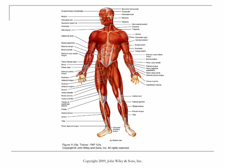
Relationship of skeletal muscles to bones

Copyright 2009, John Wiley & Sons, Inc.
Lever Systems
A lever is a rigid structure that can move around a fixed point called a fulcrum.
A lever is acted on at two different points by two different forces: the effort, which causes movement, and the load or resistance, which opposes movement.
The effort is the force due to muscular contraction; the load is the weight that is moved or some resistance an object to being moved (e.g., weight of a book to be overcome before you can pick it up).
Motion occurs when the effort applied to the bone at the insertion exceeds the load.
Copyright 2009, John Wiley & Sons, Inc.
Types of levers
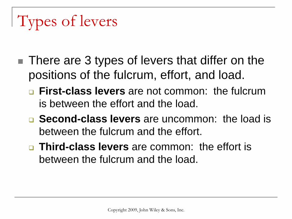
There are 3 types of levers that differ on the positions of the fulcrum, effort, and load. First-class levers are not common: the fulcrum
is between the effort and the load. Second-class levers are uncommon: the load is
between the fulcrum and the effort. Third-class levers are common: the effort is
between the fulcrum and the load.

Copyright 2009, John Wiley & Sons, Inc.
Types of levers

Copyright 2009, John Wiley & Sons, Inc.
Effects of muscle fascicle arrangement
All muscle fibers are parallel to one another within a single fascicle.
Fascicles, however, form patterns with respect to the tendons. Parallel Fusiform Circular Triangular Pennate
Copyright 2009, John Wiley & Sons, Inc.
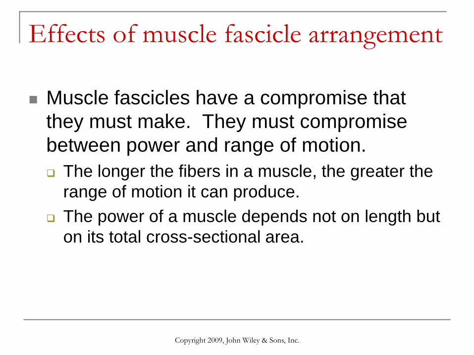
Muscle fascicles have a compromise that they must make. They must compromise between power and range of motion. The longer the fibers in a muscle, the greater the
range of motion it can produce. The power of a muscle depends not on length but
on its total cross-sectional area.
Effects of muscle fascicle arrangement

Copyright 2009, John Wiley & Sons, Inc.
Coordination among muscles
It is common to attribute a specific action at a joint to a single muscle bundle, but remember that muscles do not work in isolation.
Movements usually result from several skeletal muscles acting as a group. Most skeletal muscles are arranged in opposing (antagonistic) pairs at joints (e.g., flexors vs. extensors).
In an opposing muscle pair, one is called the prime mover or agonist and is responsible for the action, while the other muscle called the antagoniststretches and yields to the effects of the agonist.

Copyright 2009, John Wiley & Sons, Inc.
Coordination among muscles
To prevent unwanted movements at other joints or to otherwise aid the movement of the agonist, muscles called synergists contract and stabilize the intermediate joints.
Other muscles act as fixators, stabilizing the origin of the agonist so that the agonist is more efficiently.
Depending upon the movement required, many muscles may act as prime movers, antagonists, synergists, or fixators.
Copyright 2009, John Wiley & Sons, Inc.

Copyright 2009, John Wiley & Sons, Inc.

Copyright 2009, John Wiley & Sons, Inc.
Muscles of facial expression
Muscles of facial expression lie within the subcutaneous layer usually originate in the fascia or skull bones &
insert into the skin. Because of their insertions, the muscles of
facial expression move the skin rather than a joint when they contract.

Copyright 2009, John Wiley & Sons, Inc.
Muscles of Facial Expression

Copyright 2009, John Wiley & Sons, Inc.
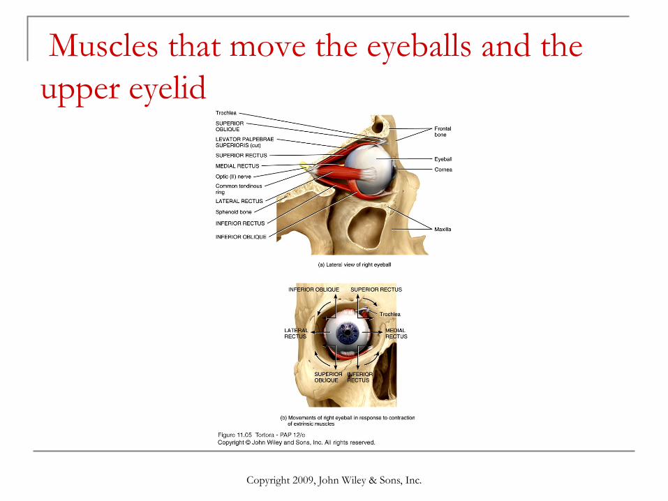
Extrinsic Eye Muscles
Six extrinsic eye muscles control movements of each eyeball. They are called extrinsic because they originate on the outside of the eyeballs in the bony orbit and insert on the outer surface of the sclera.
Those muscles with the word “rectus” in their name have obvious actions (the inferior rectus muscle moves the eye inferiorly so that you would be looking downward).
The actions of the two oblique muscles cannot be deduced from their names. To understand how they move the eye, you must know the origin, insertion, and the unusual ‘path’ that each follows (see 11.5b).
Copyright 2009, John Wiley & Sons, Inc.
Muscles that move the eyeballs and the upper eyelid

Copyright 2009, John Wiley & Sons, Inc.
Muscles that move the mandible
Four pairs of muscles move the mandible, and are known as ‘muscles of mastication’.
The masseter, temporalis, and medial pterygoid account for the strength of the bite.
The medial and lateral pterygoid muscles help to chew by moving the mandible from side to side. Additionally, these muscles protract (protrude) the mandible.

Copyright 2009, John Wiley & Sons, Inc.
Muscles that move the mandible
Copyright 2009, John Wiley & Sons, Inc.
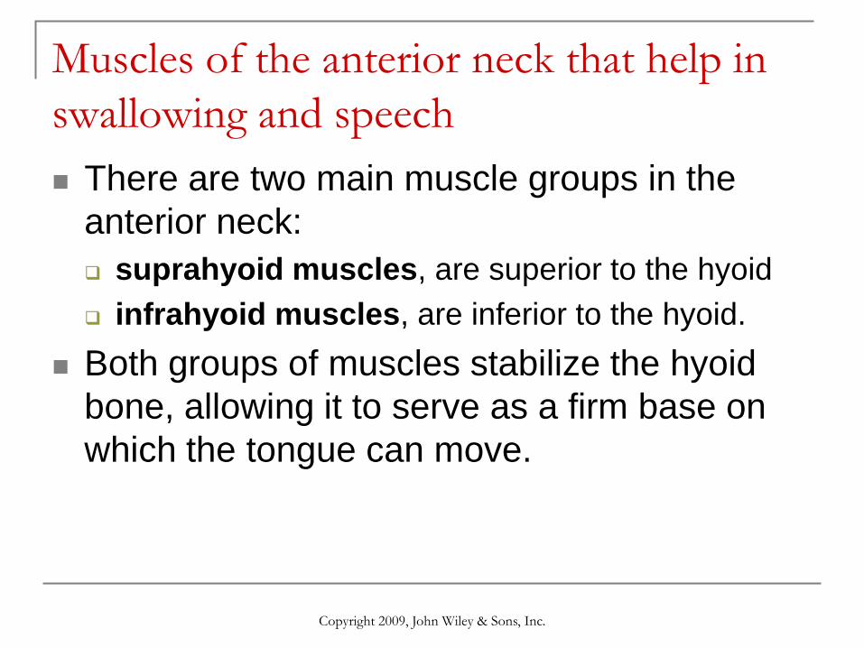
Muscles of the anterior neck that help in swallowing and speech There are two main muscle groups in the
anterior neck: suprahyoid muscles, are superior to the hyoid infrahyoid muscles, are inferior to the hyoid.
Both groups of muscles stabilize the hyoid bone, allowing it to serve as a firm base on which the tongue can move.

Copyright 2009, John Wiley & Sons, Inc.
Muscles of the anterior neck that help in swallowing and speech

Copyright 2009, John Wiley & Sons, Inc.
Muscles of the neck that move the head
The head articulates with the vertebral column at joints formed by the atlas & occipital bone.
Balance and movement of the head involves several neck muscles.
An important landmark (the sternocleidomastoid muscle) divides the sides of the neck into two major triangles: anterior and posterior. The triangles are important anatomically and
surgically because of the structures that lie within their boundaries.

Copyright 2009, John Wiley & Sons, Inc.
Muscles of the neck that move the head

Copyright 2009, John Wiley & Sons, Inc.
Muscles of the Abdomen

Copyright 2009, John Wiley & Sons, Inc.
Muscles of the abdomen that protect the viscera and move the vertebral column The anterolateral abdominal wall includes the
external oblique, internal oblique, and transversus abdominis muscles which form three protective layers around the abdomen.
The muscle fascicles of each layer extend in a different direction, conferring considerable protection to the abdominal viscera.
The aponeuroses of these 3 muscles form the rectus sheaths which enclose the rectus abdominis muscles. The sheaths form the linea alba, a connective tissue band
extending from the xiphoid process to the pubic symphysis.

Copyright 2009, John Wiley & Sons, Inc.
Muscles of the Thorax that Assist in Breathing

Copyright 2009, John Wiley & Sons, Inc.
Muscles of the Thorax that Assist in Breathing Respiratory muscles alter the size of the thoracic
cavity which affects the pressure in the lungs, and that determines whether we inhale or exhale.
The diaphragm is the most important respiratory muscle.
Other important respiratory muscles include the external and internal intercostal muscles.
There are also a number of accessory muscles useful in forced breathing.
Copyright 2009, John Wiley & Sons, Inc.
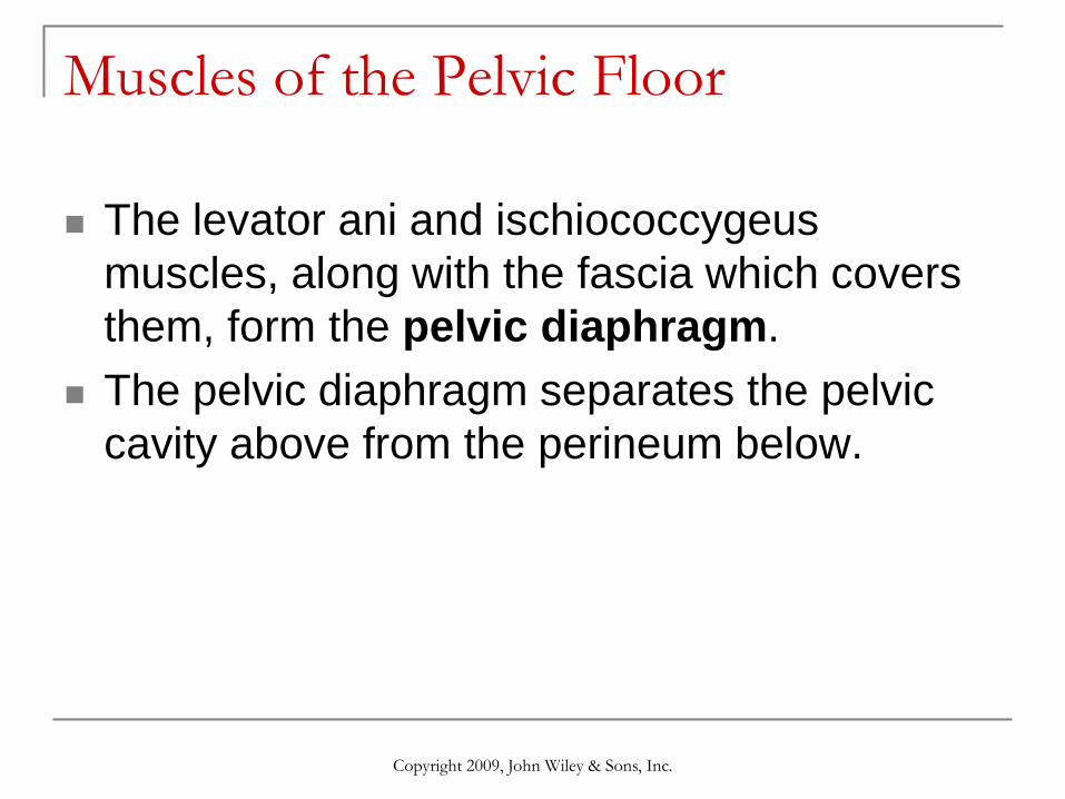
Muscles of the Pelvic Floor
The levator ani and ischiococcygeus muscles, along with the fascia which covers them, form the pelvic diaphragm.
The pelvic diaphragm separates the pelvic cavity above from the perineum below.

Copyright 2009, John Wiley & Sons, Inc.
Muscles of the Pelvic Floor
Copyright 2009, John Wiley & Sons, Inc.
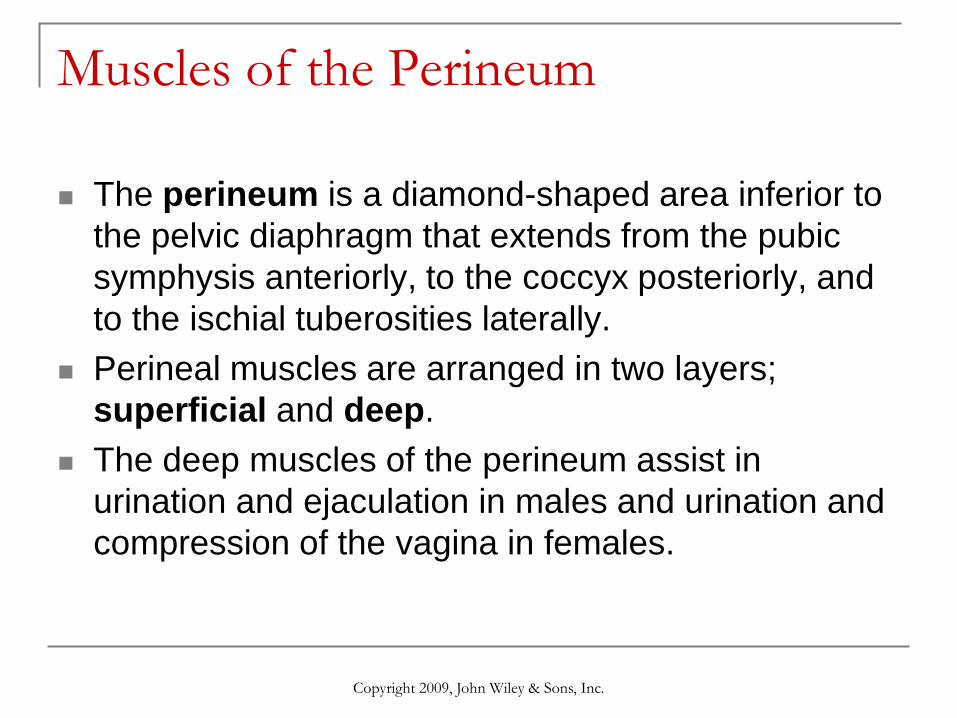
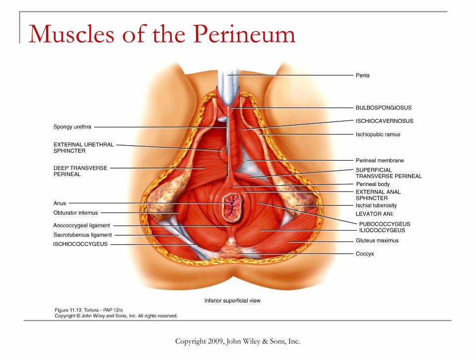
Muscles of the Perineum
The perineum is a diamond-shaped area inferior to the pelvic diaphragm that extends from the pubic symphysis anteriorly, to the coccyx posteriorly, and to the ischial tuberosities laterally.
Perineal muscles are arranged in two layers; superficial and deep.
The deep muscles of the perineum assist in urination and ejaculation in males and urination and compression of the vagina in females.
Copyright 2009, John Wiley & Sons, Inc.
Muscles of the Perineum

Copyright 2009, John Wiley & Sons, Inc.
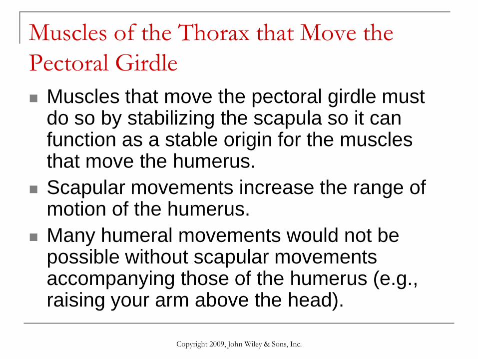
Muscles of the Thorax that Move the Pectoral Girdle
Copyright 2009, John Wiley & Sons, Inc.
Muscles of the Thorax that Move the Pectoral Girdle Muscles that move the pectoral girdle must
do so by stabilizing the scapula so it can function as a stable origin for the muscles that move the humerus.
Scapular movements increase the range of motion of the humerus.
Many humeral movements would not be possible without scapular movements accompanying those of the humerus (e.g., raising your arm above the head).

Copyright 2009, John Wiley & Sons, Inc.
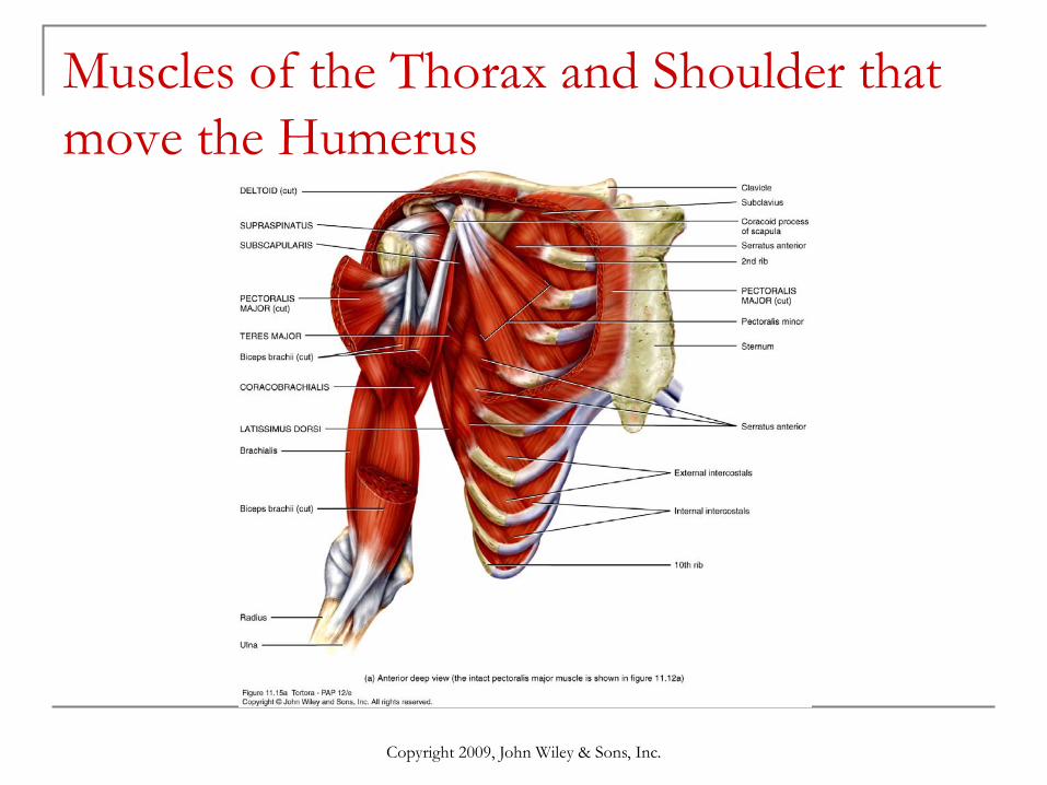
Muscles of the Thorax and Shoulder that move the Humerus Seven of nine muscles that cross the shoulder joint
originate on the scapula, except the pectoralis major and latissimus dorsi.
It is for this reason that the pectoralis major and latissimus dorsi are considered axial muscles.
Four deep shoulder muscles strengthen and stabilize the shallow shoulder joint, and act to join the scapula to the humerus. They form the rotator cuff, a nearly complete circle of tendons around the shoulder joint, like the cuff on a shirtsleeve.
Copyright 2009, John Wiley & Sons, Inc.
Muscles of the Thorax and Shoulder that move the Humerus

Copyright 2009, John Wiley & Sons, Inc.
Muscles of the Thorax that Move the Humerus

Copyright 2009, John Wiley & Sons, Inc.
Muscles of the Arm that Move the Radiusand Ulna Most muscles that move the forearm cause flexion
and extension at the elbow. The biceps brachii, brachialis, and brachioradialis
are flexors. The extensors are the triceps brachii and the anconeus.
Some muscles that move the forearm are involved in pronation and supination. The pronators are the pronator teres and pronator quadratus. Only the supinator can supinate. You use the powerful action of the supinator when you twist a corkscrew or turn a screw with a screwdriver.

Copyright 2009, John Wiley & Sons, Inc.
Muscles of the Arm that Move the Radius and Ulna

Copyright 2009, John Wiley & Sons, Inc.
Muscles of the Forearm that Move the Wrist, Hand, Thumb and Fingers Muscles in this group are known as extrinsic
muscles of the hand because they originate outside the hand and insert within it.
Based on location and function, these muscles are divided into an anterior, and a posterior compartment group.
The tendons of these muscles that continue into the hand are held close to the bones by strong fascial bands called retinacula.

Copyright 2009, John Wiley & Sons, Inc.
Muscles of the Forearm that Move the Wrist, Hand, Thumb and Fingers
Copyright 2009, John Wiley & Sons, Inc.
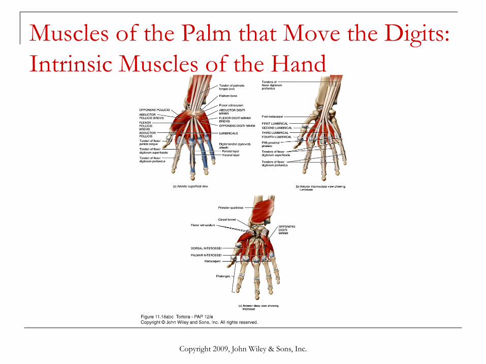
Muscles of the Palm that Move the Digits: Intrinsic Muscles of the Hand

Copyright 2009, John Wiley & Sons, Inc.
Muscles of the Palm that Move the Digits: Intrinsic Muscles of the Hand Intrinsic muscles of the hand produce weak but
precise movements. Intrinsic hand muscles are split into 3 groups:
thenar, hypothenar, & intermediate. The thenar muscles plus the adductor pollicis form the
thenar eminence. Hypothenar muscles act on the little finger and form the
hypothenar eminence. Movements of the thumb are defined in different
planes compared to other digits because the thumb is positioned at a right angle to the other digits.

Copyright 2009, John Wiley & Sons, Inc.
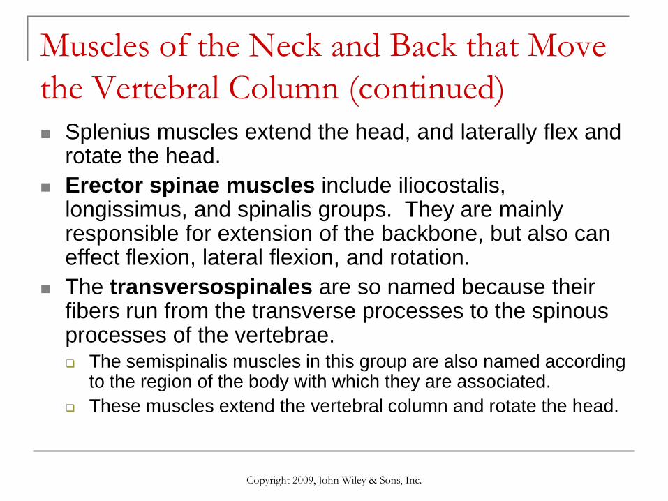
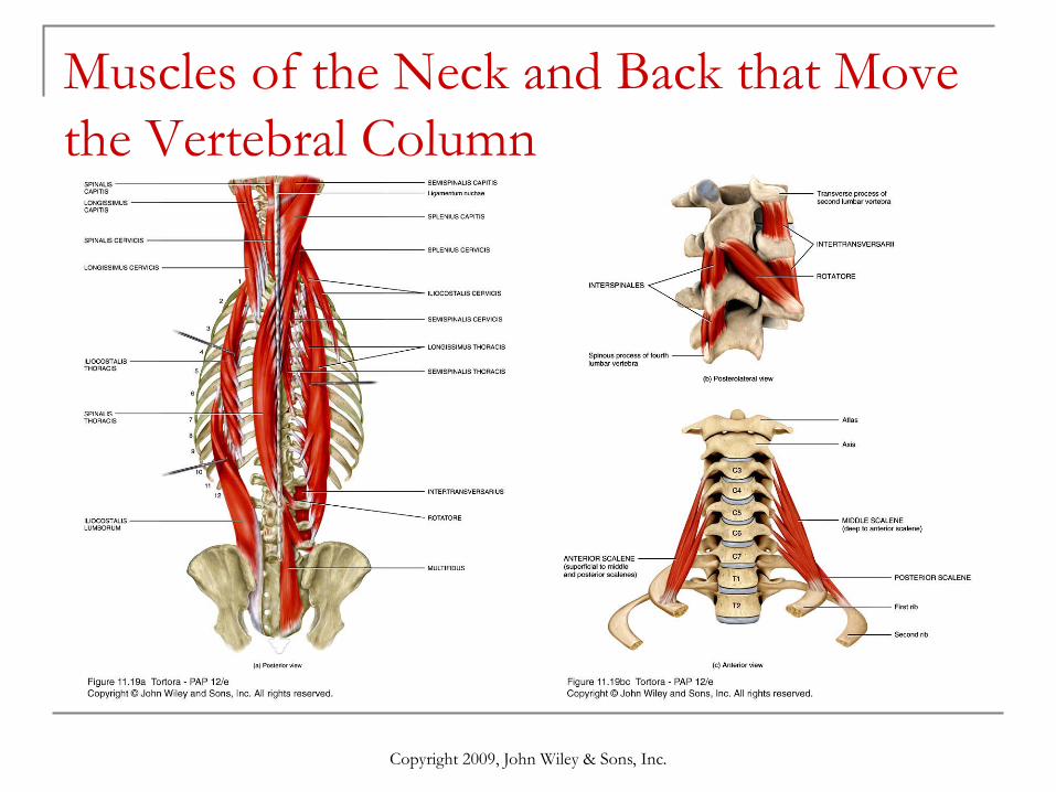
Muscles of the Neck and Back that Move the Vertebral Column Muscles that move the backbone are quite
complex having multiple origins/insertions with considerable overlap among them.
One way to simplify this is to group muscles based on the general direction of the muscle bundles and their lengths.
Many of these muscles are name for the position of the superior attachment site (e.g., splenius capitus is attached to the head).
Copyright 2009, John Wiley & Sons, Inc.
Muscles of the Neck and Back that Move the Vertebral Column (continued) Splenius muscles extend the head, and laterally flex and
rotate the head. Erector spinae muscles include iliocostalis,
longissimus, and spinalis groups. They are mainly responsible for extension of the backbone, but also can effect flexion, lateral flexion, and rotation.
The transversospinales are so named because their fibers run from the transverse processes to the spinous processes of the vertebrae. The semispinalis muscles in this group are also named according
to the region of the body with which they are associated. These muscles extend the vertebral column and rotate the head.
Copyright 2009, John Wiley & Sons, Inc.
Muscles of the Neck and Back that Move the Vertebral Column

Copyright 2009, John Wiley & Sons, Inc.
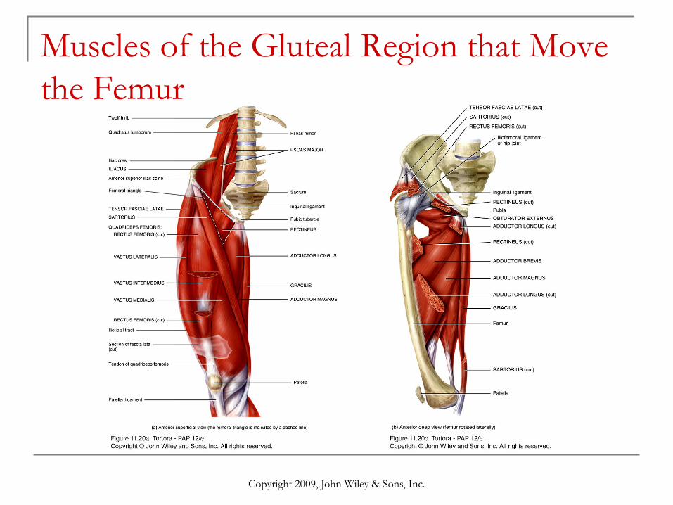
Muscles of the Gluteal Region that Move the Femur Lower limb muscles function in stability,
locomotion, and maintenance of posture. In contrast, upper limb muscles are characterized by versatility of movement.
Muscles of the lower limbs often cross two joints and can act equally on both.
Most muscles that move the femur originate on the pelvic girdle and insert on the femur.
Major muscle groups that move the thigh include the gluteals, and adductor muscles.
Copyright 2009, John Wiley & Sons, Inc.
Muscles of the Gluteal Region that Move the Femur
Copyright 2009, John Wiley & Sons, Inc.

Copyright 2009, John Wiley & Sons, Inc.
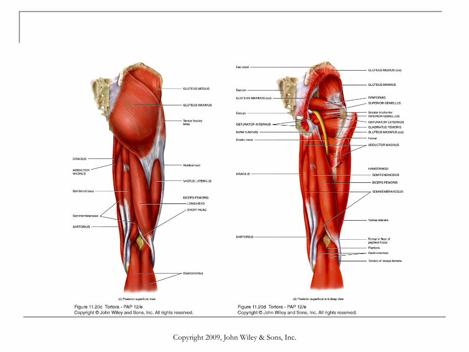
Muscles of the Thigh
Deep fascia separate muscles that act on the femur, and tibia and fibula into medial, anterior, and posterior compartments. medial (adductor) compartment of the thigh
adduct the femur at the hip joint. anterior (extensor) compartment of the thigh
extend the leg (and flex the thigh). posterior (flexor) compartment of the thigh flex
the leg (and extend the thigh).
Copyright 2009, John Wiley & Sons, Inc.
Muscles of the Leg that Move the Foot and Toes Leg muscles, like those of the thigh, are
divided by deep fascia into three compartments: anterior, lateral, and posterior. Anterior compartment muscles dorsiflex the foot. Lateral compartment muscles plantar flex & evert the
foot. Posterior compartment muscles are split between
superficial (e.g., gastrocnemius) and deep (e.g., tibialis posterior) groups. Superficial muscles share a common tendon of insertion, the calcaneal tendon.

Copyright 2009, John Wiley & Sons, Inc.
Muscles of the Leg that Move the Foot and Toes (Fig. 11.24)
Copyright 2009, John Wiley & Sons, Inc.
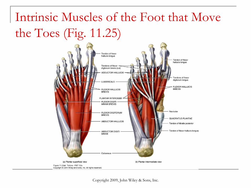
Intrinsic Muscles of the Foot that Move the Toes These muscles are termed intrinsic because
they originate & insert within the foot. These muscles are limited designed for
support and locomotion, and are split into dorsal and plantar groups.
There is only one dorsal muscle which extends toes 2–5 at the MTP joints.
Plantar muscles are arranged in four layers with the most superficial of these called the first layer, etc.
Copyright 2009, John Wiley & Sons, Inc.
Intrinsic Muscles of the Foot that Move the Toes (Fig. 11.25)

Copyright 2009, John Wiley & Sons, Inc.
End of Chapter 11
Copyright 2009 John Wiley & Sons, Inc.All rights reserved. Reproduction or translation of this work beyond that permitted in section 117 of the 1976 United States Copyright Act without express permission of the copyright owner is unlawful. Request for further information should be addressed to the Permission Department, John Wiley & Sons, Inc. The purchaser may make back-up copies for his/her own use only and not for distribution or resale. The Publishers assumes no responsibility for errors, omissions, or damages caused by the use of theses programs or from the use of the information herein