chapter 11 slides 2017 - calvin university
TRANSCRIPT

11/2/17
1
• Biomolecules are large, complex organic molecules that are present in all living things.
• There are FOUR major classes:1. Proteins2. Carbohydrates3. Lipids4. Nucleic acids (DNA and RNA)
• They are BIG and COMPLEX…….but still follow all of the chemical rules you’ve learned so far!
CHAPTER 11: Proteins: Structure and Function
BIOMOLECULES
CHAPTER 11: Proteins: Structure and Function
PROTEIN FUNCTION IS DEPENDENT ON PROPER STRUCTURE!
• What is this molecule?
• What type of biomolecule is it?
• What is its function?
• 11.1 Amino Acids
• 11.2 Chirality and Amino Acids
• 11.3 Peptides
• 11.4 Protein Architecture
• 11.5 Enzymes
CHAPTER 11: Proteins: Structure and Function
OUTLINE

11/2/17
2
• Proteins have some of the most diverse functions of all biological molecules:
• Proteins are composed of amino acids linked together in chains, folded into complex structures
• Key concepts :
CHAPTER 11: Proteins: Structure and Function
OVERVIEW OF PROTEINS
Ø Chemical properties of functional groups
Ø Effect of pH on acidic and basic groups
Ø CatalysisØ Structure
Ø TransportØ Information storage
• All amino acids consist of: 1. a central atom, the a-carbon2. an amine 3. a carboxylic acid4. a hydrogen atom5. a side chain (R group)
CHAPTER 11: Proteins: Structure and Function
PARTS OF AMINO ACIDS
• The identity of an amino acid is determined by its R-groupØ There are 20 different
R-groups possible
• The amino group is basic; the carboxylic acidgroup is acidic:
Ø --NH2 + H2O à --NH3+ + OH-
Ø --COOH + H2O à --COO- + H3O+
• At neutral pH (7.0) both groups are ionized:Ø Compounds containing both a negative and a positive
charge are called zwitterionsØ The charge on amino acids changes with solution pH.
CHAPTER 11: Proteins: Structure and Function
AMINO ACID STRUCTURE AND pH

11/2/17
3
• There are 20 “common” amino acids that can be differentiated by their side chains (R-groups):
• Amino acid properties mostly depend on whether their R-groups are polar or nonpolar:Ø Nonpolar side chains are generally alkanes or
aromatics in structure.Ø Polar side chains may be divided into acidic, basic, or
neutral side chains.
• At physiological pH (~7.4):Ø the 3 basic side chains have positive chargeØ the 2 acidic side chains have negative charge
CHAPTER 11: Proteins: Structure and Function
AMINO ACID SIDE CHAINS
CHAPTER 11: Proteins: Structure and Function
NONPOLAR AMINO ACIDS
*Essential Amino Acid
Alkanes
Aromatics
CHAPTER 11: Proteins: Structure and Function
POLAR AMINO ACIDS
Key functional group:
Carboxylic Acids
Key functional groups:
AlcoholsThiol
Amide
Key functional group:
Amines
Acidic Basic
Neutral
*Essential Amino Acid

11/2/17
4
• About half of all the amino acids can be synthesized by our bodies.
• Essential amino acids are amino acids required in our diets:
Ø The ones we cannot synthesize ourselves
CHAPTER 11: Proteins: Structure and Function
ESSENTIAL AMINO ACIDS
For each of the following amino acids check the boxes that apply to understand their classification:
CHAPTER 11: Proteins: Structure and Function
AFTER CLASS EXERCISE
• Chirality is a property associated with many organic molecules:
Chiral molecules have at least one tetrahedral atom with four different atoms or groups attached
• Chiral molecules have mirror images that cannot be superimposable (think of two hands…)
CHAPTER 11: Proteins: Structure and Function
DEFINING CHIRALITY
Ø Arrangement of the four atoms around the central carbon can occur in two distinct ways
11/2/17
5
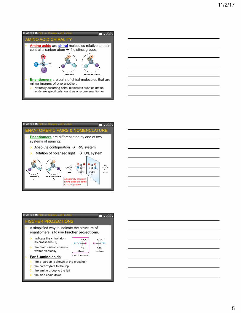
• Amino acids are chiral molecules relative to their central a-carbon atom à 4 distinct groups:
• Enantiomers are pairs of chiral molecules that are mirror images of one another:Ø Naturally occurring chiral molecules such as amino
acids are specifically found as only one enantiomer
CHAPTER 11: Proteins: Structure and Function
AMINO ACID CHIRALITY
• Enantiomers are differentiated by one of two systems of naming:Ø Absolute configuration à R/S systemØ Rotation of polarized light à D/L system
CHAPTER 11: Proteins: Structure and Function
ENANTOMERIC PAIRS & NOMENCLATURE
All naturally occurring amino acids are in the L- configuration
• A simplified way to indicate the structure of enantiomers is to use Fischer projections.
• For L-amino acids:1. the a-carbon is shown at the crosshair 2. the carboxylate to the top3. the amino group to the left4. the side chain down
CHAPTER 11: Proteins: Structure and Function
FISCHER PROJECTIONS
Ø Indicate the chiral atom as crosshairs (+)
Ø the main carbon chain is written vertically

11/2/17
6
• The physical & chemical properties of enantiomers in normal solutions are nearly identical:Ø Except rotation in polarized light
• In biological systems (cells), chiral molecules are differentiated by their interactions with biological molecules, which are also chiral:
CHAPTER 11: Proteins: Structure and Function
PROPERTIES OF ENANTIONMERS
Ø Proteins are composed exclusively of L-amino acids, so they are chiral molecules……SHAPE MATTERS!!
These two drugs are enantiomers with dramatically different effects on human physiology.
CHAPTER 11: Proteins: Structure and Function
A PAIR OF DRUG ENANTIONMERS
Thalidomide
This drug changed the pharmaceutical industry worldwide & strengthened FDA regulations in the United States
Sedative & anti-nausea medicine
Immune & vascular modulator
• Peptides are molecules formed by joining two or more amino acids together into a polymerØ A polymer is a “chain” of repeating chemical units
• Peptides may be: Ø small oligopeptides (2-15 amino acids)
Ø larger polypeptides (usually >50 amino acids)
Ø fully folded polypeptides are called proteins
• To form polymers, amino acids are joined by a special amide bond called a peptide bonds:
CHAPTER 11: Proteins: Structure and Function
LINKING AMINO ACIDS TOGETHER

11/2/17
7
• A peptide bond is an amide bond between two amino acidsØ Peptide bond
formation is a condensation reaction
Ø Peptide bond breakage is hydrolysis
CHAPTER 11: Proteins: Structure and Function
PEPTIDE BOND FORMATION
• The nomenclature for small oligopeptides depends on amino acid number: Ø Two amino acids constitute a dipeptide.Ø Three amino acids constitute a tripeptide.
• The ends of the peptide chain are constant:Ø The N-terminus is always an ammonium ion (-NH3
+) Ø The C-terminus is always carboxylate ion (-COO-)
CHAPTER 11: Proteins: Structure and Function
PEPTIDE STRUCTURE
• Glutathione, an important antioxidant in cells, is a tripeptide:
CHAPTER 11: Proteins: Structure and Function
BIOLOGICALLY RELEVANT OLIGOPEPTIDES
• The endorphin, met-enkephalin, has 5 amino acids. It helps reduce pain following injury.
• Aspartame, commercially the artificial sweetener Nutrasweet™, is s dipeptide (Asp-Phe)

11/2/17
8
• Polypeptides are long polymers of amino acids (>50 amino acids) that can assume complex 3D shapes during the process of folding:
• Proteins are properly folded polypeptides
CHAPTER 11: Proteins: Structure and Function
OVERVIEW OF PROTEINS
Ø Additional molecules (coenzymes, metal ions) are often added to polypeptides to give them function
Proteins are not just “strings”—they have 3D shape!
1. Primary structureØ Sequence of amino acids from N-terminus to C-terminus
2. Secondary structureØ Localized regular folding stabilized by hydrogen bonds
3. Tertiary structureØ Complex irregular folding
of entire protein
4. Quaternary structureØ Association of two or
more subunits
CHAPTER 11: Proteins: Structure and Function
FOUR LEVELS OF PROTEIN STRUCTURE
• The sequence of amino acids determines all other aspects of a protein’s structure and function.Ø If this sequence is altered, the protein may not function
properly.Ø Genetic diseases often involve disruption of a single
amino acid in a protein’s primary structure
• An amino acid is located in sequence by the number of its position from the N-terminus.
CHAPTER 11: Proteins: Structure and Function
PRIMARY STRUCTURE OF A PROTEIN
Normal protein
Mutant protein

11/2/17
9
• Secondary protein structure refers to the regular folding patterns in localized regions of a protein.
• The driving force for the folding of secondary structures is hydrogen bonding:Ø H-bonds in the peptide backbone of proteins are the
most important factor
CHAPTER 11: Proteins: Structure and Function
SECONDARY STRUCTURE
Ø Between carbonyl oxygensand amide hydrogens
Ø Interactions between R-groups is less important in secondary structures
• There are two common secondary structures:
CHAPTER 11: Proteins: Structure and Function
PATTERNS IN SECONDARY STRUCTURE
a-helix = coiled segment of polypeptide held in place by vertical hydrogen bondsbetween adjacent amino acid residues
b-pleated sheet = linear segment of polypeptide held in place by
horizontal hydrogen bondsbetween adjacent strands of
amino acids
Note placement of R-groups in each structure
R-groups point outward
R-groups alternate up & down
• Ribbon diagrams of proteins identify secondary structures with specific shapes:Ø a-helicies are indicated with twisted (red) ribbons
indicating the coiled structureØ b-pleated sheets are indicated with broad flat
arrows, pointing in the direction of the C-terminus
CHAPTER 11: Proteins: Structure and Function
RIBBON DRAWING SECONDARY STRUCTURES
A protein may have any combination of secondary structures, including multiple a-helicies and b-sheets

11/2/17
10
1. Label the partial positive and negative charges in the atoms involved in hydrogen bonding.
2. Why are there partial charges?
3. Orient the peptide structures below to show how they would form hydrogen bonds, and add dashes to show the corresponding hydrogen bonds.
CHAPTER 11: Proteins: Structure and Function
PRACTICE PROBLEMS
• The tertiary structure of proteins describes the entire folding pattern of a single polypeptide:Ø It consists of complex and irregular folding patternsØ Not easily predicted from primary/secondary structure
• Tertiary structure is determined by interactions between the R-groups of amino acid residues:Ø May be interactions between amino acid residues that
are far apart in the primary sequence
CHAPTER 11: Proteins: Structure and Function
TERTIARY STRUCTURE
• There are 4 main types of intermolecular forces between R-groups that provide tertiary structure:1. Dispersion forces (hydrophobic)
2. Hydrogen bonding (hydrophilic)
3. Salt bridges (ionic bond, hydrophilic)
4. Disulfide bridges (covalent bond, hydrophobic)
CHAPTER 11: Proteins: Structure and Function
TYPES OF ELECTROSTATIC INTERACTIONS IN PROTEINS
Note: all four of these interactions are occurring at the same time to stabilize a protein tertiary structure
Increasing bond
strength

11/2/17
11
General, Organic, and Biochemistry Chapter 11: Figure 11.17
Disulfide bridges are covalent bonds between two cysteines.
Hydrogen bonding occurs between polar residues when one residue contains either an N-H or an O-H bond
Salt bridges are ionic bonds between oppositely charged amino acid residues
Dispersion forces are weak interactions between non-polar (hydrophobic residues)
• Thiols (-SH) in adjacent cysteine residues react to form a disulfide bridges (-S-S-). Ø This is an example of an Redox ReactionØ “Perming hair” involves redox of the disulfide bonds in
keratin — a hair protein, giving the hair the shape of the rollers
CHAPTER 11: Proteins: Structure and Function
DISULFIDE BRIDGES
• Dispersion forces are the weakest of the interactions stabilizing protein conformation, but play a critical role in early protein folding steps:Ø Because nonpolar groups only interact with other
nonpolar groups, these amino acids are only in the interior, away from the aqueous environment.
CHAPTER 11: Proteins: Structure and Function
DISPERSION FORCES IN PROTEINS
Rosales, et al. Soft Matter, 2013,9, 8400-8414.
Unfolded polypeptide
Globular structure
Hydrophobic collapse
Ø An early phase in folding is “hydrophobic collapse”of non-polar amino acids into a hydrophobic sphere called a “globule”

11/2/17
12
Indicate the type of bond that would likely form between each of the following pairs of amino acids if they were closely located in a protein.
Choose from the following: disulfide bond, salt bridge, hydrogen bonding, or dispersion forces.
1. aspartic acid and histidine à2. serine and lysine à3. leucine and valine à
4. two cysteines à5. tryptophan and isoleucine à
CHAPTER 11: Proteins: Structure and Function
PRACTICE PROBLEMS
• For some proteins, tertiary structure also includes prosthetic groups:Ø Non-peptide organic molecules or metal ions (or a
combination) that are strongly bound to the proteinØ They are essential to a protein’s function
CHAPTER 11: Proteins: Structure and Function
PROSTHETIC GROUPS
Example
Heme is an organic/iron (Fe2+) cofactor involved in oxygen transport by globin proteins
• Some proteins consist of more than one polypeptide chain; each is termed a subunit
• Quaternary structure is the relative arrangement and position of the subunits within the protein. Ø The subunits are stabilized by the same interactions
responsible for tertiary structure
CHAPTER 11: Proteins: Structure and Function
QUATERNARY STRUCTURE
One subunit(monomer)
Natively folded hemoglobin is
a tetramer

11/2/17
13
• Denaturation of a protein occurs by any process that disrupts its folding:Ø Denaturation disrupts secondary & tertiary structuresØ Protein unfolding is usually irreversible
CHAPTER 11: Proteins: Structure and Function
PROTEIN DENATURATION
• Things that can denature proteins include:1. Heat2. pH changes (acid or base)3. Detergents (“soap”)4. Some metal ions (lead, Pb+2
and mercury, Hg+2)
• Partial denaturation of proteins alters their shape to form abnormal structures:Ø Partially denatured proteins are “sticky”Ø Collections of denatured proteins form
insoluble clusters called aggregates
• Several different neurological diseases are caused by protein misfolding:Ø Mad cow disease (CJD)Ø Alzheimer’s diseaseØ Parkinson’s diseaseØ Amylotrophic lateral sclerosis (ALS)
General, Organic, and Biochemistry Chapter 11: Unnumbered 11.11
PROTEIN MISFOLDING CAN CAUSE DISEASE
• There are several ways to categorize proteins:Ø StructureØ Function
• Categories that combine some of these features: 1. Fibrous proteins
Ø Poorly soluble, consists of long fibers or sheetsØ Primarily function is structural (extracellular matrix; cytoskeleton)
2. Globular proteinsØ Highly soluble; compact, “spherical”Ø Most enzymes fit into this category
3. Membrane proteinsØ Embedded within cell membrane
CHAPTER 11: Proteins: Structure and Function
TYPES OF PROTEINS
Ø SolubilityØ Size

11/2/17
14
• The different types of proteins & other molecules found in a cell can be detected by a method called fluorescence microscopy
CHAPTER 11: Proteins: Structure and Function
TYPES OF PROTEINS
Fibrous proteins (actin) Globular proteins
(mitochondrial enzymes)
Nucleic acids (DNA)
• Enzymes are globular proteins that function as biological catalysts to increase reaction rate.
• An enzyme can increase reaction rate by >1 million (1x10⁶) times!
CHAPTER 11: Proteins: Structure and Function
HOW DO ENZYMES WORK?
Ø Enzymes work by lowering the activation energy(EA) for a reaction.
Ø Enzymes do not alter the overall change in bond energy of a reaction (∆H)
• Enzymes are typically named after the reaction they catalyze, with the suffix –ase added.
• The six classes of enzymes describe all of the types of biochemical reactions that are possible!
CHAPTER 11: Proteins: Structure and Function
ENZYME CLASSIFICATIONS

11/2/17
15
• The active site of an enzyme is where the reaction it catalyzes occurs:
CHAPTER 11: Proteins: Structure and Function
THE ENZYME-SUBSTRATE COMPLEX
Ø Substrates are bound in the active site of an enzyme
Ø The shape of the substrate is complementary to the active site.
• Binding occurs through weak intermolecular forces: Ø Hydrogen bonds, dipole-dipole
interactions, and dispersion forcesØ Example: phosphatases
active site
Phosphoryl group
Hobiger & Friedrich. Front. Pharmacol., 10 February 2015
• The action of an enzyme can be thought of in three distinct steps:1. Enzyme (E) + substrate (S) binding: E + S à ES 2. Chemical reaction: ES à EP3. Release of the product from the enzyme: EP à E + P
CHAPTER 11: Proteins: Structure and Function
THE ENZYME-SUBSTRATE COMPLEX
transition state
• Enzymes often require additional ions or small organic molecules to carry out their reaction:Ø Amino acid R-groups may not be sufficiently reactive
• Cofactors are generally metal ions required by the enzyme for its catalytic function:
Ø Examples include Mg2+, Zn2+, and Fe2+
Ø Especially useful as oxidants in redox reactions
• Coenzymes are small organic molecules required by enzymes for catalytic function:
Ø Examples include NAD+, FAD and coenzyme AØ These often function in redox reactions, too
CHAPTER 11: Proteins: Structure and Function
COFACTORS AND COENZYMES
11/2/17
16
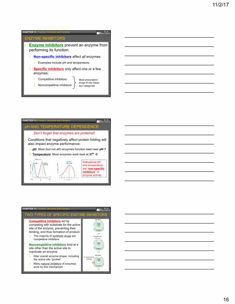
• Enzyme inhibitors prevent an enzyme from performing its function. • Non-specific inhibitors affect all enzymes.
Ø Examples include pH and temperature.
• Specific inhibitors only affect one or a few enzymes:1. Competitive inhibitors
2. Noncompetitive inhibitors
CHAPTER 11: Proteins: Structure and Function
ENZYME INHIBITORS
Most prescription drugs fit into these two categories
Don’t forget that enzymes are proteins!!
• Conditions that negatively affect protein folding will also impact enzyme performance:Ø pH: Most (but not all!) enzymes function best near pH 7Ø Temperature: Most enzymes work best at 37°C
CHAPTER 11: Proteins: Structure and Function
pH AND TEMPERATURE DEPENDENCE
Suboptimal pH and temperature are “non-specific inhibitors” of enzyme activity
• Competitive inhibitors act by competing with substrate for the active site of the enzyme, preventing their binding, and thus formation of product:Ø The majority of synthetic drugs are
competitive inhibitors
• Noncompetitive inhibitors bind at a site other than the active site to inactivate an enzyme:Ø Alter overall enzyme shape, including
the active site “pocket”Ø Many natural inhibitors of enzymes
work by this mechanism
CHAPTER 11: Proteins: Structure and Function
TWO TYPES OF SPECIFIC ENZYME INHIBITORS