chapter 1 - jones & bartlett learning
TRANSCRIPT

1
3RD PAGES
What Is a Cell?
1.1 TheBigPicture
1.2 TheCellIstheFundamentalUnitofLife
1.3 AnOverviewoftheMolecularBuildingBlocksofCellularStructures
1.4 CellsContainDistinctStructuresThatPerformSpecializedFunctions
1.5 CellsinMulticellularOrganismsCanBeHighlySpecializedtoPerformaSubsetoftheFunctionsNecessaryforLife
1.6 ModelOrganismsAreOftenStudiedtoUnderstandMoreComplexOrganisms
1.7 ChapterSummary
1
© Jones & Bartlett Learning, LLC. NOT FOR SALE OR DISTRIBUTION.
© Jones & Bartlett Learning, LLCNOT FOR SALE OR DISTRIBUTION
© Jones & Bartlett Learning, LLCNOT FOR SALE OR DISTRIBUTION
© Jones & Bartlett Learning, LLCNOT FOR SALE OR DISTRIBUTION
© Jones & Bartlett Learning, LLCNOT FOR SALE OR DISTRIBUTION
© Jones & Bartlett Learning, LLCNOT FOR SALE OR DISTRIBUTION
© Jones & Bartlett Learning, LLCNOT FOR SALE OR DISTRIBUTION
© Jones & Bartlett Learning, LLCNOT FOR SALE OR DISTRIBUTION
© Jones & Bartlett Learning, LLCNOT FOR SALE OR DISTRIBUTION
© Jones & Bartlett Learning, LLCNOT FOR SALE OR DISTRIBUTION
© Jones & Bartlett Learning, LLCNOT FOR SALE OR DISTRIBUTION
© Jones & Bartlett Learning, LLCNOT FOR SALE OR DISTRIBUTION
© Jones & Bartlett Learning, LLCNOT FOR SALE OR DISTRIBUTION
© Jones & Bartlett Learning, LLCNOT FOR SALE OR DISTRIBUTION
© Jones & Bartlett Learning, LLCNOT FOR SALE OR DISTRIBUTION
© Jones & Bartlett Learning, LLCNOT FOR SALE OR DISTRIBUTION
© Jones & Bartlett Learning, LLCNOT FOR SALE OR DISTRIBUTION
© Jones & Bartlett Learning, LLCNOT FOR SALE OR DISTRIBUTION
© Jones & Bartlett Learning, LLCNOT FOR SALE OR DISTRIBUTION
© Jones & Bartlett Learning, LLCNOT FOR SALE OR DISTRIBUTION
© Jones & Bartlett Learning, LLCNOT FOR SALE OR DISTRIBUTION
2 Chapter1 WhatIsaCell?
3RD PAGES 3RD PAGES
1.1 The Big PictureTo begin implementing the study strategy outlined in the To the Student section of this book, let’s start by exploring the overall organization of the text as a whole, and this chap-ter in particular. The first 4 chapters of the book address the question, “What are cells made of?” The remaining chapters of the book deal with 10 Principles of Cell Biology. While it is not essential to read the chapters in order, each builds on the material covered in earlier chapters.
This chapter is organized into five major sections, which serves two purposes. First, it provides an overall introduction to the fundamentals of cell biology, including the basic chemistry that helps define how cells are built and how they function. In particular, this introduction focuses on the structural organization of the simplest of the four cellular build-ing blocks: sugars. Second, this chapter includes an overview of the common structures we will encounter in more detail in later chapters. The five major sections are as follows:
�� The first section, The Cell Is the Fundamental Unit of Life, serves as an introduc-tion to some of the essential concepts that form the foundation for supporting life. This includes the definition of life we will use for this book, as well as an examination of the three domains of living organisms. This section also intro-duces the concept that evolution by natural selection takes place at even the most basic levels of living organisms. We will return to this concept repeatedly throughout the book.�� The second section, An Overview of the Molecular Building Blocks of Cellular Structures, introduces some of the basic chemistry that governs how molecules in living organisms interact. This section also lists some of the most common chemical structures found in biomolecules. These form a critical part of the structure–function relationship, another essential concept in biology that we will refer to often.
This section is also where we take our closest look at sugars. This information will be put to immediate use in Chapter 2, where we examine how they are used to synthesize nucleotides, another class of cellular building blocks. Pay particular attention to how sugars are assembled into polymers, as we will be referring to these polymers in several chapters.�� The third section, Cells Contain Distinct Structures That Perform Specialized Functions, focuses on the regions in eukaryotic cells called organelles. The descrip-tions of these structures are intended only to serve as an overview, since the remainder of the chapters will examine each of them in closer detail. It is impor-tant to keep in mind that these organelles are multitaskers, so most will appear in several chapters. Many students are tempted to map an organelle to one or two specific cellular functions (e.g., nucleus = DNA replication), when in fact every organelle can perform many functions at the same time. Thus, when we discuss DNA packing (Chapter 2), DNA replication (Chapter 7), nuclear pore transport (Chapter 8), and regulation of gene expression (Chapter 12), remember that these events are all occurring simultaneously in the nucleus, and they often influ-ence one another. It is important to keep an eye on this big picture of a cell as we explore each of the Principles.�� The fourth section, Cells in Multicellular Organisms Can Be Highly Specialized to Perform a Subset of the Functions Necessary for Life, introduces another impor-tant concept in biology: that the organizing principles we will use to explain how individual cells function can be applied across many different scales of biologi-cal complexity. Here, we will take a quick look at the structure and function of
© Jones & Bartlett Learning, LLC. NOT FOR SALE OR DISTRIBUTION.
© Jones & Bartlett Learning, LLCNOT FOR SALE OR DISTRIBUTION
© Jones & Bartlett Learning, LLCNOT FOR SALE OR DISTRIBUTION
© Jones & Bartlett Learning, LLCNOT FOR SALE OR DISTRIBUTION
© Jones & Bartlett Learning, LLCNOT FOR SALE OR DISTRIBUTION
© Jones & Bartlett Learning, LLCNOT FOR SALE OR DISTRIBUTION
© Jones & Bartlett Learning, LLCNOT FOR SALE OR DISTRIBUTION
© Jones & Bartlett Learning, LLCNOT FOR SALE OR DISTRIBUTION
© Jones & Bartlett Learning, LLCNOT FOR SALE OR DISTRIBUTION
© Jones & Bartlett Learning, LLCNOT FOR SALE OR DISTRIBUTION
© Jones & Bartlett Learning, LLCNOT FOR SALE OR DISTRIBUTION
© Jones & Bartlett Learning, LLCNOT FOR SALE OR DISTRIBUTION
© Jones & Bartlett Learning, LLCNOT FOR SALE OR DISTRIBUTION
© Jones & Bartlett Learning, LLCNOT FOR SALE OR DISTRIBUTION
© Jones & Bartlett Learning, LLCNOT FOR SALE OR DISTRIBUTION
© Jones & Bartlett Learning, LLCNOT FOR SALE OR DISTRIBUTION
© Jones & Bartlett Learning, LLCNOT FOR SALE OR DISTRIBUTION
© Jones & Bartlett Learning, LLCNOT FOR SALE OR DISTRIBUTION
© Jones & Bartlett Learning, LLCNOT FOR SALE OR DISTRIBUTION
© Jones & Bartlett Learning, LLCNOT FOR SALE OR DISTRIBUTION
© Jones & Bartlett Learning, LLCNOT FOR SALE OR DISTRIBUTION

3RD PAGES 3RD PAGES
1.2 theCellIstheFundamentalUnitofLife 3
tissues, the next highest order (after cells) of biological organization. We will need to keep this in mind as we make our way through the chapters, as it helps explain why cells expend so much effort to adhere to and communicate with their neighbors. In Chapter 14, we will look at this scaling issue in much greater detail.�� The fifth section, Model Organisms Are Often Studied to Understand More Com-plex Organisms, explains where most of the information we will examine in later chapters comes from. While we will not be examining any of these organisms in detail in this book, it is important to recognize that at the cellular level, seem-ingly different organisms (such as bacteria, plants, flies, worms, and fish) share nearly as many similarities with us as does the familiar lab mouse, because they are all built on the same principles we will discuss in this book.
This chapter is one of the longest in this book, simply because there are many differ-ent topics to introduce. With the exception of sugars, we will review each of these topics in much greater detail in the chapters to come. While most of the remaining chapters will be somewhat shorter, they will contain a much higher density of critical details. Tackling these will provide students an opportunity to apply the study skills they will develop in this chapter. Welcome to the realm of cell biology! Let’s get started.
1.2 The Cell Is the Fundamental Unit of Life
KeyConCepts
�Cellsareself-replicatingstructuresthatarecapableofrespondingtochangesintheirenvironment.�prokaryotesarethesimplestformsofcells.�eukaryotesarecomplexcellscapableofformingmulticellularorganisms.
For most cell biologists, the definition of the word life is fairly straightforward: it is a chemical system capable of Darwinian evolution. Objects that are alive must therefore have the ability to both generate nearly exact copies of themselves and self-correct (i.e., restore themselves to a defined state by repairing damage). In the hierarchy of biologi-cal complexity, individual molecules, such as proteins or DNA, are not considered to be alive, even if they exist inside a living organism, because they lack one or both of these abilities. Although many molecules (e.g., enzymes) have the ability to catalyze chemical reactions, no single chemical reaction can both replicate and repair these molecules.
From these observations, it is clear that life is a trait possessed only by groups of mol-ecules that work together. Biologists believe that the earliest biological molecules may have been simple molecules capable of self-replication. Once these molecules were enclosed in a selective barrier, called a membrane, they were capable of forming teams that cooper-ated to maintain a fairly constant internal environment, even when conditions outside the membrane varied greatly. Molecular teams that developed the additional ability to repair or replace damaged team members were then able to generate nearly exact replicas of them-selves, including the membrane. This membrane-enclosed team of molecules is now called a cell. The membrane is often referred to as the plasma membrane or cell membrane, and it encloses a compartment called the cytosol or cytoplasm. Because a cell is the simplest liv-ing structure, it is also referred to as the fundamental unit of life (BOX 1-1). All living beings are composed of cells. The simplest cells are called prokaryotes, to distinguish them from more complex cells known as eukaryotes. Examples of different types of cells are shown in FIgUre 1-1.
© Jones & Bartlett Learning, LLC. NOT FOR SALE OR DISTRIBUTION.
© Jones & Bartlett Learning, LLCNOT FOR SALE OR DISTRIBUTION
© Jones & Bartlett Learning, LLCNOT FOR SALE OR DISTRIBUTION
© Jones & Bartlett Learning, LLCNOT FOR SALE OR DISTRIBUTION
© Jones & Bartlett Learning, LLCNOT FOR SALE OR DISTRIBUTION
© Jones & Bartlett Learning, LLCNOT FOR SALE OR DISTRIBUTION
© Jones & Bartlett Learning, LLCNOT FOR SALE OR DISTRIBUTION
© Jones & Bartlett Learning, LLCNOT FOR SALE OR DISTRIBUTION
© Jones & Bartlett Learning, LLCNOT FOR SALE OR DISTRIBUTION
© Jones & Bartlett Learning, LLCNOT FOR SALE OR DISTRIBUTION
© Jones & Bartlett Learning, LLCNOT FOR SALE OR DISTRIBUTION
© Jones & Bartlett Learning, LLCNOT FOR SALE OR DISTRIBUTION
© Jones & Bartlett Learning, LLCNOT FOR SALE OR DISTRIBUTION
© Jones & Bartlett Learning, LLCNOT FOR SALE OR DISTRIBUTION
© Jones & Bartlett Learning, LLCNOT FOR SALE OR DISTRIBUTION
© Jones & Bartlett Learning, LLCNOT FOR SALE OR DISTRIBUTION
© Jones & Bartlett Learning, LLCNOT FOR SALE OR DISTRIBUTION
© Jones & Bartlett Learning, LLCNOT FOR SALE OR DISTRIBUTION
© Jones & Bartlett Learning, LLCNOT FOR SALE OR DISTRIBUTION
© Jones & Bartlett Learning, LLCNOT FOR SALE OR DISTRIBUTION
© Jones & Bartlett Learning, LLCNOT FOR SALE OR DISTRIBUTION

3RD PAGES 3RD PAGES
4 Chapter1 WhatIsaCell?
BOX 1-1 FAQ Are viruses alive?oneissuethatcontinuestobedebatedamongbiologistsiswhetherviruses,whichmustinfectalivingcelltoreplicate,arelivingorganisms.acommonargumentsupportingthe livingorganismdefinition is that virusesare capableof accurately replicating themselvesbyfollowing thesametypeof instructions(thegeneticcodeofDnaandrna) thatallother livingorganismsuse.proponentsofthelivingdefinitionalsopointoutthatviraldependenceonacellforreplicationisnotunlikethedependenceofindividualcellsinamulticellularorganismonthewholeorganism(e.g.,foraskincelltodivide,itrequiresthewholeanimaltobealive).thosewhodisagreewiththispointofviewarguethatvirusescannotself-correct;ifavirusparticleisdamaged,itwillneverrepair thedamage—itcanonlyattempt tocreatenewvirusparticles.Muchof thisdebatestemsfromthefactthatthereisnouniversallyaccepteddefinitionoflife.
FIgUre 1-1 examplesofdifferentcelltypes,includingstructuresspecifictoeach.
(d) Fibroblast
Vacuole
Cell wallChloroplast
(e) Plant cell
Mitochondrion
Rough endoplasmicreticulum
Smooth endoplasmic reticulum
Golgi apparatus
Centrioles Ribosomes
Cytosol
Plasma membrane
Microtubule organizing center
Secretory vesicle
Microvilli
Cytoskeletalfilament
Transportvesicle
(a) Epithelial cell
Nuclearmembrane
ChromatinNucleolus
Nucleus Uterine tube
Uterus
Muscle
Tendon
© Jones & Bartlett Learning, LLC. NOT FOR SALE OR DISTRIBUTION.
© Jones & Bartlett Learning, LLCNOT FOR SALE OR DISTRIBUTION
© Jones & Bartlett Learning, LLCNOT FOR SALE OR DISTRIBUTION
© Jones & Bartlett Learning, LLCNOT FOR SALE OR DISTRIBUTION
© Jones & Bartlett Learning, LLCNOT FOR SALE OR DISTRIBUTION
© Jones & Bartlett Learning, LLCNOT FOR SALE OR DISTRIBUTION
© Jones & Bartlett Learning, LLCNOT FOR SALE OR DISTRIBUTION
© Jones & Bartlett Learning, LLCNOT FOR SALE OR DISTRIBUTION
© Jones & Bartlett Learning, LLCNOT FOR SALE OR DISTRIBUTION
© Jones & Bartlett Learning, LLCNOT FOR SALE OR DISTRIBUTION
© Jones & Bartlett Learning, LLCNOT FOR SALE OR DISTRIBUTION
© Jones & Bartlett Learning, LLCNOT FOR SALE OR DISTRIBUTION
© Jones & Bartlett Learning, LLCNOT FOR SALE OR DISTRIBUTION
© Jones & Bartlett Learning, LLCNOT FOR SALE OR DISTRIBUTION
© Jones & Bartlett Learning, LLCNOT FOR SALE OR DISTRIBUTION
© Jones & Bartlett Learning, LLCNOT FOR SALE OR DISTRIBUTION
© Jones & Bartlett Learning, LLCNOT FOR SALE OR DISTRIBUTION
© Jones & Bartlett Learning, LLCNOT FOR SALE OR DISTRIBUTION
© Jones & Bartlett Learning, LLCNOT FOR SALE OR DISTRIBUTION
© Jones & Bartlett Learning, LLCNOT FOR SALE OR DISTRIBUTION
© Jones & Bartlett Learning, LLCNOT FOR SALE OR DISTRIBUTION

3RD PAGES 3RD PAGES
1.2 theCellIstheFundamentalUnitofLife 5
Cells Are Self-replicating Structures That Are Capable of responding to Changes in Their environment
Division of labor is a common theme in cell biology. Since no single molecule is capable of both self-replication and self-maintenance, these tasks are tackled by cooperating groups of molecules that specialize in specific activities. All cells contain molecular teams respon-sible for accomplishing these essential tasks (BOX 1-2):
�� Maintenance of the internal environment. Living organisms must capture and store energy, and this is accomplished by forming and maintaining chemical dis-equilibrium with the external environment. To remain alive, cells must continually
Nucleoid
Bacteria
Plasmid
Plasma membrane
Outer lipopolysaccharidelayer
Cell envelope
Mesosome
Pilus (fimbria)
CytoplasmCapsule or slime layer(partiallyremoved)
Capsule or slime layer
Flagellum
Cell wall + peptidoglycan
(f) Prokaryotic cell
Muscle
Fascicle
Muscle fiber (cell)
Nuclei
MitochondrionMyofibrils
Sacroplasmicreticulum
Sarcolemma
Transversetubule
(b) Muscle cell
Dendrites
Cell body
Schwann cell
Node of Ranvier
Axon
Axon terminal
(c) Nerve cell
FIgUre 1-1 (Continued)
© Jones & Bartlett Learning, LLC. NOT FOR SALE OR DISTRIBUTION.
© Jones & Bartlett Learning, LLCNOT FOR SALE OR DISTRIBUTION
© Jones & Bartlett Learning, LLCNOT FOR SALE OR DISTRIBUTION
© Jones & Bartlett Learning, LLCNOT FOR SALE OR DISTRIBUTION
© Jones & Bartlett Learning, LLCNOT FOR SALE OR DISTRIBUTION
© Jones & Bartlett Learning, LLCNOT FOR SALE OR DISTRIBUTION
© Jones & Bartlett Learning, LLCNOT FOR SALE OR DISTRIBUTION
© Jones & Bartlett Learning, LLCNOT FOR SALE OR DISTRIBUTION
© Jones & Bartlett Learning, LLCNOT FOR SALE OR DISTRIBUTION
© Jones & Bartlett Learning, LLCNOT FOR SALE OR DISTRIBUTION
© Jones & Bartlett Learning, LLCNOT FOR SALE OR DISTRIBUTION
© Jones & Bartlett Learning, LLCNOT FOR SALE OR DISTRIBUTION
© Jones & Bartlett Learning, LLCNOT FOR SALE OR DISTRIBUTION
© Jones & Bartlett Learning, LLCNOT FOR SALE OR DISTRIBUTION
© Jones & Bartlett Learning, LLCNOT FOR SALE OR DISTRIBUTION
© Jones & Bartlett Learning, LLCNOT FOR SALE OR DISTRIBUTION
© Jones & Bartlett Learning, LLCNOT FOR SALE OR DISTRIBUTION
© Jones & Bartlett Learning, LLCNOT FOR SALE OR DISTRIBUTION
© Jones & Bartlett Learning, LLCNOT FOR SALE OR DISTRIBUTION
© Jones & Bartlett Learning, LLCNOT FOR SALE OR DISTRIBUTION
© Jones & Bartlett Learning, LLCNOT FOR SALE OR DISTRIBUTION

6 Chapter1 WhatIsaCell?
3RD PAGES 3RD PAGES
adjust their internal activities to maintain a consistent environment that differs from conditions outside the cell membrane.�� Sensing the external environment. It is essential that cells be “aware” of changes in the external environment that may impact their own internal environment (e.g., changes in temperature, acidity, nutrient levels, osmotic pressure). Cells contain sensors for relevant environmental conditions such as these and ignore the rest.�� Controlling the flow of molecules into and out of the cell. Cells communicate with their external environment mainly through selective transport of molecules (e.g., cells import nutrients and export metabolic waste). Controlling this molec-ular traffic also helps cells maintain chemical disequilibrium and sense their surroundings.�� Catalyzing chemical reactions. In order to maintain a consistent internal environ-ment, cells must be able to control the chemical reactions taking place within them. Molecules called enzymes play an important role in regulating these reactions.�� Generating useful energy. To catalyze most chemical reactions and do any form of work, cells must expend energy. Many molecules in cells are devoted to capturing energy from outside of the cell (e.g., sunlight and “food”) and converting it into a small number of energy forms that cells can use directly. A well-known example of a useful energy form is adenosine triphosphate, otherwise known as ATP.�� Storing genetic information. Cells contain instructions for manufacturing most of the biological molecules necessary to stay alive. These instructions are stored in the form of a simple molecular polymer called deoxyribonucleic acid, or DNA.�� Synthesis of biological molecules. A considerable amount of the energy cap-tured by cells is used to construct new biological molecules inside cells. These molecules may serve to replace damaged molecules, permit new functions in the cell, or generate sufficient copies of a molecule for the cell to replicate. To gener-ate a nearly exact copy of itself, a cell must ensure that all information stored in its DNA is present in the newly created daughter cell. Cells possess molecular teams responsible for accurately replicating DNA and properly segregating it during cell division.�� Regulating information flow. Much like teams of people that communicate with one another to accomplish a complex task, molecular teams in a cell communi-cate with one another as well. Some molecules specialize in transferring informa-tion from one team to another.
BOX 1-2 The “cell as a busy city” analogy.Becausemoststudentsnewtocellbiologyareunfamiliarwithcells,theycouldtrythinkingofacellassomethingsimilartowhatisalreadyfamiliar:averybusycity.Considerhowtodescribetheconceptofacitytosomeonewhohasneverbeenin(orevenseen)one.It’sadauntingtask.hoveringoveranylarge,busycityinahelicopterandlookingdownatit,wemightstartbydescribingthegeneralfunction(s)ofthelargeststructureswecansee(factories,officebuildings,schools,roads,etc.)withoutdelvingintohowtheyallworktogethertokeepthecityfunctioning.Likewise,wemightdescribethegeneralconceptof“people”inourinitialdescriptionofacity,butwecertainlywouldnotwanttostartdescribingeverysinglepersoninthatcity.thepurposeofthischapter,then,isto(1)introducethebuildingblocksofthe“bigbuildings”incells(organelles,proteins,andotherstructures),(2)surveysomeofthemostcommon“bigbuildings”(nucleus,plasmamembrane,mitochondria,etc.),and(3)introducetheconceptthatnotall“cities”are alike (i.e., they can be clustered into different regions—tissues—that work together). We’llreturntothisanalogyinotherchapters,andasweaddmoredetailtotheanalogy,wecanbringthiscity/celltolife,withthegoalofarrivingataclearpictureofhowcellsfunctionatthemolecularlevel.
© Jones & Bartlett Learning, LLC. NOT FOR SALE OR DISTRIBUTION.
© Jones & Bartlett Learning, LLCNOT FOR SALE OR DISTRIBUTION
© Jones & Bartlett Learning, LLCNOT FOR SALE OR DISTRIBUTION
© Jones & Bartlett Learning, LLCNOT FOR SALE OR DISTRIBUTION
© Jones & Bartlett Learning, LLCNOT FOR SALE OR DISTRIBUTION
© Jones & Bartlett Learning, LLCNOT FOR SALE OR DISTRIBUTION
© Jones & Bartlett Learning, LLCNOT FOR SALE OR DISTRIBUTION
© Jones & Bartlett Learning, LLCNOT FOR SALE OR DISTRIBUTION
© Jones & Bartlett Learning, LLCNOT FOR SALE OR DISTRIBUTION
© Jones & Bartlett Learning, LLCNOT FOR SALE OR DISTRIBUTION
© Jones & Bartlett Learning, LLCNOT FOR SALE OR DISTRIBUTION
© Jones & Bartlett Learning, LLCNOT FOR SALE OR DISTRIBUTION
© Jones & Bartlett Learning, LLCNOT FOR SALE OR DISTRIBUTION
© Jones & Bartlett Learning, LLCNOT FOR SALE OR DISTRIBUTION
© Jones & Bartlett Learning, LLCNOT FOR SALE OR DISTRIBUTION
© Jones & Bartlett Learning, LLCNOT FOR SALE OR DISTRIBUTION
© Jones & Bartlett Learning, LLCNOT FOR SALE OR DISTRIBUTION
© Jones & Bartlett Learning, LLCNOT FOR SALE OR DISTRIBUTION
© Jones & Bartlett Learning, LLCNOT FOR SALE OR DISTRIBUTION
© Jones & Bartlett Learning, LLCNOT FOR SALE OR DISTRIBUTION
© Jones & Bartlett Learning, LLCNOT FOR SALE OR DISTRIBUTION
3RD PAGES 3RD PAGES
1.2 theCellIstheFundamentalUnitofLife 7
Prokaryotes Are the Simplest Form of Cells
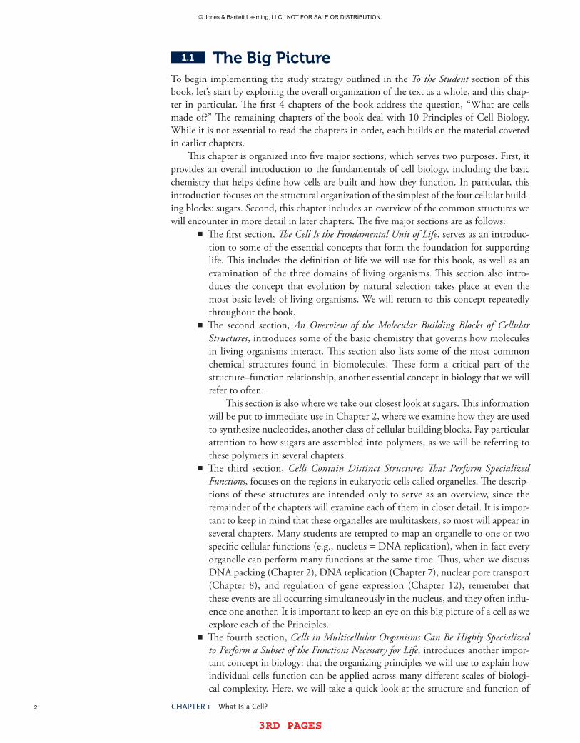
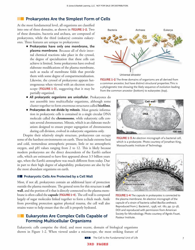
At the most fundamental level, all organisms are classified into one of three domains, as shown in FIgUre 1-2. Two of these domains, bacteria and archaea, are composed of prokaryotes, while the third (eukarya) contains eukary-otes. Three features are unique to prokaryotes:
�� Prokaryotes have only one membrane, the plasma membrane. Because all of their inter-nal chemical reactions take place in the cytosol, the degree of specialization that these cells can achieve is limited. Some prokaryotes have evolved elaborate modifications of the plasma membrane, such as stacks of membrane folds that provide them with some degree of compartmentalization. Likewise, the cytosol of prokaryotes appears het-erogeneous when viewed with an electron micro-scope (FIgUre 1-3), suggesting that it may be partially organized.�� All prokaryotic organisms are unicellular. Prokaryotes do not assemble into multicellular organisms, although some cluster together to form enormous structures called biofilms.�� Prokaryotes do not divide by mitosis. Most genetic informa-tion in prokaryotic cells is contained in a single circular DNA molecule called the chromosome, while eukaryotic cells con-tain several chromosomes. Mitosis, which is an elaborate mech-anism designed to ensure proper segregation of chromosomes during cell division, evolved in eukaryotic organisms only.
Despite their relatively simple structure, prokaryotes can occupy some of the harshest environments on earth. These include extreme heat and cold, tremendous atmospheric pressure, little or no atmospheric oxygen, and pH values ranging from 2 to 12. This is likely because modern prokaryotes are the direct descendents of the Earth’s earliest cells, which are estimated to have first appeared about 3.5 billion years ago, when the Earth’s atmosphere was much different from today. Due in part to their high degree of adaptability, prokaryotes are also by far the most abundant organisms on earth.
Prokaryotic Cells Are Protected by a Cell Wall
Most, if not all, prokaryotes contain an additional layer of protection outside the plasma membrane. The general term for this structure is cell wall, and the portion of it that is directly connected to the plasma mem-brane is often called the capsule (FIgUre 1-4). The cell wall is composed largely of sugar molecules linked together to form a thick mesh. Aside from providing protection against physical trauma, the cell wall also retains water to help ensure that the cell is properly hydrated.
eukaryotes Are Complex Cells Capable of Forming Multicellular Organisms
Eukaryotic cells comprise the third, and most recent, domain of biological organisms shown in Figure 1-2. When viewed under a microscope, the most striking feature of
Bacteria Archaea Eukarya
Universal ancestor
FIgUre 1-2 thethreedomainsoforganismsareallderivedfromacommonancestor,buthavedistinctstructuralproperties.thisisaphylogenetictreeshowingthelikelysequenceofevolutionleadingfromthecommonancestor(bottom)toeukaryotes(top).
100 nm
Cell wall
DNA
FIgUre 1-3 anelectronmicrographofabacterialcell,whichisaprokaryote.photocourtesyofJonathanKing,MassachusettsInstituteoftechnology.
Capsule
S-layer
Peptidoglycan
Plasmic membrane
250250500 nm nm nmnmnmnmmm250 nm
FIgUre 1-4 thecapsuleinprokaryotesisconnectedtotheplasmamembrane.anelectronmicrographofthecapsuleofastrainofbacteriacalledBacillus anthracis.reproducedfromJ.Bacteriol.,1998,vol.180,pp.52–58,DoIandreproducedwithpermissionfromamericansocietyforMicrobiology.photocourtesyofagnésFouet,pasteurInstitute.
© Jones & Bartlett Learning, LLC. NOT FOR SALE OR DISTRIBUTION.
© Jones & Bartlett Learning, LLCNOT FOR SALE OR DISTRIBUTION
© Jones & Bartlett Learning, LLCNOT FOR SALE OR DISTRIBUTION
© Jones & Bartlett Learning, LLCNOT FOR SALE OR DISTRIBUTION
© Jones & Bartlett Learning, LLCNOT FOR SALE OR DISTRIBUTION
© Jones & Bartlett Learning, LLCNOT FOR SALE OR DISTRIBUTION
© Jones & Bartlett Learning, LLCNOT FOR SALE OR DISTRIBUTION
© Jones & Bartlett Learning, LLCNOT FOR SALE OR DISTRIBUTION
© Jones & Bartlett Learning, LLCNOT FOR SALE OR DISTRIBUTION
© Jones & Bartlett Learning, LLCNOT FOR SALE OR DISTRIBUTION
© Jones & Bartlett Learning, LLCNOT FOR SALE OR DISTRIBUTION
© Jones & Bartlett Learning, LLCNOT FOR SALE OR DISTRIBUTION
© Jones & Bartlett Learning, LLCNOT FOR SALE OR DISTRIBUTION
© Jones & Bartlett Learning, LLCNOT FOR SALE OR DISTRIBUTION
© Jones & Bartlett Learning, LLCNOT FOR SALE OR DISTRIBUTION
© Jones & Bartlett Learning, LLCNOT FOR SALE OR DISTRIBUTION
© Jones & Bartlett Learning, LLCNOT FOR SALE OR DISTRIBUTION
© Jones & Bartlett Learning, LLCNOT FOR SALE OR DISTRIBUTION
© Jones & Bartlett Learning, LLCNOT FOR SALE OR DISTRIBUTION
© Jones & Bartlett Learning, LLCNOT FOR SALE OR DISTRIBUTION
© Jones & Bartlett Learning, LLCNOT FOR SALE OR DISTRIBUTION

8 Chapter1 WhatIsaCell?
3RD PAGES 3RD PAGES
eukaryotic cells is that their cytosol is highly organized. Even the simplest microscope reveals the presence of a large, often oval-shaped structure called the nucleus. In fact, the presence of a nucleus is the defining feature of eukaryotic cells. Closer examination with more powerful microscopes allows us to see additional distinct structures in the cytosol (FIgUre 1-5). These structures are generally classified into two groups:
�� Cytosolic structures that are surrounded by at least one distinct membrane are called organelles. The presence of these membranes allows the cell to create specialized compartments in the cytosol that are devoted to performing a subset of cellular tasks under optimized conditions. Like the plasma membrane, these membranes are selectively permeable, which helps to create a unique internal environment optimally suited to the molecules contained inside. Because it is membrane bound, the nucleus is classified as an organelle. Additional organelles found in eukaryotic cells include the endoplasmic reticulum, Golgi apparatus, mitochondria, chloroplasts, endosomes, lysosomes, and peroxisomes. Each of these organelles contains unique molecules and performs a separate set of functions.�� Large molecular complexes that are not enclosed in a separate membrane do not have a generic name (like organelle), but they do share one important trait: they represent specialized regions in the cytosol that are devoted to a subset of cellular tasks. For example, eukaryotic cells contain three different types of fibers that serve as a scaffold for the organization of the cytosol, thereby earning them the name cytoskeleton. Careful arrangement of the cytoskeleton is essential for proper cell function; without it, muscle cells would not contract and nerve cells would fall silent.
Some eukaryotic Cells Contain Structures That Allow Them to Form Multicellular Clusters
One obvious advantage to possessing such a high degree of compartmentalization is that eukaryotic cells can customize their cytosol to generate a tremendous number of different cell types. Approximately one billion years after cells first appeared, some eukaryotic cells acquired the ability to work together in groups, giving rise to multicellular organisms. The structural basis for multicellularity is revealed by a relatively small number of structures that hold adjacent cells together. Many of these structures are also linked to the cytoskeleton, thereby creating a supercytoskeleton that spans multiple cells and helps integrate them into a single functioning unit.
ConCeptCheCK#1
explainwhyalllivingorganismsareenclosedbyatleastonemembrane.
1.3 An Overview of the Molecular Building Blocks of Cellular Structures
KeyConCepts
�thestructure–functionrelationshipisapowerfultoolforunderstandingcellularorganization.�Wateristhemostcommoncompoundfoundincells.�thestudyofcellularchemistrybeginswithanexaminationofthecarbonatom.�Complexbiomoleculesaremostlycomposedofchemicalbuildingblockscalledfunctionalgroups.�Lipids,sugars,aminoacids,andnucleicacidsarethemostcommonbiomoleculesincells.
© Jones & Bartlett Learning, LLC. NOT FOR SALE OR DISTRIBUTION.
© Jones & Bartlett Learning, LLCNOT FOR SALE OR DISTRIBUTION
© Jones & Bartlett Learning, LLCNOT FOR SALE OR DISTRIBUTION
© Jones & Bartlett Learning, LLCNOT FOR SALE OR DISTRIBUTION
© Jones & Bartlett Learning, LLCNOT FOR SALE OR DISTRIBUTION
© Jones & Bartlett Learning, LLCNOT FOR SALE OR DISTRIBUTION
© Jones & Bartlett Learning, LLCNOT FOR SALE OR DISTRIBUTION
© Jones & Bartlett Learning, LLCNOT FOR SALE OR DISTRIBUTION
© Jones & Bartlett Learning, LLCNOT FOR SALE OR DISTRIBUTION
© Jones & Bartlett Learning, LLCNOT FOR SALE OR DISTRIBUTION
© Jones & Bartlett Learning, LLCNOT FOR SALE OR DISTRIBUTION
© Jones & Bartlett Learning, LLCNOT FOR SALE OR DISTRIBUTION
© Jones & Bartlett Learning, LLCNOT FOR SALE OR DISTRIBUTION
© Jones & Bartlett Learning, LLCNOT FOR SALE OR DISTRIBUTION
© Jones & Bartlett Learning, LLCNOT FOR SALE OR DISTRIBUTION
© Jones & Bartlett Learning, LLCNOT FOR SALE OR DISTRIBUTION
© Jones & Bartlett Learning, LLCNOT FOR SALE OR DISTRIBUTION
© Jones & Bartlett Learning, LLCNOT FOR SALE OR DISTRIBUTION
© Jones & Bartlett Learning, LLCNOT FOR SALE OR DISTRIBUTION
© Jones & Bartlett Learning, LLCNOT FOR SALE OR DISTRIBUTION
© Jones & Bartlett Learning, LLCNOT FOR SALE OR DISTRIBUTION

3RD PAGES 3RD PAGES
1.3 anoverviewoftheMolecularBuildingBlocksofCellularstructures 9
10X magnification
10X magnification
10X magnification
10X magnification
FIgUre 1-5 subcellularstructurescanbevisualizedwithlightmicroscopesorelectronmicroscopes.©sebastianKaulitzki/shutterstock,Inc.,©Dr.thomasDeerinck/VisualsUnlimited,Inc.,©peterarnold,Inc./alamy,©CnrI/photoresearchers,Inc.,©Keithr.porter/photoresearchers,Inc.
© Jones & Bartlett Learning, LLC. NOT FOR SALE OR DISTRIBUTION.
© Jones & Bartlett Learning, LLCNOT FOR SALE OR DISTRIBUTION
© Jones & Bartlett Learning, LLCNOT FOR SALE OR DISTRIBUTION
© Jones & Bartlett Learning, LLCNOT FOR SALE OR DISTRIBUTION
© Jones & Bartlett Learning, LLCNOT FOR SALE OR DISTRIBUTION
© Jones & Bartlett Learning, LLCNOT FOR SALE OR DISTRIBUTION
© Jones & Bartlett Learning, LLCNOT FOR SALE OR DISTRIBUTION
© Jones & Bartlett Learning, LLCNOT FOR SALE OR DISTRIBUTION
© Jones & Bartlett Learning, LLCNOT FOR SALE OR DISTRIBUTION
© Jones & Bartlett Learning, LLCNOT FOR SALE OR DISTRIBUTION
© Jones & Bartlett Learning, LLCNOT FOR SALE OR DISTRIBUTION
© Jones & Bartlett Learning, LLCNOT FOR SALE OR DISTRIBUTION
© Jones & Bartlett Learning, LLCNOT FOR SALE OR DISTRIBUTION
© Jones & Bartlett Learning, LLCNOT FOR SALE OR DISTRIBUTION
© Jones & Bartlett Learning, LLCNOT FOR SALE OR DISTRIBUTION
© Jones & Bartlett Learning, LLCNOT FOR SALE OR DISTRIBUTION
© Jones & Bartlett Learning, LLCNOT FOR SALE OR DISTRIBUTION
© Jones & Bartlett Learning, LLCNOT FOR SALE OR DISTRIBUTION
© Jones & Bartlett Learning, LLCNOT FOR SALE OR DISTRIBUTION
© Jones & Bartlett Learning, LLCNOT FOR SALE OR DISTRIBUTION
© Jones & Bartlett Learning, LLCNOT FOR SALE OR DISTRIBUTION

10 Chapter1 WhatIsaCell?
3RD PAGES 3RD PAGES
Much of the remainder of this book is devoted to exploring the organization and molec-ular function of membranes, organelles, and cytosolic structures in eukaryotic cells. Along the way, we will be guided by a very powerful paradigm in biology called the structure–function relationship. The central tenet of the structure–function relationship is that the function of any biological entity (ranging from an individual molecule to a vast ecosystem) can be determined by understanding its structure. We will use the structure–function rela-tionship to understand how molecules in cells function, but to do so, we must have a good understanding of the chemical principles that govern molecular structure (BOX 1-3). We will review the fundamentals here (BOX 1-4).
Water Is the Most Common Compound Found in Cells
All living cells, including the driest plant seeds and fungal spores, contain water. In most cells, water is the most abundant molecule. It is likely that the very first chemical reactions that lead to the formation of cells occurred in some form of liquid water (the so-called pri-mordial soup). Consequently, every chemical reaction that takes place in living organisms reflects this ancestry. To understand life, we need to understand water.
Water is unusual in at least five aspects, as shown in FIgUre 1-6:�� First, it is the only molecule of its size that exists as a liquid at room temperature and normal atmospheric pressure (compounds of a similar mass, such as meth-ane and ammonia, form a gas under these conditions).�� This can be explained by its second unusual trait: it is a very polar molecule. Specifically, the high electronegativity of oxygen causes the hydrogen electron involved in the covalent bond to spend the majority of its time circling the nucleus of the oxygen atom. This creates an imbalanced bond; because the electron orbits mostly around the oxygen, the oxygen acquires a partial negative charge (typi-cally represented by the symbol d-), while each hydrogen atom becomes par-tially positive (represented as d+). The imbalance in charge is what holds water
BOX 1-3 TIP Overcoming the jargon barrier.agoodportionofthischapterdiscussesthechemicalprinciplesthatgoverncellstructureandfunction,andintroducesafairlylargenumberofspecializedwords.thefieldsofbiologyandchemistryhavetheirownjargon,andthesewordsoftenmakeitdifficultforstudentstoseetheconnectionsbetweenbasicconcepts.hereisatip:studentsshouldbeabletoexplaintherelationshipsdiscussedhereintheirownwordsbeforetheystartmemorizingthejargon.It isoftendifficult to recall specificwordswhen theunderlyingconcept isunclear, and thesheernumberoftechnicaltermsbiologistsusewilloverwhelmtheshort-termmemorycapacityofeventhebeststudents.Concept before vocabularyisagoodhabitinthisarea.
BOX 1-4 FAQ How much chemistry do I need to know to understand the subjects in this book?thischaptercontainsarelativelyshortsummaryofabroadrangeofchemicalprinciples,includingtopicsoftencoveredinorganicchemistrycourses.this istohelpstudentsthathaveorganicchemistryexperienceapplythisknowledgetothesubjects incellbiology,butthisdoesnotmeanstudentsmustmasterorganicchemistrytolearnfromthisbook.Infact,onecanexploreagreatdealofcellbiologywithanunderstandingofchemistryatthelevelcoveredinmostintroductorycollegecoursesingeneralchemistry.Ifstudentshaveanyconcernsaboutthelevelofchemistryexpectedintheircourse,theyshouldcheckwiththeirinstructor.
© Jones & Bartlett Learning, LLC. NOT FOR SALE OR DISTRIBUTION.
© Jones & Bartlett Learning, LLCNOT FOR SALE OR DISTRIBUTION
© Jones & Bartlett Learning, LLCNOT FOR SALE OR DISTRIBUTION
© Jones & Bartlett Learning, LLCNOT FOR SALE OR DISTRIBUTION
© Jones & Bartlett Learning, LLCNOT FOR SALE OR DISTRIBUTION
© Jones & Bartlett Learning, LLCNOT FOR SALE OR DISTRIBUTION
© Jones & Bartlett Learning, LLCNOT FOR SALE OR DISTRIBUTION
© Jones & Bartlett Learning, LLCNOT FOR SALE OR DISTRIBUTION
© Jones & Bartlett Learning, LLCNOT FOR SALE OR DISTRIBUTION
© Jones & Bartlett Learning, LLCNOT FOR SALE OR DISTRIBUTION
© Jones & Bartlett Learning, LLCNOT FOR SALE OR DISTRIBUTION
© Jones & Bartlett Learning, LLCNOT FOR SALE OR DISTRIBUTION
© Jones & Bartlett Learning, LLCNOT FOR SALE OR DISTRIBUTION
© Jones & Bartlett Learning, LLCNOT FOR SALE OR DISTRIBUTION
© Jones & Bartlett Learning, LLCNOT FOR SALE OR DISTRIBUTION
© Jones & Bartlett Learning, LLCNOT FOR SALE OR DISTRIBUTION
© Jones & Bartlett Learning, LLCNOT FOR SALE OR DISTRIBUTION
© Jones & Bartlett Learning, LLCNOT FOR SALE OR DISTRIBUTION
© Jones & Bartlett Learning, LLCNOT FOR SALE OR DISTRIBUTION
© Jones & Bartlett Learning, LLCNOT FOR SALE OR DISTRIBUTION
© Jones & Bartlett Learning, LLCNOT FOR SALE OR DISTRIBUTION

3RD PAGES 3RD PAGES
1.3 anoverviewoftheMolecularBuildingBlocksofCellularstructures 11
It takes more heat energy to raise the temperature of water than is needed to raise the temperature of CH4 or CO2.
(a) Water is a liquid at room temperature.
Other molecules of similar size (CH4, CO2) are gasses at room temperature.
Hydrogen bonds form between partially charged atoms of opposite polarity.
(b) Water is a polar molecule.
H2O
H2O
CH4 CO2
CH4 CO2H2O
CH4 CO2
CH4 CO2
(c) The liquid phase of water is more dense than its solid phase (ice).
(d) Water has a high specific heat and is a good insulator.
(e) Water has a high heat of evaporation.
H2O
δ−
δ−
δ−
δ+
δ+
δ+
δ+
δ+
δ+
Ice cubes
C HH
H
H
C OO
CH4 and CO2 do not have partially charged atoms
and therefore do not form hydrogen bonds.
When liquid water is cooled enough to form a solid, the solid water is less dense, so ice floats on liquid water.
If liquid CH4 or CO2 were cooled enough to form solids, these solids would sink to the bottom of the liquid phase.
FIgUre 1-6 Fiveunusualtraitsofwater.
molecules together so tightly: the d- of the oxygen atom attracts the d+ of the hydrogen atoms in nearby water molecules, and vice versa. This phenomenon occurs in other molecules as well, and it is called hydrogen bonding.�� A third unusual trait of water is that its liquid phase has a higher density than its solid phase in standard conditions (i.e., ice floats in water). Again, this can be explained by the ubiquitous hydrogen bonding that occurs in liquid water, as
© Jones & Bartlett Learning, LLC. NOT FOR SALE OR DISTRIBUTION.
© Jones & Bartlett Learning, LLCNOT FOR SALE OR DISTRIBUTION
© Jones & Bartlett Learning, LLCNOT FOR SALE OR DISTRIBUTION
© Jones & Bartlett Learning, LLCNOT FOR SALE OR DISTRIBUTION
© Jones & Bartlett Learning, LLCNOT FOR SALE OR DISTRIBUTION
© Jones & Bartlett Learning, LLCNOT FOR SALE OR DISTRIBUTION
© Jones & Bartlett Learning, LLCNOT FOR SALE OR DISTRIBUTION
© Jones & Bartlett Learning, LLCNOT FOR SALE OR DISTRIBUTION
© Jones & Bartlett Learning, LLCNOT FOR SALE OR DISTRIBUTION
© Jones & Bartlett Learning, LLCNOT FOR SALE OR DISTRIBUTION
© Jones & Bartlett Learning, LLCNOT FOR SALE OR DISTRIBUTION
© Jones & Bartlett Learning, LLCNOT FOR SALE OR DISTRIBUTION
© Jones & Bartlett Learning, LLCNOT FOR SALE OR DISTRIBUTION
© Jones & Bartlett Learning, LLCNOT FOR SALE OR DISTRIBUTION
© Jones & Bartlett Learning, LLCNOT FOR SALE OR DISTRIBUTION
© Jones & Bartlett Learning, LLCNOT FOR SALE OR DISTRIBUTION
© Jones & Bartlett Learning, LLCNOT FOR SALE OR DISTRIBUTION
© Jones & Bartlett Learning, LLCNOT FOR SALE OR DISTRIBUTION
© Jones & Bartlett Learning, LLCNOT FOR SALE OR DISTRIBUTION
© Jones & Bartlett Learning, LLCNOT FOR SALE OR DISTRIBUTION
© Jones & Bartlett Learning, LLCNOT FOR SALE OR DISTRIBUTION

12 Chapter1 WhatIsaCell?
3RD PAGES 3RD PAGES
these bonds pack water molecules more closely together than the regular, repeating arrangement found in crystals such as ice. If liquid water forms its most common solid, known as ice (Ih) in cells, the increased volume of the solid water can tear membranes apart and rupture cells.�� Fourth, the extensive hydrogen bonding in water allows it to absorb a great deal of heat before it changes temperature: 1 calorie of heat is required to heat 1 mL of water by 1°C. The technical term for this is the specific heat, and the value for water is much higher than for most other liquids. In practical terms, this means that water is a good insulator against any heat generated by chemical reactions in a cell.�� Finally, the high polarity of water molecules also means that it takes a relatively large amount of heat to vaporize liquid water; this is called the heat of vaporization. Some multicellular organisms take advantage of this property by using water as a coolant (e.g., sweat) or as a means of transporting molecules (e.g., transpira-tion in plants).
The Chemical Properties of Water Impact Nearly All Molecular Interactions in Cells
Why does this matter? Because it allows us to better understand why molecules behave as they do in cells. Since water is polar, every other molecule that interacts with it can be clas-sified as compatible (hydrophilic) or incompatible (hydrophobic). Hydrophilic molecules include ions (H+, Na+, Cl-, etc.) as well as other polar molecules (e.g., sugars, ammonia); the charge imbalance in these compounds attracts the polar atoms in water. Hydrophobic molecules are nonpolar (and contain no charged atoms); therefore, they do not attract water. This can be easily demonstrated by placing a drop of oil in a cup of water: the oil does not disperse, but instead remains clustered together in an attempt to avoid contact with the water molecules.
At first glance, one might expect that every molecule in a cell must be hydrophilic if life began in water. However, this is not true, and cells can in fact benefit from having hydrophobic molecules. There are three major advantages that hydrophobic molecules offer cells, as shown in FIgUre 1-7:
�� First, hydrophobic molecules spontaneously cluster together in water. This principle is the underlying reason why phospholipids, which form the bulk of biological membranes, spontaneously assemble into a bilayer when submerged in water: the fatty acid tails in phospholipids are hydrophobic and thus cluster together. In short, cells have to expend little or no energy to organize phospho-lipids into a membrane.�� A second advantage is that this spontaneous assembly process permits membranes to automatically reseal if they are punctured (remember that self maintenance is a critical feature of all cells).�� Finally, a membrane composed of hydrophobic molecules repels most hydro-philic molecules, thereby creating an effective barrier to hydrophilic molecules between the two sides of that membrane. This is the main reason why cells can create their own internal environment.
The advantages of water’s hydrogen bonding to other hydrophilic molecules are quite clear: polar (and ionic) molecules dissolve readily in water, so cells can concentrate a large number of them on one side of a membrane. As we will see in later chapters, the concen-tration and movement of ions across membranes is a common activity in all cells, and is responsible for conducting the electrical signals in our nervous system.
© Jones & Bartlett Learning, LLC. NOT FOR SALE OR DISTRIBUTION.
© Jones & Bartlett Learning, LLCNOT FOR SALE OR DISTRIBUTION
© Jones & Bartlett Learning, LLCNOT FOR SALE OR DISTRIBUTION
© Jones & Bartlett Learning, LLCNOT FOR SALE OR DISTRIBUTION
© Jones & Bartlett Learning, LLCNOT FOR SALE OR DISTRIBUTION
© Jones & Bartlett Learning, LLCNOT FOR SALE OR DISTRIBUTION
© Jones & Bartlett Learning, LLCNOT FOR SALE OR DISTRIBUTION
© Jones & Bartlett Learning, LLCNOT FOR SALE OR DISTRIBUTION
© Jones & Bartlett Learning, LLCNOT FOR SALE OR DISTRIBUTION
© Jones & Bartlett Learning, LLCNOT FOR SALE OR DISTRIBUTION
© Jones & Bartlett Learning, LLCNOT FOR SALE OR DISTRIBUTION
© Jones & Bartlett Learning, LLCNOT FOR SALE OR DISTRIBUTION
© Jones & Bartlett Learning, LLCNOT FOR SALE OR DISTRIBUTION
© Jones & Bartlett Learning, LLCNOT FOR SALE OR DISTRIBUTION
© Jones & Bartlett Learning, LLCNOT FOR SALE OR DISTRIBUTION
© Jones & Bartlett Learning, LLCNOT FOR SALE OR DISTRIBUTION
© Jones & Bartlett Learning, LLCNOT FOR SALE OR DISTRIBUTION
© Jones & Bartlett Learning, LLCNOT FOR SALE OR DISTRIBUTION
© Jones & Bartlett Learning, LLCNOT FOR SALE OR DISTRIBUTION
© Jones & Bartlett Learning, LLCNOT FOR SALE OR DISTRIBUTION
© Jones & Bartlett Learning, LLCNOT FOR SALE OR DISTRIBUTION

3RD PAGES 3RD PAGES
1.3 anoverviewoftheMolecularBuildingBlocksofCellularstructures 13
+
Long hydophobic molecules such as lipids spontaneously form a highly organized arrangement, without any additional energy added.
If one vigorously mixes water and hydrophobic molecules such as lipids, the lipids will disperse.
When submerged in water, phospholipids form a bilayer membrane with their lipid "tails" oriented away from the water.
If the bilayer is disrupted for any reason (e.g., a change in pressure on one side of the membrane), this could form a "tear" in the membrane
The hydrophobic nature of the phospholipid tails allows them to spontaneously reassemble into a bilayer, because this is the lowest energy configuration for lipids submerged in water.
Spontaneous reformation allows the membrane to fully recover its bilayer arrangement without the input of any energy.
While large charged molecules cannot penetrate the phospholipid bilayer, small neutral molecules such as O2 can diffuse freely across the membrane.
Once the liquid comes to rest, the lipids spontaneously form a layer at the top of the liquid, thereby minimizing interaction with the polar water molecules.
(a) Hydrophobic molecules spontaneously cluster in water.
Phospholipid membranes spontaneously repel large hydrophilic molecules.
(c)
(b) Phospholipid membranes spontaneously reseal
Water
Water
H2O
H2O
O2
O2
FIgUre 1-7 threeadvantagesofusinghydrophobicmoleculesincells.
The Study of Cellular Chemistry Begins with an examination of the Carbon Atom
Carbon is an especially important element in cells, for three reasons:1. Aside from water, carbon-containing compounds are the most abundant mol-
ecules in cells.2. These compounds exist in a dizzying array of variations: of all known molecules,
those containing carbon vastly outnumber all of the rest, combined.3. Carbon atoms can attach to one another more readily than the atoms of any other
element, giving rise to molecules of tremendous size (some contain several thou-sand carbons) and structural complexity.
No wonder, then, that organisms are often referred to as carbon-based life forms.
© Jones & Bartlett Learning, LLC. NOT FOR SALE OR DISTRIBUTION.
© Jones & Bartlett Learning, LLCNOT FOR SALE OR DISTRIBUTION
© Jones & Bartlett Learning, LLCNOT FOR SALE OR DISTRIBUTION
© Jones & Bartlett Learning, LLCNOT FOR SALE OR DISTRIBUTION
© Jones & Bartlett Learning, LLCNOT FOR SALE OR DISTRIBUTION
© Jones & Bartlett Learning, LLCNOT FOR SALE OR DISTRIBUTION
© Jones & Bartlett Learning, LLCNOT FOR SALE OR DISTRIBUTION
© Jones & Bartlett Learning, LLCNOT FOR SALE OR DISTRIBUTION
© Jones & Bartlett Learning, LLCNOT FOR SALE OR DISTRIBUTION
© Jones & Bartlett Learning, LLCNOT FOR SALE OR DISTRIBUTION
© Jones & Bartlett Learning, LLCNOT FOR SALE OR DISTRIBUTION
© Jones & Bartlett Learning, LLCNOT FOR SALE OR DISTRIBUTION
© Jones & Bartlett Learning, LLCNOT FOR SALE OR DISTRIBUTION
© Jones & Bartlett Learning, LLCNOT FOR SALE OR DISTRIBUTION
© Jones & Bartlett Learning, LLCNOT FOR SALE OR DISTRIBUTION
© Jones & Bartlett Learning, LLCNOT FOR SALE OR DISTRIBUTION
© Jones & Bartlett Learning, LLCNOT FOR SALE OR DISTRIBUTION
© Jones & Bartlett Learning, LLCNOT FOR SALE OR DISTRIBUTION
© Jones & Bartlett Learning, LLCNOT FOR SALE OR DISTRIBUTION
© Jones & Bartlett Learning, LLCNOT FOR SALE OR DISTRIBUTION
© Jones & Bartlett Learning, LLCNOT FOR SALE OR DISTRIBUTION

14 Chapter1 WhatIsaCell?
3RD PAGES 3RD PAGES
Carbon Forms Characteristic Bonds with Hydrogen, Oxygen, Nitrogen, and Other Carbons
The number of carbon-containing compounds is so vast that they are classified into groups (or families) according to their structure. We will have a closer look at some of these groups later in this chapter, but first we need to recognize an important concept: that despite their tremendous complexity, carbon-containing compounds are typically constructed from a small number of basic chemical shapes. In cells, carbon atoms are typically covalently bound to only four other atoms: hydrogen, oxygen, nitrogen, and other carbons. Most carbon-based compounds in cells are built with these simple structures.
For those interested in the details, let’s examine these carbon building blocks more carefully. (As discussed in BOX 1-4, readers are encouraged to check with their instructor if this material is new; some courses may not require this level of detail.) Since each contains at least one carbon, we have to understand how covalent bonds with carbon are formed. The carbon atom contains six electrons, arranged in three orbital configurations, as shown in FIgUre 1-8. Two electrons are present in the 1s orbital (the innermost shell), filling it. The other four are located in the second (valence, or outermost) shell: two are in the 2s orbital, and in a single (unbound) carbon atom, the other two are unpaired and occupy two of the three 2p orbitals, as shown in Figure 1-8A. In chemistry, the octet rule states that nonmetallic atoms tend to gain, lose, or share electrons until they are surrounded by eight valence electrons. Because carbon has only four electrons in its valence shell, it “needs” four additional electrons. It fills the remaining positions in the two 2p orbitals by forming four covalent bonds with other atoms. This results in a rearrangement of the valence shell, as shown in Figure 1-8B: one electron in the 2s shell is “borrowed” by the 2p orbitals, result-ing in the formation four orbitals called sp3 hybrids.
These four bonds adopt characteristic shapes for each configuration, as seen in FIgUre 1-9. When carbon binds to four other atoms, these four bonds are arranged in a tetrahedral pattern, with bond angles of 109.5°. When carbon binds to three other atoms, one of these atoms forms a double bond, and this forces the other two atoms
The nucleus contains 6 protons and 6 neutrons.
Two electrons fill the 1s shell.
Four unpaired electrons occupy four sp3 hybrid orbitals.
The four sp3 orbitals are arranged as a tetrahedron.
Two electrons fill the 1s orbital.
Two unpaired electrons fill two of the three 2p orbitals, leaving room for four additional electrons. These four electrons are donated by other atoms, which share them with carbon via covalent bonds.
The four atoms bound to this carbon share one electron each, thereby forming a single covalent bond and filling each sp3 orbital. They attach to the carbon at the four points of the tetrahedron.
Two electrons fill the 2s orbital.
(a) (b)
Atom 4
Atom 1
Atom 2
Atom 3
FIgUre 1-8 (a)Modelofasinglecarbonatom.(b)Modelofacarbonatomboundtofourotheratoms.
© Jones & Bartlett Learning, LLC. NOT FOR SALE OR DISTRIBUTION.
© Jones & Bartlett Learning, LLCNOT FOR SALE OR DISTRIBUTION
© Jones & Bartlett Learning, LLCNOT FOR SALE OR DISTRIBUTION
© Jones & Bartlett Learning, LLCNOT FOR SALE OR DISTRIBUTION
© Jones & Bartlett Learning, LLCNOT FOR SALE OR DISTRIBUTION
© Jones & Bartlett Learning, LLCNOT FOR SALE OR DISTRIBUTION
© Jones & Bartlett Learning, LLCNOT FOR SALE OR DISTRIBUTION
© Jones & Bartlett Learning, LLCNOT FOR SALE OR DISTRIBUTION
© Jones & Bartlett Learning, LLCNOT FOR SALE OR DISTRIBUTION
© Jones & Bartlett Learning, LLCNOT FOR SALE OR DISTRIBUTION
© Jones & Bartlett Learning, LLCNOT FOR SALE OR DISTRIBUTION
© Jones & Bartlett Learning, LLCNOT FOR SALE OR DISTRIBUTION
© Jones & Bartlett Learning, LLCNOT FOR SALE OR DISTRIBUTION
© Jones & Bartlett Learning, LLCNOT FOR SALE OR DISTRIBUTION
© Jones & Bartlett Learning, LLCNOT FOR SALE OR DISTRIBUTION
© Jones & Bartlett Learning, LLCNOT FOR SALE OR DISTRIBUTION
© Jones & Bartlett Learning, LLCNOT FOR SALE OR DISTRIBUTION
© Jones & Bartlett Learning, LLCNOT FOR SALE OR DISTRIBUTION
© Jones & Bartlett Learning, LLCNOT FOR SALE OR DISTRIBUTION
© Jones & Bartlett Learning, LLCNOT FOR SALE OR DISTRIBUTION
© Jones & Bartlett Learning, LLCNOT FOR SALE OR DISTRIBUTION

3RD PAGES 3RD PAGES
1.3 anoverviewoftheMolecularBuildingBlocksofCellularstructures 15
into a triagonal, planar configuration. When carbon binds to two other atoms, the three atoms adopt a linear arrangement. Carbon can be connected to two atoms by a pair of double bonds (e.g., carbon dioxide) or by a triple bond and a single bond (e.g., cyanide). Carbon does not form four covalent bonds with a single atom.
Complex Biomolecules Are Mostly Composed of Chemical Building Blocks Called Functional groups
Because carbon always binds to at least two other atoms, these atoms and their associated binding partners can be easily combined to create a wide variety of structures. The field of organic chemistry, which is devoted to the study of chemical compounds in organ-isms, subdivides them according to their chemical structure. The different classes are called functional groups, and TABLe 1-1 lists some of the more common functional groups we will encounter throughout this book. Note that while not all functional groups contain carbon, those that do are by far the most abundant in cells.
Lipids Are Carbon-rich Polymers That Are Insoluble in Water
When carbon forms covalent bonds with oxygen or nitrogen, these bonds are classified as polar (oxygen and nitrogen are more electronegative than carbon). By contrast, carbon– carbon bonds are nonpolar, and covalent bonds between carbon and hydrogen have so little polarity that most molecules consisting only of carbon and hydrogen do not attract water. These compounds, often called hydrocarbons, are hydrophobic and do not dissolve in water.
Lipids are a class of hydrocarbons commonly found in cells; FIgUre 1-10 shows the generalized structures of some common cellular lipids (see also BOX 1-5). Because they are insoluble in the cytosol, most lipids in cells are attached to hydrophilic functional groups that confer some degree of water solubility. These modified lipids are sometimes referred to as being amphipathic (derived from Greek, meaning “having both properties”).
Common modified lipids include:�� Phospholipids, which are by far the most common form of modified lipids in cells. They constitute most of the mass of cellular membranes, as we shall see in Chapter 4.�� A second modified lipid common in cells is cholesterol. Cholesterol is an essen-tial component of cellular membranes. Because it is largely hydrophobic, it is most commonly found in the middle (hydrophobic) zone of membranes, where
When carbon is covalently bound to four other atoms, the bonds form a tetrahedron.
When carbon is covalently bound to three other atoms, the bonds form a triangle.
When carbon is covalently bound to two other atoms, the bonds form a straight line.
Atom 4
Atom 1
Atom 1
Atom 2120°
180°
Or
Atom 3
Atom 2
109.5°
Atom 3
CC
C Atom 2
Atom 1
180°
C Atom 2
Atom 1
FIgUre 1-9 theorientationofcovalentbondsformedbycarbon.
© Jones & Bartlett Learning, LLC. NOT FOR SALE OR DISTRIBUTION.
© Jones & Bartlett Learning, LLCNOT FOR SALE OR DISTRIBUTION
© Jones & Bartlett Learning, LLCNOT FOR SALE OR DISTRIBUTION
© Jones & Bartlett Learning, LLCNOT FOR SALE OR DISTRIBUTION
© Jones & Bartlett Learning, LLCNOT FOR SALE OR DISTRIBUTION
© Jones & Bartlett Learning, LLCNOT FOR SALE OR DISTRIBUTION
© Jones & Bartlett Learning, LLCNOT FOR SALE OR DISTRIBUTION
© Jones & Bartlett Learning, LLCNOT FOR SALE OR DISTRIBUTION
© Jones & Bartlett Learning, LLCNOT FOR SALE OR DISTRIBUTION
© Jones & Bartlett Learning, LLCNOT FOR SALE OR DISTRIBUTION
© Jones & Bartlett Learning, LLCNOT FOR SALE OR DISTRIBUTION
© Jones & Bartlett Learning, LLCNOT FOR SALE OR DISTRIBUTION
© Jones & Bartlett Learning, LLCNOT FOR SALE OR DISTRIBUTION
© Jones & Bartlett Learning, LLCNOT FOR SALE OR DISTRIBUTION
© Jones & Bartlett Learning, LLCNOT FOR SALE OR DISTRIBUTION
© Jones & Bartlett Learning, LLCNOT FOR SALE OR DISTRIBUTION
© Jones & Bartlett Learning, LLCNOT FOR SALE OR DISTRIBUTION
© Jones & Bartlett Learning, LLCNOT FOR SALE OR DISTRIBUTION
© Jones & Bartlett Learning, LLCNOT FOR SALE OR DISTRIBUTION
© Jones & Bartlett Learning, LLCNOT FOR SALE OR DISTRIBUTION
© Jones & Bartlett Learning, LLCNOT FOR SALE OR DISTRIBUTION

3RD PAGES 3RD PAGES
16 Chapter1 WhatIsaCell?
TABLe 1-1
Functional Group Structure Functional Group Structure
Acetyl
Alkyl
Aldehyde
Alcohol
Alkenyl
Carboxylic acid(aka Carboxyl)
Carboxylate
Carbonyl
Amino
Amide
HC
H
H
C
O
C
H
H
HC
O
O HC
CC
O HC
O
O–C
O
C
O
N H
H
N+ H
H
H
NC
O
Phosphate
Ethyl
Methyl
Sulfide/Sulfydryl
Disulfide
Ether
Ketone
Ester
Imino
Phenyl
O
O P
O–
O–
HC
H
H
C
H
H
HC
H
H
HS
SS
O
C
O
OC
O
NC
C
C
C
C
C
C
H
H
H
HH
Commonfunctionalgroupsfoundinbiologicalmolecules.Inthisabbreviatedversionofchemicalstructure,thebondanglesformostatomsareignored,andtheatomsareusuallyarrangedatrightangles.
© Jones & Bartlett Learning, LLC. NOT FOR SALE OR DISTRIBUTION.
© Jones & Bartlett Learning, LLCNOT FOR SALE OR DISTRIBUTION
© Jones & Bartlett Learning, LLCNOT FOR SALE OR DISTRIBUTION
© Jones & Bartlett Learning, LLCNOT FOR SALE OR DISTRIBUTION
© Jones & Bartlett Learning, LLCNOT FOR SALE OR DISTRIBUTION
© Jones & Bartlett Learning, LLCNOT FOR SALE OR DISTRIBUTION
© Jones & Bartlett Learning, LLCNOT FOR SALE OR DISTRIBUTION
© Jones & Bartlett Learning, LLCNOT FOR SALE OR DISTRIBUTION
© Jones & Bartlett Learning, LLCNOT FOR SALE OR DISTRIBUTION
© Jones & Bartlett Learning, LLCNOT FOR SALE OR DISTRIBUTION
© Jones & Bartlett Learning, LLCNOT FOR SALE OR DISTRIBUTION
© Jones & Bartlett Learning, LLCNOT FOR SALE OR DISTRIBUTION
© Jones & Bartlett Learning, LLCNOT FOR SALE OR DISTRIBUTION
© Jones & Bartlett Learning, LLCNOT FOR SALE OR DISTRIBUTION
© Jones & Bartlett Learning, LLCNOT FOR SALE OR DISTRIBUTION
© Jones & Bartlett Learning, LLCNOT FOR SALE OR DISTRIBUTION
© Jones & Bartlett Learning, LLCNOT FOR SALE OR DISTRIBUTION
© Jones & Bartlett Learning, LLCNOT FOR SALE OR DISTRIBUTION
© Jones & Bartlett Learning, LLCNOT FOR SALE OR DISTRIBUTION
© Jones & Bartlett Learning, LLCNOT FOR SALE OR DISTRIBUTION
© Jones & Bartlett Learning, LLCNOT FOR SALE OR DISTRIBUTION

3RD PAGES 3RD PAGES
1.3 anoverviewoftheMolecularBuildingBlocksofCellularstructures 17
FATTY ACIDS
TRIGLYCERIDES (FATS)
CHOLESTEROL
PHOSPHOLIPIDS
HORMONES
Glycerol “backbone” in all triglycerides
Palmitic acid
Oleic acid
Or
Or
H O
C (CH2)14H
H
O–CH
CH CH CH2 CH2 CH2 CH
H
H
C H
H
H
C H
HO–
C
O
C CO
O–C
O
C
H
C OH
H H
C (CH2)6 (CH2)7H
HH
C C
O
O–C
O
C
H
C OH
O
C
H
H
C OH
O
C
H
C OH
O
C
H
H
CN
CH2
CH2
C OH
H
C HO
O
PO
HO
CH3
CH3
OH
HO
CH3
Can be modified to form
Estradiol
O–
O CH3
S
H
Lipid anchor on protein
Head group(multiple types)
Commonly abreviated as:
S-Farnesyl
Protein
This carboxylic acid group is what gives these molecules the name fatty acid.
This is an example of a triglyceride containing two saturated fatty acids and one unsaturated fatty acid.
The presence of this phosphate group is why
these molecules are named phospholipids.
The presence of a double bond between carbons indicates that this fatty acid is unsaturated because each carbon is not fully saturated with hydrogens. Palmitic acid is a saturated fatty acid because it conatins no double bonds between carbons.
FIgUre 1-10 Commontypesoflipidsincells.Commonabbreviationsoforganicstructuresareshown.
BOX 1-5 TIP Understanding molecular structure diagrams. Most of the diagrams that biologists andchemists use to depict molecular structures are shorthand simplifications of the real three-dimensionalstructures.Forexample,acarbonatom’sfourbondsareoftendrawninanup–down–left–rightorientation,eventhoughweknowthatthebondsareneverat90°anglestooneanother.Inothercases,thebondsaren’tevendrawn:amethylgroupisoftenabbreviatedas–Ch3.Instillotherinstances,eventhelettersaremissing;inmostdrawingsoflargeorganicmoleculessuchaslipids,simplelinesareusedtorepresentbondsbetweencarbons,andthecarbon–hydrogenbondsaren’tincludedatall.thus,azigzaglinecanrepresentaseriesofalkyl(–Ch2–)groups.therearemixturesofmanydifferentversionsofchemicalstructureshorthandincellbiology,butdon’tletitbeconfusing.analkylgroupisalwaysanalkylgroup,nomatterhowitisdrawn.
it interacts with phospholipids to change the mechanical properties of the mem-brane. In some animals, derivatives of cholesterol are used as hormones that circulate in the body to permit communication between even very distant cells.�� A third form of modified lipid is a triglyceride, commonly known as fat. Tri-glycerides serve as an important form of energy storage in animals. Because they are largely insoluble in water, they form distinct droplets that are, in some cells,
© Jones & Bartlett Learning, LLC. NOT FOR SALE OR DISTRIBUTION.
© Jones & Bartlett Learning, LLCNOT FOR SALE OR DISTRIBUTION
© Jones & Bartlett Learning, LLCNOT FOR SALE OR DISTRIBUTION
© Jones & Bartlett Learning, LLCNOT FOR SALE OR DISTRIBUTION
© Jones & Bartlett Learning, LLCNOT FOR SALE OR DISTRIBUTION
© Jones & Bartlett Learning, LLCNOT FOR SALE OR DISTRIBUTION
© Jones & Bartlett Learning, LLCNOT FOR SALE OR DISTRIBUTION
© Jones & Bartlett Learning, LLCNOT FOR SALE OR DISTRIBUTION
© Jones & Bartlett Learning, LLCNOT FOR SALE OR DISTRIBUTION
© Jones & Bartlett Learning, LLCNOT FOR SALE OR DISTRIBUTION
© Jones & Bartlett Learning, LLCNOT FOR SALE OR DISTRIBUTION
© Jones & Bartlett Learning, LLCNOT FOR SALE OR DISTRIBUTION
© Jones & Bartlett Learning, LLCNOT FOR SALE OR DISTRIBUTION
© Jones & Bartlett Learning, LLCNOT FOR SALE OR DISTRIBUTION
© Jones & Bartlett Learning, LLCNOT FOR SALE OR DISTRIBUTION
© Jones & Bartlett Learning, LLCNOT FOR SALE OR DISTRIBUTION
© Jones & Bartlett Learning, LLCNOT FOR SALE OR DISTRIBUTION
© Jones & Bartlett Learning, LLCNOT FOR SALE OR DISTRIBUTION
© Jones & Bartlett Learning, LLCNOT FOR SALE OR DISTRIBUTION
© Jones & Bartlett Learning, LLCNOT FOR SALE OR DISTRIBUTION
© Jones & Bartlett Learning, LLCNOT FOR SALE OR DISTRIBUTION

18 Chapter1 WhatIsaCell?
3RD PAGES 3RD PAGES
easy to identify with a microscope. Often, triglycerides are transported through the circulatory system, bound to proteins to form a lipoprotein. Some lipids are permanently attached to proteins, where they play an important role in targeting these proteins for cell membranes; these structures are sometimes referred to as lipid tails, and they serve as a form of anchor by interacting with the hydrophobic region of the membrane to keep the protein in place.
Sugars Are Simple Carbohydrates
Many molecules in cells are composed entirely of carbon, hydrogen, and oxygen, and often, these atoms are present in a ratio of CnH2nOn. Chemists who first characterized these com-pounds speculated that they might be arranged such that the carbon atoms are attached to water molecules, and so they referred to them as carbohydrates (we now know that this is not the arrangement of these compounds, but the name stuck). Carbohydrates typically form rings or linear strands of linked carbon atoms; the oxygen and hydrogen atoms are present in the functional groups (with names such as hydroxyls, carbonyls, aldehydes, alkanes, etc.) formed by the carbons.
Sugars, illustrated in FIgUre 1-11, are common carbohydrates found in cells. All sugars contain at least three carbons. (For those interested in the technical terms organic chemists use to describe them, one carbon forms a carbonyl group, which exists as either an aldehyde or a ketone, and the remainder of the carbons are attached to hydroxyl groups.) The most important sugars in cellular metabolism contain between three and seven carbons.
D-glucose
C
C
OHH
HO
C HHO
HHO
C OHH
C OHH
C OHH
H
H
OH
C
C
H
H
CH
HO
C
H
OH
C
O OH
C
H
1′
1′
2′
2′
3′
3′
4′4′
5′
5′
6′
1′
2′
3′
4′
5′
6′
1′
2′
3′
D-glyceraldehyde
C
C
OHH
H
H
O
CH OH
D-fructose
C
H
OHH
C OHH
C OHH
C HHO
C HHO
C O
H
6′
D-ribose
C
C
OHH
OHH
H O
CH
H
HO
C OHH
C OHH
H
HH
OHOH
C
C
H
C
C
OH
C
H
1′
1′
2′
3′
3′ 2′
4′4′
5′
5′O
H
H
HO
OHH
HH
OH
C
C
HO
C
C
H
C 2′
1′3′4′
5′
6′
H OHC
O
FIgUre 1-11 Commonmonosaccharidesincells.thecarbonsarenumberedbyconventionwith“primes”asshown.
© Jones & Bartlett Learning, LLC. NOT FOR SALE OR DISTRIBUTION.
© Jones & Bartlett Learning, LLCNOT FOR SALE OR DISTRIBUTION
© Jones & Bartlett Learning, LLCNOT FOR SALE OR DISTRIBUTION
© Jones & Bartlett Learning, LLCNOT FOR SALE OR DISTRIBUTION
© Jones & Bartlett Learning, LLCNOT FOR SALE OR DISTRIBUTION
© Jones & Bartlett Learning, LLCNOT FOR SALE OR DISTRIBUTION
© Jones & Bartlett Learning, LLCNOT FOR SALE OR DISTRIBUTION
© Jones & Bartlett Learning, LLCNOT FOR SALE OR DISTRIBUTION
© Jones & Bartlett Learning, LLCNOT FOR SALE OR DISTRIBUTION
© Jones & Bartlett Learning, LLCNOT FOR SALE OR DISTRIBUTION
© Jones & Bartlett Learning, LLCNOT FOR SALE OR DISTRIBUTION
© Jones & Bartlett Learning, LLCNOT FOR SALE OR DISTRIBUTION
© Jones & Bartlett Learning, LLCNOT FOR SALE OR DISTRIBUTION
© Jones & Bartlett Learning, LLCNOT FOR SALE OR DISTRIBUTION
© Jones & Bartlett Learning, LLCNOT FOR SALE OR DISTRIBUTION
© Jones & Bartlett Learning, LLCNOT FOR SALE OR DISTRIBUTION
© Jones & Bartlett Learning, LLCNOT FOR SALE OR DISTRIBUTION
© Jones & Bartlett Learning, LLCNOT FOR SALE OR DISTRIBUTION
© Jones & Bartlett Learning, LLCNOT FOR SALE OR DISTRIBUTION
© Jones & Bartlett Learning, LLCNOT FOR SALE OR DISTRIBUTION
© Jones & Bartlett Learning, LLCNOT FOR SALE OR DISTRIBUTION

3RD PAGES 3RD PAGES
1.3 anoverviewoftheMolecularBuildingBlocksofCellularstructures 19
The Common Sugars glucose and ribose Serve Several Different Functions in Cells
For many students, mentioning sugar immediately brings to mind its important role as a source of metabolic energy. But sugars are far more than food. For example, ribose is a five-carbon sugar found in all cells. Ribose and its derivative, known as deoxyribose, form the backbone of the nucleic acids RNA and DNA, respectively. We will discuss nucleic acids in more detail. Glucose is a six-carbon sugar that serves as the building block for complex molecules such as starch, cellulose (a major component of the cell wall in plants), and chitin (found in the exoskeleton of arthropods).
Many Sugars exist as Disaccharides in Cells
Glucose and ribose are examples of the simplest form of a sugar, called a monosaccharide. In cells, sugars are commonly found in linked pairs called disaccharides. For example, the common table sugar, sucrose, consists of glucose and another six-carbon sugar, fructose. Lactose and maltose are other common disaccharides. Two important properties of dis-accharides help determine their function in cells. The first of these is simply the types of monosaccharides they contain.
The second is how they are connected, as illustrated in FIgUre 1-12. All mono-saccharides are linked by a glycosidic bond (a form of ether bond specific to sugars) between the carbon atoms on each sugar, but these bonds are not all the same. All disac-charides are formed between sugars in a ring form. The two sides of the ring are never identical, because each carbon in the ring has two different atoms (–H, –OH, or –CHO)
1α′ 1α′
1α′
1β′ 1β′
2′3′
4′
5′
6′
2′
2′
1′
3′ 3′
4′
4′
5′
5′
6′
6′
2′3′
4′
5′
6′
1β′ 1α′
2′3′
4′
5′
6′
2′3′
4′
5′
6′
1α′
2′3′
4′
5′
6′
2′3′
5′
6′CH2OH
H
H
H
HO
H
O
O
C
C
C C
O
OH
OH
C
H
CH2OH
H
H
H
H
OHC
C
C C
O
OH
OH
C
H
CH2OH
CH2OH
CH2OH
H
H
H
HO
HC
C
C C
O
OH
OH
C
H H
H
C
C C
O
OH H
C
HO
Maltose
Sucrose
Glucose Glucose
Glucose
α(1,4) glycosidic bond
CH2OH
H
H
H
HO H
O
C
C
C C
O
OH
OH
C
H
CH2OH
H
H
H H
OH
C
C
C C
O
OH
OH
C
H
Glucose Glucose
β(1,4) glycosidic bond
CH2OH
H
H
H
HO HO
HC
C
C C
O
OH
OH
OHC
H
CH2OH
H
H
H H
OH
C
C
C C
O
OH
OH
C
H
Cellulose
Glucose Glucose
α(1,2) glycosidic bond
Fructose
Notice that the 1′ carbon that forms the bond is in the α configuration, so the bond is called an α bond.
If we flip the orientation of the -H and -OH atoms attached to the 1′ carbon, we will create a β orientation.
The bond formed by a carbon in the β configuration is called a β bond.
FIgUre 1-12 a and b glycosidicbondsincommondisaccharides.
© Jones & Bartlett Learning, LLC. NOT FOR SALE OR DISTRIBUTION.
© Jones & Bartlett Learning, LLCNOT FOR SALE OR DISTRIBUTION
© Jones & Bartlett Learning, LLCNOT FOR SALE OR DISTRIBUTION
© Jones & Bartlett Learning, LLCNOT FOR SALE OR DISTRIBUTION
© Jones & Bartlett Learning, LLCNOT FOR SALE OR DISTRIBUTION
© Jones & Bartlett Learning, LLCNOT FOR SALE OR DISTRIBUTION
© Jones & Bartlett Learning, LLCNOT FOR SALE OR DISTRIBUTION
© Jones & Bartlett Learning, LLCNOT FOR SALE OR DISTRIBUTION
© Jones & Bartlett Learning, LLCNOT FOR SALE OR DISTRIBUTION
© Jones & Bartlett Learning, LLCNOT FOR SALE OR DISTRIBUTION
© Jones & Bartlett Learning, LLCNOT FOR SALE OR DISTRIBUTION
© Jones & Bartlett Learning, LLCNOT FOR SALE OR DISTRIBUTION
© Jones & Bartlett Learning, LLCNOT FOR SALE OR DISTRIBUTION
© Jones & Bartlett Learning, LLCNOT FOR SALE OR DISTRIBUTION
© Jones & Bartlett Learning, LLCNOT FOR SALE OR DISTRIBUTION
© Jones & Bartlett Learning, LLCNOT FOR SALE OR DISTRIBUTION
© Jones & Bartlett Learning, LLCNOT FOR SALE OR DISTRIBUTION
© Jones & Bartlett Learning, LLCNOT FOR SALE OR DISTRIBUTION
© Jones & Bartlett Learning, LLCNOT FOR SALE OR DISTRIBUTION
© Jones & Bartlett Learning, LLCNOT FOR SALE OR DISTRIBUTION
© Jones & Bartlett Learning, LLCNOT FOR SALE OR DISTRIBUTION

20 Chapter1 WhatIsaCell?
3RD PAGES 3RD PAGES
projecting outward. This means that the bond formed between two carbons oriented in the same direction is different from the bond that joins two carbons facing in opposite direc-tions, even if the same carbons are involved. Organic chemists have developed a shorthand way of defining these bonds. First, each carbon in a monosaccharide is assigned a number, followed by an apostrophe to designate the term “prime” (1′: one prime, 2′: two prime, 3′: three prime, etc.). Next, the bond between carbons in two monosaccharides is identified by simply listing the involved carbons, separated by a comma (e.g., 1′, 4′). Finally, these bonds are classified as either a or b according to the specific orientation of the 1′ carbon. Notice, for example, the a and b orientations of glucose in Figure 1-12, and the resulting disaccharides formed by a1,4 and b1,4 bonds between the two glucose molecules.
A seemingly minor point such as the orientation of a single atom has profound effects for cells. All of the chemical reactions necessary to make and break bonds between sugars are catalyzed by enzymes, which are proteins that contain a binding site where these chemi-cal reactions take place. Each enzyme has its own specific binding site; this means that an enzyme that catalyzes the formation of an a1,4 glycosidic bond cannot also catalyze the formation of an a1,6 bond or a b1,4 bond because these bonds are shaped differently. Likewise, the enzymes that degrade a specific bond cannot degrade the others. Practically speaking, this difference determines which disaccharides cells can make and degrade, based on the type of enzymes they contain. This fact also explains why humans can digest many a1,4 bonds (e.g., in sucrose and maltose) but not most b1,4 bonds (e.g., in chitin and cel-lulobiose): human cells contain few enzymes capable of binding and breaking b1,4 glyco-sidic bonds. The enzyme lactase, which breaks the b1,4 bond in the disaccharide lactose, is expressed only in infancy in most mammals.
Oligosaccharides and Polysaccharides: The Storage, Structural, and Signaling Components of Cells
Just as cells can build disaccharides with monosaccharides, they can also combine sugars to form larger polymers. Many of these polymers are composed of repeating disaccharide units, and some are assembled into complex branching structures. There are two different types of these complexes: oligosaccharides and polysaccharides. The term oligosaccharide (“oligo-” means many) is typically used to describe the sugars attached to cellular proteins and lipids, and they play an important role in determining the shape and function of the molecules they are attached to. We will see numerous examples of this in Chapter 9. Polysaccharides (“poly-” means even more than oligo) are tremendously large complexes of sugar that lie outside of cells in the extracellular space. These polysaccharides play a number of different roles in organisms, including long-term storage of food sugars and as a reinforcing material in plant cell walls. In many animals, both oligosaccharides and polysaccharides are important components of the extracellular matrix, a dense network of molecules that provide structural support to tissues; we’ll examine the extracellular matrix in Chapter 6. These complexes can also contribute to cellular signaling networks, as we will see in later chapters, especially Chapter 11. FIgUre 1-13 illustrates some important features of oligo- and polysaccharides.
Amino Acids Are Carbon-rich Polymers That Contain an Amino group and a Carboxylic Acid group
Amino acids are the building blocks of proteins. Each amino acid contains a central car-bon atom, called an a-carbon, attached to four different molecular structures. Two of these are functional groups (an amino group and a carboxylic acid group, hence the term amino acid ), and one is a simple hydrogen atom. The fourth structure, often called an amino acid side chain (or “R” group), differs in each different amino acid. The proteins in cells are constructed from 20 different amino acids; these 20 amino acids are classified
© Jones & Bartlett Learning, LLC. NOT FOR SALE OR DISTRIBUTION.
© Jones & Bartlett Learning, LLCNOT FOR SALE OR DISTRIBUTION
© Jones & Bartlett Learning, LLCNOT FOR SALE OR DISTRIBUTION
© Jones & Bartlett Learning, LLCNOT FOR SALE OR DISTRIBUTION
© Jones & Bartlett Learning, LLCNOT FOR SALE OR DISTRIBUTION
© Jones & Bartlett Learning, LLCNOT FOR SALE OR DISTRIBUTION
© Jones & Bartlett Learning, LLCNOT FOR SALE OR DISTRIBUTION
© Jones & Bartlett Learning, LLCNOT FOR SALE OR DISTRIBUTION
© Jones & Bartlett Learning, LLCNOT FOR SALE OR DISTRIBUTION
© Jones & Bartlett Learning, LLCNOT FOR SALE OR DISTRIBUTION
© Jones & Bartlett Learning, LLCNOT FOR SALE OR DISTRIBUTION
© Jones & Bartlett Learning, LLCNOT FOR SALE OR DISTRIBUTION
© Jones & Bartlett Learning, LLCNOT FOR SALE OR DISTRIBUTION
© Jones & Bartlett Learning, LLCNOT FOR SALE OR DISTRIBUTION
© Jones & Bartlett Learning, LLCNOT FOR SALE OR DISTRIBUTION
© Jones & Bartlett Learning, LLCNOT FOR SALE OR DISTRIBUTION
© Jones & Bartlett Learning, LLCNOT FOR SALE OR DISTRIBUTION
© Jones & Bartlett Learning, LLCNOT FOR SALE OR DISTRIBUTION
© Jones & Bartlett Learning, LLCNOT FOR SALE OR DISTRIBUTION
© Jones & Bartlett Learning, LLCNOT FOR SALE OR DISTRIBUTION
© Jones & Bartlett Learning, LLCNOT FOR SALE OR DISTRIBUTION

3RD PAGES 3RD PAGES
1.3 anoverviewoftheMolecularBuildingBlocksofCellularstructures 21
into three groups, according to the chemical nature of the side chains (nonpolar, polar, or ionic), as shown in FIgUre 1-14.
Proteins are composed of long, linear sequences of amino acids. These sequences are created by forming covalent bonds (here called peptide bonds) between the carboxylic acid group of one amino acid and the amino group of another. Depending upon the num-ber of amino acids linked together, the names of the polymers differ; two and three amino acids linked together are called dipeptide and tripeptide, respectively, while the term polypeptide typically refers to polymers of 10 or more amino acids (some polypeptides contain over 1,500 amino acids). When polypeptides fold into a stable configuration that is useful to cells, they are called proteins. A more in-depth discussion of the naming schemes for polypeptides, proteins, and protein subunits is provided in Chapter 3.
Chemical Modifications of Amino Acids Help Control Protein Function
The structure–function relationship predicts that any condition that alters the shape of a protein also impacts its function. Changes in a protein’s environment (temperature, pH, ionic strength, etc.) have global effects on protein folding and function that are difficult to control. In contrast, cells use targeted chemical modifications of individual amino
FIgUre 1-13 oligosaccharidesandpolysaccharidesplaymanyimportantrolesincells.Mostoligosaccharidesandpolysaccharidesarelocatedonthecellsurfaceandintheextracellularspace.
Polysaccharides in the extracellular space can "trap" some proteins, a form of storage. This property also permits them to inhibit the movement of pathogens in tissues.
Some polysaccharides are bound to central core proteins, and they project outward in a brush-like pattern to fill the space between cells.
Some polysaccharides exist as extremely long linear chains.
Oligosaccharides are attached to proteins in the plasma membrane, and they help determine the proper shape of these proteins.
Some oligosac-charides serve as binding sites for proteins.
Some oligosaccha-rides are attached to lipids in cell membranes. They are called glycolipids.
Oligosaccharides are attached to structural proteins in the extracellular space.
Extracellular space
Cytosol
Plasmamembrane
© Jones & Bartlett Learning, LLC. NOT FOR SALE OR DISTRIBUTION.
© Jones & Bartlett Learning, LLCNOT FOR SALE OR DISTRIBUTION
© Jones & Bartlett Learning, LLCNOT FOR SALE OR DISTRIBUTION
© Jones & Bartlett Learning, LLCNOT FOR SALE OR DISTRIBUTION
© Jones & Bartlett Learning, LLCNOT FOR SALE OR DISTRIBUTION
© Jones & Bartlett Learning, LLCNOT FOR SALE OR DISTRIBUTION
© Jones & Bartlett Learning, LLCNOT FOR SALE OR DISTRIBUTION
© Jones & Bartlett Learning, LLCNOT FOR SALE OR DISTRIBUTION
© Jones & Bartlett Learning, LLCNOT FOR SALE OR DISTRIBUTION
© Jones & Bartlett Learning, LLCNOT FOR SALE OR DISTRIBUTION
© Jones & Bartlett Learning, LLCNOT FOR SALE OR DISTRIBUTION
© Jones & Bartlett Learning, LLCNOT FOR SALE OR DISTRIBUTION
© Jones & Bartlett Learning, LLCNOT FOR SALE OR DISTRIBUTION
© Jones & Bartlett Learning, LLCNOT FOR SALE OR DISTRIBUTION
© Jones & Bartlett Learning, LLCNOT FOR SALE OR DISTRIBUTION
© Jones & Bartlett Learning, LLCNOT FOR SALE OR DISTRIBUTION
© Jones & Bartlett Learning, LLCNOT FOR SALE OR DISTRIBUTION
© Jones & Bartlett Learning, LLCNOT FOR SALE OR DISTRIBUTION
© Jones & Bartlett Learning, LLCNOT FOR SALE OR DISTRIBUTION
© Jones & Bartlett Learning, LLCNOT FOR SALE OR DISTRIBUTION
© Jones & Bartlett Learning, LLCNOT FOR SALE OR DISTRIBUTION

22 Chapter1 WhatIsaCell?
3RD PAGES 3RD PAGES
FIgUre 1-14 the20mostcommonaminoacidsareclassifiedinto3classesbasedonthestructureoftheirsidechains.examplesofchemicallymodifiedsidechainsareshowninparte.
The carboxylic acid group can also exist as a negatively charged carboxylate (-COO–) group.
The amino group can also exist as a positively charged -NH3
+ group.
HH
H C OHN
α carbon
O
C
R
H3N+ HC
COO–
CH2OH
H3N+ HC
COO–
CH2SH
H3N+ HC
OHH C
COO–
CH3
H3N+ HC
COO–
CH2
OH
H3N+ HC
C
COO–
CH2
O NH2
H3N+ HC
C
COO–
CH2
CH2
O NH2
Serine(Ser or S)
Cysteine(Cys or C)
Threonine(Thr or T)
Tyrosine(Tyr or T)
Asparagine(Asn or N)
(c) Polar hydrophilic side chains
Glutamine(Gln or Q)
H3N+ HC
COO–
CH2
H2N+ HC
CH2CH2
CH2
COO–
H3N+ HC
COO–
H
H3N+ HC
COO–
H3N+ H
N
C
COO–
CH2
CH3
CH3CH3
H3N+ HC
COO–
CH
CH3
CH2
CH3
H3N+ HC
COO–
CH
CH3
H3N+ HC
COO–
H C
CH2
CH3
H3N+ HC
COO–
CH2
S
CH2
CH3H
Gylcine(Gly or G)
Alanine(Ala or A)
Valine(Val or V)
(b) Hydrophobic (nonpolar) side chains
Leucine(Leu or L)
Isoleucine(Ile or I)
Methionine(Met or M)
Phenylalanine(Phe or F)
Proline(Pro or P)
Tryptophan(Trp or W)
(a) General amino acid structure
Proline is an unusual amino acid because the end of its
side chain loops back to form a covalent bond with the nitrogen
in the amino group. Note that this causes the nitrogen to
become positively charged.
H3N+ HC
COO–
CH2
CH2
CH2
CH2
NH3+
H3N+ H
HC NH
HN+ CH
C
COO–
CH2
C
H3N+ HC
C
COO–
CH2
CH2
CH2
NH2 NH2+
NH
Lysine(Lys or L)
Histidine(His or H)
Arginine(Arg or A)
(d) Charged hydrophilic side chains
H3N+ HC
COO–
CH2
COO–
H3N+ HC
COO–
CH2
CH2
COO–
Aspartic acid(Asp or D)
Glutamic acid(Glu or E)
© Jones & Bartlett Learning, LLC. NOT FOR SALE OR DISTRIBUTION.
© Jones & Bartlett Learning, LLCNOT FOR SALE OR DISTRIBUTION
© Jones & Bartlett Learning, LLCNOT FOR SALE OR DISTRIBUTION
© Jones & Bartlett Learning, LLCNOT FOR SALE OR DISTRIBUTION
© Jones & Bartlett Learning, LLCNOT FOR SALE OR DISTRIBUTION
© Jones & Bartlett Learning, LLCNOT FOR SALE OR DISTRIBUTION
© Jones & Bartlett Learning, LLCNOT FOR SALE OR DISTRIBUTION
© Jones & Bartlett Learning, LLCNOT FOR SALE OR DISTRIBUTION
© Jones & Bartlett Learning, LLCNOT FOR SALE OR DISTRIBUTION
© Jones & Bartlett Learning, LLCNOT FOR SALE OR DISTRIBUTION
© Jones & Bartlett Learning, LLCNOT FOR SALE OR DISTRIBUTION
© Jones & Bartlett Learning, LLCNOT FOR SALE OR DISTRIBUTION
© Jones & Bartlett Learning, LLCNOT FOR SALE OR DISTRIBUTION
© Jones & Bartlett Learning, LLCNOT FOR SALE OR DISTRIBUTION
© Jones & Bartlett Learning, LLCNOT FOR SALE OR DISTRIBUTION
© Jones & Bartlett Learning, LLCNOT FOR SALE OR DISTRIBUTION
© Jones & Bartlett Learning, LLCNOT FOR SALE OR DISTRIBUTION
© Jones & Bartlett Learning, LLCNOT FOR SALE OR DISTRIBUTION
© Jones & Bartlett Learning, LLCNOT FOR SALE OR DISTRIBUTION
© Jones & Bartlett Learning, LLCNOT FOR SALE OR DISTRIBUTION
© Jones & Bartlett Learning, LLCNOT FOR SALE OR DISTRIBUTION

3RD PAGES 3RD PAGES
1.3 anoverviewoftheMolecularBuildingBlocksofCellularstructures 23
acids to slightly alter protein shape; these modifications are carried out by other proteins, and so are comparatively easy to control. Examples of these modifications include the addition of phosphate, methyl, and acetyl groups to the side chains of amino acids, as shown in Figure 1-14. These modifications are easily reversible, which allows cells to fine-tune the shape of individual proteins with great precision. Other modifications are permanent, and essential for protein function. Two good examples of permanent modi-fications are the creation of disulfide bonds between the sulfhydryl groups in cysteine amino acid side chains and the addition of oligosaccharides to some asparagine and serine amino acid side chains.
Nucleotides Are Complex Structures Containing a Sugar, a Phosphate group, and a Base
Nucleotides are the building blocks for DNA and RNA, the genetic material of cells. The nucleotides that compose RNA contain a five-carbon sugar called ribose, and those found in DNA contain a modified ribose called deoxyribose, as shown in FIgUre 1-15. The sugar
(e) Modified side chains
H3N+
P–O
–O
O
O
CH COO–CH2
Phosphoserine
H3N+
P–O
–O
O
O
CH COO–CH
CH3
Phosphothreonine
H3N+
P–O
–O
O
O
CH COO–CH2
Phosphotyrosine
H3N+
CH COO–CH2CH2CH2CH2HN
C O
CH3 AcetyllysineH3N+
CH COO–CH2CH2CH2CH2CH3 NH
Methyllysine
FIgUre 1-14 (Continued)
Note that in RNA, the 2′ carbon of ribose is attached to an -OH group. In DNA, this -OH group is replaced by a hydrogen, thereby "deoxygenating" the ribose.
N
NH
NH2
O
HNCH3
NH
O
O
N
NH
N
N
NH2
N
NH
HN
NH2N
O
Thymine (T)Adenine (A) Guanine (G) Cytosine (C)
Pyrimidine bases Purine bases Phosphate group
Nucleotide
HN
NH
O
O
Uracil (U)
N
N
NH2
O
O
O
PO
O–
O
O
PO
O–
O
O
P
O–
O–
O
O
P
O–
O–
OHH
OH OH(H)
C
CH2
H
C
C H
H H C
O~~
Base
Sugar
Phosphate groups
FIgUre 1-15 nucleotidesarecomposedofasugar,abase,andonetothreephosphategroups.
© Jones & Bartlett Learning, LLC. NOT FOR SALE OR DISTRIBUTION.
© Jones & Bartlett Learning, LLCNOT FOR SALE OR DISTRIBUTION
© Jones & Bartlett Learning, LLCNOT FOR SALE OR DISTRIBUTION
© Jones & Bartlett Learning, LLCNOT FOR SALE OR DISTRIBUTION
© Jones & Bartlett Learning, LLCNOT FOR SALE OR DISTRIBUTION
© Jones & Bartlett Learning, LLCNOT FOR SALE OR DISTRIBUTION
© Jones & Bartlett Learning, LLCNOT FOR SALE OR DISTRIBUTION
© Jones & Bartlett Learning, LLCNOT FOR SALE OR DISTRIBUTION
© Jones & Bartlett Learning, LLCNOT FOR SALE OR DISTRIBUTION
© Jones & Bartlett Learning, LLCNOT FOR SALE OR DISTRIBUTION
© Jones & Bartlett Learning, LLCNOT FOR SALE OR DISTRIBUTION
© Jones & Bartlett Learning, LLCNOT FOR SALE OR DISTRIBUTION
© Jones & Bartlett Learning, LLCNOT FOR SALE OR DISTRIBUTION
© Jones & Bartlett Learning, LLCNOT FOR SALE OR DISTRIBUTION
© Jones & Bartlett Learning, LLCNOT FOR SALE OR DISTRIBUTION
© Jones & Bartlett Learning, LLCNOT FOR SALE OR DISTRIBUTION
© Jones & Bartlett Learning, LLCNOT FOR SALE OR DISTRIBUTION
© Jones & Bartlett Learning, LLCNOT FOR SALE OR DISTRIBUTION
© Jones & Bartlett Learning, LLCNOT FOR SALE OR DISTRIBUTION
© Jones & Bartlett Learning, LLCNOT FOR SALE OR DISTRIBUTION
© Jones & Bartlett Learning, LLCNOT FOR SALE OR DISTRIBUTION

24 Chapter1 WhatIsaCell?
3RD PAGES 3RD PAGES
portion of nucleotides is always in a ring conformation. One, two, or three phosphates are attached to the 5′ carbon, and a nitrogen-containing base is attached to the 1′ carbon. Polymers of nucleotides are assembled by joining the 3′ carbon of one nucleotide to the single phosphate attached to the 5′ carbon of another. We will examine the structure of DNA and RNA more closely in Chapters 2 and 7.
ConCeptCheCK#2
Imagineforamomentthatlifeisdiscoveredonanotherplanet.Whileitisneithercarbonnorwaterbased,inmanywaysitresemblesterrestriallifeforms,includingtheuseofequivalentsoflipids,sugars,aminoacids,andnucleotides.Whatcriticalchemicalpropertieswouldyouexpectthesealienmoleculestohave?
1.4 Cells Contain Distinct Structures That Perform Specialized Functions
KeyConCepts
�toaccomplishthetremendousnumberoftasksnecessarytoremainalive,cellsspecializeandpracticedivisionoflabor.�Membranesseparatecellularcontentsintodistinctregionsthatperformspecificfunctions.�allcellscontainthousandsofdifferentproteins,andeachproteinperformsasmallnumberofcellulartasks.�someproteinsclustertogethertoformcomplexstructuresontheplasmamembraneorinthecytosol.�eukaryoticcellscontainmembrane-boundorganellesinthecytosolthatarehighlyspecialized.
Cells expend a tremendous amount of energy to synthesize the polymers we have been discussing. To ensure that these molecules function optimally, cells also devote a great deal of effort toward assembling them into functional teams. Both prokaryotes and eukaryotes organize cytosolic proteins into clusters; eukaryotes however, go a step further by enclosing some of these teams in membranes, thereby creating compartments called organelles. This concept of compartmentalization is key to understanding cells, as it not only permits cells to delegate related tasks to subcompartments, but it also permits entire cells to specialize in a subset of tasks required in a multicellular organism. Because most of the specialized structures occur in eukaryotic cells, we will focus largely on eukaryotes in this book. Let’s briefly discuss the highlights of the most common structures found in these cells as a way of surveying some of the major topics covered in the other chapters (BOX 1-6).
The Plasma Membrane Is a Semipermeable Barrier between a Cell and the external environment
The primary function of the plasma membrane is to permit cells to create a chemical envi-ronment that is distinct from the outside world. Cells are continuously striving to maintain this internal environment at some optimal set point, a process known as homeostasis. A large part of this effort is accomplished by the hydrophobic portions of membrane phos-pholipids, which serve as a barrier to most hydrophilic molecules. But preventing passage of
BOX 1-6 TIP Becausethisisaquicksurvey,itleavesoutmanyofthedetailsnecessarytoreallyunderstandhowthesestructuresfunction;iftheshortdescriptionsherearesomewhatconfusing,restassuredwewillspendmoretimedevelopingtheminthelaterchapters.
© Jones & Bartlett Learning, LLC. NOT FOR SALE OR DISTRIBUTION.
© Jones & Bartlett Learning, LLCNOT FOR SALE OR DISTRIBUTION
© Jones & Bartlett Learning, LLCNOT FOR SALE OR DISTRIBUTION
© Jones & Bartlett Learning, LLCNOT FOR SALE OR DISTRIBUTION
© Jones & Bartlett Learning, LLCNOT FOR SALE OR DISTRIBUTION
© Jones & Bartlett Learning, LLCNOT FOR SALE OR DISTRIBUTION
© Jones & Bartlett Learning, LLCNOT FOR SALE OR DISTRIBUTION
© Jones & Bartlett Learning, LLCNOT FOR SALE OR DISTRIBUTION
© Jones & Bartlett Learning, LLCNOT FOR SALE OR DISTRIBUTION
© Jones & Bartlett Learning, LLCNOT FOR SALE OR DISTRIBUTION
© Jones & Bartlett Learning, LLCNOT FOR SALE OR DISTRIBUTION
© Jones & Bartlett Learning, LLCNOT FOR SALE OR DISTRIBUTION
© Jones & Bartlett Learning, LLCNOT FOR SALE OR DISTRIBUTION
© Jones & Bartlett Learning, LLCNOT FOR SALE OR DISTRIBUTION
© Jones & Bartlett Learning, LLCNOT FOR SALE OR DISTRIBUTION
© Jones & Bartlett Learning, LLCNOT FOR SALE OR DISTRIBUTION
© Jones & Bartlett Learning, LLCNOT FOR SALE OR DISTRIBUTION
© Jones & Bartlett Learning, LLCNOT FOR SALE OR DISTRIBUTION
© Jones & Bartlett Learning, LLCNOT FOR SALE OR DISTRIBUTION
© Jones & Bartlett Learning, LLCNOT FOR SALE OR DISTRIBUTION
© Jones & Bartlett Learning, LLCNOT FOR SALE OR DISTRIBUTION

3RD PAGES 3RD PAGES
1.4 CellsContainDistinctstructuresthatperformspecializedFunctions 25
molecules through the plasma membrane is not enough to ensure homeostasis, because cells must also have a way to allow molecules to enter and exit the cytosol in a controlled fashion.
Proteins in the plasma membrane control nearly all molecular traffic between a cell and its external environment (FIgUre 1-16). Many of these proteins actually span the membrane, and are therefore called transmembrane proteins. Classes of proteins called channels, carriers, and pumps act as selective passageways that permit transit of ions and small molecules such as sugars and amino acids. Most large molecules entering a cell are captured by transmembrane receptor proteins at the cell surface, and are then clustered into a patch on the plasma membrane and internalized by a process known as endocytosis (see Figure 9-1, in Chapter 9). Similarly, molecules synthesized by cells can be expelled by a related process called exocytosis. These subjects are discussed in Chapters 9 and 10.
Protein Complexes in and near the Plasma Membrane Control a Cell’s Attachment to the external environment and to Other Cells
A second important function of the plasma membrane is to provide a means for attach-ing to and interacting with molecules in the external environment (often referred to as the extracellular space). This task is handled entirely by proteins in or near the plasma membrane that form stable complexes with proteins capable of generating and/or resisting mechanical force. The bulk of the proteins that perform this task are present in the cyto-sol, and they bind to the cytoplasmic portion of a specific set of transmembrane receptor proteins. In multicellular organisms, similar complexes form around different types of transmembrane proteins that act as receptors for neighboring cells. Both types of pro-tein complexes can associate with the cytoskeleton, which is discussed in greater detail in Chapter 5.
FIgUre 1-16 Commonmethodsforcontrollingtransportofmoleculesacross/throughacellmembrane.
+
+
+
+
++
ADPATP
Pi
+
Hydrophobic molecules and small neutral molecules can freely diffuse through a membrane.
Polar organic molecules, ions, and proteins cannot diffuse through a membrane.
Channel proteins form a selective "pore" through a membrane that permits specific molecules to pass through.
Carrier proteins "capture" molecules on one side of a membrane, then change shape to allow the molecules to pass through.
Pump proteins function much like carrier proteins, but require the input of energy, usually in the form of ATP hydrolysis.
CO2
H2O
H2OCO2
© Jones & Bartlett Learning, LLC. NOT FOR SALE OR DISTRIBUTION.
© Jones & Bartlett Learning, LLCNOT FOR SALE OR DISTRIBUTION
© Jones & Bartlett Learning, LLCNOT FOR SALE OR DISTRIBUTION
© Jones & Bartlett Learning, LLCNOT FOR SALE OR DISTRIBUTION
© Jones & Bartlett Learning, LLCNOT FOR SALE OR DISTRIBUTION
© Jones & Bartlett Learning, LLCNOT FOR SALE OR DISTRIBUTION
© Jones & Bartlett Learning, LLCNOT FOR SALE OR DISTRIBUTION
© Jones & Bartlett Learning, LLCNOT FOR SALE OR DISTRIBUTION
© Jones & Bartlett Learning, LLCNOT FOR SALE OR DISTRIBUTION
© Jones & Bartlett Learning, LLCNOT FOR SALE OR DISTRIBUTION
© Jones & Bartlett Learning, LLCNOT FOR SALE OR DISTRIBUTION
© Jones & Bartlett Learning, LLCNOT FOR SALE OR DISTRIBUTION
© Jones & Bartlett Learning, LLCNOT FOR SALE OR DISTRIBUTION
© Jones & Bartlett Learning, LLCNOT FOR SALE OR DISTRIBUTION
© Jones & Bartlett Learning, LLCNOT FOR SALE OR DISTRIBUTION
© Jones & Bartlett Learning, LLCNOT FOR SALE OR DISTRIBUTION
© Jones & Bartlett Learning, LLCNOT FOR SALE OR DISTRIBUTION
© Jones & Bartlett Learning, LLCNOT FOR SALE OR DISTRIBUTION
© Jones & Bartlett Learning, LLCNOT FOR SALE OR DISTRIBUTION
© Jones & Bartlett Learning, LLCNOT FOR SALE OR DISTRIBUTION
© Jones & Bartlett Learning, LLCNOT FOR SALE OR DISTRIBUTION
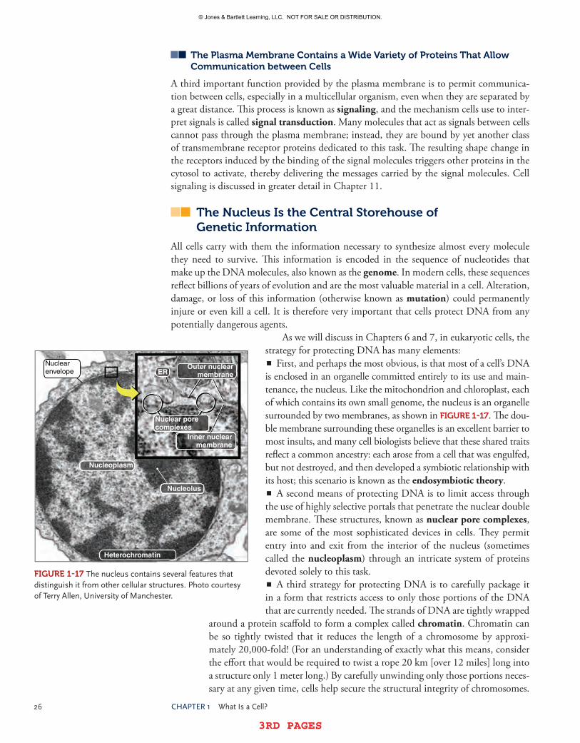
26 Chapter1 WhatIsaCell?
3RD PAGES 3RD PAGES
The Plasma Membrane Contains a Wide Variety of Proteins That Allow Communication between Cells
A third important function provided by the plasma membrane is to permit communica-tion between cells, especially in a multicellular organism, even when they are separated by a great distance. This process is known as signaling, and the mechanism cells use to inter-pret signals is called signal transduction. Many molecules that act as signals between cells cannot pass through the plasma membrane; instead, they are bound by yet another class of transmembrane receptor proteins dedicated to this task. The resulting shape change in the receptors induced by the binding of the signal molecules triggers other proteins in the cytosol to activate, thereby delivering the messages carried by the signal molecules. Cell signaling is discussed in greater detail in Chapter 11.
The Nucleus Is the Central Storehouse of genetic Information
All cells carry with them the information necessary to synthesize almost every molecule they need to survive. This information is encoded in the sequence of nucleotides that make up the DNA molecules, also known as the genome. In modern cells, these sequences reflect billions of years of evolution and are the most valuable material in a cell. Alteration, damage, or loss of this information (otherwise known as mutation) could permanently injure or even kill a cell. It is therefore very important that cells protect DNA from any potentially dangerous agents.
As we will discuss in Chapters 6 and 7, in eukaryotic cells, the strategy for protecting DNA has many elements:�� First, and perhaps the most obvious, is that most of a cell’s DNA
is enclosed in an organelle committed entirely to its use and main-tenance, the nucleus. Like the mitochondrion and chloroplast, each of which contains its own small genome, the nucleus is an organelle surrounded by two membranes, as shown in FIgUre 1-17. The dou-ble membrane surrounding these organelles is an excellent barrier to most insults, and many cell biologists believe that these shared traits reflect a common ancestry: each arose from a cell that was engulfed, but not destroyed, and then developed a symbiotic relationship with its host; this scenario is known as the endosymbiotic theory.�� A second means of protecting DNA is to limit access through
the use of highly selective portals that penetrate the nuclear double membrane. These structures, known as nuclear pore complexes, are some of the most sophisticated devices in cells. They permit entry into and exit from the interior of the nucleus (sometimes called the nucleoplasm) through an intricate system of proteins devoted solely to this task.�� A third strategy for protecting DNA is to carefully package it
in a form that restricts access to only those portions of the DNA that are currently needed. The strands of DNA are tightly wrapped
around a protein scaffold to form a complex called chromatin. Chromatin can be so tightly twisted that it reduces the length of a chromosome by approxi-mately 20,000-fold! (For an understanding of exactly what this means, consider the effort that would be required to twist a rope 20 km [over 12 miles] long into a structure only 1 meter long.) By carefully unwinding only those portions neces-sary at any given time, cells help secure the structural integrity of chromosomes.
FIgUre 1-17 thenucleuscontainsseveralfeaturesthatdistinguishitfromothercellularstructures.photocourtesyofterryallen,UniversityofManchester.
Nuclear pore complexes
ER
Heterochromatin
Inner nuclear membrane
Nucleoplasm
Nucleolus
Outer nuclear membrane
Nuclear envelope
© Jones & Bartlett Learning, LLC. NOT FOR SALE OR DISTRIBUTION.
© Jones & Bartlett Learning, LLCNOT FOR SALE OR DISTRIBUTION
© Jones & Bartlett Learning, LLCNOT FOR SALE OR DISTRIBUTION
© Jones & Bartlett Learning, LLCNOT FOR SALE OR DISTRIBUTION
© Jones & Bartlett Learning, LLCNOT FOR SALE OR DISTRIBUTION
© Jones & Bartlett Learning, LLCNOT FOR SALE OR DISTRIBUTION
© Jones & Bartlett Learning, LLCNOT FOR SALE OR DISTRIBUTION
© Jones & Bartlett Learning, LLCNOT FOR SALE OR DISTRIBUTION
© Jones & Bartlett Learning, LLCNOT FOR SALE OR DISTRIBUTION
© Jones & Bartlett Learning, LLCNOT FOR SALE OR DISTRIBUTION
© Jones & Bartlett Learning, LLCNOT FOR SALE OR DISTRIBUTION
© Jones & Bartlett Learning, LLCNOT FOR SALE OR DISTRIBUTION
© Jones & Bartlett Learning, LLCNOT FOR SALE OR DISTRIBUTION
© Jones & Bartlett Learning, LLCNOT FOR SALE OR DISTRIBUTION
© Jones & Bartlett Learning, LLCNOT FOR SALE OR DISTRIBUTION
© Jones & Bartlett Learning, LLCNOT FOR SALE OR DISTRIBUTION
© Jones & Bartlett Learning, LLCNOT FOR SALE OR DISTRIBUTION
© Jones & Bartlett Learning, LLCNOT FOR SALE OR DISTRIBUTION
© Jones & Bartlett Learning, LLCNOT FOR SALE OR DISTRIBUTION
© Jones & Bartlett Learning, LLCNOT FOR SALE OR DISTRIBUTION
© Jones & Bartlett Learning, LLCNOT FOR SALE OR DISTRIBUTION

3RD PAGES 3RD PAGES
1.4 CellsContainDistinctstructuresthatperformspecializedFunctions 27
�� Fourth, cells make RNA copies of DNA sequences (a process known as transcription), then use them to assemble the machinery necessary for cell function. This strategy is effective because any mistakes that arise from misreading the RNA, damage to the RNA, and so on, take place outside of the nucleus. With the exception of ribosomes (which are formed by the nucleolus, a specialized sub-region in the nucleus), all macromolecules other than DNA and RNA are assembled outside the nucleus.
Chloroplasts Build Food Molecules from CO2 and H2O Using Light energy
The function of chloroplasts (illustrated in FIgUre 1-18 and discussed in Chapter 10) is to harness light energy from the sun to create the carbon polymers necessary for building all of the other macromolecules in cells. Only autotrophic cells (literally, cells that nourish themselves), such as those found in the leaves of plants, contain chloroplasts. The biochemical
Cartoon representation of the chloroplast shown in the micrograph on the left. Note that the thylakoids, which appear as fibers in electron micrographs, are actually stacks of interconnected discs, also called grana (singular = granum).
An electron micrograph of a chloroplast at low magnification. The names of the major structures/ compartments are shown.
Higher magnification image of the chloroplast shown above. Note that there are two forms of thylakoids: those that form parallel stacks (grana), and those that form interconnecting bridges between grana; the latter are called stroma thylakoids.
Notice that each thylakoid contains an interior space, called the thylakoid lumen. The thylakoid membrane is where light energy is captured and converted into cellular energy.
Granum
Higher magnification
Cutting plane
Outer membrane
Inner membrane
Stroma
Thylakoidlumen
Thylakoidmembrane
FIgUre 1-18 thechloroplastisspecializedtoconvertCo2,water,andlightenergyintocomplexcarboncompounds.©Dr.JeremyBurgess/photoresearchers,Inc.,©Dr.Kennethr.Miller/photoresearchers,Inc.
© Jones & Bartlett Learning, LLC. NOT FOR SALE OR DISTRIBUTION.
© Jones & Bartlett Learning, LLCNOT FOR SALE OR DISTRIBUTION
© Jones & Bartlett Learning, LLCNOT FOR SALE OR DISTRIBUTION
© Jones & Bartlett Learning, LLCNOT FOR SALE OR DISTRIBUTION
© Jones & Bartlett Learning, LLCNOT FOR SALE OR DISTRIBUTION
© Jones & Bartlett Learning, LLCNOT FOR SALE OR DISTRIBUTION
© Jones & Bartlett Learning, LLCNOT FOR SALE OR DISTRIBUTION
© Jones & Bartlett Learning, LLCNOT FOR SALE OR DISTRIBUTION
© Jones & Bartlett Learning, LLCNOT FOR SALE OR DISTRIBUTION
© Jones & Bartlett Learning, LLCNOT FOR SALE OR DISTRIBUTION
© Jones & Bartlett Learning, LLCNOT FOR SALE OR DISTRIBUTION
© Jones & Bartlett Learning, LLCNOT FOR SALE OR DISTRIBUTION
© Jones & Bartlett Learning, LLCNOT FOR SALE OR DISTRIBUTION
© Jones & Bartlett Learning, LLCNOT FOR SALE OR DISTRIBUTION
© Jones & Bartlett Learning, LLCNOT FOR SALE OR DISTRIBUTION
© Jones & Bartlett Learning, LLCNOT FOR SALE OR DISTRIBUTION
© Jones & Bartlett Learning, LLCNOT FOR SALE OR DISTRIBUTION
© Jones & Bartlett Learning, LLCNOT FOR SALE OR DISTRIBUTION
© Jones & Bartlett Learning, LLCNOT FOR SALE OR DISTRIBUTION
© Jones & Bartlett Learning, LLCNOT FOR SALE OR DISTRIBUTION
© Jones & Bartlett Learning, LLCNOT FOR SALE OR DISTRIBUTION

28 Chapter1 WhatIsaCell?
3RD PAGES 3RD PAGES
steps required are numerous and organized into two classes. The first, sometimes called the light reactions, is concerned with capturing photons of light and using this energy to strip electrons from water molecules and add them to carbon dioxide (CO2) molecules, which results in the release of molecular oxygen (O2) and the accumulation of two types of three-carbon sugars: glyceraldehyde 3-phosphate and dihydroxyacetone phosphate. The second (dark reactions) is concerned with converting these sugars into starch and sucrose. In turn, these provide the necessary molecules for building all of the other biomolecules necessary for cell function.
Chloroplasts are also an excellent example of a common theme in cell biology: that cells capitalize on the selective permeability of membranes to build molecular gradients across them as a form of intermediate energy storage. Many of these are ion gradients, built by ion pump proteins. The potential energy stored in these gradients can later be used by membrane proteins to drive energy-dependent chemical reactions, such as the synthesis of ATP from ADP and inorganic phosphate that occurs in chloroplasts and mitochondria. These topics are discussed in Chapter 10.
Mitochondria Convert Food to Cellular energy
Whereas chloroplasts are committed to the creation of many complex biomolecules, mitochondria are committed to breaking them apart. This process is not as easy as it might seem, because mitochondria must dismantle these molecules in a way that allows them to harness as much of the released energy as pos-sible. And, like in chloroplasts, the numerous biochemical steps are organized into two classes. The first is concerned with strip-ping the high-energy electrons off of food molecules and storing them in a set of molecules designated as highenergy electron carriers. In the second set of reactions, these electrons are com-bined with protons and molecular oxygen to create water; during this process, the energy of the electrons is temporarily stored as a proton gradient across the inner membrane of the mitochondria, and then used to generate ATP. These reactions take place in dis-tinct regions of the mitochondria, as shown in FIgUre 1-19, and are discussed in detail in Chapter 10.
The endoplasmic reticulum and golgi Apparatus Collaborate to Modify, Sort, and Transport Proteins and Phospholipids to Their Final Destinations
An excellent example of teamwork in cells is the way in which the endoplasmic reticulum (ER) and Golgi apparatus work together to handle much of the membrane trafficking that takes
place inside cells. These structures are subdivided into specialized regions that perform a subset of tasks, as shown in FIgUre 1-20 and discussed in Chapter 9.
The endoplasmic reticulum Is Composed of the Smooth endoplasmic reticulum and the rough endoplasmic reticulum
The membrane defining the endoplasmic reticulum is attached to the outer membrane of the nucleus and forms two distinct structures that perform different functions. One
Outer membrane
Innermembrane
Intermembrane space
Matrix: This is where enzymes convert food molecules into small, high-energy compounds.
Cristae: This is where high-energy compounds are converted into ATP.
FIgUre 1-19 themitochondrionisspecializedtoconvertcomplexbiomoleculesintousefulmetabolicenergy.
© Jones & Bartlett Learning, LLC. NOT FOR SALE OR DISTRIBUTION.
© Jones & Bartlett Learning, LLCNOT FOR SALE OR DISTRIBUTION
© Jones & Bartlett Learning, LLCNOT FOR SALE OR DISTRIBUTION
© Jones & Bartlett Learning, LLCNOT FOR SALE OR DISTRIBUTION
© Jones & Bartlett Learning, LLCNOT FOR SALE OR DISTRIBUTION
© Jones & Bartlett Learning, LLCNOT FOR SALE OR DISTRIBUTION
© Jones & Bartlett Learning, LLCNOT FOR SALE OR DISTRIBUTION
© Jones & Bartlett Learning, LLCNOT FOR SALE OR DISTRIBUTION
© Jones & Bartlett Learning, LLCNOT FOR SALE OR DISTRIBUTION
© Jones & Bartlett Learning, LLCNOT FOR SALE OR DISTRIBUTION
© Jones & Bartlett Learning, LLCNOT FOR SALE OR DISTRIBUTION
© Jones & Bartlett Learning, LLCNOT FOR SALE OR DISTRIBUTION
© Jones & Bartlett Learning, LLCNOT FOR SALE OR DISTRIBUTION
© Jones & Bartlett Learning, LLCNOT FOR SALE OR DISTRIBUTION
© Jones & Bartlett Learning, LLCNOT FOR SALE OR DISTRIBUTION
© Jones & Bartlett Learning, LLCNOT FOR SALE OR DISTRIBUTION
© Jones & Bartlett Learning, LLCNOT FOR SALE OR DISTRIBUTION
© Jones & Bartlett Learning, LLCNOT FOR SALE OR DISTRIBUTION
© Jones & Bartlett Learning, LLCNOT FOR SALE OR DISTRIBUTION
© Jones & Bartlett Learning, LLCNOT FOR SALE OR DISTRIBUTION
© Jones & Bartlett Learning, LLCNOT FOR SALE OR DISTRIBUTION

3RD PAGES 3RD PAGES
1.4 CellsContainDistinctstructuresthatperformspecializedFunctions 29
portion, called the smooth endoplasmic reticulum (or SER), is specialized to perform four important functions:
�� It is the site of phospholipid biosynthesis. Phospholipids are synthesized on the cytoplasmic face of the SER membrane, and then distributed to the rest of the cell membranes.�� The SER serves as a storage site for calcium ions. These ions are released in brief bursts to control the activities of calcium-dependent proteins, and then quickly pumped back into the SER to shut these proteins off.�� In some cells, such as liver and muscle cells, proteins associated with the SER mem-brane play an important role in converting stored energy (in the form of glycogen, a polymer of glucose held together by a1,4 bonds) into individual glucose mol-ecules for immediate use by cells.�� The SER also contains enzymes that inactivate biochemical toxins; these are found in great abundance in liver cells, which remove these toxins from the bloodstream.
The rough endoplasmic reticulum (or RER) is responsible for controlling the syn-thesis, modification, and assembly of two important classes of cellular proteins. When viewed with an electron microscope (FIgUre 1-21), the RER can be easily distinguished from the SER by the presence of numerous ribosomes, which dot the cytoplasmic surface of the RER, giving it a comparatively “rough” appearance. Ribosomes attached to the sur-face of the RER are actively synthesizing polypeptides that pass from the cytosol through a complex of membrane proteins called the translocon and into the interior, or lumen, of the RER. Many of these polypeptides do not pass entirely into the lumen, because they con-tain one or more regions of hydrophobic amino acids that remain in the RER membrane. Polypeptides like these that have one or more transmembrane sequences form transmem-brane proteins, the first class of proteins. The second class of polypeptides synthesized by ribosomes on the RER consists of those that pass entirely through the translocon without attaching to the RER membrane. A generic term for proteins synthesized in this fashion is
The endoplasmic reticulum synthesizes membrane phospholip-ids and is where secreted proteins and many membrane proteins are synthesized. These are carried by
vesicles to the Golgi apparatus.
Many proteins passing through the Golgi apparatus are post translationally
modified, for example, by adding sugars, phophate groups, or sulfate
groups to amino acids in the proteins.
The molecules leave the Golgi apparatus in vesicles destined for a variety of intracellular locations. The term exocytosis refers to the targeting of these vesicles to the plasma membrane, which causes the contents of the vesicles to be secreted into the extracellular space.
ExocyticpathwayExocyticpathway
Endoplasmicreticulum
Golgiapparatus
FIgUre 1-20 theendoplasmicreticulumandGolgiapparatuscollaboratetocontrolproteinandphospholipidtrafficincells.
© Jones & Bartlett Learning, LLC. NOT FOR SALE OR DISTRIBUTION.
© Jones & Bartlett Learning, LLCNOT FOR SALE OR DISTRIBUTION
© Jones & Bartlett Learning, LLCNOT FOR SALE OR DISTRIBUTION
© Jones & Bartlett Learning, LLCNOT FOR SALE OR DISTRIBUTION
© Jones & Bartlett Learning, LLCNOT FOR SALE OR DISTRIBUTION
© Jones & Bartlett Learning, LLCNOT FOR SALE OR DISTRIBUTION
© Jones & Bartlett Learning, LLCNOT FOR SALE OR DISTRIBUTION
© Jones & Bartlett Learning, LLCNOT FOR SALE OR DISTRIBUTION
© Jones & Bartlett Learning, LLCNOT FOR SALE OR DISTRIBUTION
© Jones & Bartlett Learning, LLCNOT FOR SALE OR DISTRIBUTION
© Jones & Bartlett Learning, LLCNOT FOR SALE OR DISTRIBUTION
© Jones & Bartlett Learning, LLCNOT FOR SALE OR DISTRIBUTION
© Jones & Bartlett Learning, LLCNOT FOR SALE OR DISTRIBUTION
© Jones & Bartlett Learning, LLCNOT FOR SALE OR DISTRIBUTION
© Jones & Bartlett Learning, LLCNOT FOR SALE OR DISTRIBUTION
© Jones & Bartlett Learning, LLCNOT FOR SALE OR DISTRIBUTION
© Jones & Bartlett Learning, LLCNOT FOR SALE OR DISTRIBUTION
© Jones & Bartlett Learning, LLCNOT FOR SALE OR DISTRIBUTION
© Jones & Bartlett Learning, LLCNOT FOR SALE OR DISTRIBUTION
© Jones & Bartlett Learning, LLCNOT FOR SALE OR DISTRIBUTION
© Jones & Bartlett Learning, LLCNOT FOR SALE OR DISTRIBUTION

30 Chapter1 WhatIsaCell?
3RD PAGES 3RD PAGES
soluble proteins; a subset of these are ultimately released into the extracellular space, and are therefore called secreted (or secretory) proteins.
Most proteins synthesized by ribosomes on the RER undergo a series of chemical modi-fications (often called posttranslational modifications) catalyzed by enzymes in the RER. The most striking of these modifications is the addition of branched oligosaccharides to some asparagine and serine side chains, a process known as glycosylation. As discussed previ-ously, these oligosaccharides play an important role in controlling the structure and function of the proteins they are attached to. A second type of modification is the formation of a cova-lent bond between the sulfur atoms in two cysteine amino acid side chains, called a disulfide bond. Because it is covalent, it is very difficult to break and essentially locks the two cysteines together, which helps stabilize protein structure. Some proteins receive a third form of modi-fication, the addition of a phospholipid to the carboxyl terminus of the polypeptide, which anchors the protein to the luminal face of the endoplasmic reticulum membrane.
Traffic between the rer and golgi Apparatus Is Carried by Membrane-Bounded Vesicles
With the exception of those proteins destined to remain in the RER to assist with the translocation and modification of newly synthesized polypeptides (often called ER-resident proteins), all other polypeptides synthesized by RER-associated ribosomes are carried to the Golgi apparatus. The mechanism cells use to accomplish this is quite complex and only
FIgUre 1-21 theendoplasmicreticulumisorganizedintotwolinkedsubcompartments.Bothimagesby©Dr.DonaldFawcett/VisualsUnlimited,Inc.
The rough endoplastic reticulum has numerous
ribosomes attached to its cytosolic surface,
where protein synthesis takes place.
The smooth endoplasmic reticulum lacks ribosomes and is the location where most membrane phospholip-ids are synthesized.
© Jones & Bartlett Learning, LLC. NOT FOR SALE OR DISTRIBUTION.
© Jones & Bartlett Learning, LLCNOT FOR SALE OR DISTRIBUTION
© Jones & Bartlett Learning, LLCNOT FOR SALE OR DISTRIBUTION
© Jones & Bartlett Learning, LLCNOT FOR SALE OR DISTRIBUTION
© Jones & Bartlett Learning, LLCNOT FOR SALE OR DISTRIBUTION
© Jones & Bartlett Learning, LLCNOT FOR SALE OR DISTRIBUTION
© Jones & Bartlett Learning, LLCNOT FOR SALE OR DISTRIBUTION
© Jones & Bartlett Learning, LLCNOT FOR SALE OR DISTRIBUTION
© Jones & Bartlett Learning, LLCNOT FOR SALE OR DISTRIBUTION
© Jones & Bartlett Learning, LLCNOT FOR SALE OR DISTRIBUTION
© Jones & Bartlett Learning, LLCNOT FOR SALE OR DISTRIBUTION
© Jones & Bartlett Learning, LLCNOT FOR SALE OR DISTRIBUTION
© Jones & Bartlett Learning, LLCNOT FOR SALE OR DISTRIBUTION
© Jones & Bartlett Learning, LLCNOT FOR SALE OR DISTRIBUTION
© Jones & Bartlett Learning, LLCNOT FOR SALE OR DISTRIBUTION
© Jones & Bartlett Learning, LLCNOT FOR SALE OR DISTRIBUTION
© Jones & Bartlett Learning, LLCNOT FOR SALE OR DISTRIBUTION
© Jones & Bartlett Learning, LLCNOT FOR SALE OR DISTRIBUTION
© Jones & Bartlett Learning, LLCNOT FOR SALE OR DISTRIBUTION
© Jones & Bartlett Learning, LLCNOT FOR SALE OR DISTRIBUTION
© Jones & Bartlett Learning, LLCNOT FOR SALE OR DISTRIBUTION
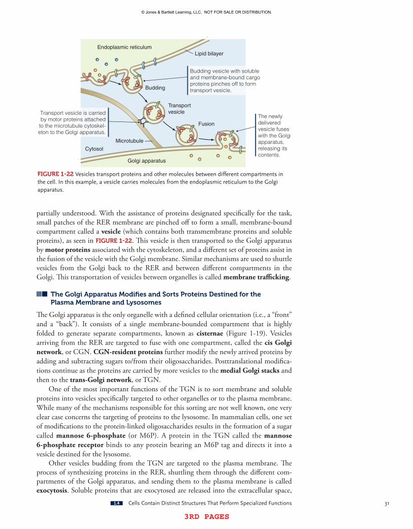
3RD PAGES 3RD PAGES
1.4 CellsContainDistinctstructuresthatperformspecializedFunctions 31
partially understood. With the assistance of proteins designated specifically for the task, small patches of the RER membrane are pinched off to form a small, membrane-bound compartment called a vesicle (which contains both transmembrane proteins and soluble proteins), as seen in FIgUre 1-22. This vesicle is then transported to the Golgi apparatus by motor proteins associated with the cytoskeleton, and a different set of proteins assist in the fusion of the vesicle with the Golgi membrane. Similar mechanisms are used to shuttle vesicles from the Golgi back to the RER and between different compartments in the Golgi. This transportation of vesicles between organelles is called membrane trafficking.
The golgi Apparatus Modifies and Sorts Proteins Destined for the Plasma Membrane and Lysosomes
The Golgi apparatus is the only organelle with a defined cellular orientation (i.e., a “front” and a “back”). It consists of a single membrane-bounded compartment that is highly folded to generate separate compartments, known as cisternae (Figure 1-19). Vesicles arriving from the RER are targeted to fuse with one compartment, called the cis Golgi network, or CGN. CGNresident proteins further modify the newly arrived proteins by adding and subtracting sugars to/from their oligosaccharides. Posttranslational modifica-tions continue as the proteins are carried by more vesicles to the medial Golgi stacks and then to the transGolgi network, or TGN.
One of the most important functions of the TGN is to sort membrane and soluble proteins into vesicles specifically targeted to other organelles or to the plasma membrane. While many of the mechanisms responsible for this sorting are not well known, one very clear case concerns the targeting of proteins to the lysosome. In mammalian cells, one set of modifications to the protein-linked oligosaccharides results in the formation of a sugar called mannose 6phosphate (or M6P). A protein in the TGN called the mannose 6phosphate receptor binds to any protein bearing an M6P tag and directs it into a vesicle destined for the lysosome.
Other vesicles budding from the TGN are targeted to the plasma membrane. The process of synthesizing proteins in the RER, shuttling them through the different com-partments of the Golgi apparatus, and sending them to the plasma membrane is called exocytosis. Soluble proteins that are exocytosed are released into the extracellular space,
Fusion
Budding vesicle with soluble and membrane-bound cargo proteins pinches off to form transport vesicle.
Transport vesicle is carried by motor proteins attached to the microtubule cytoskel-eton to the Golgi apparatus.
The newly delivered vesicle fuses with the Golgi apparatus, releasing its contents.
Transportvesicle
Budding
Endoplasmic reticulum
Golgi apparatus
Cytosol
Microtubule
Lipid bilayer
FIgUre 1-22 Vesiclestransportproteinsandothermoleculesbetweendifferentcompartmentsinthecell.Inthisexample,avesiclecarriesmoleculesfromtheendoplasmicreticulumtotheGolgiapparatus.
© Jones & Bartlett Learning, LLC. NOT FOR SALE OR DISTRIBUTION.
© Jones & Bartlett Learning, LLCNOT FOR SALE OR DISTRIBUTION
© Jones & Bartlett Learning, LLCNOT FOR SALE OR DISTRIBUTION
© Jones & Bartlett Learning, LLCNOT FOR SALE OR DISTRIBUTION
© Jones & Bartlett Learning, LLCNOT FOR SALE OR DISTRIBUTION
© Jones & Bartlett Learning, LLCNOT FOR SALE OR DISTRIBUTION
© Jones & Bartlett Learning, LLCNOT FOR SALE OR DISTRIBUTION
© Jones & Bartlett Learning, LLCNOT FOR SALE OR DISTRIBUTION
© Jones & Bartlett Learning, LLCNOT FOR SALE OR DISTRIBUTION
© Jones & Bartlett Learning, LLCNOT FOR SALE OR DISTRIBUTION
© Jones & Bartlett Learning, LLCNOT FOR SALE OR DISTRIBUTION
© Jones & Bartlett Learning, LLCNOT FOR SALE OR DISTRIBUTION
© Jones & Bartlett Learning, LLCNOT FOR SALE OR DISTRIBUTION
© Jones & Bartlett Learning, LLCNOT FOR SALE OR DISTRIBUTION
© Jones & Bartlett Learning, LLCNOT FOR SALE OR DISTRIBUTION
© Jones & Bartlett Learning, LLCNOT FOR SALE OR DISTRIBUTION
© Jones & Bartlett Learning, LLCNOT FOR SALE OR DISTRIBUTION
© Jones & Bartlett Learning, LLCNOT FOR SALE OR DISTRIBUTION
© Jones & Bartlett Learning, LLCNOT FOR SALE OR DISTRIBUTION
© Jones & Bartlett Learning, LLCNOT FOR SALE OR DISTRIBUTION
© Jones & Bartlett Learning, LLCNOT FOR SALE OR DISTRIBUTION

32 Chapter1 WhatIsaCell?
3RD PAGES 3RD PAGES
while membrane proteins that reach the plasma membrane via this route remain associated with it after vesicle fusion. This is how cell surface proteins are delivered to the plasma membrane.
The endosome Sorts and Condenses the Contents of endocytic Vesicles
The formation of vesicles at the plasma membrane usually occurs after cell surface recep-tors have bound to their ligands and clustered into a patch, and is therefore often called receptormediated endocytosis; the vesicles created are called endocytic vesicles. The contents of endocytic vesicles undergo sorting in an organelle called the endosome. In the endosome, the ligands detach from their receptors and some of the cell surface receptors are sorted into a vesicle that returns them to the plasma membrane for reuse. Vesicles from the TGN bearing M6P receptors and their cargo also fuse with endosomes, causing the dissociation of the M6P receptors and the M6P-tagged proteins. Endosomes sort the M6P receptors into a vesicle that returns them to the TGN so they can be reused.
The mechanisms responsible for this sorting by endosomes are not well understood, but it is clear that acidification of the endosome lumen plays an important role. Some of the M6P-tagged proteins arising from the TGN are proton pumps, and once they arrive in the endosome, they pump protons into the lumen. The resulting drop in pH alters the shape of the proteins in the endosome, allowing them to change their functions: cell surface receptors and M6P receptors release their cargo and cluster into separate vesicles, leaving behind only M6P-tagged proteins and any material captured by the cell surface receptors.
The Lysosome Digests Proteins, Lipids, and Nucleic Acids
The end product of endosomal sorting is called a lysosome. Due to the activity of the proton pumps they contain, lysosomes are characteristically very acidic. Most of the other M6P-tagged proteins that originate in the RER are hydrolytic enzymes. These are pro-teins that break large molecules (virus particles, proteins, lipids, nucleic acids, oligosac-charides, etc.), or sometimes even entire cells, into their simpler building blocks (amino acids, fatty acids, sugars) by simply adding back the water that was removed during the dehydration reactions used to construct them. Glycosidic bonds, phosphoester bonds, and peptide bonds are common targets of these enzymes. The resulting building blocks are then transported into the cytosol for reuse. Lysosomes are especially prevalent in many cells that are active in the immune system, such as macrophages and lymphocytes, and literally engulf invaders such as bacteria and viruses.
The Peroxisome Contributes to a Number of Metabolic Activities in Cells
As its name suggests, the peroxisome is an organelle that oxidizes molecules and generates hydrogen peroxide as a byproduct. Hydrogen peroxide (H202) is a potentially dangerous reactant in cells, which may explain why it is generated in its own organelle. Peroxisomes contain an enzyme called catalase that efficiently degrades hydrogen peroxide into water and molecular oxygen. Many of the oxidation reactions taking place in peroxisomes con-vert long-chain fatty acids into simpler compounds that can be more easily metabolized by cells. Peroxisomes are also capable of oxidizing toxic chemicals, thereby inactivating them. At least some membrane proteins in peroxisomes are synthesized in the RER and delivered by vesicles, while proteins destined for the interior of the peroxisome (called the peroxisomal matrix) are synthesized by free ribosomes in the cytosol and pass through the peroxisomal membrane after their synthesis is complete.
© Jones & Bartlett Learning, LLC. NOT FOR SALE OR DISTRIBUTION.
© Jones & Bartlett Learning, LLCNOT FOR SALE OR DISTRIBUTION
© Jones & Bartlett Learning, LLCNOT FOR SALE OR DISTRIBUTION
© Jones & Bartlett Learning, LLCNOT FOR SALE OR DISTRIBUTION
© Jones & Bartlett Learning, LLCNOT FOR SALE OR DISTRIBUTION
© Jones & Bartlett Learning, LLCNOT FOR SALE OR DISTRIBUTION
© Jones & Bartlett Learning, LLCNOT FOR SALE OR DISTRIBUTION
© Jones & Bartlett Learning, LLCNOT FOR SALE OR DISTRIBUTION
© Jones & Bartlett Learning, LLCNOT FOR SALE OR DISTRIBUTION
© Jones & Bartlett Learning, LLCNOT FOR SALE OR DISTRIBUTION
© Jones & Bartlett Learning, LLCNOT FOR SALE OR DISTRIBUTION
© Jones & Bartlett Learning, LLCNOT FOR SALE OR DISTRIBUTION
© Jones & Bartlett Learning, LLCNOT FOR SALE OR DISTRIBUTION
© Jones & Bartlett Learning, LLCNOT FOR SALE OR DISTRIBUTION
© Jones & Bartlett Learning, LLCNOT FOR SALE OR DISTRIBUTION
© Jones & Bartlett Learning, LLCNOT FOR SALE OR DISTRIBUTION
© Jones & Bartlett Learning, LLCNOT FOR SALE OR DISTRIBUTION
© Jones & Bartlett Learning, LLCNOT FOR SALE OR DISTRIBUTION
© Jones & Bartlett Learning, LLCNOT FOR SALE OR DISTRIBUTION
© Jones & Bartlett Learning, LLCNOT FOR SALE OR DISTRIBUTION
© Jones & Bartlett Learning, LLCNOT FOR SALE OR DISTRIBUTION

3RD PAGES 3RD PAGES
1.4 CellsContainDistinctstructuresthatperformspecializedFunctions 33
The Plasma Membrane, endoplasmic reticulum, golgi Apparatus, endosomes, Lysosomes, and Peroxisomes Form a Protein-Trafficking Network Called the endomembrane System
Despite the fact that each different organelle performs its own specific set of cellular activi-ties, most of the organelles found in eukaryotic cells are linked to one another via the vesicles that shuttle between them. This network, which consists of the plasma membrane, endoplas-mic reticulum, Golgi apparatus, endosomes, lysosomes, and peroxisomes, is called the endomembrane system. (This system is the primary subject of Chapter 9.) The only organelles that do not participate in this system are the nucleus, mitochondrion, and chloroplast. Recall that the endosymbiotic theory suggests these three organelles arose from the engulfment of one cell by another, and that prokaryotes sometimes exhibit elaborate extensions of the plasma membrane. Taken together, these observations suggest that all organelles are derived from specialized regions of the plasma membrane of an ancestral prokaryotic cell.
The Cytoskeleton and Motor Proteins Determine the Shape and Motion of Prokaryotic and eukaryotic Cells
Recall from our discussion of membrane structure that the hydrophobic property of phospholipids allows them to spontaneously assemble in an aqueous environment. In the absence of any other force, these membranes will form a sphere to minimize the surface-to-volume ratio. But it is important that these membranes actively participate in the selec-tive transport of molecules across them, and this favors an elaboration of the membrane surface. Cells resolve this apparent paradox by employing proteins that generate force and stabilize the resulting deformations in membranes. These proteins are the subject of Chapter 5.
The eukaryotic Cytoskeleton Is Composed of Three Types of Filamentous Proteins
The structural components of this system are called cytoskeletal proteins, or simply the cytoskeleton, as illustrated in FIgUre 1-23. Prokaryotic cells contain a cytoskeleton that permits them to adopt a myriad of shapes, and forms the cilia and flagella they use to move through their environment. In eukaryotic cells, these relatively primitive cytoskel-etal proteins evolved into a larger number of proteins that are grouped into three classes (though all three form filamentous polymers). Actin filaments are used in many ways, including cell crawling and the final division of a cell body after mitosis (called cytokinesis), and they also permit cells to flatten and spread to achieve a tremendous variety of shapes. Microtubules serve as tracks for the transport of cytosolic com-ponents, including the vesicles that link the endomembrane system. Microtubules also form the mitotic spindle that ensures the proper seg-regation of replicated chromosomes during mitosis. Proteins in the third class of filaments are called intermediate filaments; these filaments serve as a strong reinforcing material to hold a cell in place once it has adopted its preferred shape. Members of all three classes of cytoskeletal proteins are present in each eukaryotic cell.
The proteins that generate force in this system are called motor proteins. Motor proteins bind to actin filaments or microtubules, but
Intermediate filamentsActin filaments
Microtubules
FIgUre 1-23 Cytoskeletalproteinsformadensemeshworkinthecytosol.seeasmallregionofafibroblastcellviewedbyelectronmicroscopy(leftpanel).numerousfilamentsarevisible.Intherightpanel,thethreetypesofcytoskeletalpolymerspresentineukaryoticcellshavebeencoloredsothattheycanbeeasilydistinguishedfromoneanother.reprintedfromJ.struct.Biol.,vol.115,M.t.svitkina,a.B.Verkhovsky,andG.G.Borisy,ImprovedproceduresforelectronMicrosopicVisualization...,pp.290–303,Copyright(1995)withpermissionfromelsevier[http://www.sciencedirect.com/science/journal/10478477].photoscourtesyoftatyanasvitkina,Universityofpennsylvania.
© Jones & Bartlett Learning, LLC. NOT FOR SALE OR DISTRIBUTION.
© Jones & Bartlett Learning, LLCNOT FOR SALE OR DISTRIBUTION
© Jones & Bartlett Learning, LLCNOT FOR SALE OR DISTRIBUTION
© Jones & Bartlett Learning, LLCNOT FOR SALE OR DISTRIBUTION
© Jones & Bartlett Learning, LLCNOT FOR SALE OR DISTRIBUTION
© Jones & Bartlett Learning, LLCNOT FOR SALE OR DISTRIBUTION
© Jones & Bartlett Learning, LLCNOT FOR SALE OR DISTRIBUTION
© Jones & Bartlett Learning, LLCNOT FOR SALE OR DISTRIBUTION
© Jones & Bartlett Learning, LLCNOT FOR SALE OR DISTRIBUTION
© Jones & Bartlett Learning, LLCNOT FOR SALE OR DISTRIBUTION
© Jones & Bartlett Learning, LLCNOT FOR SALE OR DISTRIBUTION
© Jones & Bartlett Learning, LLCNOT FOR SALE OR DISTRIBUTION
© Jones & Bartlett Learning, LLCNOT FOR SALE OR DISTRIBUTION
© Jones & Bartlett Learning, LLCNOT FOR SALE OR DISTRIBUTION
© Jones & Bartlett Learning, LLCNOT FOR SALE OR DISTRIBUTION
© Jones & Bartlett Learning, LLCNOT FOR SALE OR DISTRIBUTION
© Jones & Bartlett Learning, LLCNOT FOR SALE OR DISTRIBUTION
© Jones & Bartlett Learning, LLCNOT FOR SALE OR DISTRIBUTION
© Jones & Bartlett Learning, LLCNOT FOR SALE OR DISTRIBUTION
© Jones & Bartlett Learning, LLCNOT FOR SALE OR DISTRIBUTION
© Jones & Bartlett Learning, LLCNOT FOR SALE OR DISTRIBUTION
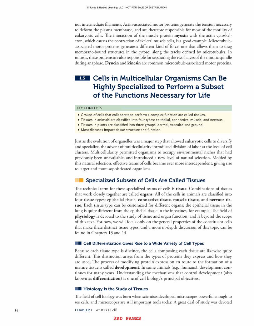
34 Chapter1 WhatIsaCell?
3RD PAGES 3RD PAGES
not intermediate filaments. Actin-associated motor proteins generate the tension necessary to deform the plasma membrane, and are therefore responsible for most of the motility of eukaryotic cells. The interaction of the muscle protein myosin with the actin cytoskel-eton, which causes the contraction of skeletal muscle cells, is a good example. Microtubule-associated motor proteins generate a different kind of force, one that allows them to drag membrane-bound structures in the cytosol along the tracks defined by microtubules. In mitosis, these proteins are also responsible for separating the two halves of the mitotic spindle during anaphase. Dynein and kinesin are common microtubule-associated motor proteins.
1.5 Cells in Multicellular Organisms Can Be Highly Specialized to Perform a Subset of the Functions Necessary for Life
KeyConCepts
�Groupsofcellsthatcollaboratetoperformacomplexfunctionarecalledtissues.�tissuesinanimalsareclassifiedintofourtypes:epithelial,connective,muscle,andnervous.�tissuesinplantsareclassifiedintothreegroups:dermal,vascular,andground.�Mostdiseasesimpacttissuestructureandfunction.
Just as the evolution of organelles was a major step that allowed eukaryotic cells to diversify and specialize, the advent of multicellularity introduced division of labor at the level of cell clusters. Multicellularity permitted organisms to occupy environmental niches that had previously been unavailable, and introduced a new level of natural selection. Molded by this natural selection, effective teams of cells became ever more interdependent, giving rise to larger and more sophisticated organisms.
Specialized Subsets of Cells Are Called Tissues
The technical term for these specialized teams of cells is tissue. Combinations of tissues that work closely together are called organs. All of the cells in animals are classified into four tissue types: epithelial tissue, connective tissue, muscle tissue, and nervous tissue. Each tissue type can be customized for different organs: the epithelial tissue in the lung is quite different from the epithelial tissue in the intestines, for example. The field of physiology is devoted to the study of tissue and organ function, and is beyond the scope of this text. For now, we will focus only on the general properties of the constituent cells that make these distinct tissue types, and a more in-depth discussion of this topic can be found in Chapters 13 and 14.
Cell Differentiation gives rise to a Wide Variety of Cell Types
Because each tissue type is distinct, the cells composing each tissue are likewise quite different. This distinction arises from the types of proteins they express and how they are used. The process of modifying protein expression en route to the formation of a mature tissue is called development. In some animals (e.g., humans), development con-tinues for many years. Understanding the mechanisms that control development (also known as differentiation) is one of cell biology’s principal objectives.
Histology Is the Study of Tissues
The field of cell biology was born when scientists developed microscopes powerful enough to see cells, and microscopes are still important tools today. A great deal of study was devoted
© Jones & Bartlett Learning, LLC. NOT FOR SALE OR DISTRIBUTION.
© Jones & Bartlett Learning, LLCNOT FOR SALE OR DISTRIBUTION
© Jones & Bartlett Learning, LLCNOT FOR SALE OR DISTRIBUTION
© Jones & Bartlett Learning, LLCNOT FOR SALE OR DISTRIBUTION
© Jones & Bartlett Learning, LLCNOT FOR SALE OR DISTRIBUTION
© Jones & Bartlett Learning, LLCNOT FOR SALE OR DISTRIBUTION
© Jones & Bartlett Learning, LLCNOT FOR SALE OR DISTRIBUTION
© Jones & Bartlett Learning, LLCNOT FOR SALE OR DISTRIBUTION
© Jones & Bartlett Learning, LLCNOT FOR SALE OR DISTRIBUTION
© Jones & Bartlett Learning, LLCNOT FOR SALE OR DISTRIBUTION
© Jones & Bartlett Learning, LLCNOT FOR SALE OR DISTRIBUTION
© Jones & Bartlett Learning, LLCNOT FOR SALE OR DISTRIBUTION
© Jones & Bartlett Learning, LLCNOT FOR SALE OR DISTRIBUTION
© Jones & Bartlett Learning, LLCNOT FOR SALE OR DISTRIBUTION
© Jones & Bartlett Learning, LLCNOT FOR SALE OR DISTRIBUTION
© Jones & Bartlett Learning, LLCNOT FOR SALE OR DISTRIBUTION
© Jones & Bartlett Learning, LLCNOT FOR SALE OR DISTRIBUTION
© Jones & Bartlett Learning, LLCNOT FOR SALE OR DISTRIBUTION
© Jones & Bartlett Learning, LLCNOT FOR SALE OR DISTRIBUTION
© Jones & Bartlett Learning, LLCNOT FOR SALE OR DISTRIBUTION
© Jones & Bartlett Learning, LLCNOT FOR SALE OR DISTRIBUTION

3RD PAGES 3RD PAGES
1.5 CellsinMulticellularorganismsCanBehighlyspecializedtoperformasubsetoftheFunctionnecessaryforLife 35
to inventing better ways to study cells in tissues, and one area grew so rapidly that biologists coined a new term for this discipline: histology. The word histology literally means the study of tissues, but it actually refers to a specific set of microscope-based techniques. Histologists treat cell and tissue samples with special contrast agents, called stains, to better see structural organization in the samples with light and electron microscopes. While the heyday of histology has largely passed, the lessons learned form the structural foundation for modern cell biology.
Let’s now have a brief look at each of the four tissue types in animals, using Figures 1-24, 1-25, 1-26, and 1-27 as visual aids.
epithelial Tissues Form Selectively Permeable Barriers between Distinct Body Compartments
The most striking feature of epithelial tissues is that they are arranged as sheets, as shown in FIgUre 1-24. On one side of the sheet, called the basal surface, the cells are in contact with a special form of fibrous material called the basement membrane. On the opposite side of the sheet, called the apical surface, the cells are in contact with an entirely different environment, which varies from organ to organ. In the lung, for example, the apical surface of the epithelium is in contact with air, while the apical surface of the epithelium in intestines interacts with digested food from the stomach. Many epithelial tissues are composed of multiple layers of epithelial cells, each lying directly on top of the layer below it. The function of epithelial tis-sues in organs is analogous to the function of the plasma membrane in a single cell: to act as a selectively permeable barrier that separates and protects the tissues on either side of the epithelial cell layer. And, just as individual cells use proteins to regulate traffic across the plasma membrane, epithelial sheets use protein complexes to regulate the traf-ficking of molecules between cells and across the cell layer. A general term used to describe these protein complexes is the junctional complex.
Connective Tissues Provide Structural Stability to Organs
For organs to be effective, cells in tissues must be organized in a specific three-dimensional arrangement. Connective tis-sues, such as those shown in FIgUre 1-25, supply the strong material necessary to maintain this arrangement, much like the cytoskeleton maintains the shape and organization of a single cell. This structural material is concentrated in the space between cells, and is called the extracellular matrix. The extracellular matrix is a complex mixture of organic and
FIgUre 1-24 epithelialtissuesformselectivelypermeablebarriersbetweendistinctbodycompartments.Inthesephotos,theepithelialtissuesliebetweenthebrightspaces(topoftoppanel,rightofmiddlepanel,leftofbottompanel)andthedarkpinkspaces.allthreeimagesby©DonnaBeerstolz,phD,CenterforBiologicImaging,UniversityofpittsburghMedicalschool.
© Jones & Bartlett Learning, LLC. NOT FOR SALE OR DISTRIBUTION.
© Jones & Bartlett Learning, LLCNOT FOR SALE OR DISTRIBUTION
© Jones & Bartlett Learning, LLCNOT FOR SALE OR DISTRIBUTION
© Jones & Bartlett Learning, LLCNOT FOR SALE OR DISTRIBUTION
© Jones & Bartlett Learning, LLCNOT FOR SALE OR DISTRIBUTION
© Jones & Bartlett Learning, LLCNOT FOR SALE OR DISTRIBUTION
© Jones & Bartlett Learning, LLCNOT FOR SALE OR DISTRIBUTION
© Jones & Bartlett Learning, LLCNOT FOR SALE OR DISTRIBUTION
© Jones & Bartlett Learning, LLCNOT FOR SALE OR DISTRIBUTION
© Jones & Bartlett Learning, LLCNOT FOR SALE OR DISTRIBUTION
© Jones & Bartlett Learning, LLCNOT FOR SALE OR DISTRIBUTION
© Jones & Bartlett Learning, LLCNOT FOR SALE OR DISTRIBUTION
© Jones & Bartlett Learning, LLCNOT FOR SALE OR DISTRIBUTION
© Jones & Bartlett Learning, LLCNOT FOR SALE OR DISTRIBUTION
© Jones & Bartlett Learning, LLCNOT FOR SALE OR DISTRIBUTION
© Jones & Bartlett Learning, LLCNOT FOR SALE OR DISTRIBUTION
© Jones & Bartlett Learning, LLCNOT FOR SALE OR DISTRIBUTION
© Jones & Bartlett Learning, LLCNOT FOR SALE OR DISTRIBUTION
© Jones & Bartlett Learning, LLCNOT FOR SALE OR DISTRIBUTION
© Jones & Bartlett Learning, LLCNOT FOR SALE OR DISTRIBUTION
© Jones & Bartlett Learning, LLCNOT FOR SALE OR DISTRIBUTION

36 Chapter1 WhatIsaCell?
3RD PAGES 3RD PAGES
inorganic molecules that completely fills the space between cells in connective tissues and also forms the basement membrane under sheets of epithelial cells. One character-istic common to all connective tissues is that they consist largely of extracellular matrix molecules, populated with a relatively low density of cells. Familiar examples of connec-tive tissues include bone, tendon, and cartilage.
Muscle Tissue Provides the Force Necessary to Move the Body and Pump Blood
There are three different types of muscle tissue in animals, each composed of a different type of muscle cell, as shown in FIgUre 1-26. Skeletal muscles are composed of muscle cells called myocytes, which have a characteristic organization of their cytoskeleton. The bulk of the cytosol in these cells is occupied by overlapping fibers of actin and myosin, all ori-ented in one direction; these regions of overlap are called sarcomeres. Typically, hundreds of sarcomeres are lined up to form a myofibril that extends the entire length of the muscle cell. When viewed with a microscope, the sarcomeres appear
as alternating stripes (or striations) of light and dark material, so these cells are also called striated muscle. Upon stimulation by a nerve cell, the actin and myosin proteins slide past one another to shorten the length of the sarcomere. When hundreds of sarcomeres contract in this fashion, the muscle cell shortens considerably; when thousands of sarcomeres con-tract together, a skeletal muscle can generate tremendous force. Note that because all of the actin and myosin filaments are aligned in the same direction, skeletal muscles generate force only in that direction. Thus, to move a bone in many different directions, several skeletal muscles must be employed, each oriented in a different direction.
Cardiac muscle cells, also called cardiomyocytes, are found in the heart and resemble skeletal myocytes in many ways, including a striated appearance. However, one important difference is that these cells can branch, so the sarcomeres can be ori-ented in more than one direction in a single cell. A second important difference is that these cells can stimulate their own contraction, without the need of a nerve. Third, cardiomyocytes are connected by one of the special protein complexes found in epithe-lial cells, called the gap junction, which permits them to pass small molecules directly from the cytosol of one cell into the cytosol of a neighboring cell. These junctions per-mit rapid spreading of any contraction signal, so the entire heart works together during each beat to pump blood in one direction.
The third type of muscle cell, called a smooth muscle cell, is quite different from the other two. Smooth muscle cells contract using actin and myosin, but do not organize these proteins into sarcomeres. Instead, these proteins are oriented in every direction, so these cells can contract in any direction necessary. Their contractions can also last much longer than those in striated muscle cells. This versatility comes at a cost, however. Smooth muscle cells typically take longer to contract, and do not generate the magnitude of force that striated muscles can. Smooth muscle tissue is commonly found beneath the basal surface of epithelia. Many epithelial tissues line tubes (airways in the lung, blood vessels, intestines), and the smooth muscle layer that surrounds them helps to control the diam-eter of these tubes. In intestines, the periodic contraction of the smooth muscle, called peristalsis, is essential to move food through the digestive tract.
FIgUre 1-25 Connectivetissuesprovidestructuralstabilitytoorgans.©DonnaBeerstolz,ph.D.,CenterforBiologicImaging,UniversityofpittsburghMedicalschool.
© Jones & Bartlett Learning, LLC. NOT FOR SALE OR DISTRIBUTION.
© Jones & Bartlett Learning, LLCNOT FOR SALE OR DISTRIBUTION
© Jones & Bartlett Learning, LLCNOT FOR SALE OR DISTRIBUTION
© Jones & Bartlett Learning, LLCNOT FOR SALE OR DISTRIBUTION
© Jones & Bartlett Learning, LLCNOT FOR SALE OR DISTRIBUTION
© Jones & Bartlett Learning, LLCNOT FOR SALE OR DISTRIBUTION
© Jones & Bartlett Learning, LLCNOT FOR SALE OR DISTRIBUTION
© Jones & Bartlett Learning, LLCNOT FOR SALE OR DISTRIBUTION
© Jones & Bartlett Learning, LLCNOT FOR SALE OR DISTRIBUTION
© Jones & Bartlett Learning, LLCNOT FOR SALE OR DISTRIBUTION
© Jones & Bartlett Learning, LLCNOT FOR SALE OR DISTRIBUTION
© Jones & Bartlett Learning, LLCNOT FOR SALE OR DISTRIBUTION
© Jones & Bartlett Learning, LLCNOT FOR SALE OR DISTRIBUTION
© Jones & Bartlett Learning, LLCNOT FOR SALE OR DISTRIBUTION
© Jones & Bartlett Learning, LLCNOT FOR SALE OR DISTRIBUTION
© Jones & Bartlett Learning, LLCNOT FOR SALE OR DISTRIBUTION
© Jones & Bartlett Learning, LLCNOT FOR SALE OR DISTRIBUTION
© Jones & Bartlett Learning, LLCNOT FOR SALE OR DISTRIBUTION
© Jones & Bartlett Learning, LLCNOT FOR SALE OR DISTRIBUTION
© Jones & Bartlett Learning, LLCNOT FOR SALE OR DISTRIBUTION
© Jones & Bartlett Learning, LLCNOT FOR SALE OR DISTRIBUTION

3RD PAGES 3RD PAGES
1.5 CellsinMulticellularorganismsCanBehighlyspecializedtoperformasubsetoftheFunctionnecessaryforLife 37
(b) Cardiac muscle
(c) Smooth muscle
(a) Skeletal muscle
Muscle
FascicleMuscle fiber (cell)
Myofibril
FIgUre 1-26 Muscletissueprovidestheforcenecessarytomovethebodyandpumpblood.©DonnaBeerstolz,phD,CenterforBiologicImaging,UniversityofpittsburghMedicalschool.,©Biologypics/photoresearchers,Inc.,©DonnaBeerstolz,phD,CenterforBiologicImaging,UniversityofpittsburghMedicalschool.
© Jones & Bartlett Learning, LLC. NOT FOR SALE OR DISTRIBUTION.
© Jones & Bartlett Learning, LLCNOT FOR SALE OR DISTRIBUTION
© Jones & Bartlett Learning, LLCNOT FOR SALE OR DISTRIBUTION
© Jones & Bartlett Learning, LLCNOT FOR SALE OR DISTRIBUTION
© Jones & Bartlett Learning, LLCNOT FOR SALE OR DISTRIBUTION
© Jones & Bartlett Learning, LLCNOT FOR SALE OR DISTRIBUTION
© Jones & Bartlett Learning, LLCNOT FOR SALE OR DISTRIBUTION
© Jones & Bartlett Learning, LLCNOT FOR SALE OR DISTRIBUTION
© Jones & Bartlett Learning, LLCNOT FOR SALE OR DISTRIBUTION
© Jones & Bartlett Learning, LLCNOT FOR SALE OR DISTRIBUTION
© Jones & Bartlett Learning, LLCNOT FOR SALE OR DISTRIBUTION
© Jones & Bartlett Learning, LLCNOT FOR SALE OR DISTRIBUTION
© Jones & Bartlett Learning, LLCNOT FOR SALE OR DISTRIBUTION
© Jones & Bartlett Learning, LLCNOT FOR SALE OR DISTRIBUTION
© Jones & Bartlett Learning, LLCNOT FOR SALE OR DISTRIBUTION
© Jones & Bartlett Learning, LLCNOT FOR SALE OR DISTRIBUTION
© Jones & Bartlett Learning, LLCNOT FOR SALE OR DISTRIBUTION
© Jones & Bartlett Learning, LLCNOT FOR SALE OR DISTRIBUTION
© Jones & Bartlett Learning, LLCNOT FOR SALE OR DISTRIBUTION
© Jones & Bartlett Learning, LLCNOT FOR SALE OR DISTRIBUTION
© Jones & Bartlett Learning, LLCNOT FOR SALE OR DISTRIBUTION

38 Chapter1 WhatIsaCell?
3RD PAGES 3RD PAGES
Nervous Tissue Provides for rapid Communication between Body Parts in Animals
The function of nervous tissue is to transmit signals from one part of the body to another. This is not the only means of communication between cells, but it is certainly the fast - est. These signals are transmitted along the length of a single nerve cell much like an electrical signal passes through a wire. The imbalance of ions across the plasma mem-brane (called a resting potential) is used as a tool to transmit signals. At one end of a nerve cell (or neuron), a stimulus causes proteins to permit a brief exchange of ions (called a depolarization) across the plasma membrane, and this triggers nearby pro-teins to do so as well. This propagation of ion movement, called an action potential, travels to the opposite end of the nerve cell at rapid speed (10–100 m/s) and is then converted into a chemical signal that diffuses to a nearby nerve cell and initiates a new action potential.
Anatomists classify nerve tissues into two types, as shown in FIgUre 1-27. The central nervous system, or CNS, is composed of the brain and spinal cord, while the peripheral nervous system, or PNS, consists of the rest of the nervous tissues in the body. In humans, this includes nerves in the arms and legs, as well as bundles of nerves that lie outside but near the spinal cord, called ganglia. Nerves can be classified according to the direction their action potential travels with respect to the central nervous system: efferent nerves carry action potentials toward the CNS, while afferent nerves (also called motor nerves) carry action potentials away from the CNS and toward effectors, which include muscles. The term nerve actually refers to a bundle of two cell types. Not all cells in nervous tissue conduct action potentials. Support cells, called glial cells, actually outnumber the conducting cells. Their function is to insulate, protect, and nourish the nerve conducting cells.
FIgUre 1-27 nervoustissueisorganizedintotwotypesandiscomposedofmanydifferentcelltypesworkingtogethertoprocessandtransmitnerveimpulses.CourtesyofDr.ChrisBjornsson.
Central nervous system
Peripheral nervous system
© Jones & Bartlett Learning, LLC. NOT FOR SALE OR DISTRIBUTION.
© Jones & Bartlett Learning, LLCNOT FOR SALE OR DISTRIBUTION
© Jones & Bartlett Learning, LLCNOT FOR SALE OR DISTRIBUTION
© Jones & Bartlett Learning, LLCNOT FOR SALE OR DISTRIBUTION
© Jones & Bartlett Learning, LLCNOT FOR SALE OR DISTRIBUTION
© Jones & Bartlett Learning, LLCNOT FOR SALE OR DISTRIBUTION
© Jones & Bartlett Learning, LLCNOT FOR SALE OR DISTRIBUTION
© Jones & Bartlett Learning, LLCNOT FOR SALE OR DISTRIBUTION
© Jones & Bartlett Learning, LLCNOT FOR SALE OR DISTRIBUTION
© Jones & Bartlett Learning, LLCNOT FOR SALE OR DISTRIBUTION
© Jones & Bartlett Learning, LLCNOT FOR SALE OR DISTRIBUTION
© Jones & Bartlett Learning, LLCNOT FOR SALE OR DISTRIBUTION
© Jones & Bartlett Learning, LLCNOT FOR SALE OR DISTRIBUTION
© Jones & Bartlett Learning, LLCNOT FOR SALE OR DISTRIBUTION
© Jones & Bartlett Learning, LLCNOT FOR SALE OR DISTRIBUTION
© Jones & Bartlett Learning, LLCNOT FOR SALE OR DISTRIBUTION
© Jones & Bartlett Learning, LLCNOT FOR SALE OR DISTRIBUTION
© Jones & Bartlett Learning, LLCNOT FOR SALE OR DISTRIBUTION
© Jones & Bartlett Learning, LLCNOT FOR SALE OR DISTRIBUTION
© Jones & Bartlett Learning, LLCNOT FOR SALE OR DISTRIBUTION
© Jones & Bartlett Learning, LLCNOT FOR SALE OR DISTRIBUTION

3RD PAGES 3RD PAGES
1.6 ModelorganismsareoftenstudiedtoUnderstandMoreComplexorganisms 39
Plants Contain Three Classes of Tissues
Plant cells are organized into three tissues that are consider-ably different from those in animals, as illustrated in FIg-
Ure 1-28. Dermal tissue forms the outer layers of plants, and is specialized for each plant organ (leaves, stems, and roots). It consists of epidermal cells (usually one layer thick) that provide the outermost layer of protection, and guard cells that form stomata on the underside of leaves to permit gas exchange. In roots, dermal tissue regulates uptake of ions and water. Vascular tissue, composed of phloem and xylem, pro-vides a vascular system to move water and nutrients through-out the plant. Phloem and xylem form channels that conduct water, organic solutes, and minerals. They are typically orga-nized into parallel channels called vascular bundles. Ground tissue contains three different cell types that together supply new cells for growth, and provide rope-like fibers for strength.
Most Diseases Are Manifested at the Level of Tissues
Understanding tissue structure and function remains a significant challenge in biology. Cancer and heart disease, the two leading causes of death in the United States, are not dangerous when they only affect individual cells. These diseases typically are not diagnosed until they begin to affect the teams of cells in tissues, as shown in FIgUre 1-29. For example, cancer cells multiply and invade surrounding tissues, disrupting their organization and function. Often, by the time cancer is diagnosed, the tumor cells have invaded a tissue and damaged its cells so badly they no longer function properly. Similarly, heart disease causes the death of heart muscle cells; loss of a few cells is not typically noticed, but when enough cells die such that the heart no longer contracts properly, this damage is largely irreversible. Treating both of these diseases remains a significant challenge because we do not yet fully under-stand how cells cooperate to form a tissue.
ConCeptCheCK#3
Biologistscommonlyrefertodifferentscales,orlevels,ofbiologicalorganization.onecommonorganizingscheme,basedonincreasingcomplexity,is:moleculesorganellescellstissuesorgans,andsoon.asidefromtheirobviousdifferencesinsize,whatotherimportantdifferencesaretherebetweencellsandtissues?
1.6 Model Organisms Are Often Studied to Understand More Complex Organisms
KeyConCepts
�asmallnumberoforganisms,calledmodelorganisms,areoftenstudiedasrepresentativesofalllivingorganisms.�Becausemodelorganismsarecomposedofcells,themechanismscontrollingtheirbehaviorareverysimilartothoseinotherorganisms.
Palisademesophyll cell
Vascular tissue
Dermal tissue
Ground tissue
Spongymesophyll cell
Lowerepidermis
Upperepidermis
FIgUre 1-28 plantscontainthreeclassesoftissues.
FIgUre 1-29 Mostdiseasesaremanifestedattheleveloftissues.Leiomyosarcomacancermetastatictolungisshown.©GarryDeLong/photoresearchers,Inc.
© Jones & Bartlett Learning, LLC. NOT FOR SALE OR DISTRIBUTION.
© Jones & Bartlett Learning, LLCNOT FOR SALE OR DISTRIBUTION
© Jones & Bartlett Learning, LLCNOT FOR SALE OR DISTRIBUTION
© Jones & Bartlett Learning, LLCNOT FOR SALE OR DISTRIBUTION
© Jones & Bartlett Learning, LLCNOT FOR SALE OR DISTRIBUTION
© Jones & Bartlett Learning, LLCNOT FOR SALE OR DISTRIBUTION
© Jones & Bartlett Learning, LLCNOT FOR SALE OR DISTRIBUTION
© Jones & Bartlett Learning, LLCNOT FOR SALE OR DISTRIBUTION
© Jones & Bartlett Learning, LLCNOT FOR SALE OR DISTRIBUTION
© Jones & Bartlett Learning, LLCNOT FOR SALE OR DISTRIBUTION
© Jones & Bartlett Learning, LLCNOT FOR SALE OR DISTRIBUTION
© Jones & Bartlett Learning, LLCNOT FOR SALE OR DISTRIBUTION
© Jones & Bartlett Learning, LLCNOT FOR SALE OR DISTRIBUTION
© Jones & Bartlett Learning, LLCNOT FOR SALE OR DISTRIBUTION
© Jones & Bartlett Learning, LLCNOT FOR SALE OR DISTRIBUTION
© Jones & Bartlett Learning, LLCNOT FOR SALE OR DISTRIBUTION
© Jones & Bartlett Learning, LLCNOT FOR SALE OR DISTRIBUTION
© Jones & Bartlett Learning, LLCNOT FOR SALE OR DISTRIBUTION
© Jones & Bartlett Learning, LLCNOT FOR SALE OR DISTRIBUTION
© Jones & Bartlett Learning, LLCNOT FOR SALE OR DISTRIBUTION
© Jones & Bartlett Learning, LLCNOT FOR SALE OR DISTRIBUTION
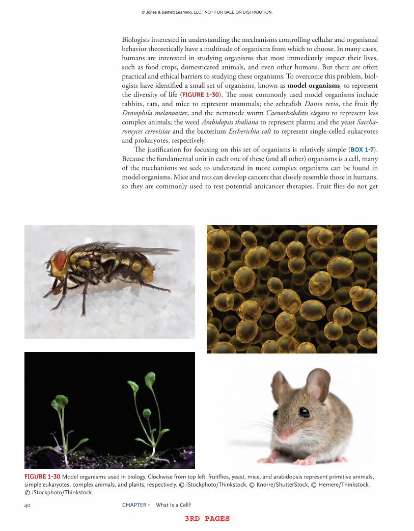
40 Chapter1 WhatIsaCell?
3RD PAGES 3RD PAGES
Biologists interested in understanding the mechanisms controlling cellular and organismal behavior theoretically have a multitude of organisms from which to choose. In many cases, humans are interested in studying organisms that most immediately impact their lives, such as food crops, domesticated animals, and even other humans. But there are often practical and ethical barriers to studying these organisms. To overcome this problem, biol-ogists have identified a small set of organisms, known as model organisms, to represent the diversity of life (FIgUre 1-30). The most commonly used model organisms include rabbits, rats, and mice to represent mammals; the zebrafish Danio rerio, the fruit fly Drosophila melanoaster, and the nematode worm Caenorhabditis elegans to represent less complex animals; the weed Arabidopsis thaliana to represent plants; and the yeast Saccha-romyces cerevisiae and the bacterium Escherichia coli to represent single-celled eukaryotes and prokaryotes, respectively.
The justification for focusing on this set of organisms is relatively simple (BOX 1-7). Because the fundamental unit in each one of these (and all other) organisms is a cell, many of the mechanisms we seek to understand in more complex organisms can be found in model organisms. Mice and rats can develop cancers that closely resemble those in humans, so they are commonly used to test potential anticancer therapies. Fruit flies do not get
FIgUre 1-30 Modelorganismsusedinbiology.Clockwisefromtopleft:fruitflies,yeast,mice,andarabidopsisrepresentprimitiveanimals,simpleeukaryotes,complexanimals,andplants,respectively.©istockphoto/thinkstock,©Knorre/shutterstock,©hemere/thinkstock,©istockphoto/thinkstock.
© Jones & Bartlett Learning, LLC. NOT FOR SALE OR DISTRIBUTION.
© Jones & Bartlett Learning, LLCNOT FOR SALE OR DISTRIBUTION
© Jones & Bartlett Learning, LLCNOT FOR SALE OR DISTRIBUTION
© Jones & Bartlett Learning, LLCNOT FOR SALE OR DISTRIBUTION
© Jones & Bartlett Learning, LLCNOT FOR SALE OR DISTRIBUTION
© Jones & Bartlett Learning, LLCNOT FOR SALE OR DISTRIBUTION
© Jones & Bartlett Learning, LLCNOT FOR SALE OR DISTRIBUTION
© Jones & Bartlett Learning, LLCNOT FOR SALE OR DISTRIBUTION
© Jones & Bartlett Learning, LLCNOT FOR SALE OR DISTRIBUTION
© Jones & Bartlett Learning, LLCNOT FOR SALE OR DISTRIBUTION
© Jones & Bartlett Learning, LLCNOT FOR SALE OR DISTRIBUTION
© Jones & Bartlett Learning, LLCNOT FOR SALE OR DISTRIBUTION
© Jones & Bartlett Learning, LLCNOT FOR SALE OR DISTRIBUTION
© Jones & Bartlett Learning, LLCNOT FOR SALE OR DISTRIBUTION
© Jones & Bartlett Learning, LLCNOT FOR SALE OR DISTRIBUTION
© Jones & Bartlett Learning, LLCNOT FOR SALE OR DISTRIBUTION
© Jones & Bartlett Learning, LLCNOT FOR SALE OR DISTRIBUTION
© Jones & Bartlett Learning, LLCNOT FOR SALE OR DISTRIBUTION
© Jones & Bartlett Learning, LLCNOT FOR SALE OR DISTRIBUTION
© Jones & Bartlett Learning, LLCNOT FOR SALE OR DISTRIBUTION
© Jones & Bartlett Learning, LLCNOT FOR SALE OR DISTRIBUTION

3RD PAGES 3RD PAGES
1.7 Chaptersummary 41
cancer, but many of the mechanisms governing the growth and differentiation of their cells are closely related to those found in more complex animals. Model organisms also have relatively short life cycles, which permits researchers to study the mechanisms governing all stages of life with relative ease; this also helps researchers understand the genetic basis of organismal variation, because large numbers of offspring can be maintained over several generations. Finally, the cost of maintaining model organisms in a laboratory setting is typically much lower than that for more complex organisms.
1.7 Chapter SummaryCells, which are the fundamental units of life, are complex yet highly organized struc-tures that possess two traits common to all living beings: the ability to self-replicate and the ability to self-correct. The remaining chapters in this book focus on the cellular mecha-nisms responsible for these two abilities, but before we explore the structure and function of cells in greater detail, it is important to understand that cells share common features. All cells contain water and are composed primarily of carbon-based molecules that can be categorized into four structural classes: lipids, sugars, amino acids, and nucleic acids. These simple building blocks form a tremendous number of different molecules, each of which performs a very small number of the tasks necessary for cells to remain alive. Over the course of evolution, cells acquired a common strategy to simplify these tasks: they clus-tered these molecules into teams that cooperate to perform a subset of cellular activities. In many cases, the structure–function relationship helps explain how these teams work. For example, eukaryotic cells acquired the ability to enclose many of these teams in membrane-bound compartments called organelles, and many of the structural features found in an organelle play an important role in that organelle’s function. Other functionally related teams of molecules are not in organelles, but play critical roles in cell behavior, such as attachment to extracellular molecules and neighboring cells, or replicating and separating chromosomes during cell growth. These ideas form the foundation for understanding how and why cells look and act the way they do.
ConCeptCheCKansWers#1
Membranesareessentialtoalllivingthingsbecausetheycreatetheboundarythatpermitscellstoestablishchemicaldisequilibrium,andtherebystoreenergy.Withoutthisenergy,lifeisnotpossible.
BOX 1-7 FAQ Is animal research ethical?Itiscriticaltopointoutthattheselectionofmodelorganismswasnotmadearbitrarily,andthatresearchersdonottaketheseorganismsforgranted.Internationalguidelineshavebeenestablishedforthecareandmaintenanceoftheseorganisms,andallotherorganisms used in biological research, and they are strictly enforced. Just as it is unethical tomistreathumanbeings,sotooisitunethicaltomistreatorganismsthatundergoexperimentationina laboratory.For thoseorganismsthatcanexperiencepainandsuffering, theutmosteffort ismadetokeepthemcomfortablethroughouttheirlifetime.
© Jones & Bartlett Learning, LLC. NOT FOR SALE OR DISTRIBUTION.
© Jones & Bartlett Learning, LLCNOT FOR SALE OR DISTRIBUTION
© Jones & Bartlett Learning, LLCNOT FOR SALE OR DISTRIBUTION
© Jones & Bartlett Learning, LLCNOT FOR SALE OR DISTRIBUTION
© Jones & Bartlett Learning, LLCNOT FOR SALE OR DISTRIBUTION
© Jones & Bartlett Learning, LLCNOT FOR SALE OR DISTRIBUTION
© Jones & Bartlett Learning, LLCNOT FOR SALE OR DISTRIBUTION
© Jones & Bartlett Learning, LLCNOT FOR SALE OR DISTRIBUTION
© Jones & Bartlett Learning, LLCNOT FOR SALE OR DISTRIBUTION
© Jones & Bartlett Learning, LLCNOT FOR SALE OR DISTRIBUTION
© Jones & Bartlett Learning, LLCNOT FOR SALE OR DISTRIBUTION
© Jones & Bartlett Learning, LLCNOT FOR SALE OR DISTRIBUTION
© Jones & Bartlett Learning, LLCNOT FOR SALE OR DISTRIBUTION
© Jones & Bartlett Learning, LLCNOT FOR SALE OR DISTRIBUTION
© Jones & Bartlett Learning, LLCNOT FOR SALE OR DISTRIBUTION
© Jones & Bartlett Learning, LLCNOT FOR SALE OR DISTRIBUTION
© Jones & Bartlett Learning, LLCNOT FOR SALE OR DISTRIBUTION
© Jones & Bartlett Learning, LLCNOT FOR SALE OR DISTRIBUTION
© Jones & Bartlett Learning, LLCNOT FOR SALE OR DISTRIBUTION
© Jones & Bartlett Learning, LLCNOT FOR SALE OR DISTRIBUTION
© Jones & Bartlett Learning, LLCNOT FOR SALE OR DISTRIBUTION

42 Chapter1 WhatIsaCell?
3RD PAGES 3RD PAGES
ConCeptCheCKansWers#2
Ifanalienlifeformcontainsequivalentsofcellularbuildingblocks,thentheywouldlikelyhavethefollowingproperties:
�alienlipidswouldspontaneouslyaggregateintheprevailingsolventinwhichthisorganismlives,andthisaggregatewouldformasemipermeablebarriersurroundingthecontentsofanalienequivalentofacell.theyneednotbeamphipathic—theysimplyneedtocreatesomeformofselectivebarrier.�aliensugarswouldbelinkedtooneanothertogeneratestructurallycomplexpolymersthatcouldprovidestructuralsupporttothelivingorganismandgeneratestructuraldiversityintheothermoleculestowhichtheyareattached.�alienaminoacidswouldlinktogetherinlinearpolymersthatarechemicallyverystableandstructurallyflexible,andthepolymerwouldhavetheabilitytobindtootherbiologicallyimportantmoleculeswithgreatspecificity.�aliennucleotideswouldservetostoreinformationinalinearsequence.thenucleotidesneednotbeasstructurallycomplexasterrestrialnucleotidesare;theysimplyneedtoformstablepolymersthatcanstoreinformation.
ConCeptCheCKansWers#3
Wecanaddressthisquestionbyfirstbrieflydefiningthetwowords:�Cellsarethesmallestunitoflife,andaredefinedbythepresenceofatleastonebiologicalmembranethatseparatesthemfromtheexternalenvironment.Inthehierarchyofbiologicalcomplexity,thecelllevelreferstosinglecellsonly,sothismeansacellmustbeabletoperformallthenecessaryfunctionstoremainalive.�tissuesareclustersofinterconnectedcellsthatcooperatetoperformafunctionthatindividualcellscannot.Itisassumedthatthecellscomposingthetissuearecapableofremainingalive.
thus,animportantdifferencebetweencellsandtissuesisthefunctionstheyperform.Ifafunctioncanbeperformedbyanindividualcell(e.g.,growth,generatingatp,buildingproteins),byourdefinitionitisnotatissueproperty.Likewise,functionsperformedonlybyteamsofcells(e.g.,transmittinganelectricalsignalfromonepartofthebodytoanother,contractingtoshrinkthediameterofatube,formingabarriertoprotectagainstphysicaltraumathatwouldkillanysinglecell)belonginthetissue-levelproperties.
asecondimportantdifferenceisthatcellsareassumedtobeself-sustaining,whiletissuesarenot.onceaclusterofcellsformsaspecializedteam,itrequireshelpfromothercellstoremainalive.thisiswhyaclusterofsingle-celledorganisms(e.g.,agroupofbacteriainadropofwater)isnotconsideredatissue.tissuesarefarmorethanagroupofsimilarcells.
? CHAPTER STUDY QUESTIONS
Q. Usingthebulletedlistof“essentialtasks”performedbycellsasaguide,whatadditionaltasksdocellshavetoperform?Whatadditionaltasksmightbeespeciallyimportantforunicellularorganisms,orforprokaryotes,orfortissuecells(e.g.,skin,heartmuscle,brain)?
A. someadditionaltaskscellsmightperforminclude(1)determiningwheretheyarewithrespecttoothercells,(2)movingfromonelocationtoanother,and(3)modifyingtheirexternalenvironment.Unicellularand/orprokaryoticorganismsmightneedto(1)ingestanddigestfood,(2)evadepredators,or(3)fightoffattacksbyviruses.tissuecellsmustbeableto(1)communicatewithothercellsinthesametissueandintherestofthebody,(2)growanddifferentiatewhenneeded,and(3)dieattheappropriatetimetokeepthetissuehealthy.typically,muscleandnervecellsintheadultdonotdieintentionally,butskincellsdoonaregularbasis.
Q. Mostprokaryotesaregenerallyconsideredtobelesscomplexthanmosteukaryoticcells,yettheyarethemostabundantcellsonearth.howwouldoneexplainwhysimpleprokaryotesaresoabundant?
A. theseorganismsarerepresentativeofsomeoftheoldestcellsonearth,andthushavebeensubjecttoevolutionbynaturalselectionforthelongesttime.Becausetheirlifecycleistypicallyshorterthanmosteukaryotes,theyarecapableofgeneratinglargernumbersofoffspringwithgreaterphenotypicvariationthaneukaryotes,andthusareabletoadapttoenvironmentalchangesmorerapidlythanmorecomplexcells.
© Jones & Bartlett Learning, LLC. NOT FOR SALE OR DISTRIBUTION.
© Jones & Bartlett Learning, LLCNOT FOR SALE OR DISTRIBUTION
© Jones & Bartlett Learning, LLCNOT FOR SALE OR DISTRIBUTION
© Jones & Bartlett Learning, LLCNOT FOR SALE OR DISTRIBUTION
© Jones & Bartlett Learning, LLCNOT FOR SALE OR DISTRIBUTION
© Jones & Bartlett Learning, LLCNOT FOR SALE OR DISTRIBUTION
© Jones & Bartlett Learning, LLCNOT FOR SALE OR DISTRIBUTION
© Jones & Bartlett Learning, LLCNOT FOR SALE OR DISTRIBUTION
© Jones & Bartlett Learning, LLCNOT FOR SALE OR DISTRIBUTION
© Jones & Bartlett Learning, LLCNOT FOR SALE OR DISTRIBUTION
© Jones & Bartlett Learning, LLCNOT FOR SALE OR DISTRIBUTION
© Jones & Bartlett Learning, LLCNOT FOR SALE OR DISTRIBUTION
© Jones & Bartlett Learning, LLCNOT FOR SALE OR DISTRIBUTION
© Jones & Bartlett Learning, LLCNOT FOR SALE OR DISTRIBUTION
© Jones & Bartlett Learning, LLCNOT FOR SALE OR DISTRIBUTION
© Jones & Bartlett Learning, LLCNOT FOR SALE OR DISTRIBUTION
© Jones & Bartlett Learning, LLCNOT FOR SALE OR DISTRIBUTION
© Jones & Bartlett Learning, LLCNOT FOR SALE OR DISTRIBUTION
© Jones & Bartlett Learning, LLCNOT FOR SALE OR DISTRIBUTION
© Jones & Bartlett Learning, LLCNOT FOR SALE OR DISTRIBUTION
© Jones & Bartlett Learning, LLCNOT FOR SALE OR DISTRIBUTION

3RD PAGES 3RD PAGES
1.7 Chaptersummary 43
Q. thehumanbodyiscomposedofatleast100differentcelltypes.Ifoneweretocompareaskin(epithelial)cellwithaheart(cardiac)musclecell,whatstructuralandfunctionalsimilaritiesanddifferenceswouldoneexpecttofind?
A. Functionalsimilaritieswouldincludetheessentialtasks,andstructuralsimilaritiesincludethepresenceoftheorganelles.Becauseepithelialtissueandmuscletissueperformdistinctfunctionsinthebody,thedifferencesbetweenthetwocelltypeswouldreflecttheseroles.skinepithelialcellsseparatethebodyfromtheoutsideenvironmentandbindtightlytooneanothertoformastrongprotectivebarrieragainstharmfulagentsintheenvironment(chemicals,viruses,bacteria,etc.).Cardiac(heart)musclecellsworktogethertopumpbloodthroughthebody,andthereforehaveaverydifferentstructuralorganization:theircytoskeletonisorganizedtomaximizethecontractionofeachcellinacoordinatedfashiontopumpbloodthroughtheheartandintothecirculatorysystem.assuch,thesecellsalsoformstrongconnectionswithoneanother,butarenotorganizedtoformabarrier;instead,theydeliverconcentratedforce.
Q. explainthestructure–functionrelationship.then,developanexampleofthestructure–functionrelationshipineveryday(non-biological)life.howapplicableisthisconcepttothenonbiologicalstructuresthatpeopleinteractwithduringatypicalday?
A. asimplewayofdescribingthestructure–functionrelationshipisthatadeviceisbuilttoaccomplishatask,soifoneunderstandsthestructureofthedevice,itispossibletoguessthetaskitperforms.almostanysimpledeviceinourdailylivesfollowsthislineofreasoning.Forexample,kitchenutensilshavedifferentshapesbecausetheyperformdifferenttasks—onewouldnotuseaspoontocutfoodintopieces,oraknifetoeatsoup.thisconceptpervadesoureverydaylives,andisequallyaspowerfulincellbiology.agoodhabittoformwhenlearningcellbiologyistoask,“Whatcanthestructureofthisitem(protein,organelle,cell)tellmeaboutthetasksitperforms?”
Q. applythestructure–functionrelationshiptothenucleus:howdothefunctionsofthenucleusinfluenceitsstructure,andvice-versa?
A. oneofthemostimportantfunctionsofthenucleusistosafelystoreDna.thisisreflectedbythefactthatitismembranebound(asallorganellesare),andthatthemembranetightlyregulatesmoleculartrafficinandoutthroughnuclearporecomplexes.thenucleusisalsothestartingpointfortransferofgeneticinformationineukaryotes;henceitisthesitewhereDnaistranscribedtorna.Itsharesitsoutermembranewiththeendoplasmicreticulum,wheremanymrnasaretranslatedintoproteins.thus,theDna→rna→proteinprogressionofgeneticinformationisphysicallylinkedtogether.
animportantstructuralpropertyofthenucleusisthatitisenclosedbyadoublemembrane.ratherthanbeingsimplyredundant,thetwomembranesfunctionquitedifferently.theinnernuclearmembranecontainsproteinsthatconnecttochromosomesandhelpcontroltheirspatialorganizationwithinthenucleoplasm.theouternuclearmembraneextendsbeyondtheinnermembrane,intothecytosoltoincludetheendoplasmicreticulum,wheremanyofthemrnaproductsofgenetranscriptionaretranslated.thislinkstranscriptionandtranslationtogether.
Q. Whatpropertiesofthecarbonatompermitittoformawidervarietyofmoleculesthananyotheratom?A. accordingtotheoctetruleinorganicchemistry,carbonmustaddfourelectronstofillitsvalenceshell.thisencourages
carbonstoformfourcovalentbonds,whichisalargernumberofbondsthanisfoundintheotherabundantatomsincells(hydrogen,nitrogen,oxygen).thisallowscarbontobuildavarietyofcomplexmolecules.Inaddition,carbonhaslowelectronegativity,whichpermitsittoformlargechainsheldtogetherbynonpolarcovalentbonds;nitrogenandoxygenhavecomparativelyhighelectronegativity,andthusformpolarcovalentbondsthatdonoteasilypolymerize.
Q. Carbonisoneofthemostabundantatomsinsugarsandlipids,butsugarsandlipidshaveverydifferentchemicalproperties.howwouldoneexplainthis?
A. sugarisverysolubleinwater,butlipidsarenot.oneexplanationforthisisthatmostcarbonatomsinsugarsarecovalentlyboundtooxygen,creatingapolarbondthatattractswater.Mostcarbonatomsinlipidsarecovalentlyboundtohydrogen,creatingnonpolarbondsthatarehydrophobic.
Q. Cowsandpeoplearemammals,butcowscanusemostgrasses(whichcontaintheb1,4linkedpolysaccharidecellulose)asafoodsource,whilehumanscannot.Whatspecialabilitiesmustcowshavetoenablethemtoeatgrass?(hint:cowsandhumanshavedifferentbacteriagrowingintheirgastrointestinaltract.)
A. Manymammalsdonotpossestheenzymesnecessaryforbreakingb1,4glycosidicbondsincellulose,andthereforecannotbreakcelluloseintotheglucosemonomerscellsuseforgeneratingmetabolicenergy.Cowsovercomethislimitationbyhavingbacteriaintheirstomachsthatarecapableofdigestingthesebonds;thebacteriathatliveinhumansdonothavethisability.
© Jones & Bartlett Learning, LLC. NOT FOR SALE OR DISTRIBUTION.
© Jones & Bartlett Learning, LLCNOT FOR SALE OR DISTRIBUTION
© Jones & Bartlett Learning, LLCNOT FOR SALE OR DISTRIBUTION
© Jones & Bartlett Learning, LLCNOT FOR SALE OR DISTRIBUTION
© Jones & Bartlett Learning, LLCNOT FOR SALE OR DISTRIBUTION
© Jones & Bartlett Learning, LLCNOT FOR SALE OR DISTRIBUTION
© Jones & Bartlett Learning, LLCNOT FOR SALE OR DISTRIBUTION
© Jones & Bartlett Learning, LLCNOT FOR SALE OR DISTRIBUTION
© Jones & Bartlett Learning, LLCNOT FOR SALE OR DISTRIBUTION
© Jones & Bartlett Learning, LLCNOT FOR SALE OR DISTRIBUTION
© Jones & Bartlett Learning, LLCNOT FOR SALE OR DISTRIBUTION
© Jones & Bartlett Learning, LLCNOT FOR SALE OR DISTRIBUTION
© Jones & Bartlett Learning, LLCNOT FOR SALE OR DISTRIBUTION
© Jones & Bartlett Learning, LLCNOT FOR SALE OR DISTRIBUTION
© Jones & Bartlett Learning, LLCNOT FOR SALE OR DISTRIBUTION
© Jones & Bartlett Learning, LLCNOT FOR SALE OR DISTRIBUTION
© Jones & Bartlett Learning, LLCNOT FOR SALE OR DISTRIBUTION
© Jones & Bartlett Learning, LLCNOT FOR SALE OR DISTRIBUTION
© Jones & Bartlett Learning, LLCNOT FOR SALE OR DISTRIBUTION
© Jones & Bartlett Learning, LLCNOT FOR SALE OR DISTRIBUTION
© Jones & Bartlett Learning, LLCNOT FOR SALE OR DISTRIBUTION

44 Chapter1 WhatIsaCell?
3RD PAGES
Q. allorganellesareboundbymembranes.Whatmakeseachtypeoforganelledistinctfromtherest?A. eachorganellepossessesitsowncomplementofdistinctproteins.proteinsfacilitatemostofthechemicalreactionstaking
placeinorganelles,andthishelpseachorganelleperformadistinctsetoftasksincells.
Q. thecytoskeletonisagoodexampleofproteinsworkingasateam.Whataresomeotherteamsofproteins?A. otherteamsincludetheproteinsthatreplicateandrepairDnainthenucleus,theproteinsthatcapturesunlightandconvert
itsenergyintoorganiccompoundsinchloroplasts,andtheproteinsthatoxidizetheseorganiccompoundsinmitochondria.
© Jones & Bartlett Learning, LLC. NOT FOR SALE OR DISTRIBUTION.
© Jones & Bartlett Learning, LLCNOT FOR SALE OR DISTRIBUTION
© Jones & Bartlett Learning, LLCNOT FOR SALE OR DISTRIBUTION
© Jones & Bartlett Learning, LLCNOT FOR SALE OR DISTRIBUTION
© Jones & Bartlett Learning, LLCNOT FOR SALE OR DISTRIBUTION
© Jones & Bartlett Learning, LLCNOT FOR SALE OR DISTRIBUTION
© Jones & Bartlett Learning, LLCNOT FOR SALE OR DISTRIBUTION
© Jones & Bartlett Learning, LLCNOT FOR SALE OR DISTRIBUTION
© Jones & Bartlett Learning, LLCNOT FOR SALE OR DISTRIBUTION
© Jones & Bartlett Learning, LLCNOT FOR SALE OR DISTRIBUTION
© Jones & Bartlett Learning, LLCNOT FOR SALE OR DISTRIBUTION
© Jones & Bartlett Learning, LLCNOT FOR SALE OR DISTRIBUTION
© Jones & Bartlett Learning, LLCNOT FOR SALE OR DISTRIBUTION
© Jones & Bartlett Learning, LLCNOT FOR SALE OR DISTRIBUTION
© Jones & Bartlett Learning, LLCNOT FOR SALE OR DISTRIBUTION
© Jones & Bartlett Learning, LLCNOT FOR SALE OR DISTRIBUTION
© Jones & Bartlett Learning, LLCNOT FOR SALE OR DISTRIBUTION
© Jones & Bartlett Learning, LLCNOT FOR SALE OR DISTRIBUTION
© Jones & Bartlett Learning, LLCNOT FOR SALE OR DISTRIBUTION
© Jones & Bartlett Learning, LLCNOT FOR SALE OR DISTRIBUTION
© Jones & Bartlett Learning, LLCNOT FOR SALE OR DISTRIBUTION