chamber quantitation guidelines: what is...
TRANSCRIPT

3/30/2017
1
Roberto M Lang, MD
Chamber Quantitation Guidelines:
What is New?
J AM Soc Echocardiogr 2005; 18:1440-1463

3/30/2017
2
• Approximately 10,000 citations
iASE in iTUNE
RT3DE
Database
Deformation Imaging
Cardiac Chamber Quantification: What is
New?

3/30/2017
3
J Am Soc Echocardiogr 2015;28:1-39Eur Heart J Cardiovasc Imaging. 2015 Mar;16(3):233-71.
In Chinese ………..
3/30/2017
4
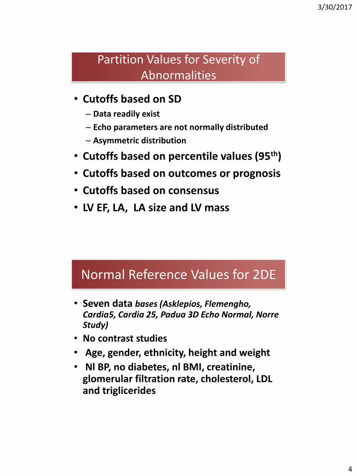
Partition Values for Severity of Abnormalities
• Cutoffs based on SD– Data readily exist
– Echo parameters are not normally distributed
– Asymmetric distribution
• Cutoffs based on percentile values (95th)
• Cutoffs based on outcomes or prognosis
• Cutoffs based on consensus
• LV EF, LA, LA size and LV mass
Normal Reference Values for 2DE
• Seven data bases (Asklepios, Flemengho, Cardia5, Cardia 25, Padua 3D Echo Normal, NorreStudy)
• No contrast studies
• Age, gender, ethnicity, height and weight
• Nl BP, no diabetes, nl BMI, creatinine, glomerular filtration rate, cholesterol, LDL and triglicerides

3/30/2017
5
Left Ventricle and Left Atrium
Subjective
Experience dependent
Lack of standardization
Large inter- and intra-observer variability
Qualitative
Assessment
Eye ball
How do we Assess LV Function ?

3/30/2017
6
Left Ventricular Linear Measurement
TEICHHOLZ Formula
Left Ventricular Volumetric Measurement

3/30/2017
7
Left Ventricular Volumetric Measurement
Biplane Disk Summation
Area Length Method
1
2

3/30/2017
8
Normal Mild Moderate Severe
2015 >52 51-41 40-30 <30
2005 >55 54-45 44-30 <30
Left Ventricular Ejection Fraction

3/30/2017
9
Normal Mildly Moderately Severely
LVEF 52-72 41-51 30-40 <30
Normal Mildly Moderately Severely
LVEF 54-74 41-53 30-40 <30
Female
Male
LV Ejection Fraction
3

3/30/2017
10
A4C
7
8
9
10
11
2D 3D
lon
g a
xis
(cm
)
*
Mor-Avi V, Lang RM et al., Circulation
2004. 110: 1814-1818.
Why is 3D More
Accurate?
Validation by MRI
• Jacobs LD, et al. Eur Heart J 2005; 27:460-8
• Sugeng L, et al. Circulation 2006; 114:654-61
• Jenkins C, et al. J Am Soc Echocardiogr 2007; 20:962-8
• Soliman OI, et al. Am Soc Echocardiogr 2007; 20:1042-9
EDV, ESV
Excellent correlation(r²>0.85)
but RT3DE
underestimates
volumes

3/30/2017
11
Sources of error Latex balloon:
• Mor-Avi V. et al, JACC Cardiovasc Img 2008: 1: 413-423
Human ventricles:
True volume: 150 ml
• Tracing error is the most
important factor contributing to
LV volume underestimation
Patient A Patient B
LV Volumes: 3DE
Disadvantages• Low temporal
resolution• Less data on
normals
Advantages• Avoid image
foreshortening
• No geometric assumptions
• More accurate and reproducible
✓
✗

3/30/2017
12
Men Women
LV Mass/BSA 49-115 43-95
RWT, cm 0.24-0.42 0.22-0.42
Septal WT, cm 0.6-1.0 0.6-0.9
PWT, cm 0.6-1.0 0.6-0.9
Cubed Formula
2D Methods
Area Length
Truncated ellipsoid
Men Women
LV mass/BSA, g/m2 50-102 41-88
Linear Method
• Direct measurement without geometrical assumptions about cavity shape and hypertrophy distribution
• More accurate that the linear or the 2D measurements
• Higher inter-measurement and test/retest reproducibility
• Better discriminates small changes within a patient
Normal values less well establishedDependent on image qualityPatients cooperation required
3D Methods

3/30/2017
13
RWT = 2PW TH / LVIDd
Lang RM, JASE 2005; 18: 1440-63
LV Global Longitudinal Strain
Peak GLS in the
range of -20% can
be expected in a
healthy person
Low Flow AS
Cardio-oncology
Valvular
Regurgitation

3/30/2017
14
LV Global Longitudinal Strain
-30
-25
-20
-15
-10
-5
0
5
LV Segmentation: Regional Deformation
• Quantitative assessment of the magnitude of regional LV deformation is not recommended – lack of reference values
– suboptimal reproducibility
– considerable inter-vendor measurement variability

3/30/2017
15
1. Normal or
Hyperkinetic
2. Hypokinetic
(reduced
thickening)
3. Akinetic
(absent or
negligible
thickening
4. Dyskinetic
(systolic
thinning or
stretching)

3/30/2017
16
The Left Atrium
Booster PumpReservoir Conduit
Mehrzad et al. Int. J. Mol. Sci. 2014, 15, 15146-15160
15-30% LV SV
Left atrial function – 3DE
LV V
olu
me
Reservoir
Conduit
Booster

3/30/2017
17
Left atrial function
• Conduit volume = LV SV – LA max – LA min
• Max = End-systole, just before mitral valve opening
• Min = End-diastole, when the mitral valve closes
• Pre-A = Immediately before atrial systole (p-wave)
Hoit BD. J Am Coll Cardiol 2014;63:493–505
Left atrial function – 2DE
• 2D Speckle-tracking analysisReservoir function
Conduit function
Booster function
Singh A, Addetia K…Lang RM ASE 2015

3/30/2017
18
Diastolic Dysfunction
Hypertension
Ischemia
Sleep Apnea
Mitral /aortic valve disease
Volume/Pressure Overload
LA Enlargement
Clinical Outcomes
• atrial fibrillation
• systolic heart failure
• diastolic dysfunction
• chronic coronary artery disease
• myocardial infarction
• mitral regurgitation
• systemic hypertension
• stroke
• hypertrophic cardiomyopathy
• renal failureTsang, T.S.M. et al. J Am Coll Cardiol 2006
LA size has a powerful prognostic value in a variety of clinical conditions:
3D Echo for Assessing the Left Atrium

3/30/2017
19
Diameters
- M-mode
- 2D guided
Area
- 4Ch
Volume
- Calculated from 2D
- Measured by 3D
TIME EVOLUTION
Assesment of Left Atrial Size/Volumes

3/30/2017
20
Time
• LA enlargement does not occur uniformly in all directions
3D Echo for Assessing the Left Atrium
Assymmetrical LA Remodelling
LA Linear Dimension

3/30/2017
21
LA Volume
Accuracy of 2DE is limited: – View-dependent – Geometrical assumptions– Measured on apical views optimized for LV
Left atrial volume on 2DE

3/30/2017
22
LAVi 38.4 mL/m2LAVi 34.0 mL/m2
View optimized
for LA
View optimized
for LV
LV axis
LA axis
Left atrial volume on 2DE
LA volume assessment on 2DE
Biplane method of disksBiplane area-length
Single planearea-length
Single plane method of disks
ASE/EACVI Chamber Quantification Guidelines 2015
3D Echo

3/30/2017
23
Standard views
A4C3DE-derived views
A2C
Biplane volume: 82 ml
Atrial-focused views
Biplane volume: 87 ml
A4C A2C
3D volume: 88 ml
Left atrial volume on 2DE
Left atrial volume on 2DE
• LA volumes obtained from non-foreshortened LA-focused views correlated highly with those obtained
from conventional A4C views (r=0.94), but were larger (Bland Altman bias 7 ml, limits of agreement ±19 ml).
V. Mor-Avi, Addetia K and Lang RML work in progress

3/30/2017
24
34
Normal Mildly Moderately Severely
LA Vol/BSA
16-34 35-41 42-48 >48
Normal Mildly Moderately Severely
LA Vol/BSA
16-28 29-33 34-39 >40
LA Volume
Lang RM et al; J Am Soc Echocardiogr 2015; 28:1-39
Lang RM et al; J Am Soc Echocardiogr 2005; 18:1440-1463
3DE2DE vs. 3DE for LA Volume Quantification
Mor-Avi V ,Lang RM et al.: Real-time 3D echocardiographic quantification of left atrial volume:
Multicenter study for validation with magnetic resonance imaging. JACC Imaging 2012.

3/30/2017
25
Left atrial function
• Conduit volume = LV SV – LA max – LA min
• Max = End-systole, just before mitral valve opening
• Min = End-diastole, when the mitral valve closes
• Pre-A = Immediately before atrial systole (p-wave)
Hoit BD. J Am Coll Cardiol 2014;63:493–505
Left atrial function – 2DE
• 2D Speckle-tracking analysisReservoir function
Conduit function
Booster function
Singh A, Addetia K…Lang RM ASE 2015

3/30/2017
26
Aorta
NCC LCC
Aortic Annulus Measurements
When: mid-systole: slightly larger and rounder Where: mid right coronary cusp and the edge of the commissures between the LCC and NCC from inner edge to inner edge
RCC

3/30/2017
27
• Sinuses of Valsalva (End-diastole)
• Sino-tubular junction (End-diastole)
• Maximal diameter of the proximal Asc Ao (End-diastole)
Leading edge to leading edge

3/30/2017
28
Aortic Root Measurements(Sinus of Valsalva)

3/30/2017
29
Summary
1. Reference ranges for left ventricular volumes and ejection fraction as well as LA volumes have changed in the recent guidelines due to the use of large echo databases.
2. Left ventricular wall motion scoring has changed to a 4-grade system.
3. Three-dimensional echocardiography is recommended for measurement of left and right ventricular volumes if possible.
4. If global longitudinal strain is being used to follow patients, it should be using the same vendors machine and analysis package.
Lang et al. Recommendations for Cardiac Chamber Quantification by Echocardiography in Adults: An Update from the American Society of Echocardiography and the European Association of Cardiovascular Imaging. J Am. Soc. Echocardiogr. 2015;28:1-39.
http://asecho.org/wordpress/wp-content/uploads/2015/01/ChamberQuantification2015.pdf
Summary

3/30/2017
30