challenges in x-ray diagnosis: a review of referrals for ... · challenges in x-ray diagnosis: a...
TRANSCRIPT
Challenges in X-ray diagnosis: a review of referrals for specialist opinion
Manas Dave BSc (Hons)
Keith Horner BChD, MSc, PhD, Odont Dr (hc), FDSRCPSGlasg, FRCR, DDR
Division of Dentistry,
School of Medical Sciences,
University of Manchester

Abstract
The aim of this study was to determine the common reasons why a dental
professional might request a second opinion on a dental radiograph from a Dental
and Maxillofacial (DMF) radiologist. The study was a retrospective analysis of
consecutive referrals for an opinion received by post or email by one DMF radiologist
based in a UK Dental Hospital. The study period was from March 2009 to November
2015. Referrals came from a mixture of sources: mainly from general dental
practitioners and specialists working in primary care, but with some referrals from
hospital-based practitioners. An enormous range in diagnoses were made by the
DMF radiologist, but the ten most frequent diagnostic categories contributed 57.5%
of the total. Normal anatomy and anatomical variations in normal anatomy made up
the largest category. Common dental disease was often diagnosed, but idiopathic
osteosclerosis and maxillary antrum pathosis were both frequent reasons for seeking
a second opinion. This service evaluation may assist in developing curricula for
undergraduates and in designing continuing education courses. It also highlights a
service that may avoid unnecessary referrals to hospital specialists but which
currently is not commissioned by the NHS.

INTRODUCTION
Radiography is one of the commonest special tests a General Dental Practitioner
(GDP) can perform, with over 11 million dental x-ray examinations conducted in
2008/2009 in the UK.1 Compared to their medical colleagues, GDPs are required not
only to justify the need for a particular type of radiograph but also to report on the
images obtained. Although radiological interpretation is part of the undergraduate
dental curriculum and, more recently, of the training given to hygienists and
therapists, referrals are sometimes made by dental professionals to Dental and
Maxillofacial (DMF) radiologists requesting a second opinion.
DMF radiology is a speciality recognised by the GDC with only 27 registrants on the
specialists list.2 There is no current literature from the UK examining the reasons
why dental professionals may refer for a second radiological opinion. One study,
carried out in Canada, reviewed 430 referrals made to two DMF radiologists over a
three year period and found that approximately one quarter of referrals related to
interpretation of variations in normal anatomy.3 This was followed in frequency by
“bone dysplasias”, cysts and inflammatory diseases. Dental education and clinical
practice in the UK, however, is likely to have some differences to the Canadian
situation. Knowledge about the needs of UK dental professionals for specialist DMF
radiological opinions could help with design of Continuing Professional Development
(CPD) courses and tailor the undergraduate curriculum which, in turn, have the
potential to reduce future referrals and lessen the financial implications of this
service to the National Health Service (NHS).

Consequently, the aim of this service evaluation was to determine the common
reasons for a dental professional to request a second opinion on a dental radiograph
from a DMF radiologist.
MATERIAL AND METHODS
This study was a retrospective analysis of consecutive referrals for radiological
opinion, by letter or email, made to a registered specialist in DMF Radiology, who is
a clinical academic (Honorary Consultant) at a UK Dental Hospital. Data were
collected in reverse order of date of referral, starting at 6th November 2015 and
ending at 17th March 2009. The end date was selected on the basis that records of
referrals were not always available prior to that date and consecutive data collection
thus became impossible.
Exclusion criteria for referrals included medico-legal work, non-patient radiological
queries (principally about aspects of radiation protection or image quality) and
overseas requests.
For each referral, one of the authors (MD) reviewed the referrer’s communication
and the reply made by the DMF radiologist. In the case of referrals by email, images
were also available for review as attachments or embedded within the email body.
For referrals by letter, images were not available for review as film radiographs or
digital images on recordable media had been returned to the referrer at the time of
providing the opinion.
The origin of the referral (general dental practice, specialist dental practice or
hospital specialist) was recorded. The type of radiograph provided by the referrer
and the diagnosis made by the DMF radiologist was also recorded. The additional
parameters recorded included substandard quality of the radiograph supplied by the
referrer and requests for a cone beam computed tomography (CBCT) scan that were
judged not to be inconsistent with accepted referral criteria. Also recorded were
recommendations made in the response by the DMF radiologist to undertake
additional imaging (dental radiograph, CBCT) or referral to a specialist colleague for
a second clinical opinion.
All data were collated on a Microsoft Excel worksheet. No data identifying either the
patient or the referrer were recorded. Data analysis was limited to descriptive
statistics.
As this study was classified as a service evaluation using the Health Research
Authority Decision Tool (http://www.hra-decisiontools.org.uk/research/), it was
judged not to require NHS ethical approval. This was confirmed by the Research
Office of Central Manchester University Hospitals NHS Foundation Trust.
RESULTS
In total, 871 separate requests for radiological opinions were collected, 713 of which
had been made by post and the remaining 158 by email. The majority of these had
originated from GDPs (n = 600; 68.9%). Referrals also originated from a wide range
of other practitioners, with specialists in oral surgery or oral and maxillofacial surgery
(n = 107; 12.3%) and orthodontists (n = 106; 12.2%) forming two substantial groups,
followed by specialists in Restorative Dentistry or one of its sub-specialisms (n = 19;
2.2%) and Community Dental Officers (n = 16; 1.8%). The remainder (n = 23; 2.6%)
was made up of small numbers of other specialist and non-specialist groups.
The 871 referrals included 967 radiographs, of which periapical radiographs were the
most common (49.6%), followed by dental panoramic radiographs (33.6%) and
bitewing radiographs (6.9%). The remainder (9.9%) was made up of occlusal
radiographs, images from cone beam CT and multislice CT scans, sialography and
Magnetic Resonance Images. The more advanced imaging modalities came
principally from oral surgeons and oral and maxillofacial surgeons.
Substandard image quality of at least one image for each case was reported in
replies to 58 referrals (6.7%). In terms of advice on further imaging given to the
referrer, a further radiograph was proposed by the DMF radiologist for 133 cases
(15.3%) and a CBCT examination for 90 cases (10.3%). Within the 871 referrals
there were 32 cases (3.7%) in which the request for a radiological opinion had
included a request for a CBCT examination that was judged to be unnecessary by
the DMF radiologist after viewing the radiograph(s). Onward referral to a specialist
clinical colleague for management was advised by the DMF radiologist for 84 cases
(9.6%).
The replies of the DMF radiologist to the referrers often included more than one
diagnosis. Of the 871 referrals, 1428 diagnoses were made, grouped into 112
separate categories. Of these, the ten most common diagnoses made up 57.5% of
the total. This “top ten” is shown in Table 1, of which the most frequent was “normal
anatomy or anatomical variations”. This encompassed a range of conditions (Table
2), but normal variation in trabecular bone pattern/ large marrow space contributed
more than a quarter of these (27.3%). If any diagnostic sub-category within “normal
anatomy or anatomical variations” occurred on fewer than five occasions, it was
classified as “other”. The “other” category made up 25.3% of the total in this category
and included the anterior nasal spine, the hyoid bone, anomalous pulp morphology,
the inferior nasal concha and inferior alveolar canal variation, amongst others.
Figures 1 to 19 give examples of some radiographs for which an opinion was sought
by referrers.
DISCUSSION
This retrospective review of requests for second opinions on radiographs was limited
to those received by one consultant DMF radiologist in one hospital, so gives a
limited snapshot of what may be happening on a larger scale nationally.
Nonetheless, referrals were received from over a wide part of the North West of
England and sometimes from further afield. We did not attempt to identify any aspect
of the referrers’ training, such as dental school of qualification or continuing
education, as this was not the focus of the service evaluation, but it would be of
interest to try to relate aspects of education with the need for radiological opinions or
particular diagnostic challenges.
One study is available against which ours can be compared, from Ontario in
Canada.3 They found that referrers were predominantly GDPs, followed by oral
surgeons and orthodontists, a pattern which was broadly the same in our study. We
found fewer oral surgeons (12.3%), however, in our study than the 21.5% reported in
the Canadian sample. This probably reflects the greater proportion of Canadian oral
surgeons who are based in primary care, without easy access to a clinical
radiologist’s opinion, than is the case in the UK.4 In the Canadian study, panoramic
radiographs were submitted alone or in combination with intraoral or other types of
radiographs in 79.5% of cases. This proportion of panoramic radiographs is much
greater than that seen in our study. Logically, it might be anticipated that there would
be more requests for an opinion on panoramic radiographs than intraoral images
because of the larger anatomical coverage. The reason why this was not the case in
our study may be explicable by the relatively high frequency of use of panoramic
radiographs in Canada compared with the UK. There is no accurate data on the

number of dental practices, or dentists, with access to panoramic radiography
equipment in either the UK or Ontario in Canada, but amongst dental practices using
the services of Public Health England Dental X-ray Protection Service, about one
third have their own panoramic equipment,5 while the figure is likely to be much
higher than this in Ontario.4
Replies to referrers written by the DMF radiologist made a point of commenting on
limitations in image quality in 6.7% of cases. This is likely to be an underestimate of
the real frequency of poor image quality because a comment would only be made if
artefacts or distortion significantly affected the confidence of the DMF radiologist in
diagnosis. The frequency of faults on dental radiographs has been noted previously
on several occasions.6,7 Although a switch to digital radiography has eliminated
chemical processing errors, it does not eliminate errors in technique. Furthermore,
with phosphor plate systems, damage to the plate surface inevitably occurs with use
and several images in the study had severe artefacts of this kind (Figs. 1, 13, 15).
In a small proportion of referrals, where the dentist had included a request to perform
a CBCT scan if needed, the request was judged to be unnecessary after the DMF
radiologist had viewed the radiographs. Due to the higher radiation doses and
financial costs, a basic principle of using CBCT is that it should be reserved for
cases where conventional radiography is unable to provide the information required
for diagnosis or treatment.8 Although it would be wrong to draw any solid conclusion
regarding this finding, the result of the survey raises a possibility that without an
opinion from a radiologist, unnecessary CBCT scans might be performed. There is a
need for research to examine the criteria used by dentists when judging the need to
perform CBCT examinations.
Our study revealed that a very wide range of diagnoses had been made for the
referred cases, including cases with more than one diagnosis, e.g. external root
resorption in conjunction with chronic periapical periodontitis. It is notable that
anatomy and anatomical variations were a particularly common diagnosis and, within
this category, trabecular bone pattern variation or a large marrow space was the
most frequent finding leading to a referral to the DMF radiologist. There is normal
variation in the trabecular bone pattern in the jaws, both between and within
individuals.9,10 The body of mandible in particular, however, can contain large areas
free of trabecular bone which in young people may contain bone marrow, although
the radiological appearance will persist into adulthood.10 The term “focal osteoporotic
marrow defect” is sometimes used in textbooks.10 Osteoporosis is also associated
with areas of sparse trabeculation in any area of the jaws.11 The way to distinguish
between a marrow cavity and a large but asymptomatic pathological area is based
upon the margins and the effects on adjacent structures. The commoner radiolucent
pathoses in the jaws (chronic periapical inflammatory lesions and cysts) will tend to
have well-defined and often corticated margins, although there are exceptions. With
an area of sparse trabeculation that is normal variation, there are no effects on
adjacent teeth or their lamina dura,10 nor on anatomical structures such as the
inferior dental canal, inferior mandibular cortex or the maxillary antral floor. Figs. 1
and 2 show examples of cases from the current study.

There were numerous other different diagnoses classified as variation in normal
anatomy. This emphasises that normal radiological anatomy and the more common
variations are important aspects of education for dentists. Examples of common
causes of diagnostic uncertainty amongst referrers in this category are shown in
Figs. 3 to 8. Air in the mouth, nose, maxillary antra and pharynx may all be
interpreted as potentially pathological findings. Some examples from the study are
shown in Figs. 3 to 5. This can be a particular problem on panoramic radiographs,
where the lengthy exposure time can include movement of soft tissues of the palate
and tongue that lead to asymmetry of the air shadows.10
Table 2 also includes the maxillary antrum. This may cause diagnostic uncertainty
when its appearance is atypical, particularly at the floor of the antral cavity when
there are loculations due to the presence of septa (Fig. 6). Maxillary antrum size is
variable, with hypoplasia and hyperplasia both occurring.10 With the latter,
pneumatisation of the alveolar bone is often seen and can cause diagnostic
challenges (Fig. 7). Superimposition of the external (soft tissue) nose on intraoral
radiographs of the maxillary incisors was recorded several times in this study (Fig.
8), often in the context of dental trauma and concern about possible root fracture.
This phenomenon is only likely to occur with downward angled bisecting angle
technique periapical radiographs and occlusal radiographs. The submandibular
fossa was also recorded as a reason for dentists’ referrals in the study. This is the
anatomical depression on the lingual aspect of the body of the mandible below the
molar regions. It is bounded superiorly by the mylohyoid line (or ridge) where the
muscle of that name attaches to the mandible. The medio-lateral width of the
mandible below this point is less than above it, so the mandible appears relatively
radiolucent (Fig. 9). This radiolucency may be augmented by the frequent finding of
sparse trabeculation/ large marrow space in this region.
Examples of radiographs sent in for an opinion related to the nasopalatine foramen
and the zygomatic process of the maxilla are shown in Figs. 10 and 11, respectively.
Perhaps surprisingly, dental caries, periodontal bone loss and periapical
inflammatory pathosis were relatively frequent diagnoses made relating to queries
from the referring dentist. The reason was often equivocal imaging information, with
an example being differentiation of distal caries on a lower second molar from
cervical burnout. Another situation which occurred on several occasions was a
request to report the caries on bitewing radiographs. Despite this being an everyday
diagnostic task for a GDP, the implicit reason appeared to be a need for back-up in
cases of an unexpected appearance of multiple new caries lesions in children or
adolescents and where the GDP was preparing to inform the patient or parent about
the need for a large number of restorations. In such situations the referring dentist
was not demonstrating incompetence in radiological interpretation, but a need for
reassurance and support.
One radiological abnormality which was regularly referred for diagnosis was
idiopathic osteosclerosis, synonymous with enostosis or dense bone island, an entity
that has been well-described by White & Pharoah.10 This was one of the most
common diagnoses, in particular from referrals by orthodontists and oral surgeons as
a chance incidental finding (Figs. 12, 13, 14). There are no clinical signs or
symptoms. Radiologically, it presents as a well-defined, localised, radiopacity, with
either an irregular or sometimes a rounded radiopacity, often close to or in contact
with the roots of teeth. There is no bony expansion or displacement of structures. In
terms of differential diagnosis, there should be no peripheral radiolucency, such as
might be seen with retained roots or odontomes. It is most commonly found in the
mandibular molar and premolar regions, with a greater frequency in the mandible
than the maxilla. The population prevalence is reported at a range from 2.3-9.7% in
different ethnicities.12,13 Histologically, idiopathic osteosclerosis is an island of
cortical bone in an area normally occupied by trabecular bone. No treatment or
intervention is required. When closely related to teeth, the periodontal ligament
space and lamina dura normally remain intact. In some cases, however, idiopathic
osteosclerosis is associated with external root resorption of the associated tooth
(Fig.13).10 “Bony sclerosis” (Table 1) might have been combined with the idiopathic
osteosclerosis category, but was characterised by a more diffuse or generalised
increase in radiopacity with no apparent cause other than normal variation in
trabecular pattern. Sclerosing osteitis (Fig. 15) due to chronic dental inflammation
(periapical or periodontal) was included in the chronic periapical periodontitis
category (Table 1).
Resorption of teeth was a diagnosis for a significant number of referrals. This
included cases of internal resorption, inflammatory external resorption and
replacement external resorption. In many cases in the study, referrers suggested the
diagnosis and were only asking for confirmation of this. Root resorption has been
thoroughly reviewed by Darcey & Qualtrough14, but one aspect of resorption which is
less well recognised is when it affects the crowns of unerupted teeth (Fig. 16).
Patchy resorption of unerupted teeth is usually seen in older patients and it is the

authors’ impression that this is sometimes associated with atrophy of the pericoronal
follicle. It seems to be age-related change rather than pathological in nature and, in
the absence of any other pathosis, it does not require any treatment.
Inflammatory mucosal thickening and mucosal retention cysts of the maxillary antral
lining made up 7.1% of the “top ten” diagnoses (Table 1). Inflammatory mucosal
thickening is an extremely common finding on asymptomatic patients, although it
may not be visible on radiographs when present. Nonetheless, it can often be
recognised on intraoral and panoramic radiographs as a band of radiopacity between
the lamina dura of the floor of the maxillary antrum and the radiolucent air within it
(Fig. 17).10 There are no immediate management implications if this finding is
recognised, especially if the patient is asymptomatic. If the patient has symptoms of
chronic sinusitis, then advice to see their general medical practitioner is appropriate.
Occasionally, however, inflammatory mucosal thickening arises on the sinus floor
above teeth with periodontal disease or a tooth with periapical inflammatory
pathosis. It is impossible to know in any specific case whether the dental pathosis
causes the changes in the maxillary antrum or whether the findings are purely
coincidental, but there is a clear association.15
Mucosal (or mucous) retention cysts, also known as “retention pseudocysts”, appear
to be formed by blockage of secretory ducts of seromucous glands in the sinus
mucosa, although other aetiologies have been suggested.9,10 Their characteristic
radiographic appearance is of a well-defined, dome-shaped, radiopacity, most
commonly originating from the floor and extending towards the roof of the maxillary
sinus, with usually no alteration of the antral outline (Fig. 18).9 It is the most common
lesion in the maxillary sinus with its development probably associated with allergies,
infection, trauma and humidity.10 No intervention is necessary and patients should be
reviewed, with symptomatic treatment of sinusitis if required. According to White &
Pharoah10, the mucous retention cyst has no relationship with periapical dental
disease. From the GDP’s perspective, however, it is important to differentiate the
mucous retention cyst from a radicular cyst (or other benign odontogenic lesion),
growing from the maxillary alveolus into the maxillary antrum. In such cases, the
bony floor of the maxillary antrum will be raised up to form a radiopaque margin to
the mass (Fig. 19).
The study revealed a steady demand for this service, with evidence of an increase
over the timescale of the study. The availability of DMF radiologists to provide such a
service is not widely advertised, not least because of the uncertain workload
implications and the impact on costs for the employing NHS Trusts. It should be
noted that some dentists made repeated referrals for opinion, suggesting that once
such a service was known to exist, the demand grew. In the past, opinions on
radiographs have been provided as a courtesy to professional colleagues, with no
charge made, but in the present climate of financial restraint in the NHS, this
situation is unlikely to continue. Currently, NHS Commissioners do not contract with
NHS Trusts for this service and consultants do not have allocated time in their job
plans to deliver it. NHS Trust management may see a radiology reporting service for
primary care practitioners as subsidising another organisation, certainly where the
patient is being treated privately by the dentist, and charges may begin to be made.
In our centre, a charge is now levied on referrers for reporting of radiographs and no
service is offered for reporting of cone beam CT scans because of the much greater

time commitment required and the difficulties in coping with multiple different types of
viewing software.
On the other hand, for many of the cases included in this review, e.g. in the context
of interpretation of normal anatomical features, it was evident that the provision of a
quick opinion on a radiograph from a specialist DMF radiologist avoided some
patients being inappropriately referred to a hospital and the ensuing costs to the
NHS and the patient. The DMF radiologist recommended only a small proportion of
the cases (9.6%) in this review for referral to a specialist colleague, raising the
possibility that unnecessary referrals may have been avoided. The cost benefits of a
DMF radiologist service for dentists are unknown and would be worth further
investigation. It remains to be seen whether NHS dental commissioners recognise a
need for a second opinion reporting service when planning the commissioning of
specialist services in dental and maxillofacial radiology. It must also be noted that
with so few DMF radiology specialists in the UK, there may be limitations on what
service could be delivered in practice.
Although a specialist second opinion service is one approach to improving the
service to patients, improved education of clinicians is another option. The results of
this study highlight some particular diagnostic challenges that warrant greater
attention in curricula for undergraduate education and in continuing professional
development. It should be remembered that “Radiography and Radiation Protection”
is highly recommended by the General Dental Council as part of verifiable CPD;
although this is often assumed erroneously to be limited to safety aspects of X-ray

use, radiological interpretation also comes under this umbrella and this service
evaluation provides some evidence for topics to be included.

ACKNOWLEDGEMENTS
Many thanks to Susanne Perschbacher for providing information about her study in
Ontario, Canada, and to John Holroyd (Dental X-ray Protection Services, Public
Health England) for information related to panoramic radiography use.

REFERENCES
1. Hart D, Wall BF, Hillier MC, Shrimpton PC. Frequency and collective dose for
medical and dental X-ray examinations in the UK, 2008. Didcot: Health Protection
Agency, 2010, HSE-CRCE-012.
2. General Dental Council. Search the Registers. Online information available at:
http://www.gdc-uk.org/Pages/SearchRegisters.aspx (Accessed 9 September 2016).
3. Perschbacher SE, Pharoah MJ, Leake JL, Lam EW, Lee L. A retrospective
analysis of referral patterns for oral radiologic consultation over 3 years in Ontario,
Canada. Oral Surg Oral Med Oral Pathol Oral Radiol Endod 2010; 109: e86-91.
4. Personal communication. Susanne E Perschbacher, Department of Oral
Radiology, Faculty of Dentistry, University of Toronto, Toronto, ON, Canada. 23
August 2013.
5. Personal communication. John Holroyd, Dental X-ray Protection Services, Public
Health England, Cookridge, Leeds, West Yorkshire, LS16 6RW, UK. 30 August
2016.
6. Chong BS, Miller J, Sidhu S. The quality of radiographs accompanying endodontic
referrals to a health authority clinic. Br Dent J. 2015; 219: 69-72.
7. Rushton VE, Horner K, Worthington HV. The quality of panoramic radiographs in a
sample of general dental practices. Br Dent J 1999; 186: 630-633.
8. European Commission. Radiation Protection 172. Evidence Based Guidelines on
Cone Beam CT for Dental and Maxillofacial Radiology. Luxembourg: Office for
Official Publications of the European Communities, 2012. Online information
available at https://ec.europa.eu/energy/sites/ener/files/documents/172.pdf
(Accessed 9 September 2016).
9. Whaites E, Drage N. Essentials of radiography and radiology. 5th ed. Edinburgh:
Churchill Livingstone Elsevier, 2013.
10. White SC, Pharoah MJ. Oral radiology. Principles and interpretation. 7th ed. St.
Louis: Elsevier, 2014.
11. Lindh C, Horner K, Jonasson G, et al. The use of visual assessment of dental
radiographs for identifying women at risk of having osteoporosis: the OSTEODENT
project. Oral Surg Oral Med Oral Pathol Oral Radiol Endod 2008; 106: 285-293.
12. Halse A, Molven O. Idiopathic osteosclerosis of the jaws followed through a
period of 20-27 years. Int Endod J 2002; 35: 747-751.
13. MacDonald-Jankowski DS. Idiopathic osteosclerosis in the jaws of Britons and of
the Hong Kong Chinese: radiology and systematic review. Dentomaxillofac Radiol
1999; 28: 357-363.
14. Darcey J, Qualtrough A. Resorption: part 1. Pathology, classification and
aetiology. Br Dent J 2013; 214: 439-451.

15. Shanbhag S, Karnik P, Shirke P, Shanbhag V. Association between periapical
lesions and maxillary sinus mucosal thickening: a retrospective cone-beam
computed tomographic study. J Endod 2013; 39: 853-857.

Table 1: The ten most common diagnoses made by the DMF radiologist from the
871 referrals for a second opinion. There were 1428 diagnoses made. The
percentages shown are the proportions of the “top ten” diagnoses and of all
diagnoses.
Diagnostic category
Proportion of the
“top 10” diagnoses
%
Proportion of all
diagnoses
%
Variation in normal anatomy 20.1 11.6
Chronic periapical periodontitis 15.7 9.0
Idiopathic osteosclerosis 15.6 9.0
Dental caries 9.5 5.5
Periodontal bone loss 9.3 5.3
External root resorption 8.4 4.8
Inflammatory mucosal thickening of the
maxillary antrum, including mucous
retention cyst
7.1 4.1
Retained root 5.6 3.2
Bony sclerosis 4.9 2.8
Artefact 3.9 2.2
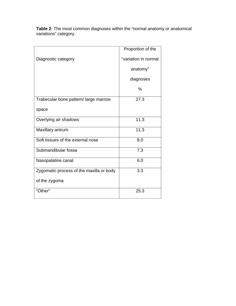
Table 2: The most common diagnoses within the “normal anatomy or anatomical variations” category.
Diagnostic category
Proportion of the
“variation in normal
anatomy”
diagnoses
%
Trabecular bone pattern/ large marrow
space
27.3
Overlying air shadows 11.3
Maxillary antrum 11.3
Soft tissues of the external nose 8.0
Submandibular fossa 7.3
Nasopalatine canal 6.0
Zygomatic process of the maxilla or body
of the zygoma
3.3
“Other” 25.3

Figure legends:
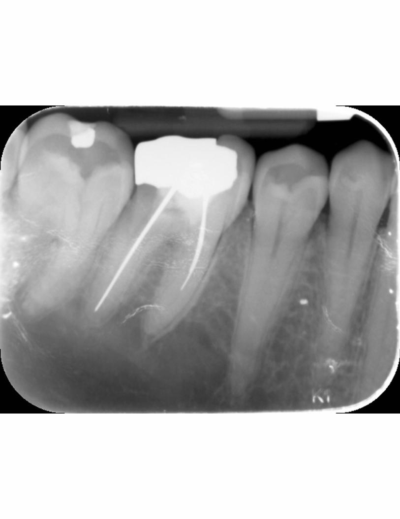
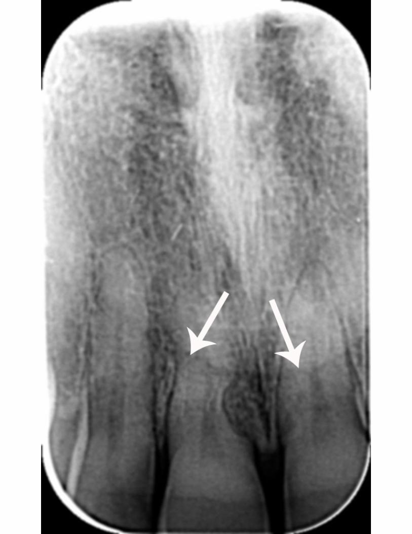
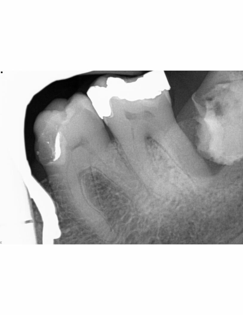
Figure 1: Variation in normal anatomy: sparse trabeculation on a periapical
radiograph of LR6. The referring dentist wanted to know whether the area of relative
radiolucency periapical to LR5, LR6 and LR7 region was cystic in nature. Note the
lamina dura of the root apices are still identifiable on the associated teeth. Note also
the multiple artefacts on the image from damage to the phosphor plate.
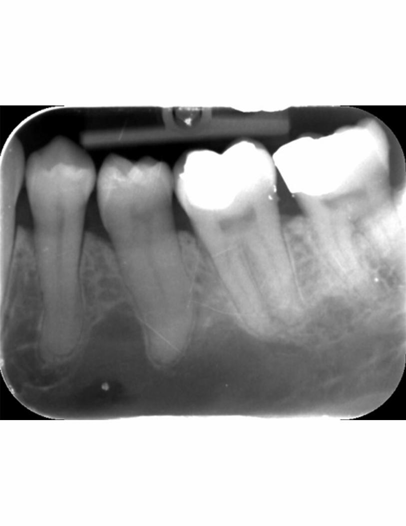
Figure 2: Variation in normal anatomy: large marrow space on a periapical
radiograph taken for LL6. The referrer was concerned that there was a cyst in the
body of mandible, extending up between the roots of LL4 and LL5. Note the lamina
dura of the root apices are still identifiable on the associated teeth.
Figure 3: Air overlying the angle of mandible on a panoramic radiograph taken of a
12 year-old female. The orthodontist noted a well-defined radiolucency with a lack of
cortication at the right angle of the mandible. She felt that this was probably an air
shadow but, as it was not symmetrical with the contralateral side, reassurance was
sought. The air lies between the posterior aspect of the tongue and the soft palate.
Figure 4: Air overlying the anterior maxilla on a periapical radiograph taken for a
discoloured UR1. The dentist requested an opinion on the radiolucent area above
and mesial to the tooth. The area of radiolucency is due to air in the right nostril,
although the nasopalatine foramen contributes the ovoid radiolucency between the
central incisor roots. Note the presence of a normal lamina dura around UR1 where
it overlies the radiolucency. Sclerosis of the pulp chamber and root canal in UR1 has
also occurred.

Figure 5: Air overlying the antero-superior part of the ramus of mandible on a
panoramic radiograph taken because of pain in the right hand side of her jaw
following trauma. The dentist requested diagnosis of whether this was a bone
fracture or artefact. The thin radiolucent band to which he referred is air between the
dorsum of the tongue and the soft palate.
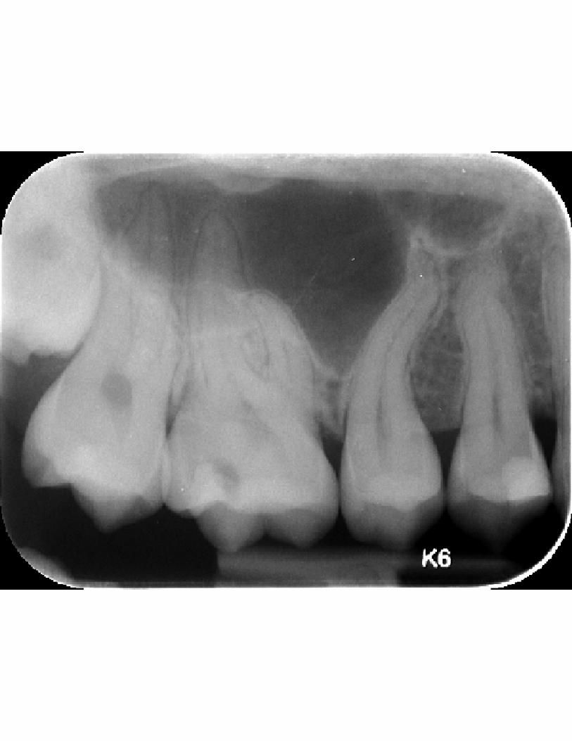
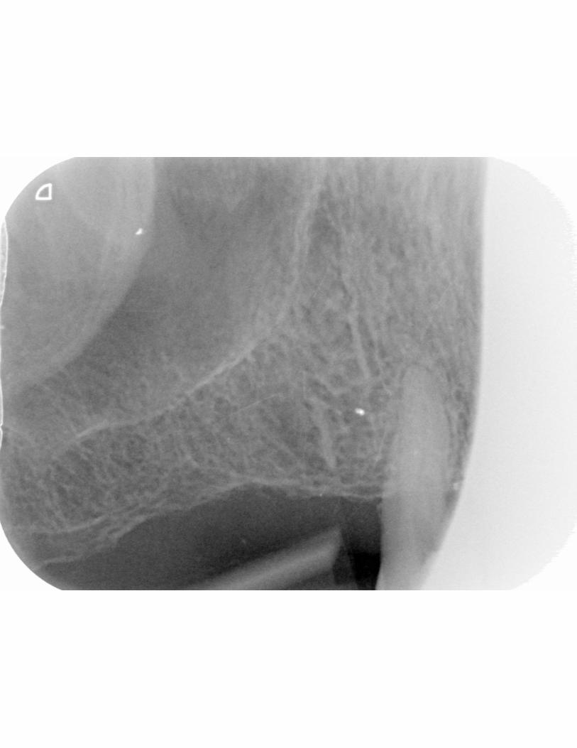
Figure 6: A locule of the maxillary antrum on a periapical radiograph taken for UL4.
The dentist asked whether the radiolucency over the upper premolars could be
cystic, or whether it was a part of the antrum. The floor of the maxillary antrum forms
the inferior edge of the radiolucency, but it continues distally across the roots of UL5
and the molars. The distal aspect of the radiolucent area is formed by a septum in
the antrum.
Figure 7: Maxillary sinus extending down into the interdental bone on a periapical
radiograph taken because of sensitivity of UR6. The dentist sought an opinion on
UR5 root and the sinus, wishing to exclude a cyst. Pneumatisation of the alveolar
bone between teeth is not unusual, but the root curvature may have accentuated the
perception of a possible lesion. Note the normal lamina dura around the teeth.
Figure 8: The soft tissues of the external nose on a periapical radiograph. The edge
of the external nose (arrowed) produces a line across the roots of the incisor teeth.
Note also the external resorption of the mesial surface of the root of UR1, with
replacement by bone.
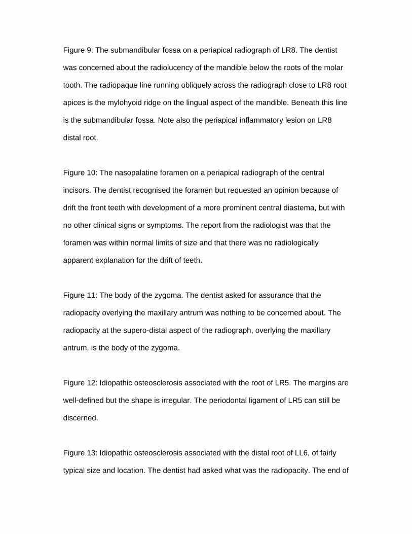
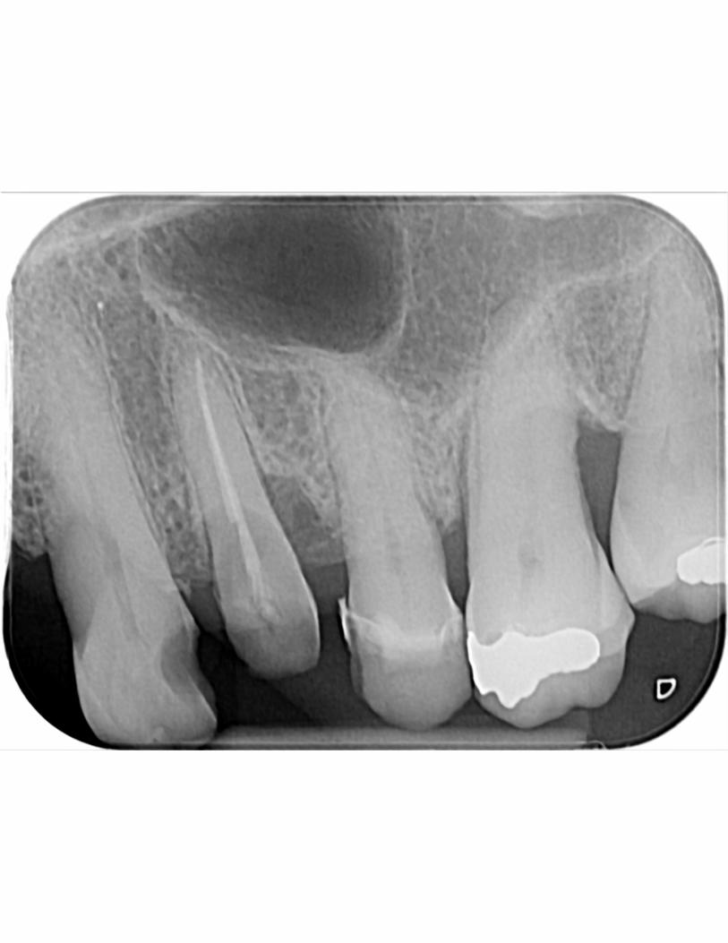
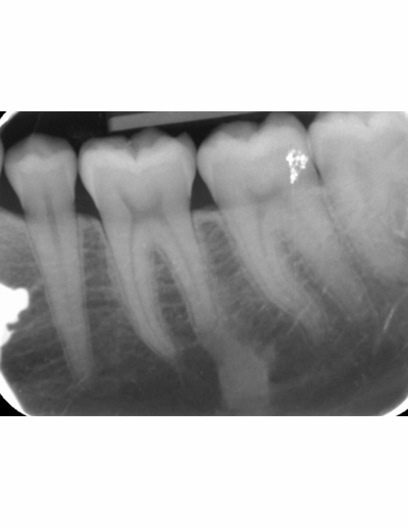
Figure 9: The submandibular fossa on a periapical radiograph of LR8. The dentist
was concerned about the radiolucency of the mandible below the roots of the molar
tooth. The radiopaque line running obliquely across the radiograph close to LR8 root
apices is the mylohyoid ridge on the lingual aspect of the mandible. Beneath this line
is the submandibular fossa. Note also the periapical inflammatory lesion on LR8
distal root.
Figure 10: The nasopalatine foramen on a periapical radiograph of the central
incisors. The dentist recognised the foramen but requested an opinion because of
drift the front teeth with development of a more prominent central diastema, but with
no other clinical signs or symptoms. The report from the radiologist was that the
foramen was within normal limits of size and that there was no radiologically
apparent explanation for the drift of teeth.
Figure 11: The body of the zygoma. The dentist asked for assurance that the
radiopacity overlying the maxillary antrum was nothing to be concerned about. The
radiopacity at the supero-distal aspect of the radiograph, overlying the maxillary
antrum, is the body of the zygoma.
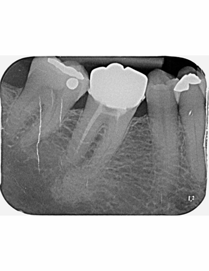
Figure 12: Idiopathic osteosclerosis associated with the root of LR5. The margins are
well-defined but the shape is irregular. The periodontal ligament of LR5 can still be
discerned.
Figure 13: Idiopathic osteosclerosis associated with the distal root of LL6, of fairly
typical size and location. The dentist had asked what was the radiopacity. The end of

the root is blunt, suggesting some resorption. Note also the phosphor plate artefact
overlying LL7 crown.
Figure 14: Section of a panoramic radiograph showing an extensive area of
idiopathic osteosclerosis associated with LL7 and LL8 region. It is unusually irregular
in shape. The sclerosis extends inferior to the inferior dental canal.
Figure 15: Periapical sclerosing osteitis associated with LR6 mesial root. The diffuse
edge and the internal trabecular character are quite different to those of idiopathic
osteosclerosis. Note also the phosphor plate artefacts, particularly over LR7.
Figure 16: External resorption of the crown of an unerupted LL8 in a 78 year-old
female who was asymptomatic but who had a small pus discharge distal to the LL7.
There is a pericoronal radiolucency affecting LL8 which is consistent with either
chronic pericoronitis or possibly a small dentigerous cyst.
Figure 17: Section of a panoramic radiograph of a patient who was complaining of
acute tenderness in upper left quadrant with no obvious diagnosis. The radiograph
shows mucosal thickening of the maxillary antrum (arrowed), as well as possibly
some root fragments in UL6 region. These were probably not the cause of the
symptoms, as a cracked tooth UL7 was subsequently suspected.
Figure 18: Section of a panoramic radiograph showing a dome-shaped radiopacity
overlying the left maxillary antrum, characteristic of a mucosal retention cyst.

Figure 19: Section of a panoramic radiograph, sent in with a request for confirmation
that the area above the root-filled UR7 was probably a cyst. Differentiation of a
periapical inflammatory cyst from a mucosal retention cyst relies on the presence in
the former of a thin bony margin, as seen here. Note that a periapical inflammatory
lesion (granuloma or cyst) appears radiopaque relative to the surrounding antral air.