chain flexibility and configurational dimensions of left handed z- dna and right handed b-dna...
TRANSCRIPT

Proc. Int. Symp. Biomol. Struct. Interactions, Suppl. J. Biosci., Vol. 8, Nos 3 & 4, August 1985, pp. 615-625. © Printed in India. Chain flexibility and configurational dimensions of left handed Z- DNA and right handed B-DNA helices †
R. MALATHI and N. YATHINDRA*
Department of Crystallography and Biophysics, University of Madras, Guindy Campus, Madras 600 025, India Abstract. The configurational behaviour of flexible helices of right handed B- and left handed Z-types have been analysed using statistical mechanical procedures. The configuration- ependent parameter, most importantly, the persistence length has been computed, using the heminucleotide scheme of treating polynucleotide chains under the approximation that perturbations in the backbone torsions produce sufficient flexibility in these helices. The values of persistence lengths obtained for Z-helices are very much higher than that of B-helices indicating that former is less flexible compared to the latter. These are in accordance with the results obtained recently on B- and Z-forms of poly(dG-dC) · (dG-dC) using light scatteringstudies. Also the persistence lengths of BII-DNA helices characterised by a skew 3'-hemi-ucleotide ( ε ~ 270°), and also when they coexist with B-DNA have been computed andthe values lie within the range of experimentally reported values on B-helices. It is argued that the decrease in the persistence length values of B-DNA at higher salt concentration is due to additional small fluctuations in sugar residue torsions induced due to neutralisation of electrostatic repulsions between adjacent phosphates of the nucleotide. Noteworthy is that these are correlated to winding angle variations and the consequent bending of the helix. Keywords. DNA flexibility; Z-DNA; B-DNA; persistence length; theoretical calculation; right and left handed helices.
Introduction The study of the nature and magnitude of flexibility associated with DNA double helices has been of considerable interest and is important to understand the underlying mechanisms responsible for DNA binding to a variety of drugs, regulatory proteins and most importantly the DNA compaction in chromatin and bacteriophages. The configurational behaviour of helical polymers, including DNA helices, in solution is described by a wormlike coil. The magnitude of persistence length (a) (defined as theaverage projection of all the bonds j (j >i) on to the direction of the initial bond i)provides a measure of helical flexibility, a larger persistence length indicating a relatively stiff and inflexible coil having a small tendency to bend. The experimental values of persistence lengths in the range of 27-84 nm are found for flexible B-DNA like helices (Bloomfield et al., 1974; Kam et al., 1981; Borochov et al., 1981; Thomas andBloomfield, 1983; Borochov and Eisenberg, 1984), the lower values occurring at higher salt concentrations. Recently a very high value of 200 nm has been obtained (Thomas and Bloomfield, 1983) for the left handed Z-helical form of poly (dG-dC) · poly(dG-dC). * To whom correspondence should be addressed.† Contribution No. 659.
615
616 Malathi and Yathindra The experimental determinations of persistence lengths generally reveal only the gross behaviour of the polymer in solution, but correlation of these with the skeletal bond conformations is important for understanding the nature of the source and magnitude of flexibility. Theoretical approaches involving statistical mechanical treatment offer convenient means of estimating persistence lengths of flexible DNA helices which are directly relatable to the skeletal bond conformational flexibility. In fact using such procedures and invoking small perturbations in the internucleotide P–O bond torsions persistence lengths have been calculated theoretically (Olson, 1979a) for B-DNA helices in agreement with experimental values obtained at low salt concentrations. Indeed such a flexibility in the sugar-phosphate backbone is shown (Yathindra, 1979; Yathindra et al.,1982; Olson, 1979b) to be even sufficient to fold the B-DNA into compact structures similar to those found in chromatin. We report here a theoretical evaluation of persistence lengths of left handed Z-helices and right handed B-DNA and BII-DNA (a variant of B-DNA possessing a tg – phosphodiester concomitant with a skew geometry for the 3'-heminucleotide) (Gupta et al., 1980a; Dickerson et al., 1982) helices usingstatistical mechanical procedures and the heminucleotide scheme (Malathi and Yathindra, 1980; Yathindra and Malathi, 1983) of treating polynucleotide backbone chains. A distinct advantage of heminucleotide treatment of polynucleotide backbone is that it permits incorporation of minor perturbations in sugar residue conformations (sugar pucker as well as exocyciic C4'–C5' bond) in addition to the phosphodiesterdirectly into the calculations. It may be relevant to invoke this secondary source of flexibility to account for the observed decrease in the persistence length values of B- DNA helices at higher salt concentrations. Materials and methods Statistical mechanical treatment of flexible helices
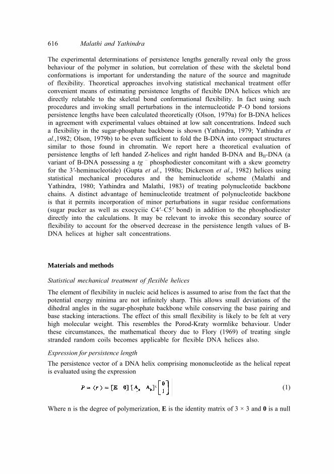
The element of flexibility in nucleic acid helices is assumed to arise from the fact that the potential energy minima are not infinitely sharp. This allows small deviations of the dihedral angles in the sugar-phosphate backbone while conserving the base pairing and base stacking interactions. The effect of this small flexibility is likely to be felt at very high molecular weight. This resembles the Porod-Kraty wormlike behaviour. Under these circumstances, the mathematical theory due to Flory (1969) of treating single stranded random coils becomes applicable for flexible DNA helices also. Expression for persistence length The persistence vector of a DNA helix comprising mononucleotide as the helical repeat is evaluated using the expression
(1) Where n is the degree of polymerization, E is the identity matrix of 3 × 3 and 0 is a null

DNA Flexibility 617
matrix of required order. Aa and Ab are 4 × 4 matrices defined as (2)
la and lb are the P-C4' and C4'-P virtual bond vectors (figure 1). The matrix Ta effects thetransformation from the coordinate system of vector lb to that of la precedingimmediately and is a function of the rotations (γ, δ). Similarly Tb effects the transformation of coordinate system of la to its preceding lb and is a function of the
Figure 1. Section of a polynucleotide chain showing the heminucleotide and the associatedvirtual bond scheme. The torsions around the various chemical bonds, the inclination of thevirtual bonds with the chemical bonds and also the length of the virtual bonds are indicated.These values correspond to the heminucleotide geometry of β = 180° and ε = 210°. Thecoordinate systems defined in order to relate successive virtual bonds are also shown.

618 Malathi and Yathindra rotations (ζ, α). The angle brackets denote the statistical mechanical averaging over the conformational states. Ta and Tb can be evaluated using the well known procedures.
The persistence vector of Z- DNA wherein a dinucleotide forms the helical repeat, theexpression (1) gets modified to
(3)
where [A1] = [Aa1Ab1] and A2 = [Aa2 A b2 ]. The matrices Aa1(γι,δ1), Ab1 (ζ1, α1),Aa2(γ2, δ2) and Ab2 (ζ2, α2) carry the same meaning as Aa and Ab of expression (2).
The projection of persistence vector along the initial axis (in this case the y-axis)⟨ y ⟩, yields the value of persistence length a.
Structure ald source of flexibility in B-DNA helices
The repeating nucleotide conformation of a B-DNA helix is described by the six backbone rotational angles α, β, γ, δ, ε and ζ and the glycosyl torsion χ (figure 1). The repeating unit of the reference helix is assigned (γ, δ) = (g+, 2E) and (ζ, α) = (g –, g –). The C5'-O5' (β) and C3'-O3' (ε) bond torsions are assigned values of 180° and 210°conforming to the preferred trans geometry for both 5'- and 3'-heminucleotides. Theseare typical of conformations found in B-form of helices. Similarly BII-DNA is assigned (γ, δ) = (60°, 151°) and (ζ, α) = (180°, 300°), whereas β and ε torsions are assigned the values of 180° and 270° respectively. Note that the 3'-heminucleotide has a skewgeometry i.e. ε = 270° concomitant with a tg– phosphodiester. The 3E and 2E sugars correspond to δ values of 76° and 151° respectively. Both the helices have the anti glycosyl conformations.
The element of flexibility in both B-DNA and BII- DNA helices is assumed as earlier (Yathindra, 1979; Olson, 1979a) to arise from minor rotations around the P–O bonds which link the successive repeating nucleotide units. Such rotations while producing a variety of helical conformations of varying helical twists, more or less ensure base pairing and adjacent base overlap interactions. A similar kind of flexibility is also feasible with minor perturbations in the sugar ring pucker (δ) and the exocyclic C4'-C5'bond torsion (γ). It should be noted that the flexibility through P-O bond rotations (ζ, α) can be effected at lesser energetic cost that (γ, δ). We have considered the influence of both on the magnitude of persistence lengths of B-DNA helices.
Structure and source of flexibility in Z-DNA helices
The structure of left handed Z-DNA has many differences compared to the familiar B-DNA. The structure is favoured by sequences which have alternation of purines and pyrimidines, especially cytosine and guanine sequences. In Z-DNA both C2' endo andC3' endo sugar puckers are found alternating in the structure. They are associated withanti cytosines and syn guanines respectively (Wang et al., 1979; Drew et al., 1980). Thephosphodiester conformation between the cytidine and the guanosine is always of g+ g+ type while that between guanosine and cytidine exhibits large variability(Crawford et al., 1980; Drew and Dickerson, 1981; Yathindra et al., 1982; Jayaraman,1982). Thus the Z-DNA structure is characterised by a dinucleotide helical repeat.

DNA flexibility 619 A new Z-DNA polymorph with a tg+ phosphodiester
We have identified (Yathindra, N., unpublished results) another possible helical polymorph for Z-DNA. This structure like others differs in the phosphodiester conformation between the dinucleotide repeat. The phosphodiester in this structure has a tg+ conformation concomitant with the trans geometry for the 3'-heminucleotide,other features in the structure being quite similar to other polymorphe. Thus one can have g– g– , g+ g+, g– t, g+ t and tg+ types of phosphodiester conformations betweenthe dinucleotide repeats while the polymeric helical conformations are very similar. This seems quite unique to Z-helices and further shows that the conformational constraints embodied in the dinucleotide repeat largely set the course of the helical path. It is because of this fact that we have assumed that the element of flexibility in Z- elices arises from minor perturbations in the phosphodiester conformations between the dinucleotide repeats.
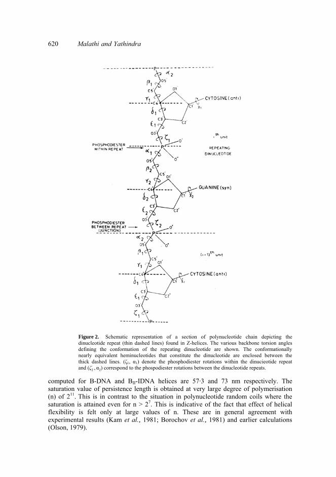
Treatment of the polynucleotide backbone of Z-helix in terms of heminucleotidesstructural repeats requires specification of four pairs of torsion angles (γ1, δ1), (ζl α1), (γ2, δ2) and (ζ2 , α2) (see figure 2). The (γ1, δ1) and (γ2, δ2) rotation pairs are assigned the(g+, 2E) and (t, 3E) conformation states respectively. The phosphodiester conformation (ζ1, α1) within the dinucleotide repeat is assigned the (g+, g+) state. It is found that the trans of γ2 and 2E for δ1 are essential requirements for this phosphodiester to be energetically feasible (Jayaraman et al., 1982). The C-O bond torsions which define the5'- and 3' heminucleotide geometry are assigned values in accordance with bothexperimental (Wang et al., 1979; Crawford et al., 1980) and theoretical (Jayaraman and Yathindra, 1980; Jayaraman, 1982) conformation analyses of Z-helices. The 3'-heminucleotide preceding the phosphodiester (ζ1, α1) is assigned a skew geometry withε1 = 270° as this is required to achieve optimum sugar-base interactions. Calculations are however made with the trans geometry i.e. with ε1 = 210°. Similarly both the preferred trans as well as skew geometries have been considered for the 3'-heminucleotide preceding (ζ2, α2) i.e. phosphodiester between the dinucleotide repeats.It is shown that the former has characteristic influence on the latter. Hence appropriate geometries of either trans and skew are assigned for this 3'-heminucleotide depending on the type of Z-helicogenic phosphodiester.
Results and discussion Persistence lengths of B-DNA and B11-DNA helices
Persistence lengths of B-DNA helix are computed by using the eq. (1) and the conformational parameters mentioned above. Average geometry (Yathindra and Sundaralingam, 1975) has been assumed for the bond angles and bond distances. For configurational averaging the phosphodiester rotations (ζ, α) are varied within the g –
g – domain from 260-290° at intervals of 3°. All the conformational states that resultdue to different combinations of (ζ, α) within this range are treated as equally probableand hence are assigned equal statistical weights. Similarly the ζ and α rotations arevaried from 150-180° and 270-300° respectively at intervals of 3° for estimation of configurational average of BII-DNA helices. The values of persistence lengths thus
620 Malathi and Yathindra
Figure 2. Schematic representation of a section of polynucleotide chain depicting the dinucleotide repeat (thin dashed lines) found in Z-helices. The various backbone torsion angles defining the conformation of the repeating dinucleotide are shown. The conformationally nearly equivalent heminucleotides that constitute the dinucleotide are enclosed between the thick dashed lines. (ζ1, α1) denote the phosphodiester rotations within the dinucieotide repeat and (ζ2 , α2) correspond to the phospodiester rotations between the dinucleotide repeats.
computed for B-DNA and BII-IDNA helices are 57·3 and 73 nm respectively. The saturation value of persistence length is obtained at very large degree of polymerisation (n) of 211. This is in contrast to the situation in polynucleotide random coils where the saturation is attained even for n > 27. This is indicative of the fact that effect of helical flexibility is felt only at large values of n. These are in general agreement with experimental results (Kam et al., 1981; Borochov et al., 1981) and earlier calculations(Olson, 1979).

DNA flexibility 621
The calculated a values of 73 nm for BII-DNA is about 30 % higher than the value obtained for B-DNA. The increase is probably due to the extended nature of tg–
hosphodiester and variation of residue height (h) in the higher ranges compared to B-DNA. Nevertheless the a values are in the range ofexperimentally determined values (Kam et al., 1981; Thomas and Bloomfield, 1983).
Persistence lengths calculated by considering 10–20% of BII-DNA to coexist with B-DNA helices are still (60 nm) within the range of experimental values. Thus it seems that small fractions of BII-DNA do not introduce any major bending or distortion ofsugar-phosphate backbone. It is found that a slight shearing of the bases occur causing slight unwinding of the helix at the B-DNA and BII- DNA junction. Existence of suchmultiple phosphodiester conformation has been noted (Dickerson et al., 1982; Shindo et al., 1984).
Invoking additional flexibility due to sugar residue variations (γ, δ), besides the P–Obond rotations, and performing similar analysis, it is found that persistence lengths decrease from a value of 57·3 nm to a value of 35·6 nm. This is very close to the values found experimentally for B-DNA like helices at higher salt concentrations (Borochov et al., 1981; Borochov and Eisenberg, 1984). It seems logical to argue then that with neutralisation of electrostatic repulsions between the adjacent phosphates, the nucleotide flexibility governed mainly by the (γ, δ) torsions becomes prominent and this additional flexibility may what determine the cause for lower values of a found at higher salt concentrations. A Monte Carlo investigation (Malathi, R. and Yathindra, N., unpublished results) of configurational properties of B-DNA helices has led to values of persistence lengths very similar to those discussed above.
Persistence lengths of Z-DNA helices
Persistence lengths of Z-DNA helices are computed by using the expression (3). The element of flexibility in them is assumed to reside in the P–O bond rotations (ζ2, α2) between the dinucleotide repeats for reasons given above. A number of Z-helical models are considered. The backbone conformational states of the various Z-helical structures are shown in table 1. The different values of (ζ2
, α2) that distinguish the various Z-models correspond to g+
g+, tg+, g+t, g– g– and g–t. Of these, the conformational states of g + t, g–
t and g – g– are found in the crystal structures of deoxyoligonucleotides of alternating pyrimidine-purine sequences. The other two, g+g+ (Gupta et al., 1980b, Jayaraman and Yathindra, 1980) and tg+ (Yathindra, unpublished results), are found in the models proposed, based on theoretical investigations. As in B-DNA helices small variations of the order of 30° in these pair of torisons well within their local minima, are considered for configurational averaging purposes. The values of persistence lengths computed for different Z- DNA helices are shown in table 1. The Ζ, ΖI-, ZII-, ZIII- and ZIV-DNA models yield a value in the range200 ± 15nm for the persistence length whereas Zv- and ZVI-DNA models possess slightly lower values of 170 nm. These values are much higher than the values obtained for B-DNA helices both from theoretical and experimental approaches. However, they are close to the experimental value of about 200 nm obtained recently (Thomas and Bloomfield, 1983) for the Z-helical form of poly (dG-dC)· poly (dG-dC). This shows that small perturbations in the phosphodiester conformations between the dinuc-

622 Malathi and Yathindra Table 1. Backbone conformational states and persistence lengths of the various Z-helices.
Note: Ideal staggered states are considered for all the backbone bonds except for (ζ, α) for which values of(90°, 90°) are chosen since they are crucial for obtaining the required geometry. Calculations performed withthe actual torsional angles reported for Z-helix also yield results very much similar to above. Values of 76° and 151° have been assigned for 2 E and 3E states of sugar ring conformations (δ). leotide helical repeat are sufficient enough to reproduce the experimentally observed values of persistence lengths. The results also show that the saturation value of persistence lengths is obtained for n higher (213) than found (211) for B-helices. Both the above data suggest that the left handed Z-DNA helices, are much more stiff than the righthanded B-DNA helices, a conclusion also reached by Thomas and Bloomfield (1983) based on hydrodynamic studies.
It is noteworthy that the above Z-helical models although have different phos-phodiester conformations between the conformationally nearly constant dinucleotide repeats, possess qualitatively similar values of persistence lengths. This is not surprising in view of the fact that all the Z-helical models have essentially similar overallconfigurations. It is also found from detailed calculations that occurrence of skew geometry (i.e. ε1 = ε2 ~ 270°) or trans geometry for 3'-heminucleotides does not havesignificant effect on the magnitude of persistence lengths. This supports the premises of treating approximately the polynucleotide backbone chain in terms of the trans heminucleotide repeat even in the unusual left handed helical conformations. Values of persistence lengths are not drastically altered when coexistence of any two Z-helical conformations are considered to occur along the chain. This shows that no major distortion or bend occur along the sugar-phosphate backbone. The results of these are shown in table 2.
Consideration of additional flexibility in the P–O bond torsions (ζ1, α1) (restricted to 30° within the g+
g+ domain) within the dinucleotide repeat besides (ζ2, α2) already incorporated in Z-DNA, shows that the persistence lengths decrease to a saturated value which may correspond to the values obtainable at much higher salt concentra- ions. The calculated values of persistence lengths obtained for different Z-models are as follows; Z-DNA = 180 nm, ZI–DNA = 151 nm, ZIII
_DNA = 149 nm, ZIV-DNA = 145 nm and ZV-DNA = 138 nm (see table 1 for further details). These values are still very much higher than those obtained for flexible B-DNA helices.

DNA flexibility 623

624 Malathi and Yathindra
Conclusion The results of the present investigation show that flexible left handed Z-DNA helices exhibit very large values which are nearly two orders of magnitude more than obtained for flexible right handed B-DNA helices. The different Z-models considered essentially lead to similar values of persistence lengths and are in very good agreement with experimental values. The large values of the persistence lengths for Z-DNA are probably due to the direct consequence of the constraints imposed on the backbone by the characteristic conformation of the dinucleotide repeat together with the higher residue height (7·4 Å) compared to B-DNA (6·8 Å). Other structural features such as the existence of only one groove (i.e. the minor groove) which has a non-uniform width may also contribute to the increase in persistence length of Z-helices. More experimental data obtainable at higher salt concentrations are required for a better understanding. The present calculations performed in isolated state for the B- and Z-helices under the same premises namely that the element of flexibility in them resides primarily in the phosphodiester rotations linking the repeating units, lead to large differences in the persistence lengths between B- and Z-helices. This indicates clearly the relative flexibility associated with these molecules and the consequent differences in their configurational behaviour.
Acknowledgement One of the authors (R.M.) thanks Council of Scientific and Industrial Research, New Delhi, for a fellowship. References Bloomfield, V. Α., Crothers, D. M. and Tinoco, Jr. I. (1974) Physical Chemistry of Nucleic Acids, (New York:
Harper and Row) p. 294. Borochov, N., Eisenberg, Η. and Kam, Z. (1981) Biopolymers, 20, 231. Borochov, Ν. and Eisenberg, H. (1984) Biopolymers, 23, 1757. Crawford, J. L., Kolpak, F. J., Wang, Α. Η. J., Quigley, G. J., VanBoom, J. H., Van der Marel, G. and Rich, A.
(1980) Proc. Natl. Acad. Sci. USA, 77, 4016. Dickerson, R. E., Kopaka, M. L. and Drew, Η. R. (1982) in Conformation in Biology (eds R. Srinivasan and
R. H. Sarma) (New York: Adenine Press) p. 227. Drew, Η. R., Takano, T., Tanaka, S., Itakura, K. and Dickerson R. E. (1980) Nature (London), 286, 567.Drew, Η. R. and Dickerson, R. E. (1981) J. Mol. Biol., 149, 761. Flory, P. J. (1969) Statistical Mechanics of Chain Molecules, (New York: Interscience). Gupta, G., Bansal, M. and Sasisekaran, V. (1980a) Proc. Nat. Acad. Sci. USA, 77, 6486. Gupta, G., Bansal, M. and Sasisekaran, V. (1980b) Biochem. Biophys. Res. Commun., 97, 1258. Jayaraman, S. (1982) Studies on the possible helical conformations of nucleic acids using the concept of
'dinucleotide repeat', Ph.D. thesis, Madras University, Madras. Jayaraman, S. and Yathindra, N. (1980) Biochem. Biophys. Res. Commun., 97, 1407. Jayaraman, S., Yathindra, N. and Sundaralingam, M. (1982) Biopolymers, 21, 1207. Kam, Ζ., Borochov, Ν. and Eisenberg, H. (1981) Biopolymers, 20, 267. Malathi, R. and Yathindra, N. (1980) Curr. Sci., 49, 803.

DNA flexibility 625 Olson, W. Κ. (1979a) Biopolymers, 18, 1213. Olson, W. K. (1979b) Biopolymers, 18, 1235. Shindo, H., Fujiwara, T., Akatsu, H., Matsumoto, V. and Shimidzu, M. (1954) J. Mol. Biol., 174, 221. Thomas, Τ. J. and Bloomfield, V. A. (1983) Nucleic Acids Res., 11, 1919. Wang, Α. Η. J., Quigley, G. J., Kolpak, P. J., van der Marel, G. and Rich, A. (1979) Nature (London), 282, 680.Yathindra, N. and Sundaralingam, M. (1975) in Structure and Conformation of Nucleic acids and Protein-
Nucleic acid Interaction (eds M. Sundaralingam and S. T. Rao) (Baltimore: Univ. Park Press) p. 649.Yathindra, N. and Malathi, R. (1983) in Nucleic Acids; The Vectors of Life, (eds B. Pullman and J. Jortner) (Holland: D. Reidel Publishing Company) p. 229. Yathindra, N., Jayaraman, S. and Malathi, R. (1982) in Conformation in Biology, (eds R. Srinivasan and R. H.
Sarma) (New York: Adenine Press) p. 299.Yathindra, N. (1979) Curr. Sci., 17, 753.