cerebellar projections to the prefrontal cortex of the primate
TRANSCRIPT

Cerebellar Projections to the Prefrontal Cortex of the Primate
Frank A. Middleton1 and Peter L. Strick1,2,3
Departments of 1Neurobiology and 2Psychiatry, University of Pittsburgh School of Medicine, Pittsburgh, Pennsylvania15261, and 3Department of Veterans Affairs Medical Center, Pittsburgh, Pennsylvania 15240
The cerebellum is known to project via the thalamus to multiplemotor areas of the cerebral cortex. In this study, we examinedthe extent and anatomical organization of cerebellar input tomultiple regions of prefrontal cortex. We first used conventionalretrograde tracers to map the origin of thalamic projections tofive prefrontal regions: medial area 9 (9m), lateral area 9 (9l),dorsal area 46 (46d), ventral area 46, and lateral area 12. Onlyareas 46d, 9m, and 9l received substantial input from thalamicregions included within the zone of termination of cerebellarefferents. This suggested that these cortical areas were thetarget of cerebellar output. We tested this possibility usingretrograde transneuronal transport of the McIntyre-B strain ofherpes simplex virus type 1 from areas of prefrontal cortex.Neurons labeled by retrograde transneuronal transport of viruswere found in the dentate nucleus only after injections into
areas 46d, 9m, and 9l. The precise location of labeled neuronsin the dentate varied with the prefrontal area injected. In addi-tion, the dentate neurons labeled after virus injections intoprefrontal areas were located in regions spatially separate fromthose labeled after virus injections into motor areas of thecerebral cortex. Our observations indicate that the cerebelluminfluences several areas of prefrontal cortex via the thalamus.Furthermore, separate output channels exist in the dentate toinfluence motor and cognitive operations. These results providean anatomical substrate for the cerebellum to be involved incognitive functions such as planning, working memory, andrule-based learning.
Key words: prefrontal cortex; cerebellum; thalamus; dentatenucleus; transneuronal transport; herpes simplex virus; primate
Which cortical areas are the target of cerebellar output? Theanswer to this question has important implications for conceptsabout cerebellar function. The traditional view of cerebrocerebel-lar loops is that they gather information from widespread corticalareas in the frontal, parietal, and temporal lobes (Brodal, 1978;Hartmann-von Monakow et al., 1981; Vilensky and van Hoesen,1981; Leichnetz et al., 1984; Glickstein et al., 1985; Schmahmannand Pandya, 1997a). The output of cerebellar processing is thenthought to be directed at a single cortical area, the primary motorcortex (M1). Thus, cerebrocerebellar circuits are believed tofunction primarily in the domain of motor control (Evarts andThach, 1969; Kemp and Powell, 1971; Allen and Tsukahara, 1974;Allen et al., 1978; Brooks and Thach, 1981; Asanuma et al., 1983).
A number of observations have raised doubts about the generalapplicability of this point of view. From an anatomical perspec-tive, it is now clear that the site of termination of cerebellarefferents is not restricted to only the subdivisions of the ventro-lateral thalamus that innervate M1. In fact, the regions of thethalamus that receive cerebellar input are now recognized as adiverse group that innervates many motor and nonmotor areas ofthe cerebral cortex (Kusama et al., 1971; Kievit and Kuypers,1972, 1977; Percheron, 1977; Sasaki et al., 1979; Stanton, 1980;
Kalil, 1981; Miyata and Sasaki, 1983; Schell and Strick, 1984;Goldman-Rakic and Porrino, 1985; Wiesendanger and Wiesend-anger, 1985a,b; Matelli et al., 1989; Orioli and Strick, 1989;Darian-Smith et al., 1990; Gonzalo-Ruiz and Leichnetz, 1990;Barbas et al., 1991; Yamamoto et al., 1992; Rouiller et al., 1994;Matelli and Luppino, 1996; Percheron et al., 1996; Sakai et al.,1996). In addition, there is physiological evidence that the activityof neurons in selected regions of the cerebellum is related moreto cognitive aspects of performance than to motor function (Kimet al., 1994; Mushiake and Strick, 1995; Gao et al., 1996; forreview, see Middleton and Strick, 1997). Furthermore, cerebellarlesions can result in cognitive as well as motor deficits (for review,see Leiner et al., 1986, 1987, 1989, 1991, 1993; Botez et al., 1989;Ivry and Keele, 1989; Schmahmann, 1991; Akshoomoff andCourchesne, 1992; Fiez et al., 1992; Grafman et al., 1992;Schmahmann and Pandya, 1997b; Schmahmann and Sherman,1998).
Motivated in part by these observations, we decided to examinethe extent and topographic organization of cerebellar input tomultiple regions of prefrontal cortex. Defining such connectionswould provide an anatomical substrate for the cerebellum toinfluence working memory and other aspects of higher executivefunction. Indeed, in a series of reviews, Leiner et al. (1986, 1987,1989, 1991, 1993) suggested the existence of a cerebellar projec-tion to higher order areas in the frontal lobe based on the parallelexpansion of the dentate nucleus and prefrontal cortex in higherprimates. In the present study we used retrograde transneuronaltransport of the McIntyre-B strain of herpes simplex virus type 1(HSV1) to determine whether five specific areas of prefrontalcortex are the target of cerebellar output: medial and lateral area9 (9m and 9l, respectively), dorsal and ventral area 46 (46d and46v, respectively), and lateral area 12 (12l). We examined these
Received July 26, 2000; revised Oct. 19, 2000; accepted Oct. 30, 2000.This work was supported by the Veterans Affairs Medical Research Service and
by United States Public Health Service Grants MH11262 (F.A.M.), MH56661(P.L.S.), and MH48185 (P.L.S.). We thank M. Page for the development of com-puter programs and W. Burnette, M. Corneille-Evans, S. Fitzpatrick, K. Hughes,and M. O’Malley-Davis for their expert technical assistance. We also thank Drs.D. I. Bernstein (Gamble Institute of Medical Research, Cincinnati, OH), R. D. Dix(Jones Eye Institute, Little Rock, AR), and J. H. LaVail (University of CaliforniaSan Francisco, San Francisco, CA) for supplying HSV1.
Correspondence should be addressed to Dr. Peter L. Strick, Departments ofNeurobiology and Psychiatry, University of Pittsburgh, W1640 Biomedical ScienceTower, 200 Lothrop Street, Pittsburgh, PA 15261. E-mail: [email protected] © 2001 Society for Neuroscience 0270-6474/01/210700-13$15.00/0
The Journal of Neuroscience, January 15, 2001, 21(2):700–712

cortical areas because studies with conventional tracers providedevidence that they receive some input from regions of the ven-trolateral thalamus that are the site of termination of cerebellarefferents. There are two major results of this study. First, wefound that the cerebellum projects via the thalamus to portions ofareas 9 and 46 in prefrontal cortex. Second, the cerebellar pro-jections to prefrontal cortex originate from dentate regions thatare spatially separate from those that influence motor areas ofcortex. Thus, separate output channels exist in the dentate toinfluence motor and cognitive operations.
Parts of this paper have been published previously (Middletonand Strick, 1994, 1996, 1997, 1998, 2000).
MATERIALS AND METHODSThis report is based on observations from 12 juvenile cebus monkeys(Cebus apella; 1.3–2.4 kg) (Table 1). The McIntyre-B strain of HSV1 wasinjected into different regions of the prefrontal cortex in 13 hemispheres.Fluorescent tracers were injected into comparable regions of cortex in 4hemispheres. The procedures adopted for this study and the care pro-vided experimental animals conformed to the regulations detailed in theNational Institutes of Health Guide for the Care and Use of LaboratoryAnimals. All protocols were reviewed and approved by the InstitutionalAnimal Care and Use committees. The biosafety precautions takenduring these experiments conformed to or exceeded the biosafety level 2(BSL-2) regulations detailed in Biosafety in Microbiolog ical and Biomed-ical Laboratories (Health and Human Services publication 93-8395). Adetailed description of the procedures for handling virus and virus-infected animals is presented in Strick and Card (1992) and Hoover andStrick (1999).
SurgeryTwelve hours before surgery, each animal was administered dexameth-asone (Decadron, 0.5 mg/kg, i.m.) and restricted from food and water.Approximately 20 min before initiating anesthesia, animals were pre-treated with either atropine sulfate (0.05 mg/kg, i.m.) or glycopyrrolate(0.01 mg/kg, i.m.). Most of the animals were anesthetized initially with
ketamine hydrochloride (Ketalar, 15–20 mg/kg, i.m.), intubated, andmaintained under gas anesthesia using a 1:1 mixture of isoflurane (En-flurane) and nitrous oxide (1.5–2.5%; 1–3 l /min). Other animals wereanesthetized with Telazol (initial dose, 20 mg/kg, i.m.; supplementaldose, 5–7 mgzkg 21zhr 21, i.m.). In these cases, the analgesic butorphenol(Torbugesic, 0.1–0.4 mg/kg, i.m.) was given every 2–4 hr to reduce theoverall amount of Telazol used. After being anesthetized, all animalswere administered dexamethasone (0.5 mg/kg, i.m.) and an antibiotic[cefazolin sodium (Kefzol, 25 mg/kg, i.m.) or ceftriaxone (Rocephin, 75mg/kg, i.m.)]. Hydration was maintained using lactated Ringer’s solutionwith 5% dextrose (6–10 cc/hr, i.v.), and temperature was maintainedwith a heating pad. Heart rate, blood oxygen saturation, body tempera-ture, and respiratory depth were continuously monitored during thesurgery.
All surgical procedures were conducted using aseptic techniques. Eachanimal’s head was positioned in a stereotaxic frame (Kopf). Ophthalmicointment was placed in the eyes. One or two large craniotomies wereperformed over the frontal lobe(s), and the dura was incised and re-flected to expose the region of interest. The cortex was kept moist usingwarmed (37–40°C) sterile saline throughout the entire procedure.
Injection sitesInjections of HSV1 or conventional tracers were made at multiple siteswithin areas 9, 46, and 12 in the prefrontal cortex (see Table 1, Figs. 1, 2).The location of each injection site was based on surface landmarks andtheir known relationship to the cytoarchitectonic borders of the prefron-tal cortex. Injections into the portions of area 46 within the banks of theprincipal sulcus were further guided by magnetic resonance images of thefrontal lobe taken at least 1 week before surgery. Injections were madewith a 5 ml Hamilton syringe, using a 28–32 gauge needle. For injectionsinto cortical gyri, the needle was oriented perpendicular to the corticalsurface, and tracer was injected ;1.5 mm below the cortical surface. Forinjections into the banks of the principal sulcus or into medial area 9, theneedle was oriented parallel to the cortical surface, and tracer wasinjected at multiple depths (1.5–6.0 mm) below the surface. After eachinjection, the microsyringe was left in place for 1–2 min. When theinjections were completed, the dura and bone flap were replaced, and theincision was closed in anatomical layers.
Table 1. Conventional tracer and HSV1 injections in prefrontal cortex
Area(s) injected CaseTraceror virus
Concentration (%w/v or pfu/ml)
Number ofinjections
Vol injected(ml)
Survivaltime (d)
Conventional tracerexperiments
9m F14 RD 10% 17 1.7 129l F27L NY 1% 10 2.5 546d F2 DY 2% 24 2.4 746v F2 FB 5% 35 3.5 712l F27R FB 5% 11 2.75 5
Virus experiments10d F20 HSV1 1.2 3 1010 48 3.5 510d F21R HSV1 1.5 3 1010 39 3.5 59m F19 HSV1 8.5 3 1010 33 3.0 59m F25 HSV1 3.0 3 109 25 2.4 59l F11L HSV1 1.0 3 109 28 2.8 59l F21L HSV1 1.5 3 1010 38 3.5 546 F1 HSV1 1.1 3 108 42 2.1 546 F6 HSV1 2.5 3 108 59 3.0 546 F11R HSV1 1.0 3 109 56 5.0 546d F24L HSV1 1.0 3 1011 25 2.4 546d F28L HSV1 3.0 3 109 21 4.0 546v F24R HSV1 1.0 3 1011 23 2.3 546v F28R HSV1 3.0 3 109 19 3.8 512l F12 HSV1 1.0 3 109 29 2.9 512l F27L HSV1 3.0 3 109 17 3.4 5
Middleton and Strick • Cerebellar Inputs to Prefrontal Cortex J. Neurosci., January 15, 2001, 21(2):700–712 701
TracersTo determine the origin of thalamic input, we injected one or twofluorescent tracers [fast blue (FB), diamidino yellow (DY), rhodaminedextran (RD), or nuclear yellow (NY)] into different sites within areas 9,46, and 12 (Table 1). Multiple small injections of tracer were made ineach cortical area (10–35 injections; 0.1–0.25 ml /site to a total volume of
1.7–3.5 ml /area; see also Table 1). To determine the origin of cerebellarinput, we used the McIntyre-B strain of HSV1. This strain travelstransneuronally in the retrograde direction in the CNS of primates(Zemanick et al., 1991; Strick and Card, 1992; Hoover and Strick, 1993,1999; Strick et al., 1993; Lynch et al., 1994; Middleton and Strick, 1994).Three different preparations of this virus were used. In four hemispheres,we injected McIntyre-B obtained from Dr. David I. Bernstein [GambleInstitute of Medical Research, Cincinnati, OH; for method of prepara-tion, see McLean et al. (1989)]. In nine hemispheres, we injected apreparation of McIntyre-B that had been passaged in African greenmonkey kidney (Vero) cells [by Dr. Richard D. Dix, Jones Eye Institute,Little Rock, AR, or by Dr. Jennifer H. LaVail, University of CaliforniaSan Francisco, San Francisco, CA; for method of preparation, see LaVailet al. (1997)]. No substantial differences were observed in the overallpatterns of labeling produced by these different preparations. Multiplesmall injections of virus were made into areas 9, 46, and 12 (17–59injections; 0.05–0.25 ml /site to a total volume of 2.1–5.0 ml /area; see alsoTable 1).
Survival periodAfter the surgery, animals injected with HSV1 were placed in a BSL-2isolation room for further observation and recovery. Animals that re-ceived injections of only fluorescent tracers were returned to the colonyroom. Observations of each animal’s appearance and behavior wererecorded every 4–8 hr, or more often as needed. All animals receiveddexamethasone (0.1–0.5 mg/kg, i.m. or p.o.) during the initial recoveryperiod. Animals that showed signs of discomfort were given butorphenol(0.01–0.4 mg/kg, i.m.) or buprenorphine (Buprenex, 0.01 mg/kg, i.m.). Ifan animal developed partial or generalized seizures, it was given Pheno-barbital (2–6 mg/kg, i.m., until the seizures were controlled; up to 40mg/kg in a 24 hr period).
After the appropriate survival period (see Table 1), each animal wasdeeply anesthetized (ketamine hydrochloride, 25 mg/kg, i.m.; pentobar-bital sodium, 36–40 mg/kg, i.p.) and transcardially perfused using athree-step procedure (Rosene and Mesulam, 1978). The perfusates in-cluded 0.1 M PBS, 4% (w/v) paraformaldehyde in PBS, and 4% parafor-maldehyde in PBS with 10% (v/v) glycerine. After the perfusion, thebrain and cerebellum were photographed, stereotaxically blocked, re-moved from the cranium, and stored in buffered 4% paraformaldehydewith 20% glycerine (4°C) for 4–7 d.
HistologyBlocks of neural tissue were frozen (Rosene et al., 1986) and seriallysectioned in the coronal plane at a thickness of 50 mm. Every 10th sectionwas counterstained with cresyl violet for cytoarchitectonic analysis [E. C.Gower; in Mesulam (1982)]. To identify neurons labeled by virus trans-port, we processed free-floating tissue sections according to the avidin–biotin–peroxidase method (ABC; Vectastain; Vector Laboratories, Bur-lingame, CA) using a commercially available antibody to HSV1 (Dako,Carpinteria, CA; 1:2000 dilution). At least every other section from theseanimals was reacted. Sections were mounted onto gelatin-coated glassslides, air dried, and then coverslipped with either Artmount or DPX. Inanimals injected with fluorescent tracers, at least every other section wasimmediately mounted onto slides. These slides were then kept refriger-ated (4°C) in darkness until examined.
Analytic proceduresWe examined at least every fourth section through the injection site,thalamus, and cerebellum of experimental animals. Material from fluo-rescent tracer experiments was examined using fluorescent illumination[Leitz filters D (355–425 nm excitation wavelength) or N2 (530–560 nmexcitation wavelength)]. Sections reacted for HSV1 were examined usingbright-field, dark-field, and polarized illumination.
Data from all experiments were plotted using a personal computer(PC)-based charting system (MD2; Minnesota Datametrics, Inc., St.Paul, MN). This system uses optical encoders to sense x–y movements ofthe microscope stage and stores the coordinates of charted structures(e.g., section outlines, injection site zones, and labeled neurons). Digitalimages of selected structures were “captured” from the microscope usinga video camera coupled to a high-resolution video-processing board in aPC. Software written in the laboratory enabled us to generate high-resolution composites from multiple images.
Determination of injection sitesConventional tracers. Three zones of labeling were evident at each fluo-rescent tracer injection site. Using established criteria (Bentivoglio et al.,
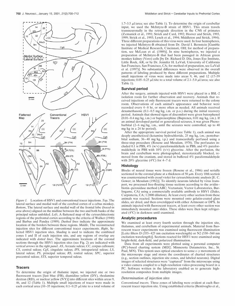
Figure 1. Location of HSV1 and conventional tracer injections. Top, Thelateral surface and medial wall of the cerebral cortex of a cebus monkey.Bottom, The lateral surface and medial wall of the frontal lobe (boxed-inarea above) aligned on the midline between the two and both banks of theprincipal sulcus unfolded. Left, A flattened map of the cytoarchitectonicregions of the prefrontal cortex according to the criteria of Walker (1940)and Barbas and Pandya (1989). Dashed lines indicate the approximatelocation of the borders between these regions. Middle, The reconstructedinjection sites for different conventional tracer experiments. Right, Se-lected HSV1 injection sites. Shading is used to indicate the combinedzones I and II of each injection site, and any regions of overlap areindicated with dotted lines. The approximate locations of the coronalsections through the HSV1 injection sites (see Fig. 2) are indicated withvertical arrows in the right panel. AS, Arcuate sulcus; CC, corpus callosum;CS, central sulcus; CgS, cingulate sulcus; IPS, intraparietal sulcus; LS,lateral sulcus; PS, principal sulcus; RS, rostral sulcus; SPC, superiorprecentral sulcus; STS, superior temporal sulcus.
702 J. Neurosci., January 15, 2001, 21(2):700–712 Middleton and Strick • Cerebellar Inputs to Prefrontal Cortex
1980; Huisman et al., 1983; Kuypers and Huisman, 1984; Conde, 1987),we defined zone I as the central region surrounding the needle track thatcontained an almost solid mass of fluorescent material. Zone II con-tained large numbers of intensely fluorescent neurons and glia amid abright background of fluorescence. Zone II gradually changed into zoneIII that contained some background tissue fluorescence and weaklyfluorescent neurons and glia. The effective area of uptake and transportof these tracers is considered to be confined to zones I and II (Bentivo-glio et al., 1980; Huisman et al., 1983; Kuypers and Huisman, 1984;Conde, 1987). Therefore, the maps of the injection sites (Figs. 1, 2) onlyillustrate these two zones.
HSV1. Three concentric zones of labeling surrounded each virusinjection site. Zone I contained the needle track and the highest densityof viral staining and pathology. In some instances, the tissue in this zonedisintegrated during tissue processing. Zone II contained a dense accu-mulation of infected neurons and glia, as well as a high degree ofbackground staining. Zone III contained large numbers of labeled neu-rons but little or no background staining. There is evidence that theactual zone of uptake for transneuronal transport of HSV1 is limited tozone I (for discussion of this issue, see Strick and Card, 1992; Hooverand Strick, 1999). Because this issue is not resolved, we included bothzones I and II in our reconstructions of injection sites (Figs. 1, 2).
Reconstruction of injection sitesThe plots of individual sections were aligned on the junction of themedial wall of the hemisphere with the lateral surface (i.e., the midlineof the hemisphere). Then, the medial wall and the lateral surface of thehemisphere, including the dorsal and ventral banks of the principalsulcus, were unfolded. This process created a flattened map of prefrontalcortex. The locations of injection sites and cytoarchitectonic borderswere added to this map (e.g., Fig. 1). Cytoarchitectonic borders weredrawn using the criteria of Walker (1940) and Barbas and Pandya (1989).
Distribution and density of cerebellar labelingCavalieri’s estimator of morphometric volume (see Rosen and Harry,1990) was used to determine the proportion of the dentate containingoutput neurons directed to each of the different prefrontal areas. Thisrule provides a statistically unbiased rectangular estimation of the vol-ume of brain structures from area measurements of regularly spacedserial sections:
Vc 5 dS Oi51
n
~ yi!D 2 ~t! ymax,
where d is the distance between the sections that are being analyzed, yiis the cross-sectional area of the ith section through the region of interest,n is the total number of sections, ymax is the maximum value of y, and tis the section thickness. A computer program was written in the labora-tory to obtain two measurements from MD2 files of sections through thedentate: (1) the total cross-sectional area of the nucleus and (2) the areaof the nucleus containing most (.90%) of the labeled neurons.
RESULTSOur results are divided into two major sections. In the firstsection, we present the results of experiments that used conven-tional tracers to examine the origin of thalamic inputs to regionsof the prefrontal cortex. We focus on the patterns of labeling inthalamic regions that are known to be the target of cerebellar orbasal ganglia efferents [e.g., nucleus ventralis anterior and latera-lis (VA/VL) and nucleus medialis dorsalis (MD)]. In the secondsection, we present the results of experiments that used HSV1 asa transneuronal tracer to define the origin of cerebellar projec-tions to the prefrontal cortex. In a subsequent report, we willpresent the patterns of retrograde transneuronal labeling ob-served in the output nuclei of the basal ganglia.
Experiments with conventional tracersInjection sitesWe injected fluorescent tracers into portions of areas 9, 46, and 12in four hemispheres (Table 1; Fig. 1, bottom middle). In general,tracers spread 400–500 mm from the needle track at injectionsites. Injections of NY and RD appeared to spread somewhat lessthan did injections of FB and DY.
Area 9. Areas 9m and 9l were separately injected in twohemispheres (F14 and F27L). In F14, the injection site primarilyinvolved the dorsal half of area 9m. It began 4.5 mm caudal to thefrontal pole, extended caudally for 5 mm, and ended 8 mm rostralto the genu of the arcuate sulcus. A portion of the peripheral zoneof this injection site extended into the most medial part of area 9l.In F27L, the injection site was entirely within the borders of 9l butwas split into two regions by the presence of a large blood vessel.Overall, the injection site began 6.5 mm caudal to the frontal pole,extended caudally for 5 mm, and ended 6.5 mm rostral to the genuof the arcuate sulcus.
Area 46. Areas 46d and 46v were injected with different tracersin a single hemisphere (F2). The injection sites filled the majorityof these areas without spreading beyond the borders of area 46.Both injection sites began ;4–5 mm caudal to the frontal pole andextended up to 2 mm rostral to the caudal tip of the principalsulcus.
Area 12. The area 12l was injected in a single hemisphere(F27R). The injection site was entirely within the borders of area12l. It began 6 mm caudal to the frontal pole, extended caudallyfor 8 mm, and ended 5 mm rostral to the genu of the arcuatesulcus.
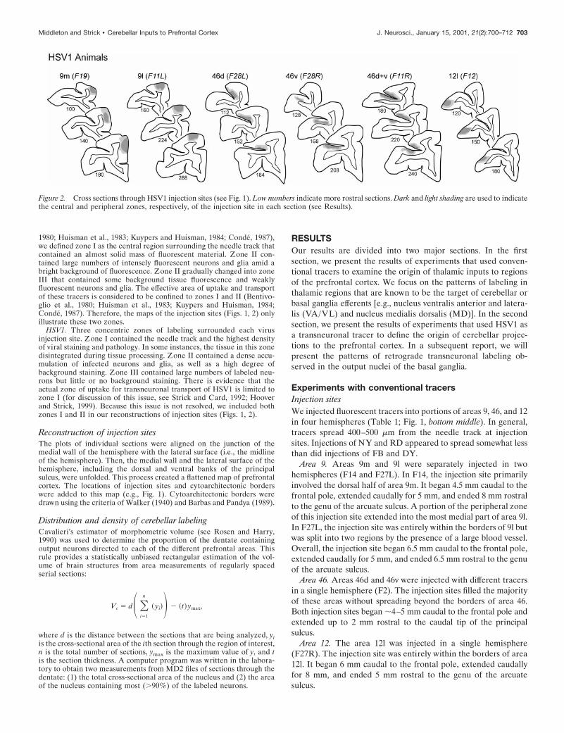
Figure 2. Cross sections through HSV1 injection sites (see Fig. 1). Low numbers indicate more rostral sections. Dark and light shading are used to indicatethe central and peripheral zones, respectively, of the injection site in each section (see Results).
Middleton and Strick • Cerebellar Inputs to Prefrontal Cortex J. Neurosci., January 15, 2001, 21(2):700–712 703

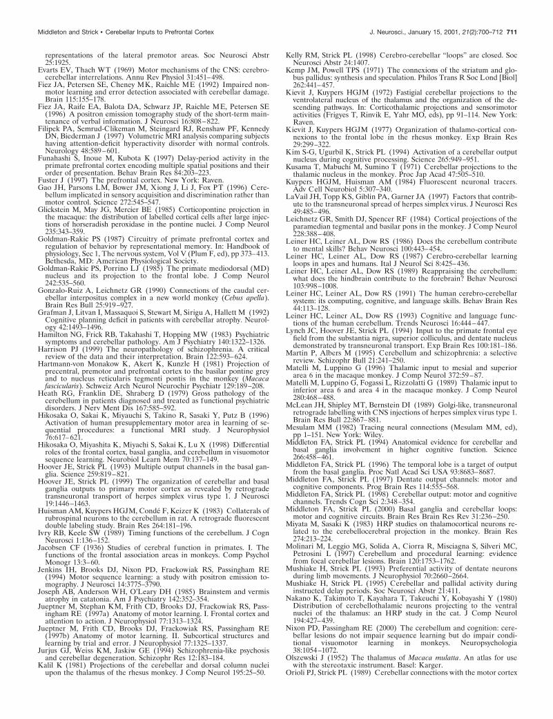
Thalamic labeling in VA/VL and MDRetrograde transport of conventional tracers from areas 9, 46,and 12 labeled many neurons in MD and in various subdivisionsof the ventrolateral thalamus (Figs. 3, 4). Because we varied theamount and type of fluorescent tracer injected in different exper-iments, the absolute numbers of labeled neurons differed fromcase to case. Thus, to compare the results from different injec-tions, we used the relative percentages of labeled neurons inspecific regions of the thalamus (Fig. 4).
Injections into the different cortical areas labeled neurons intopographically distinct regions of VA, VL, and MD. For exam-ple, neurons labeled by area 9 injections tended to be located inmore dorsal and medial regions of MD than were neurons labeledby area 46 or area 12 injections (Fig. 3). The thalamic input todifferent subdivisions of areas 9 and 46 also was distinct. Withinboth MD and subdivisions of the ventrolateral thalamus, theneurons labeled by area 9l injections tended to be located dorsalto those labeled by area 9m injections (see Fig. 3, first, secondrows). Similarly, the neurons labeled by area 46d injections tendedto be located dorsal to those labeled by area 46v injections (Fig. 4,third row). Only a small number of double-labeled neurons (,5%of the total sample) were found in the thalamus of the animal thatreceived multiple tracer injections into areas 46d and 46v of thesame hemisphere (F2, data not shown). These double-labeledneurons were most common in regions of the thalamus whereneurons projecting to the two regions of area 46 were intermin-gled (Fig. 3). Overall, our findings on the distribution of labeledneurons in the thalamus were comparable with what others havereported after similar injections of conventional tracers into areas9, 46, and 12 (Goldman-Rakic and Porrino, 1985; Barbas et al.,1991; Dermon and Barbas, 1994). The minor disparities betweenour results and those of previous studies are likely to be caused bysmall differences in the location of injection sites and the amountsof tracer injected.
Previous studies have shown that in primates the ventroante-rior, ventrolateral, and mediodorsal thalamus can be grouped intosubdivisions that receive primarily cerebellar, pallidal, or nigralinput (see Percheron, 1977; Percheron et al., 1996; Sakai et al.,1996). We analyzed the relative inputs from these subdivisions ofthe thalamus to different prefrontal cortical areas to determinewhether the anatomical substrate exists for the cerebellum andbasal ganglia to influence the prefrontal cortex. Several portionsof MD were excluded from this analysis because they are knownto receive subcortical input from multiple sources other than thebasal ganglia and cerebellum.
Area 9m. Approximately 50% of all the thalamic neurons la-beled by injections into area 9m were located in basal ganglia- orcerebellar-recipient thalamus (Fig. 4). The majority of these neu-rons were found in thalamic subdivisions that are the target ofnigral (25%, VAmc and MDmf) or pallidal (20%, VApc androstral VLc) efferents (see Figs. 3, first row, sections 439, 509; 4).
Figure 3. Thalamic input to the dorsal and lateral prefrontal cortex.Coronal cross sections through representative levels of the thalamus areshown for each conventional tracer experiment. Neurons labeled byretrograde transport from each area are indicated by dots, and nuclearborders are shown by solid or dotted lines. Thalamic nomenclature andabbreviations are according to Olszewski (1952). D, Dorsal; M, medial;Cd, caudate; c, pars caudalis; dc, pars densocellularis; Fx, fornix; IC,internal capsule; LD, nucleus lateralis dorsalis; mc, pars magnocellularis;mf, pars multiformis; pc, pars parvocellularis; m, pars medialis; R, nucleusreticularis; VPI, nucleus ventralis posterior inferior; VPL, nucleus vent-ralis posterior lateralis; X, area X.
Figure 4. Inputs from basal ganglia- and cerebellar-recipient thalamicregions. The percent of total thalamic input to different regions of theprefrontal cortex is shown for those nuclei that are well known targets ofbasal ganglia and cerebellar projections. Only those basal ganglia- andcerebellar-recipient thalamic nuclei that contained labeling are shown.VLcc, Caudal VLc; VLcr, rostral VLc; ps, pars postrema.
704 J. Neurosci., January 15, 2001, 21(2):700–712 Middleton and Strick • Cerebellar Inputs to Prefrontal Cortex

In contrast, only a few labeled neurons were located in thalamicsubdivisions that are the target of cerebellar efferents (5%, caudalVLc, VLps, and X) (Figs. 3, first row, section 539; 4). Substantialnumbers of labeled neurons also were found in a number ofthalamic nuclei that are not the target of basal ganglia or cere-bellar efferents [e.g., MDpc, 28% (Fig. 3, first row, sections 509,539), and 2–8% in each of the nucleus anterior ventralis andmedialis (Av/Am), reuniens (Re), paracentralis (Pcn), centrummedianum and parafasicularis (CM/Pf), centralis densocellularis(Cdc), centralis lateralis and centralis superior lateralis (Cl/Csl),and pulvinaris].
Area 9l. Nearly 45% of the thalamic neurons labeled by area 9linjections were found in basal ganglia- or cerebellar-recipientthalamus (Fig. 4). The largest proportion of labeled neurons(30%) was found in thalamic subdivisions that are the target ofpallidal efferents (23%; Fig. 3, second row, section 491) and nigralefferents (7%; Fig. 3, second row, section 491). Approximately halfas many neurons (15%) were found in cerebellar-recipient thal-amus (caudal portions of VLc; Fig. 3, second row, sections 577,611). Injections into area 9l labeled large numbers of neurons inMDpc (40%; Fig. 3, second row, sections 577, 611) and smallnumbers of neurons (5%) in several other thalamic nuclei [Av/Am, Cdc, Cl/Csl, Re, Pcn, and lateralis posterior (LP)].
Area 46d. Approximately 24% of the thalamic neurons labeledby area 46d injections were located in basal ganglia- or cerebellar-recipient nuclei (Fig. 4). The largest proportion was found inbasal ganglia-recipient regions of the thalamus (16%) with 10%located in subdivisions that receive input from pallidal efferents(VApc and VLcr; Fig. 3, third row, section 516) and 6% located insubdivisions that receive input from nigral efferents (MDmf andVAmc; Fig. 3, third row, section 516). However, a comparablepercentage (8%) was found in subdivisions that are the target ofcerebellar efferents (VLcc; Fig. 3, third row, section 626). Thenigral-recipient thalamus contained the smallest proportion oflabeled neurons (6%; Fig. 3, third row, section 516). On the otherhand, ;70% of the thalamic neurons labeled in this case were inMDpc. Other thalamic nuclei (MDdc, MDmc, Cl/Csl, Pcn, CM/Pf, and pulvinar) contained smaller percentages of labeled neu-rons (0.5–3%).
Area 46v. Slightly ,24% of the thalamic neurons labeled byarea 46v injections were located in basal ganglia- or cerebellar-recipient nuclei (Fig. 4). The highest percentage (20%) of labeledneurons was found in thalamic subdivisions that are the target ofnigral efferents (MDmf and VAmc; Fig. 3, third row, sections 516,556). Much smaller numbers of labeled neurons were found inthalamic nuclei that are the target of pallidal (3%) or cerebellar(1%) efferents. Over half of the total number of labeled neuronswere found in MDpc (52%), and small numbers of labeled neurons(3–4%) also were found in other thalamic nuclei (Pcn, CM/Pf, andMDmc).
Area 12l. Slightly .23% of the thalamic neurons labeled by area12l injections were located in basal ganglia- or cerebellar-recipient nuclei (Fig. 4). Nearly all of these neurons were inthalamic subdivisions that are the target of nigral efferents(MDmf and VAmc; see Fig. 3, fourth row, sections 507, 547). Incontrast, ,1% of the labeled neurons were in thalamic subdivi-sions that are the target of cerebellar or pallidal efferents. MDpccontained just .40% of the labeled neurons, and smaller percent-ages of labeled neurons (1–6%) were found in other thalamicnuclei (MDdc, MDmc, CM/Pf, pulvinar, and LP).
Overall, there were several clear trends in the patterns ofthalamic labeling observed after injections of fluorescent tracers
into different portions of prefrontal cortex. As the injection sitewas moved from ventrolateral regions of prefrontal cortex (area12l) to more dorsomedial regions (area 9m), there was a markedincrease in the number of labeled neurons in thalamic subdivi-sions that are the target of pallidal efferents. Labeling in nigral-and cerebellar-recipient subdivisions of the thalamus followeddifferent trends. The percentage of labeled neurons in the nigralthalamus was greatest after tracer injections into the most dor-somedial and ventrolateral areas examined (areas 9m and 12l),whereas labeling in cerebellar-recipient thalamus was greatestafter tracer injections into dorsolateral areas of prefrontal cortex(areas 9l and 46d). These patterns of thalamic labeling lead tosome clear predictions about the organization of basal gangliaand cerebellar “projections” to prefrontal cortex. They suggestthat basal ganglia output has a widespread influence, whereascerebellar output is more restricted. These predictions weretested using retrograde transneuronal transport of HSV1. Thecerebellar results are presented in the next section.
Experiments with HSV1Injection sitesWe injected the McIntyre-B strain of HSV1 into selected portionsof areas 9, 46, and 12 in 13 hemispheres (Table 1; Fig. 1, bottomright). In general, zones I and II of the injection sites extended500–900 mm from the needle tracks, depending on the amount ofvirus injected (Fig. 2).
Area 9m. HSV1 was injected into slightly different rostrocaudallevels of area 9m in two separate hemispheres. The first injectionsite filled up the majority of area 9m (F19, Figs. 1, 2). This injectionsite began 4 mm caudal to the frontal pole, extended caudally for 7mm, and ended 6 mm rostral to the genu of the arcuate sulcus. Asmall portion (;15%) of zone II from this injection site, but noneof zone I, extended into adjacent portions of area 9l. The secondinjection site in area 9m began 9 mm caudal to the frontal pole,extended caudally for 6 mm, and ended 2 mm rostral to the genu ofthe arcuate sulcus (F25, data not shown). As in the first area 9minjection, a small portion of the peripheral zone of this injectionsite spread to involve the most medial portion of area 9l. Althoughthe majority of this injection site was clearly within area 9m, thecaudal part of the peripheral zone of this injection site extendedinto a transitional cortical region between areas 9m, 8B, and thepresupplementary motor area. Because the patterns of transneu-ronal labeling in the output nuclei of the basal ganglia and cere-bellum did not differ significantly between these two cases, we willdescribe and illustrate the results of transport from the case thatwas most confined to area 9m (F19).
Area 9l. The HSV1 injection sites in area 9l (F11L, Figs. 1, 2;F21, data not shown) differed somewhat in their rostrocaudallocations. In each case, a small portion (,10%) of the peripheralzone of the injection site was found on the medial wall, in area9m. The injection site in F11 began 5.5 mm caudal to the frontalpole, extended caudally for 8.5 mm, and ended 4 mm rostral tothe genu of the arcuate sulcus. The injection site in F21 (data notshown) began 5 mm caudal to the frontal pole, extended caudallyfor 6 mm, and ended 5.5 mm rostral to the genu of the arcuatesulcus.
Areas 46d and 46v. Virus injections into area 46 were made intoboth banks of the principal sulcus (areas 46d and 46v combined)in three hemispheres. The precise extent of tissue within theprincipal sulcus that contained virus varied among these cases.The most complete injection of area 46 was in F11R (Fig. 2). This
Middleton and Strick • Cerebellar Inputs to Prefrontal Cortex J. Neurosci., January 15, 2001, 21(2):700–712 705
injection site was entirely within area 46 and filled up much of thetissue within the middle and caudal portions of the principalsulcus, including a portion of the fundus of the sulcus (Fig. 2). Itbegan 7 mm caudal to the frontal pole, extended caudally for 5mm, and ended 2 mm rostral to the caudal limit of the principalsulcus. The other virus injection sites in area 46 (F1 and F6, datanot shown) were similar in their rostrocaudal location but in-volved smaller portions of the principal sulcus.
Selective injections into area 46d or 46v were made in fourhemispheres (F28L, F28R, F24L, and F24R; Table 1). The injec-tion site in area 46d in F28L (Figs. 1, 2) was entirely within area46d and filled the middle and caudal portions of the dorsal bankof the principal sulcus. This injection site began 5 mm caudal tothe frontal pole, extended caudally for 7 mm, and ended 0.5 mmrostral to the caudal limit of the principal sulcus. The second
injection site in area 46d (F24, data not shown) was also com-pletely within area 46d. It began 5 mm caudal to the frontal pole,extended caudally for 5.5 mm, and ended 2 mm rostral to thecaudal limit of the PS. The two injection sites in area 46v werevery similar; they both filled a considerable amount of the middleand caudal portions of area 46v. These injection sites began ;5mm caudal to the frontal pole, extended caudally for 7 mm, andended 2 mm rostral to the caudal limit of the principal sulcus(F28R, Fig. 1; F24R, data not shown). The only significant dif-ference between these injection sites was that a portion of theinjection site in F28R extended into the white matter just beneaththe PS (F28R, Fig. 2).
Area 12l. The two HSV1 injection sites in area 12l (F12, Figs. 1,2; F27L, data not shown) were nearly identical in their dimen-sions and locations. Both injection sites began 5 mm caudal to thefrontal pole, extended caudally for ;7.5 mm, and ended 5 mmrostral to the genu of the arcuate sulcus. A small portion (,5%)of the peripheral zone of the injection site in F12 extended intothe orbital portion of area 12 (Fig. 2).
Thalamic labelingA survival time of 5 d is long enough for HSV1 to be transportedtransneuronally via two orders of synaptic connections. Thus, atthis survival time the thalamus will contain first-order neuronslabeled via retrograde transport of HSV1 directly from the injec-tion site. In addition, the thalamus will contain second-orderneurons labeled via retrograde transport from the injection site toanother brain area and then a second stage of retrograde trans-neuronal transport to thalamic neurons. In many cases, first- andsecond-order neurons can be distinguished by the intensity ofstaining and degree of cellular lysis (Hoover and Strick, 1999). Ingeneral, the distribution of first-order neurons in the thalamusafter HSV1 injections into prefrontal cortex was consistent withthat reported above for conventional tracers (compare Figs. 3, 5).
Labeling in cerebellar output nucleiMost of the cerebellar neurons labeled via retrograde transneu-ronal transport of HSV1 from prefrontal cortex were found in thedentate nucleus (Table 2). Labeled neurons had darkly stainedcell bodies, with somewhat lighter-stained dendrites radiatingfrom the cell soma (Fig. 6). These second-order neurons hadmorphological features typical of cerebellar neurons that projectto the thalamus (Tolbert et al., 1978; Nakano et al., 1980; Stantonand Orr, 1985).
Area 9m. The two animals that received injections of HSV1into area 9m displayed an average of just .90 labeled neurons inthe output nuclei of the cerebellum (Table 2). Most of theseneurons (.90%) were found in the dentate nucleus, and only asmall number (0–6%) were located in the interpositus and fasti-gial nuclei. In fact, no labeled neurons were found in the anteriorinterpositus in case F25. Over 85% of the labeled neurons in thedentate were located contralateral to the injection site. In con-trast, labeled neurons in the interpositus and fastigial nuclei weremore bilaterally distributed. In both area 9m cases, we foundlabeled neurons in a highly localized ventromedial region of thedentate, restricted to the middle and caudal third of the nucleus(Figs. 7, 8). This labeled region represented ;1% of the volumeof the dentate.
Area 9l. Injections of HSV1 into area 9l labeled an average of.400 neurons in the cerebellar output nuclei (Table 2). The vastmajority (.96%) of these labeled neurons were found in thedentate and were located contralateral to the injection site. A
Figure 5. HSV1-labeled regions of the thalamus. The patterns of HSV1labeling observed in the thalamus after injections into different regions ofthe prefrontal cortex are shown at a 5 d survival time. These sections weretaken through the most dense regions of thalamic labeling in each case.Conventions are described in Figure 3. Scale bar, 400 mm. mtt, Mamillothalamic tract.
706 J. Neurosci., January 15, 2001, 21(2):700–712 Middleton and Strick • Cerebellar Inputs to Prefrontal Cortex

small number of labeled neurons were found in the posteriorinterpositus (1%) and fastigial nuclei (2%). The labeled neuronsin the dentate were located in ventromedial regions of the caudalhalf of the nucleus. Neurons labeled by area 9l injections tendedto be located more laterally than were the neurons labeled by area9m injections (Figs. 7, 8). The dentate region that containedlabeled neurons after area 9l injections included ;8% of thevolume of the nucleus.
Area 46. Injections of HSV1 into area 46 labeled an average of.200 neurons in the cerebellar output nuclei (Table 2). Selectiveinjections into different portions of area 46 showed that nearly allof this labeling was caused by transport from area 46d (Table 2).This result is consistent with our observation that area 46d is amajor target of thalamic regions that receive input from the cere-bellum, whereas area 46v is not (see above; Fig. 4). The vastmajority (.95%) of neurons labeled after HSV1 injections intoarea 46 were found in the dentate and were located contralateral tothe injection site (97%). Labeled neurons in the dentate were mostconcentrated ventrally in the middle third of the nucleus rostro-caudally. In general, neurons that project to area 46 were locatedsomewhat more laterally in the dentate than were those that projectto area 9l (Figs. 7, 8). The dentate region that contained labeledneurons that project to area 46 represented ;6.5% of the volumeof the nucleus.
Area 12l. In contrast to the area 9 and area 46 results, injectionsof HSV1 into area 12l labeled very few neurons in the deepcerebellar nuclei. Most of the neurons that were labeled werefound in ventral regions of the dentate contralateral to the injec-tion site. The lack of labeled neurons in the dentate is consistentwith the relative absence of input to area 12 from regions of thethalamus that are the target of cerebellar efferents (see above;Fig. 4).
Overall, the patterns of labeling that we observed in the dentateafter transneuronal transport of HSV1 from prefrontal cortex areconsistent with the predictions derived from experiments withconventional tracers. Both approaches indicate that cerebellar
output targets specific portions of areas 9 and 46. In addition,virus transport uniquely demonstrates that this output originatesfrom topographically distinct portions of the ventral dentate.
DISCUSSIONIn this study, we examined the extent and topographic organiza-tion of cerebellar input to multiple regions of prefrontal cortex.The results from our studies with conventional tracers and withtransneuronal transport of HSV1 indicate that cerebellar outputdoes gain access to multiple areas of prefrontal cortex. Thesefindings clearly differ from the classical view that cerebellar out-put is focused entirely on the primary motor cortex.
The use of retrograde transneuronal transport of HSV1 en-abled us to determine the precise origin of cerebellothalamocor-tical projections to the prefrontal cortex. Clear shifts in thelocation of dense labeling in the dentate nucleus occurred withinjections into different areas of prefrontal cortex. Such shiftswere observed even after virus injections into adjacent subdivi-sions of the same cytoarchitectonic area (e.g., areas 9m and 9l;Figs. 7, 8). Thus, not only is the prefrontal cortex the target ofcerebellar output, but this output appears to be topographicallyorganized.
Comparison with cerebellar inputs to othercortical areasIn previous studies, we have used virus tracing to examine thecerebellar output to a number of motor areas of cortex. We foundthat injections of HSV1 into M1, ventral premotor area (PMv),dorsal premotor area (PMd), and frontal eyefield (FEF) all labelneurons in the dentate nucleus (Zemanick et al., 1991; Hooverand Strick, 1993, 1999; Strick et al., 1993; Lynch et al., 1994; Dumand Strick, 1999). The neurons labeled after virus injections intoM1, PMd, and PMv were all located in more dorsal regions of thedentate than were those labeled after prefrontal injections. Sim-ilarly, the neurons labeled by virus injections into the FEF werelocated in more caudal and lateral regions of the dentate than
Table 2. Numbers and percentage of total labeled neurons in the deep cerebellar nuclei
Areainjected Case
Dentatenucleus
Anteriorinterpositus
Posteriorinterpositus
Fastigialnucleus
9m F19 76 (90.5) 2 (2.4) 3 (3.6) 3 (3.6)F25 91 (90.1) 0 6 (5.9) 4 (4.0)Mean 83.5 (90.3) 1 (1.1) 4.5 (4.9) 3.5 (3.8)
9l F11L 562 (96.1) 0 6 (1.0) 17 (2.9)F21L 276 (97.5) 0 5 (1.8) 2 (0.7)Mean 419 (96.5) 0 5.5 (1.3) 9.5 (2.2)
46 F1 98 (95.1) 0 5 (4.9) 0F6 70 (97.2) 0 0 2 (2.8)F11R 486 (94.4) 2 (0.4) 9 (1.7) 18 (3.5)Mean 218 (94.8) 0.7 (0.3) 4.7 (2.0) 6.7 (2.9)
46d F24L 21 (95.5) 0 0 1 (4.5)F28L 18 (100.0) 0 0 0Mean 19.5 (97.5) 0 0 0.5 (2.5)
46v F24R 1 (100.0) 0 0 0F28R 0 0 0 0Mean 0.5 0 0 0
12l F12 2 (66.7) 0 1 (33.3) 0F27L 4 (57.1) 0 0 3 (42.9)Mean 3.0 (60.0) 0 0.5 (10.0) 1.5 (30.0)
Middleton and Strick • Cerebellar Inputs to Prefrontal Cortex J. Neurosci., January 15, 2001, 21(2):700–712 707

were those labeled by prefrontal injections. These results indicatethat cerebellar projections to prefrontal, oculomotor, and skeleto-motor areas of cortex all appear to be derived from topographi-cally distinct regions of the dentate. We have proposed that thecluster of neurons that projects to an individual cortical areacreates a distinct “output channel” in the cerebellum (Strick et al.,1993; Middleton and Strick, 1997, 1998). The present resultsindicate that the dentate contains several output channels that aredirected at different areas of prefrontal cortex.
Our results emphasize that areas 9l and 46d are major targetsof dentate output. Interestingly, these are the regions of prefron-tal cortex that project most densely on pontine nuclei that provideaccess to the input stage of cerebellar processing (Schmahmannand Pandya, 1995, 1997a). These observations suggest that amajor structural feature of cerebellar interactions with prefrontalcortex is multiple, topographically closed loops. If this arrange-ment reflects a general principle of cerebrocerebellar architec-ture, then those areas of cerebral cortex that project to the input
stage of cerebellar processing (i.e., the pontine nuclei) would alsobe the target of efferents from the output stage of cerebellarprocessing (i.e., the deep cerebellar nuclei). Widespread regionsof the cerebral cortex, including cingulate, somatic sensory, pos-terior parietal, and visual areas, are known to project to thepontine nuclei (Brodal, 1978; Hartmann-von Monakow et al.,1981; Vilensky and van Hoesen, 1981; Leichnetz et al., 1984;Glickstein et al., 1985; Schmahmann and Pandya, 1995, 1997a).Our hypothesis predicts that most of these cortical areas wouldalso be the target of cerebellar output. To date, the volume of thedentate occupied by the known output channels to skeletomotor,oculomotor, and prefrontal areas of cortex represents only ;60%of the volume of the nucleus. Consequently, the cortical targets ofa major portion of dentate output remain to be determined. Evenso, it is clear that a considerable amount of the cerebrocerebellarinteractions operate outside the domain of motor control.
Functional implicationsOur results indicate that output channels in the ventral dentateproject to specific portions of the prefrontal cortex. These obser-vations raise an important question; namely, what is the func-tional contribution of this pathway? In this section, we will de-scribe results based on a variety of experimental approaches thatsuggest that dentate output channels to prefrontal cortex areinvolved in aspects of higher executive function like planning,working memory, and sequential behavior.
Single-neuron recording in trained primatesMushiake and Strick (1993, 1995) recorded from the dentatenucleus in monkeys trained to perform sequences of movements,three elements in length. In one component of the task, threevisual cues were presented to the monkey. The monkey had toremember the position of the cues and the order in which theywere presented. Then, after a variable instructed delay period, themonkey performed the remembered sequence as quickly as pos-sible. Approximately 15% of the neurons recorded in the dentateduring this task displayed changes in activity during the in-structed delay period. The activity of some instruction-relatedneurons was specific for the particular sequence of movements theanimal had to remember. These patterns of activity resemblethose of neurons in area 46 recorded during a similar instructeddelay task that involved arm movements (Funahashi et al., 1997).The dentate neurons that displayed instruction-related activitytended to be located in ventral regions of the nucleus that projectto areas 9 and 46. These results suggest that dentate activity couldbe involved in the generation and/or maintenance of delay activ-ity in prefrontal cortex.
Imaging studies in human subjectsJueptner et al. (1997a,b) found significant activation in ventrolat-eral portions of the deep cerebellar nuclei, as well as in portionsof areas 9 and 46, during the learning of new sequences of fingermovements. They also observed peak activations laterally in thecerebellar hemispheres and at thalamic sites in caudal paralami-nar MD. Thus, every potential site in the cerebellothalamocorti-cal pathway from the neocerebellum to the prefrontal cortexdisplayed activation during a sequence-learning task in humans.
Kim et al. (1994) specifically examined dentate activation inhuman subjects by the use of high-field functional MRI. Theyfound that the magnitude and extent of activation in the dentateduring attempts to solve a Peg-Board puzzle were greater thanthat observed when subjects simply performed visually guided
Figure 6. HSV1-labeled neurons in the cerebellar output nuclei. Top,HSV1-labeled neurons in the dentate nucleus are shown after injectionsinto area 46 (F1). Bottom lef t, An example of an HSV1-labeled neuron inthe fastigial nucleus is shown after injections of area 9m. Bottom right, Anexample of an HSV1-labeled neuron in the posterior interpositus nucleusafter injection of area 9l. Scale bars, 25 mm.
708 J. Neurosci., January 15, 2001, 21(2):700–712 Middleton and Strick • Cerebellar Inputs to Prefrontal Cortex

movements of pegs. Thus, dentate activation was not simplyrelated to movement per se (see also Gao et al., 1996). Moreover,ventral regions of the dentate appeared to be especially acti-vated during attempts to solve the Peg-Board puzzle. Overall,
imaging studies in humans provide support for the suggestionthat output channels in the ventral dentate that innervateprefrontal cortex are involved in the learning of new se-quences, spatial working memory, planning, and rule-basedlearning.
Cerebellar lesionsIn addition to the classical motor deficits, there is considerableevidence that cerebellar pathology in humans can lead to deficitsin the performance of cognitive tasks that require rule-basedlearning, judgment of temporal intervals, visuospatial analysis,and shifting attention between sensory modalities, as well asworking memory and planning (for review, see Leiner et al., 1986,1987, 1989, 1991, 1993; Botez et al., 1989; Ivry and Keele, 1989;Schmahmann, 1991, 1997; Akshoomoff and Courchesne, 1992;Fiez et al., 1992; Grafman et al., 1992; Schmahmann and Sher-man, 1998). Many of these deficits reflect functions normallythought to be subserved by areas of prefrontal cortex. Based onour results, one interpretation of the origin of these deficits is thatthey result from an interruption of input to the prefrontal cortexfrom the cerebellum. Similarly, one might predict that damage tothe ventral portion of the dentate, or to the regions of cerebellarcortex that innervate it, would produce deficits that resemblethose seen after lesions of areas 9 or 46. In fact, a study by Fiez etal. (1992) provides some support for this prediction. They de-scribed a patient, designated RC1, who had circumscribed dam-age to the lateral portion of his right cerebellar cortex. Thispatient exhibited few classical signs of cerebellar damage but wasimpaired on the performance of specific types of rule-basedlanguage and memory tasks. The deficits appeared on tasks thatin normal subjects activate lateral portions of the cerebellarhemispheres and areas 9 and 46 (Petersen et al., 1988; Raichle etal., 1994; Fiez et al., 1996). Recent anatomical studies suggest thatthe portions of the cerebellum damaged in RC1 are part of thecerebellar loop with the prefrontal cortex (Kelly and Strick, 1998).Thus, the cognitive deficits in RC1 may have been a consequenceof interrupting this circuit.
Results of studies on patients with cerebellar damage suggestthat the cerebellum also is involved in sequence learning andperformance (Pascual-Leone et al., 1993; Molinari et al., 1997),behaviors that have been shown to rely, at least in part, on certain
Figure 7. Comparison of dentate labeling after HSV1 injections of area 9. Sections at the same level of the dentate are shown after injections of HSV1into area 9m (lef t), area 9l (middle), or area 46 (right). The dashed lines indicate the borders of the dentate nucleus. Note the difference in the locationof the labeled neurons within the dentate between cases. Scale bar, 500 mm.
Figure 8. Origin of dentate projections to areas 9 and 46. Sectionsthrough the middle of the dentate nucleus are shown after injections intoarea 9m (lef t), area 9l (middle), or area 46 (right). The rostrocaudaldistribution of labeled neurons in the dentate for each case is shown at thebottom of the figure, with the locations of the sections shown aboveindicated by the arrows. DN, Dentate nucleus; R, rostral; C, caudal.
Middleton and Strick • Cerebellar Inputs to Prefrontal Cortex J. Neurosci., January 15, 2001, 21(2):700–712 709

prefrontal areas (Jacobsen, 1936; Petrides and Milner, 1982; Jen-kins et al., 1994; Pascual-Leone et al., 1995, 1996; Petrides, 1995;Hikosaka et al., 1996; Funahashi et al., 1997; Jueptner et al.,1997a,b; Sakai et al., 1998). Thus, it is possible that the outputchannels that link the ventral dentate to areas of prefrontal cortexmay have a function in the learning and execution of sequentialbehavior. Two recent experimental tests of this suggestion havegiven conflicting results. Hikosaka et al. (1998) examined theeffects of focal inactivation of movement-related sites within thedentate on the learning and performance of a sequential button-press task. They concluded that the “ . . . dentate nucleus is usedfor the storage or retrieval of long-term procedural memories.”On the other hand, Nixon and Passingham (2000) examined theeffects of bilateral excitotoxic lesions of the lateral cerebellarnuclei on a similar sequential task. They suggested that thecerebellum “ . . . is not essential for learning or recall” but is“ . . . crucially involved in the process by which motor sequencesbecome automatic with extended practice.” Perhaps the disparateconclusions result, in part, from differences in the size and extentof damage within the dentate. Our anatomical data indicate thateven small shifts in the lesion site could effect different outputchannels within the dentate.
Cerebellar involvement in psychiatric disordersCerebellar abnormalities have been reported in numerous studiesof neuropsychiatric disorders, including depression, Tourette’ssyndrome, attention deficit disorder, autism, William’s syndrome,and schizophrenia, to name but a few (Heath et al., 1979; Wein-berger et al., 1979; Snider, 1982; Hamilton et al., 1983; Baumanand Kemper, 1985; Joseph et al., 1985; Shelton and Weinberger,1986; Yates et al., 1987; Courchesne et al., 1988; Volkow, 1992;Jurjus et al., 1994; Martin and Albers, 1995; Andreasen et al.,1996; Courchesne, 1997; Filipek et al., 1997; Harrison, 1999).Many of these studies have been inconclusive and often con-founded by the effects of medication. Nonetheless, it is becomingapparent that some of the changes reported in these studies couldreflect involvement of the same anatomical circuits we havedescribed. For example, among the most consistent findings instudies of schizophrenia are decreased metabolism in areas 9 and46, cytoarchitectonic alterations in thalamic-recipient layers ofareas 9 and 46, and reductions in neuron number in the MDnucleus of the thalamus (for review, see Harrison, 1999). Thepossibility that these findings could all be related to dysfunctionsof a cerebellar output channel to the prefrontal cortex was sug-gested by the work of Andreasen et al. (1996). These investigatorsreported alterations in metabolism in the cerebellum, thalamus,and dorsal prefrontal cortex of schizophrenic subjects during amemory recall task and speculated that schizophrenic subjectsmight suffer from a form of dysmetria that involved cognitiveoperations, as opposed to the dysmetria classically associated withcerebellar damage. Clearly, more detailed analyses of the cere-bellar changes in schizophrenia and a more complete map of therelations between areas of cerebral and cerebellar cortex willenable a better assessment of the potential cerebellar involvementin schizophrenia and other psychiatric disorders.
Anatomical and functional specificity of cerebellar projectionsto prefrontal cortexThe present results provide further clues about the functionalcontributions of the cerebellar projections to the prefrontal cor-tex. We found that cerebellar output targets dorsal areas of theprefrontal cortex (9 and 46d) but primarily avoids ventral pre-
frontal areas (12 and 46v). A complete discussion of the func-tional differences between dorsal and ventral areas of the pre-frontal cortex is beyond the scope of this report. However, thereis some agreement that dorsal areas of the prefrontal cortex areinvolved in spatial working memory and planning and are a majorsite of termination of the “dorsal stream” of visual processing,whereas ventral areas of the prefrontal cortex are involved in theworking memory for objects or visual discrimination learning andare a major target of the ventral stream of visual processing (seePetrides and Milner, 1982; Goldman-Rakic, 1987; Passingham,1993; Wilson et al., 1994; Petrides, 1995; Fuster, 1997; Rushworthet al., 1997). Thus, our results suggest that the cerebellar projec-tion to the prefrontal cortex is particularly concerned with spatialworking memory, planning, and other functions associated withthe dorsal stream of visual processing.
In summary, the available anatomical, physiological, and be-havioral data suggest that the cerebellum is involved not only inthe control of movement but also in many aspects of cognitivebehavior like planning, working memory, and sequential behav-ior. Our results provide the anatomical substrate for a cerebellarinfluence on processing within the prefrontal cortex. Furtherstudies are necessary to determine the full extent of the cerebralcortex that is the target of cerebellar output. Clearly, we haveonly begun to appreciate the diverse range of behaviors that couldbe influenced by the cerebellum.
REFERENCESAkshoomoff NA, Courchesne E (1992) A new role for the cerebellum in
cognitive function. Behav Neurosci 106:731–738.Allen GI, Tsukahara N (1974) Cerebrocerebellar communication sys-
tems. Physiol Rev 54:957–1006.Allen GI, Gilbert PF, Yin TC (1978) Convergence of cerebral inputs
onto dentate neurons in monkey. Exp Brain Res 32:151–170.Andreasen NC, O’Leary DS, Cizaldo T, Arndt S, Rezai K, Boles Ponto
LL, Watkins GL, Hichwa RD (1996) Schizophrenia and cognitive dys-metria: a positron-emission tomography study of dysfunctionalprefrontal-thalamic-cerebellar circuitry. Proc Natl Acad Sci USA93:9985–9990.
Asanuma C, Thach WT, Jones EG (1983) Distribution of cerebellarterminations in the ventral lateral thalamic region of the monkey. BrainRes Rev 5:237–265.
Barbas H, Pandya DN (1989) Architecture and intrinsic connections ofthe prefrontal cortex in the rhesus monkey. J Comp Neurol286:353–375.
Barbas H, Haswell Henion TH, Dermon CR (1991) Diverse thalamicprojections to the prefrontal cortex in the rhesus monkey. J CompNeurol 313:65–94.
Bauman M, Kemper TL (1985) Histoanatomic observations of the brainin early infantile autism. Neurology 35:866–874.
Bentivoglio M, Kuypers HGJM, Catsman-Berevoets CE, Loewe H, DannO (1980) Two new fluorescent retrograde neuronal tracers which aretransported over long distances. Neurosci Lett 18:25–30.
Botez MI, Botez T, Elie R, Attig E (1989) Role of the cerebellum incomplex human behavior. Ital J Neurol Sci 10:291–300.
Brodal P (1978) The corticopontine projection in the rhesus monkey.Origin and principles of organization. Brain 101:251–283.
Brooks VB, Thach WT (1981) Cerebellar control of posture and move-ment. In: Handbook of physiology, Sec 1, The nervous system, Vol 2,Motor control, Pt II (Brooks VB, ed), pp 877–946. Bethesda, MD:American Physiological Society.
Conde F (1987) Further studies on the use of the fluorescent tracers fastblue and diamidino yellow: effective uptake area and cellular storagesites. J Neurosci Methods 21:31–43.
Courchesne E (1997) Brainstem, cerebellar and limbic neuroanatomicalabnormalities in autism. Curr Opin Neurobiol 7:269–278.
Courchesne E, Yeung-Courchesne R, Press GA, Hesselink JR, JerniganTL (1988) Hypoplasia of cerebellar vermal lobules VI and VII inautism. N Engl J Med 318:1349–1354.
Darian-Smith C, Darian-Smith I, Cheema SS (1990) Thalamic projec-tions to sensorimotor cortex in the macaque monkey: use of multipleretrograde fluorescent tracers. J Comp Neurol 299:17–46.
Dermon CR, Barbas H (1994) Contralateral thalamic projections pre-dominantly reach transitional cortices in the rhesus monkey. J CompNeurol 344:508–531.
Dum RP, Strick PL (1999) Pallidal and cerebellar inputs to the digit
710 J. Neurosci., January 15, 2001, 21(2):700–712 Middleton and Strick • Cerebellar Inputs to Prefrontal Cortex
representations of the lateral premotor areas. Soc Neurosci Abstr25:1925.
Evarts EV, Thach WT (1969) Motor mechanisms of the CNS: cerebro-cerebellar interrelations. Annu Rev Physiol 31:451–498.
Fiez JA, Petersen SE, Cheney MK, Raichle ME (1992) Impaired non-motor learning and error detection associated with cerebellar damage.Brain 115:155–178.
Fiez JA, Raife EA, Balota DA, Schwarz JP, Raichle ME, Petersen SE(1996) A positron emission tomography study of the short-term main-tenance of verbal information. J Neurosci 16:808–822.
Filipek PA, Semrud-Clikeman M, Steingard RJ, Renshaw PF, KennedyDN, Biederman J (1997) Volumetric MRI analysis comparing subjectshaving attention-deficit hyperactivity disorder with normal controls.Neurology 48:589–601.
Funahashi S, Inoue M, Kubota K (1997) Delay-period activity in theprimate prefrontal cortex encoding multiple spatial positions and theirorder of presentation. Behav Brain Res 84:203–223.
Fuster J (1997) The prefrontal cortex. New York: Raven.Gao JH, Parsons LM, Bower JM, Xiong J, Li J, Fox PT (1996) Cere-
bellum implicated in sensory acquisition and discrimination rather thanmotor control. Science 272:545–547.
Glickstein M, May JG, Mercier BE (1985) Corticopontine projection inthe macaque: the distribution of labelled cortical cells after large injec-tions of horseradish peroxidase in the pontine nuclei. J Comp Neurol235:343–359.
Goldman-Rakic PS (1987) Circuitry of primate prefrontal cortex andregulation of behavior by representational memory. In: Handbook ofphysiology, Sec 1, The nervous system, Vol V (Plum F, ed), pp 373–413.Bethesda, MD: American Physiological Society.
Goldman-Rakic PS, Porrino LJ (1985) The primate mediodorsal (MD)nucleus and its projection to the frontal lobe. J Comp Neurol242:535–560.
Gonzalo-Ruiz A, Leichnetz GR (1990) Connections of the caudal cer-ebellar interpositus complex in a new world monkey (Cebus apella).Brain Res Bull 25:919–927.
Grafman J, Litvan I, Massaquoi S, Stewart M, Sirigu A, Hallett M (1992)Cognitive planning deficit in patients with cerebellar atrophy. Neurol-ogy 42:1493–1496.
Hamilton NG, Frick RB, Takahashi T, Hopping MW (1983) Psychiatricsymptoms and cerebellar pathology. Am J Psychiatry 140:1322–1326.
Harrison PJ (1999) The neuropathology of schizophrenia. A criticalreview of the data and their interpretation. Brain 122:593–624.
Hartmann-von Monakow K, Akert K, Kunzle H (1981) Projection ofprecentral, premotor and prefrontal cortex to the basilar pontine greyand to nucleus reticularis tegmenti pontis in the monkey (Macacafascicularis). Schweiz Arch Neurol Neurochir Psychiatr 129:189–208.
Heath RG, Franklin DE, Shraberg D (1979) Gross pathology of thecerebellum in patients diagnosed and treated as functional psychiatricdisorders. J Nerv Ment Dis 167:585–592.
Hikosaka O, Sakai K, Miyauchi S, Takino R, Sasaki Y, Putz B (1996)Activation of human presupplementary motor area in learning of se-quential procedures: a functional MRI study. J Neurophysiol76:617–621.
Hikosaka O, Miyashita K, Miyachi S, Sakai K, Lu X (1998) Differentialroles of the frontal cortex, basal ganglia, and cerebellum in visuomotorsequence learning. Neurobiol Learn Mem 70:137–149.
Hoover JE, Strick PL (1993) Multiple output channels in the basal gan-glia. Science 259:819–821.
Hoover JE, Strick PL (1999) The organization of cerebellar and basalganglia outputs to primary motor cortex as revealed by retrogradetransneuronal transport of herpes simplex virus type 1. J Neurosci19:1446–1463.
Huisman AM, Kuypers HGJM, Conde F, Keizer K (1983) Collaterals ofrubrospinal neurons to the cerebellum in rat. A retrograde fluorescentdouble labeling study. Brain Res 264:181–196.
Ivry RB, Keele SW (1989) Timing functions of the cerebellum. J CognNeurosci 1:136–152.
Jacobsen CF (1936) Studies of cerebral function in primates. I. Thefunctions of the frontal association areas in monkeys. Comp PsycholMonogr 13:3–60.
Jenkins IH, Brooks DJ, Nixon PD, Frackowiak RS, Passingham RE(1994) Motor sequence learning: a study with positron emission to-mography. J Neurosci 14:3775–3790.
Joseph AB, Anderson WH, O’Leary DH (1985) Brainstem and vermisatrophy in catatonia. Am J Psychiatry 142:352–354.
Jueptner M, Stephan KM, Frith CD, Brooks DJ, Frackowiak RS, Pass-ingham RE (1997a) Anatomy of motor learning. I. Frontal cortex andattention to action. J Neurophysiol 77:1313–1324.
Jueptner M, Frith CD, Brooks DJ, Frackowiak RS, Passingham RE(1997b) Anatomy of motor learning. II. Subcortical structures andlearning by trial and error. J Neurophysiol 77:1325–1337.
Jurjus GJ, Weiss KM, Jaskiw GE (1994) Schizophrenia-like psychosisand cerebellar degeneration. Schizophr Res 12:183–184.
Kalil K (1981) Projections of the cerebellar and dorsal column nucleiupon the thalamus of the rhesus monkey. J Comp Neurol 195:25–50.
Kelly RM, Strick PL (1998) Cerebro-cerebellar “loops” are closed. SocNeurosci Abstr 24:1407.
Kemp JM, Powell TPS (1971) The connexions of the striatum and glo-bus pallidus: synthesis and speculation. Philos Trans R Soc Lond [Biol]262:441–457.
Kievit J, Kuypers HGJM (1972) Fastigial cerebellar projections to theventrolateral nucleus of the thalamus and the organization of the de-scending pathways. In: Corticothalamic projections and sensorimotoractivities (Frigyes T, Rinvik E, Yahr MO, eds), pp 91–114. New York:Raven.
Kievit J, Kuypers HGJM (1977) Organization of thalamo-cortical con-nexions to the frontal lobe in the rhesus monkey. Exp Brain Res29:299–322.
Kim S-G, Ugurbil K, Strick PL (1994) Activation of a cerebellar outputnucleus during cognitive processing. Science 265:949–951.
Kusama T, Mabuchi M, Sumino T (1971) Cerebellar projections to thethalamic nucleus in the monkey. Proc Jap Acad 47:505–510.
Kuypers HGJM, Huisman AM (1984) Fluorescent neuronal tracers.Adv Cell Neurobiol 5:307–340.
LaVail JH, Topp KS, Giblin PA, Garner JA (1997) Factors that contrib-ute to the transneuronal spread of herpes simplex virus. J Neurosci Res49:485–496.
Leichnetz GR, Smith DJ, Spencer RF (1984) Cortical projections of theparamedian tegmental and basilar pons in the monkey. J Comp Neurol228:388–408.
Leiner HC, Leiner AL, Dow RS (1986) Does the cerebellum contributeto mental skills? Behav Neurosci 100:443–454.
Leiner HC, Leiner AL, Dow RS (1987) Cerebro-cerebellar learningloops in apes and humans. Ital J Neurol Sci 8:425–436.
Leiner HC, Leiner AL, Dow RS (1989) Reappraising the cerebellum:what does the hindbrain contribute to the forebrain? Behav Neurosci103:998–1008.
Leiner HC, Leiner AL, Dow RS (1991) The human cerebro-cerebellarsystem: its computing, cognitive, and language skills. Behav Brain Res44:113–128.
Leiner HC, Leiner AL, Dow RS (1993) Cognitive and language func-tions of the human cerebellum. Trends Neurosci 16:444–447.
Lynch JC, Hoover JE, Strick PL (1994) Input to the primate frontal eyefield from the substantia nigra, superior colliculus, and dentate nucleusdemonstrated by transneuronal transport. Exp Brain Res 100:181–186.
Martin P, Albers M (1995) Cerebellum and schizophrenia: a selectivereview. Schizophr Bull 21:241–250.
Matelli M, Luppino G (1996) Thalamic input to mesial and superiorarea 6 in the macaque monkey. J Comp Neurol 372:59–87.
Matelli M, Luppino G, Fogassi L, Rizzolatti G (1989) Thalamic input toinferior area 6 and area 4 in the macaque monkey. J Comp Neurol280:468–488.
McLean JH, Shipley MT, Bernstein DI (1989) Golgi-like, transneuronalretrograde labelling with CNS injections of herpes simplex virus type 1.Brain Res Bull 22:867–881.
Mesulam MM (1982) Tracing neural connections (Mesulam MM, ed),pp 1–151. New York: Wiley.
Middleton FA, Strick PL (1994) Anatomical evidence for cerebellar andbasal ganglia involvement in higher cognitive function. Science266:458–461.
Middleton FA, Strick PL (1996) The temporal lobe is a target of outputfrom the basal ganglia. Proc Natl Acad Sci USA 93:8683–8687.
Middleton FA, Strick PL (1997) Dentate output channels: motor andcognitive components. Prog Brain Res 114:555–568.
Middleton FA, Strick PL (1998) Cerebellar output: motor and cognitivechannels. Trends Cogn Sci 2:348–354.
Middleton FA, Strick PL (2000) Basal ganglia and cerebellar loops:motor and cognitive circuits. Brain Res Brain Res Rev 31:236–250.
Miyata M, Sasaki K (1983) HRP studies on thalamocortical neurons re-lated to the cerebellocerebral projection in the monkey. Brain Res274:213–224.
Molinari M, Leggio MG, Solida A, Ciorra R, Misciagna S, Silveri MC,Petrosini L (1997) Cerebellum and procedural learning: evidencefrom focal cerebellar lesions. Brain 120:1753–1762.
Mushiake H, Strick PL (1993) Preferential activity of dentate neuronsduring limb movements. J Neurophysiol 70:2660–2664.
Mushiake H, Strick PL (1995) Cerebellar and pallidal activity duringinstructed delay periods. Soc Neurosci Abstr 21:411.
Nakano K, Takimoto T, Kayahara T, Takeuchi Y, Kobayashi Y (1980)Distribution of cerebellothalamic neurons projecting to the ventralnuclei of the thalamus: an HRP study in the cat. J Comp Neurol194:427–439.
Nixon PD, Passingham RE (2000) The cerebellum and cognition: cere-bellar lesions do not impair sequence learning but do impair condi-tional visuomotor learning in monkeys. Neuropsychologia38:1054–1072.
Olszewski J (1952) The thalamus of Macaca mulatta. An atlas for usewith the stereotaxic instrument. Basel: Karger.
Orioli PJ, Strick PL (1989) Cerebellar connections with the motor cortex
Middleton and Strick • Cerebellar Inputs to Prefrontal Cortex J. Neurosci., January 15, 2001, 21(2):700–712 711

and the arcuate premotor area: analysis employing retrograde trans-neuronal transport of WGA-HRP. J Comp Neurol 288:612–626.
Pascual-Leone A, Grafman J, Clark K, Stewart M, Massaquoi S, Lou JS,Hallett M (1993) Procedural learning in Parkinson’s disease and cer-ebellar degeneration. Ann Neurol 34:594–602.
Pascual-Leone A, Grafman J, Hallett M (1995) Procedural learning andprefrontal cortex. Ann NY Acad Sci 769:61–70.
Pascual-Leone A, Wassermann EM, Grafman J, Hallett M (1996) Therole of the dorsolateral prefrontal cortex in implicit procedural learn-ing. Exp Brain Res 107:479–485.
Passingham R (1993) The frontal lobes and voluntary action. Oxford:Oxford UP.
Percheron G (1977) The thalamic territory of cerebellar afferents andthe lateral region of the thalamus of the macaque in stereotaxic ven-tricular coordinates. J Hirnforsch 18:375–400.
Percheron G, Francois C, Talbi B, Yelnik J, Fenelon G (1996) Theprimate motor thalamus. Brain Res Rev 22:93–181.
Petersen SE, Fox PT, Posner MI, Mintun M, Raichle ME (1988)Positron emission tomographic studies of the cortical anatomy ofsingle-word processing. Nature 331:585–589.
Petrides M (1995) Impairments on nonspatial self-ordered and exter-nally ordered working memory tasks after lesions of the mid-dorsal partof the lateral frontal cortex in the monkey. J Neurosci 15:359–375.
Petrides M, Milner B (1982) Deficits on subject-ordered tasks afterfrontal- and temporal-lobe lesions in man. Neuropsychologia20:249–262.
Raichle ME, Fiez JA, Videen TO, MacLeod AM, Pardo JV, Fox PT,Petersen SE (1994) Practice-related changes in human brain func-tional anatomy during nonmotor learning. Cereb Cortex 4:8–26.
Rosen GD, Harry JD (1990) Brain volume estimation from serial sectionmeasurements: a comparison of methodologies. J Neurosci Methods35:115–124.
Rosene DL, Mesulam MM (1978) Fixation variables in horseradish per-oxidase neurohistochemistry. I. The effects of fixation time and perfu-sion procedures upon enzyme activity. J Histochem Cytochem26:28–39.
Rosene DL, Roy NJ, Davis BJ (1986) A cryoprotection method thatfacilitates cutting sections of whole monkey brains for histological andhistochemical processing without freezing artifact. J Histochem Cyto-chem 34:1301–1315.
Rouiller EM, Liang F, Babalian A, Moret V, Wiesendanger M (1994)Cerebellothalamocortical and pallidothalamocortical projections to theprimary and supplementary motor cortical areas: a multiple tracingstudy in macaque monkeys. J Comp Neurol 345:185–231.
Rushworth MF, Nixon PD, Eacott MJ, Passingham R (1997) Ventralprefrontal cortex is not essential for working memory. J Neurosci17:4829–4838.
Sakai K, Hikosaka O, Miyauchi S, Takino R, Sasaki Y, Putz B (1998)Transition of brain activation from frontal to parietal areas in visuo-motor sequence learning. J Neurosci 18:1827–1840.
Sakai ST, Inase M, Tanji J (1996) Comparison of cerebellothalamic andpallidothalamic projections in the monkey (Macaca fuscata): a doubleanterograde labeling study. J Comp Neurol 368:215–228.
Sasaki K, Jinnai K, Gemba H, Hashimoto S, Mizuno N (1979) Projec-tion of the cerebellar dentate nucleus onto the frontal association cortexin monkeys. Exp Brain Res 37:193–198.
Schell GR, Strick PL (1984) The origin of thalamic inputs to the arcuatepremotor and supplementary motor areas. J Neurosci 4:539–560.
Schmahmann JD (1991) An emerging concept. The cerebellar contribu-tion to higher function. Arch Neurol 48:1178–1187.
Schmahmann JD (1997) Rediscovery of an early concept. Int Rev Neu-robiol 41:3–27.
Schmahmann JD, Pandya DN (1995) Prefrontal cortex projections to thebasilar pons in rhesus monkey: implications for the cerebellar contri-butions to higher function. Neurosci Lett 199:175–178.
Schmahmann JD, Pandya DN (1997a) Anatomic organization of thebasilar pontine projections from prefrontal cortices in rhesus monkey.J Neurosci 17:438–458.
Schmahmann JD, Pandya DN (1997b) The cerebrocerebellar system. IntRev Neurobiol 41:31–60.
Schmahmann JD, Sherman JC (1998) The cerebellar cognitive affectivesyndrome. Brain 121:561–579.
Shelton RC, Weinberger DR (1986) X-ray computerized tomographystudies in schizophrenia: a review and synthesis. In: Handbook ofschizophrenia (Nasrallah HA, Weinberger DR, eds), pp 207–250. NewYork: Elsevier.
Snider SR (1982) Cerebellar pathology in schizophrenia: cause or con-sequence? Neurosci Biobehav Rev 6:47–53.
Stanton G (1980) Topographical organization of ascending cerebellarprojections from the dentate and interposed nuclei in Macaca mulatta:an anterograde degeneration study. J Comp Neurol 190:699–731.
Stanton GB, Orr A (1985) [3H]Choline labeling of cerebellothalamicneurons with observations on the cerebello-thalamo-parietal pathwayin cats. Brain Res 335:237–243.
Strick PL, Card JP (1992) Transneuronal mapping of neural circuits withalpha herpesviruses. In: Experimental neuroanatomy: a practical ap-proach (Bolam JP, ed), pp 81–101. Oxford: Oxford UP.
Strick PL, Hoover JE, Mushiake H (1993) Evidence for “output chan-nels” in the basal ganglia and cerebellum. In: Role of the cerebellumand basal ganglia in voluntary movement (Mano N, Hamada I, DeLongMR, eds), pp 171–180. Amsterdam: Elsevier.
Tolbert DL, Bantli H, Bloedel JR (1978) Multiple branching of cerebel-lar efferent projections in cats. Exp Brain Res 31:305–316.
Vilensky JA, van Hoesen GW (1981) Corticopontine projections fromthe cingulate cortex in the rhesus monkey. Brain Res 205:391–395.
Volkow ND (1992) Low cerebellar metabolism in medicated patientswith chronic schizophrenia. Am J Psychiatry 149:686–688.
Walker AE (1940) A cytoarchitectural study of the prefrontal area of themacaque monkey. J Comp Neurol 73:59–86.
Weinberger DR, Torrey EF, Wyatt RJ (1979) Cerebellar atrophy inchronic schizophrenia. Lancet 1:718–719.
Wiesendanger R, Wiesendanger M (1985a) The thalamic connectionswith medial area 6 (supplementary motor cortex) in the monkey (Ma-caca fascicularis). Exp Brain Res 59:91–104.
Wiesendanger R, Wiesendanger M (1985b) Cerebello-cortical linkage inthe monkey as revealed by transcellular labeling with the lectin wheatgerm agglutinin conjugated to the marker horseradish peroxidase. ExpBrain Res 59:105–117.
Wilson FA, O’Scalaidhe SP, Goldman-Rakic PS (1994) Dissociation ofobject and spatial processing domains in primate prefrontal cortex.Science 260:1995–1997.
Yamamoto T, Yoshida K, Yoshikawa Y, Kishimoto Y, Oka H (1992) Themedial dorsal nucleus is one of the thalamic relays of the cerebellocerebralresponse to the frontal association cortex in the monkey: horseradishperoxidase and fluorescent dye double staining study. Brain Res579:315–320.
Yates WR, Jacoby CG, Andreasen NC (1987) Cerebellar atrophy inschizophrenia and affective disorder. Am J Psychiatry 144:465–467.
Zemanick MC, Strick PL, Dix RD (1991) Transneuronal transport ofherpes simplex virus type 1 in the primate motor system: transportdirection is strain dependent. Proc Natl Acad Sci USA 88:8048–8051.
712 J. Neurosci., January 15, 2001, 21(2):700–712 Middleton and Strick • Cerebellar Inputs to Prefrontal Cortex