cephalometrics steiner’s, wits’ & tweed’s analysis
TRANSCRIPT

CephalometricsSteiner’s, Wits’ & Tweed’s
Analysis
Dr. SushilDepartment of orthodontics

DEFINITION
• Origin: ‘Cephalo’ means head and ‘Metric’ is measurement.
• Cephalometrics is a radiographic technique for abstracting the human head into a geometric scheme.(Moyers et al - 1988)

• In 1931 Broadbent in USA & Hofrath in Germany presented a standardised Cephalometric technique using a high powered x-ray machine and a head holder called Cephalostat.

CEPHALOSTAT
• Cephalostat, also called a head-holder or cephalometer.
• The patient’s head is fixed by the two ear rods.
• The head which is centered in the cephalostat, is oriented with the Frankfort plane parallel to the floor and the midsagittal plane vertical and parallel to the cassette.

• The ear rods prevent the movement of head in the horizontal plane.
• Vertical stabilisation is brought about by an orbital pointer that contacts the lower border of the left orbit.
• Upper part of the face is supported by the forehead clamp positioned above the region of nasal bridge.

WHY LATERAL CEPH IS UNIQUE?
• The distance between the X-ray source and the mid-sagittal plane of the patient is fixed at 5 feet (152.4 cm)
• This helps in standardising the radiographs by use of constant head position and source film distance so that serial radiographs can be compared.
• The usual diamensions of the lateral cephalometric head films are 8 x 10 inches

TYPES OF CEPHALOGRAM
• Lateral cephalogram
• Frontal cephalogram

USES
• Helps in orthodontic diagnosis, by enabling study of skeletal, dental and soft tissue structures of the craniofacial region.
• Helps in classification of malocclusion.
• Helps in treatment planning.
• Evaluation of treatment results.
• Helps in predicting growth related change.
• It is also valuable aid in research work.

ANATOMIC LANDMARKS:
• These landmarks represent actual anatomic structures of the skull
DERIVED LANDMARKS:
• These are landmarks that have been obtained secondarily from anatomic structures in a cephalogram

NASION
The most anterior point of the frontonasal suture in the median plane.

SELLA
The sella point (S) is defined as the midpoint of the hypohysial fossa

POINT A
It is the deepest point in the midline between the anterior nasal spine and and alveolar crest between the two central incisors.
It is also known as subspinale

POINT B
It is the deepest point in the midline between the alveolar crest of mandible and the mental process.
It is also known as supramentale

POGONION
Most anterior point of the bony chin, in the median plane.

GNATHION
The most anterior and the most inferior point of the chin.

GONION
A constructed point at the intersection of the lines tangent to the posterior margin of the ascending ramus and the mandibular base.

MENTON
It is the most inferior midline point on the mandibular symphysis

CONDYLION
Most postero-superior point on the head of the condyle

PORION
The highest bony point on the upper margin of external auditory meatus

ORBITALE
Lowermost point of the orbit in the radiograph

ANS
Point ANS is the tip of the bony anterior nasal spine, in the median plane.

PNS
It marks the distal limit of the maxilla.

LINES AND PLANES IN CEPHALOMETRICS
• Can be obtained by connecting two land marks
• Based on orientation, they can be
VERTICAL
HORIZONTAL

S-N PLANE
It is the cranial line between the center of sella tursica(sella) and the anterior point of the fronto-nasal suture(nasion)..
It represents the anterior cranial base

FRANKFORT HORIZONTAL PLANE
This plane connects the lowest point of the orbit(orbitale) and the superior point of the externalauditory meatus (porion).

PALATAL PLANE
It is a line connecting the anterior nasal spine ofthe maxilla and the posterior nasal spine of thepalatine bone.

OCCLUSAL PLANE - 1
Line joining midpoint of overlap of mesiobuccalcusps of upper and lower first molars with point bisecting overbite of incisiors. Used by Downs and Steiner.

OCCLUSAL PLANE - 2
Used by Wits called as functional occlusal plane and is line joining the midpoint of the overlap of mesiobuccal cusp of Ist molars and buccal cusps of premolars or deciduous molars.

MANDIBULAR PLANE - 1
A line connecting gonion and gnathion(STEINER,)

MANDIBULAR PLANE - 2
Tangent to the lower border of the mandible (TWEED)

MANDIBULAR PLANE - 3
A line connecting gonion and menton (DOWNS)

ESTHETIC PLANE
It is a line between the most anteriorpoint of the soft tissue nose and softtissue chin

STEINER ANALYSIS
•was developed and promoted by Cecil C.Steiner in the 1950s
• It can be considered the first of the modern cephalometric analysis.

LANDMARKS USED BY STEINER

PLANES USED BY STEINER
SN planePalatal planeOcclusal planeMandibular plane

THE SKELETAL ANALYSIS
• In the Maxilla
• Point A - anterior limit of the apical base of upper arch
• To determine the Anterioposterior position of the upper arch. (mean 82 +/- 2 degrees)
• If angular reading is
• >84 degrees --- forward placement
• <80 degrees --- recessive location

SNA Angle
A) Normal Maxilla
B) Protrusive Maxilla
C) Retrusive Maxilla

In the Mandible
• Point B - anterior limit of the apical base of
• lower arch
• To determine the Anterioposterior position of the lower arch. (mean 80 +/- 2 degrees)
• If angular reading is
• >82 degrees --- forward placement
• <78 degrees --- recessive location

SNB ANGLE
A) Normal Mandible
B) Retrusive Mandible
C) Protrusive Mandible

ANB ANGLE
• It provides a general idea of the anteroposterior discrepancy of the maxillary to the mandibular apical bases
• The mean reading is 2 degrees
>2 degrees indicate class II tendency
<2 and below 0 indicate class III tendency
• Greater the figure ,greater the a-p discrepancy and greater the difficulty in correction

RELATIONSHIP OF MAXILLA TO MANDIBLE

THE PALATAL PLANE
• It is a plane passing through ANS and PNS
•The mean reading for normal palatal plane is 8 (+/- 2) degrees to the SN plane
•>10 indicate a steep maxillary plane
•<6 indicates a flat maxillary plane

THE PALATAL PLANE

THE OCCLUSAL PLANE
• It is drawn through the region of the overlapping cusps of the first premolars and the first molars
• The mean reading for normal occlusions is
14 (+/- 5) degrees to the SN plane
• >19 is steep plane- No extrusive mechanics possible
• <9 is a flat plane- Extrusive mechanics indicated

THE OCCLUSAL PLANE

THE MANDIBULAR PLANE
• It is drawn between Gonion and Gnathion
• The mandibular plane angle is formed by relating it to SN Plane
• The mean reading is 32 +/- 5 degrees
• <27 indicates excess horizontal growth
• >37 indicates excess vertical growth

THE MANDIBULAR PLANE

THE DENTAL ANALYSIS
The Dental Analysis is carried out under:
•Maxillary incisor position
•Mandibular incisor position
• Inter incisor angle

MAXILLARY INCISOR POSITION
• The teeth are related to the NA line
• Two measurements; one in degrees and the other in mm
• Reading in degrees indicates the relative angular relationship of upper incisors to NA
• Reading in mm indicates the a-p positioning of the teeth
• Ideal values are 22 degrees and 4mm

MANDIBULAR INCISOR POSITION
• The teeth are related to the NB line
• Two measurements; one in degrees and the other in mm
• Reading in degrees indicates the relative angular relationship of upper incisors to NB
• Reading in mm indicates the a-p positioning of the teeth
• Ideal values are 25 degrees and 4mm

INTERINCISAL ANGLE
• Relates the upper incisor to the lower incisor
• The mean angle is 130 degrees
• If angle>130 upper and/or lower incisors require advancing anteriorly or correcting of axial inclination
• The offending teeth are determined by maxillary and mandibular tooth positions

SOFT TISSUE ANALYSIS
• Steiner’s “S” line passes through the middle of ‘s’ formed by lower border of nose to the contour of chin
• Lips beyond S-line are procumbent and those within are interpreted as ‘concave’

STEINER’S S-LINE
A) Balanced lips
B) Protrusive lips
C) Recessive lips and lower facial profile

WITS APPRAISAL
• The purpose of Wits appraisal is to identify instances in which the ANB reading does not accurately reflect the extent of anteroposteriorjaw dysplasia.
• It emphasizes an awareness of the relationship of the jaws to each other and to the cranial base.
• Wits appraisal is a linear measurement and not an analysis in itself.

APPLICATION OF WITS APPRAISAL
•Assessed by dropping perpendicular lines from points A & B onto the occlusal plane to form points AO and BO respectively
• The distance between AO and BO is noted to infer the relationship between the jaws

CONSTRUCTING PERPENDICULARS

NORMAL VALUES OF WITS APPRAISAL
• For males BO ahead of AO by 1mm
• For females BO coincides with AO
• IF AO is ahead of BO it indicates Skeletal classIIpattern
• IF BO is ahead of AO it indicates Skeletal classIIIpattern

DISADVANTAGE OF WITS
•Occlusal plane cannot be identified properly in all the cases
•Occlusal plane by itself can be at fault

TWEED’S ANALYSIS
•It was developed as an aid to treatment planning, anchorage preparation and determining the prognosis of orthodontic cases

DESCRIPTION
• Dr.Tweed established that prognosis could be predicted accurately based onthe configuration of the triangle
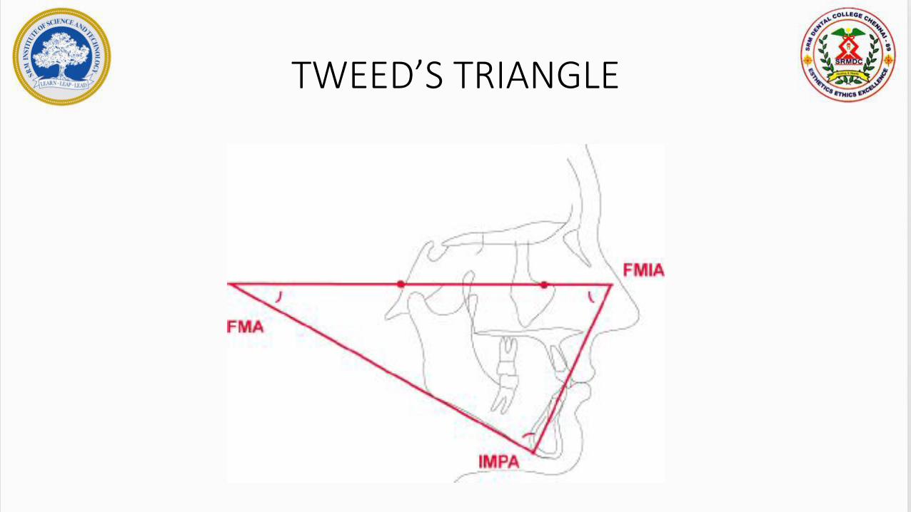
• Tweed’s triangle is formed by
1. Frankfort Horizontal Plane
2. The Mandibular plane
3. The long axis of lower incisor
TWEED’S TRIANGLE

ANGLES FORMING TWEED’S TRIANGLE
• Three angles thus formed are
a)Frankfort mandibular plane angle(FMA)
b)Lower incisor to mandibular plane(IMPA)
c)Lower incisor to Frankfort Horizontal(FMIA)
FMA = 25 degrees
IMPA= 90 degrees
FMIA= 65 degrees
