centrosome and sphere in cells of the ovarian stroma of mammals. a preliminary communication
TRANSCRIPT
Centrosome and Sphere in Cells of the Ovarian Stroma of Mammals. A PreliminaryCommunicationAuthor(s): C. M. ChildSource: Zoological Bulletin, Vol. 1, No. 2 (Sep., 1897), pp. 87-94Published by: Marine Biological LaboratoryStable URL: http://www.jstor.org/stable/1535470 .
Accessed: 23/05/2014 22:01
Your use of the JSTOR archive indicates your acceptance of the Terms & Conditions of Use, available at .http://www.jstor.org/page/info/about/policies/terms.jsp
.JSTOR is a not-for-profit service that helps scholars, researchers, and students discover, use, and build upon a wide range ofcontent in a trusted digital archive. We use information technology and tools to increase productivity and facilitate new formsof scholarship. For more information about JSTOR, please contact [email protected].
.
Marine Biological Laboratory is collaborating with JSTOR to digitize, preserve and extend access toZoological Bulletin.
http://www.jstor.org
This content downloaded from 195.78.108.179 on Fri, 23 May 2014 22:01:52 PMAll use subject to JSTOR Terms and Conditions
CENTROSOME AND SPHERE IN CELLS OF THE OVARIAN STROMA OF MAMMALS.
A PRELIMINARY COMMUNICATION.
C. M. CHILD.
THE presence of centrosome and sphere in the cell has been found in the majority of cases to present some relation to the karyokinetic processes. The number of cases, however, which
appear to indicate that the function of the centrosome and sphere may extend beyond their connection with karyokinesis is being multiplied.
Without making any attempt at completeness, a few of the references bearing on this point may be given here. Heiden- hain ('94) and Flemming ('91), among others, have studied the centrosome in leucocytes and giant cells; Dehler ('95a) and Lenhoss6k ('95b) have found them in ganglion cells of the frog; Miss Lewis ('96c) has found them in ganglion cells of an anne- lid, and they have been demonstrated in various tissue cells besides ('91).
In some of these cases it appears probable that the cells under consideration have completed their karyokinetic history, and that the centrosome is merely a relic of a past stage, or else that it possesses some additional relation to the economy of the cell which is not as yet understood.
Again Morgan's ('96b) recent work on the production of artificial astrospheres in sea-urchin eggs appears to indicate that structures at least very similar to centrosomes and spheres may appear as the result of abnormal environment. Auerbach's ('96d) latest work on spermatogenesis shows the formation of a " Nebenkern," which without doubt corresponds to the sphere in this case in each generation of sperm cells. After its appearance in the last generation at the close of cell- division, the " Nebenkern" has a special function to fulfill in the metamorphosis of this cell into the spermatozoon. These cases
This content downloaded from 195.78.108.179 on Fri, 23 May 2014 22:01:52 PMAll use subject to JSTOR Terms and Conditions
are sufficient to show that the presence of a sphere and centro- some in the cytoplasm may often be due to some other cause than an approaching or just finished mitosis. In this prelimi- nary paper it is desired to describe a case which the writer believes may be included under this head.
Some months since, in the preparation of mammalian ovaries for histological work, the ovary of a pregnant dog was sec- tioned, and, being double-stained with Delafield's haematoxylin and erythrosin, showed the stroma to consist almost entirely of
comparatively large, polyhedral cells with a coarse cytoplasmic network and a round nucleus containing clumps of chromatin of varying size and position. But the most striking feature of the preparation was the fact that each cell apparently possessed a large sphere 1 deeply stained with erythrosin. In fact the whole
appearance of the slide reminded one of an amphibian testis. The older corpora lutea showed cells somewhat similar in structure but more vacuolated, and without any trace of the sphere.
Examination of the ovaries of pregnant rabbits revealed the fact that the structure of the stroma cells was almost perfectly identical with that found in the pregnant dog. In this case, however, it was found that these cells appeared only in the ovary of thepregnant female. Rabbits which were not preg- nant showed the usual structure of the ovarian stroma without
any trace of the sphere in any of the cells. One exception must be made to this statement, viz., the case of a rabbit exam- ined during the period of lactation a few days after the young were born. The ovary of this female showed the same structure as was found in the ovaries of pregnant animals.
My observations along this line extend at present no farther than this: I have examined three pregnant rabbits and one in lactation, and in all cases have found the structure referred to in the stroma cells. I have also sectioned the ovaries of three non-pregnant rabbits, and in no case was even a trace of a sim- ilar structure to be found. I have had no opportunity to extend my observations on the dog beyond the one case mentioned, that of a pregnant female where the sphere was first seen.
Mr. W. H. Packard, a fellow of the University, who was preparing the sec- tions, was the first to observe this structure.
88 CHILD. [VOL. I.
This content downloaded from 195.78.108.179 on Fri, 23 May 2014 22:01:52 PMAll use subject to JSTOR Terms and Conditions
No. 2.] THE OVARIAN STROMA OF MAMMALS.
Ovaries of the white rat and of the cat from both pregnant and
non-pregnant animals have been examined, but without finding anything similar to the sphere in the dog and the rabbit, although the cells of the stroma show a very similar structure in other respects. It would appear, then, from the facts cited above that the presence of the sphere has some relation to the period of pregnancy, though of what nature it is impossible to state. Furthermore, a
sphere of this kind is found only in certain
mammals, though others show changes in the structure of the
observed in the dog and rabbit.
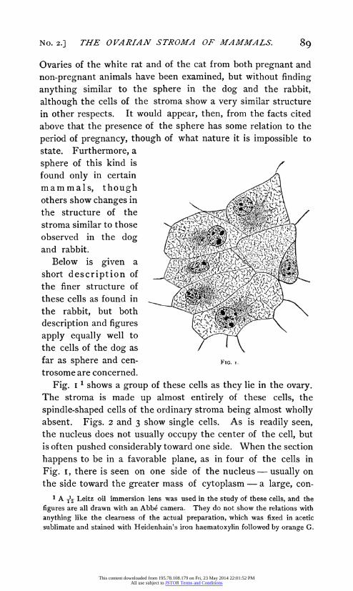
Below is given a ,:,. short description of the finer structure of t rl f e c'., these cells as found in y so, bin a s w; the rabbit, but both '"
description and figures in
apply equally well to ns the cells of the dog as far as sphere and cen- FIG. I. trosome are concerned.
Fig. i 1 shows a group of these cells as they lie in the ovary. The stroma is made up almost entirely of these cells, the
spindle-shaped cells of the ordinary stroma being almost wholly absent. Figs. 2 and 3 show single cells. As is readily seen, the nucleus does not usually occupy the center of the cell, but is often pushed considerably toward one side. When the section
happens to be in a favorable plane, as in four of the cells in
Fig. i, there is seen on one side of the nucleus - usually on the side toward the greater mass of cytoplasm - a large, con-
1 A ?z Leitz oil immersion lens was used in the study of these cells, and the figures are all drawn with an Abbe camera. They do not show the relations with
anything like the clearness of the actual preparation, which was fixed in acetic sublimate and stained with Heidenhain's iron haematoxylin followed by orange G.
89
This content downloaded from 195.78.108.179 on Fri, 23 May 2014 22:01:52 PMAll use subject to JSTOR Terms and Conditions

CHILD. 90
spicuous condensation of the cytoplasm. As stated above, the
cytoplasmic network of these cells is in general quite coarse. This statement, however, does not apply to the sphere. Al-
though I have not seen the stages of the formation of the
sphere, I believe from its appearance and relation to other
parts of the cell that it is without doubt simply a condensation of the cytoplasm. In the figures it is represented as granular, but this appearance is probably due to the condensation. In
staining qualities it resembles the cytoplasm, except that it takes the plasma stains much more deeply, and thus stands out
very distinctly from the rest of the cytoplasm. Radiating fibers
-V,:.
FIG. 2. FIG. 3.
like those so common in astrospheres and spindles in the vari- ous stages of karyokinesis are not present, but the sphere resembles very closely the structure described by Auerbach ('96d) as "Nebenkern" in the spermatogonia and spermatocytes of
Paludina, and, I believe, arises in the same way. But, though no distinct fibers are visible, the structure of the cytoplasm around the sphere proper is more or less distinctly radiate. Two of the cells in Fig. I show this to some extent, as does also Fig. 2. Sometimes this radiate arrangement is visible, though indistinctly, nearly to the periphery of the cell, as I have attempted to show in Fig. 3. It is probable that the dif- ference between a sphere of this sort and a distinctly radiate
sphere with fibers is one of degree and not of kind.
Perhaps even more striking than the presence of this sphere in these cells is the presence of a perfectly distinct centrosome - or in some cases two - in its center. Two of the cells in
Fig. I show two centrosomes, the rest in which the sphere is in the plane of section show one.
[VOL. I.
This content downloaded from 195.78.108.179 on Fri, 23 May 2014 22:01:52 PMAll use subject to JSTOR Terms and Conditions

No. 2.] THE OVARIAN STROMA OF MAiMMiALS.
I have been able to demonstrate this centrosome beyond all doubt by staining with Heidenhain's iron haematoxylin and then with orange G. or Bordeaux red, the best results being obtained with the haematoxylin and orange G. The extraction of the haematoxylin is carried just to the point where the cyto- plasm has lost the last traces of the stain. Then after the con- trast-stain and mounting, the haematoxylin is seen to be confined to the nucleus and to a single (or double) tiny but perfectly distinct granule in the center of the sphere. Error seems to be impossible here because by proper extraction the centrosome is the only extra-nuclear body in the cell that shows a trace of
haematoxylin, and in contrast to the orange about it it is per- fectly sharp and well defined. Moreover, the abundance of these cells and the uniformity with which the centrosome is visible renders the sections most striking. The group in Fig. I is drawn as seen in the section, and probably a hundred other similar groups could have been selected from the same section. I cannot state positively that every sphere contains a centro- some, but from the very large proportion of cases that I have observed it appears extremely probable that such is the case.
It is possible to obtain fairly good preparations by staining with gentian violet or with Flemming's triple stain, safranin, gentian violet and orange G., but the pictures thus obtained do not compare with those given by iron haematoxylin and
orange. Now the question arises as to the origin of these cells and
their relation to the ordinary ovarian stroma. At first I
regarded them as belonging to the corpora lutea, but further
study has shown that they occupy the position, not of the cor- pora lutea, but of the stroma itself. Moreover, the cells of the corpora lutea are perfectly distinguishable from them.
As stated above, the details of their origin have not yet been worked out, but the following observations may throw some light on the matter.
In the ovary of the adult non-pregnant rabbit the stroma is composed principally of two kinds of cells. One of these is the elongated fiber cells, some of the shorter of which are shown in the spindle-shaped cells with deeply staining nuclei
9I
This content downloaded from 195.78.108.179 on Fri, 23 May 2014 22:01:52 PMAll use subject to JSTOR Terms and Conditions
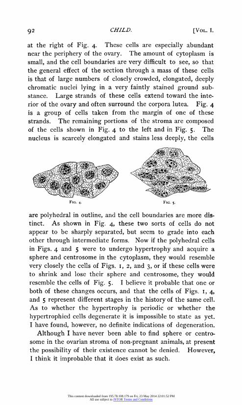
at the right of Fig. 4. These cells are especially abundant near the periphery of the ovary. The amount of cytoplasm is
small, and the cell boundaries are very difficult to see, so that the general effect of the section through a mass of these cells is that of large numbers of closely crowded, elongated, deeply chromatic nuclei lying in a very faintly stained ground sub- stance. Large strands of these cells extend toward the inte- rior of the ovary and often surround the corpora lutea. Fig. 4 is a group of cells taken from the margin of one of these strands. The remaining portions of the stroma are composed of the cells shown in Fig. 4 to the left and in Fig. 5. The nucleus is scarcely elongated and stains less deeply, the cells
FIG. 4. FIG. 5.
are polyhedral in outline, and the cell boundaries are more dis- tinct. As shown in Fig. 4, these two sorts of cells do not
appear to be sharply separated, but seem to grade into each other through intermediate forms. Now if the polyhedral cells in Figs. 4 and 5 were to undergo hypertrophy and acquire a
sphere and centrosome in the cytoplasm, they would resemble
very closely the cells of Figs. i, 2, and 3, or if these cells were to shrink and lose their sphere and centrosome, they would resemble the cells of Fig. 5. I believe it probable that one or both of these changes occurs, and that the cells of Figs. I, 4, and 5 represent different stages in the history of the same cell. As to whether the hypertrophy is periodic or whether the
hypertrophied cells degenerate it is impossible to state as yet. I have found, however, no definite indications of degeneration.
Although I have never been able to find sphere or centro- some in the ovarian stroma of non-pregnant animals, at present the possibility of their existence cannot be denied. However, I think it improbable that it does exist as such.
CHILD. [VOL. I. 92
This content downloaded from 195.78.108.179 on Fri, 23 May 2014 22:01:52 PMAll use subject to JSTOR Terms and Conditions
No. 2.] THE OVARIAN STROMA OF MAMMALS.
I have never found any indications of karyokinesis in any of the stroma cells outside the corpora lutea, so that the presence of the centrosome and sphere is probably not associated with
karyokinesis in this case. If my suggestion that the cells of Fig. I arise by hypertrophy
from the cells of Figs. 4 and 5 be correct, then it is probable that the appearance of the centrosome and sphere is in some manner connected with this hypertrophy.
In the rat and the cat there is a somewhat similar change in the structure of the stroma cells during pregnancy, but no cen- trosome or sphere appears. Sobotta ('96a) also mentions noth-
ing of the sort in connection with the mouse. Whether there is less hypertrophy in these cases I cannot as yet state.
The results of my observations thus far are then that in the stroma cells of the ovary of the dog and the rabbit a distinct centrosome and sphere appear during pregnancy, but apparently are not in any way connected with karyokinesis.
The presence of these structures under these conditions opens up a number of problems, viz., Is the centrosome a permanent organ of these cells ? How is the appearance and disappearance of the sphere and centrosome in the cytoplasm related to the other changes occurring in the cell ? How generally do these structures appear among mammals, and lastly, What is the relation of these changes of the ovarian stroma to pregnancy ?
I hope to be able to throw further light on some of these questions, but it seemed that a short account of the results obtained thus far might be of interest.
93
This content downloaded from 195.78.108.179 on Fri, 23 May 2014 22:01:52 PMAll use subject to JSTOR Terms and Conditions
94 CHILD.
LITERATURE.
'96d AUERBACH, L. Untersuchungen liber die Spermatogenese von Pal- udina vivipara. Jen. Zeitschr. f. Naturwiss. Bd. xxx (N. F. xiii). 1896.
'95a DEHLER, A. Beitrag zur Kenntniss vom feineren Bau der Sympath- ischen Ganglienzelle des Frosches. Arch. f. mikr. Anat. Bd. xlvi.
I895. '91 FLEMMING, W. Attraktionspharen und Centralkorper in Gewebezellen
und Wanderzellen. Anat. Anz. Bd. vi. 189I. '94 HEIDENHAIN. Neue Untersuchungen uber die Centralkbrper und ihre
Beziehungen zum Kern und Zellprotoplasma. Arch. f. mikr. Anat. Bd. xliii. 1894.
'95b VON LENHOSSEK. Centrosom und Sphare in den Spinalganglien zellen des Frosches. Arch.f. mikr. Anat. Bd. xlvi. I895.
'96c LEWIS, MARGARET. Centrosome and Sphere in Certain of the Nerve Cells of an Invertebrate. Anat. Anz. Bd. xii. I896.
'96b MORGAN, T. H. The Production of Artificial Astrospheres. Arch. f. Entwicklungsmechanik der Organismen. Bd. iii. I896.
'96a SOBOTTA, J. Ueber die Bildung des Corpus luteum bei der Maus. Arch. f. mikr. Anat. Bd. xlvii. 1896.
ZOOLOGICAL LABORATORY, UNIVERSITY OF CHICAGO,
November, I896.
This content downloaded from 195.78.108.179 on Fri, 23 May 2014 22:01:52 PMAll use subject to JSTOR Terms and Conditions