cells chapter 6 - st. anthony's high school... maintenance compartments • are membrane ......
TRANSCRIPT

CELLS CHAPTER 6
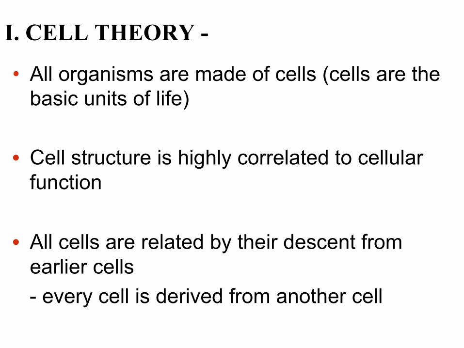
• All organisms are made of cells (cells are the basic units of life)
• Cell structure is highly correlated to cellular
function
• All cells are related by their descent from earlier cells
- every cell is derived from another cell
I. CELL THEORY -

Electron microscopes (EMs) are microscopes that use accelerated electrons as a source of illumination 1. Scanning electron microscopes (SEMs) focus a beam of electrons onto the surface of a specimen, providing images that look 3-D 2. Transmission electron microscopes (TEMs) use a high voltage beam of electrons through a specimen. It makes it possible to get a resolution 1,000 better than a light microscope TEMs are used mainly to study the internal structures of cells. Structures that measure in micrometers or smaller.

SEM 3D picture of a water bear about 1 mm in length

TEM picture of a chloroplast

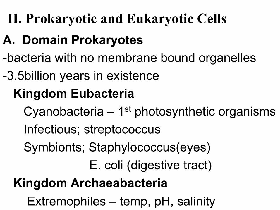
II. Prokaryotic and Eukaryotic Cells A. Domain Prokaryotes -bacteria with no membrane bound organelles -3.5billion years in existence Kingdom Eubacteria Cyanobacteria – 1st photosynthetic organisms Infectious; streptococcus Symbionts; Staphylococcus(eyes) E. coli (digestive tract) Kingdom Archaeabacteria Extremophiles – temp, pH, salinity
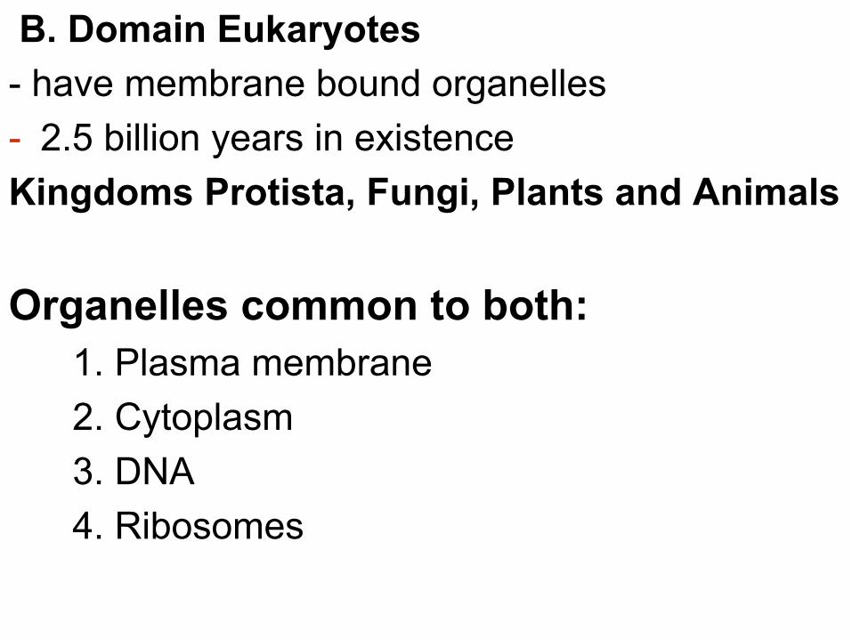
B. Domain Eukaryotes - have membrane bound organelles - 2.5 billion years in existence Kingdoms Protista, Fungi, Plants and Animals Organelles common to both:
1. Plasma membrane 2. Cytoplasm 3. DNA 4. Ribosomes

A. Prokaryotic Cell (Bacteria) Structure
1. No membrane-bound organelles; 2. DNA in an unbound region called the nucleoid 3. Cytoplasm enclosed by the plasma membrane; the liquid portion is known as the cytosol 4. Cell wall 5. Ribosomes 6. sometimes flagella

B. Eukaryotic Cells
1. DNA in a nucleus that is encapsulated by a membranous nuclear envelope 2. membrane-bound organelles; mitochondria, chloroplasts, endoplasmic reticulum, Golgi apparatus, lysosomes, vacuoles, vesicles, cilia, flagella 3. Cytoplasm in the region between the plasma membrane and nucleus 4. Ribosomes • These cells are generally much larger and more
complex than prokaryotic cells • Evolved after prokaryotic cells

ENDOPLASMIC RETICULUM (ER) Rough
ER Smooth
ER Nuclear envelope Nucleolus Chromatin
Plasma membrane
Ribosomes
Golgi apparatus
Lysosome Mitochondrion
Peroxisome
Microvilli
Microtubules Intermediate filaments
Microfilaments
Centrosome
CYTOSKELETON:
Flagellum NUCLEUS

NUCLEUS
Nuclear envelope
Nucleolus Chromatin
Golgi apparatus
Mitochondrion Peroxisome
Plasma membrane
Cell wall
Wall of adjacent cell
Plasmodesmata
Chloroplast
Microtubules
Intermediate filaments
Microfilaments
CYTOSKELETON
Central vacuole
Ribosomes
Smooth endoplasmic reticulum
Rough endoplasmic
reticulum

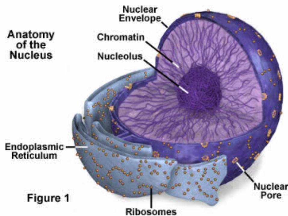
III. ORGANELLES A. Nucleus • Contains the DNA in a cell and is usually the
most conspicuous organelle • is enclosed by a nuclear envelope Function: heredity, protein synthesis, and reproduction B. Nucleolus • located within the nucleus Function: produces ribosomal RNA (rRNA) and ribosomes

C. Ribosomes: Protein Factories
• are small particles made of ribosomal RNA (rRNA) and protein
• Function: the site of protein synthesis • found in two locations
– in the cytosol (free ribosomes) – on the membrane of endoplasmic reticulum
(bound ribosomes)

D. ENDOMEMBRANE SYSTEM • Components of the endomembrane system:
- nuclear envelope - plasma membrane - endoplasmic reticulum - Golgi apparatus - lysosomes - vacuoles - transport vesicles

1. nuclear envelope – a bilayer lipid membrane that encloses the nucleus, separating it from the cytoplasm
• pores in the envelope regulate the movement of molecules in or out of the nucleus
2. plasma/cell membrane- Structure: a bilayer of phospholipids with proteins within the bilayer Function: a selective barrier that regulates the passage of materials in or out of cells

Outside of cell
Inside of cell 0.1 µm
(a) TEM of a plasma membrane
Hydrophilic region
Hydrophobic region
Hydrophilic region
Carbohydrate side chains
Proteins Phospholipid
(b) Structure of the plasma membrane
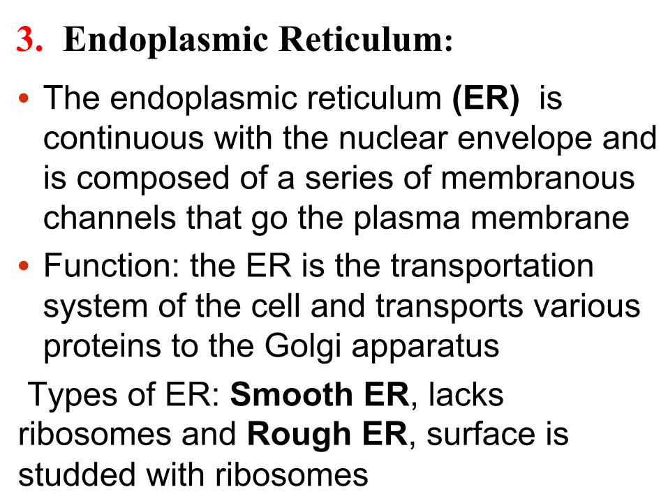
3. Endoplasmic Reticulum:
• The endoplasmic reticulum (ER) is continuous with the nuclear envelope and is composed of a series of membranous channels that go the plasma membrane
• Function: the ER is the transportation system of the cell and transports various proteins to the Golgi apparatus
Types of ER: Smooth ER, lacks ribosomes and Rough ER, surface is studded with ribosomes

Figure 6.11a
Smooth ER
Rough ER
Ribosomes Transport vesicle
Nuclear envelope

a. Functions of Smooth ER
1) Synthesizes lipids 2) Stores calcium ions 3) Metabolizes carbohydrates 4) Detoxifies drugs and poisons in the cell
b. Function of Rough ER • is based on the existence of ribosomes on the ER • Function: protein synthesis of secretory proteins and
glycoproteins • Secretory proteins are proteins secreted by the cell.
Ex: hormones or enzymes after secretory proteins are formed they leave the ER and ultimately the cell in transport vesicles Example: certain pancreatic cells make the hormone insulin in their ER, which is then transported to the plasma membrane by a vesicle, where it is secreted out of the cell into the bloodstream.

• consists of flattened membranous sacs called cisternae
• Functions of the Golgi apparatus: receiving, modifying, sorting, shipping and even some manufacturing of materials.
1) Modifies products from the ER; Ex: adding the carbohydrates on proteins to make glycoproteins. 2) Manufactures lysosomes. 3) Sorts, packages and ships out proteins & lipids via transport vesicles to discharge their contents as secretions out of the cell (exocytosis).
4. Golgi Apparatus – named for Camillo Golgi, who discovered it in 1898.

Figure 6.12
cis face (“receiving” side of Golgi apparatus)
trans face (“shipping” side of Golgi apparatus)
0.1 µm
TEM of Golgi apparatus
Cisternae

5. Lysosomes: • A lysosome is a membranous sac that contains hydrolytic enzymes
• Lysosomal enzymes can hydrolyze proteins, fats, polysaccharides, and nucleic acids
• Lysosomal enzymes are acidic; if they leak out into the cytoplasm, they can destroy the cell (self-digestion).
• In unicellular organisms, lysosomes will fuse with food vacuoles and digest the molecules within.
• WBC that engulf bacteria use lysosomes to degrade bacteria infecting an organism
• in cells with damaged organelles and proteins, lysosomes will degrade these by a process called autophagy.

Figure 6.13a Nucleus
Lysosome
1 µm
Digestive enzymes
Digestion
Food vacuole
Lysosome Plasma membrane
(a) Phagocytosis

6. Vacuoles: Maintenance Compartments • are membrane bound organelles for storage of
materials • Central vacuoles, found in many mature plant
cells, hold organic compounds and water that will be recycled in the plant.
• In some protists, food vacuoles are formed by
phagocytosis • Contractile vacuoles, found in freshwater
protists, pump excess water out of cells

Figure 6.14
Central vacuole
Cytosol
Nucleus
Cell wall
Chloroplast
Central vacuole
5 µm


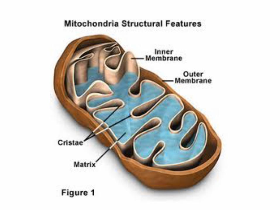
E. Mitochondria • Mitochondria are in eukaryotic cells only • They have a smooth outer membrane and an inner
folded membrane. The folds are called cristae. Cristae folds increase the surface area for the synthesis of ATP (cellular respiration) • Have ribosomes and DNA known as mtDNA and
therefore are capable of reproducing when energy needs increase.
• mtDNA does contain genetic information for inherited traits
only mitochondria from the egg is past on to the offspring during fertilization and therefore mtDNA is inherited from the maternal side.

• are double membrane bound organelles that are the site of photosynthesis
• The chloroplast is one of a group of plant organelles, called plastids
• are found mainly in leaves of plants and in algae, which is a protist
• Chloroplast structure includes: Thylakoids - stacked membranous sacs with chlorophyll Stroma- the internal fluid; analogous to the cytoplasm DNA ribosomes
F. Chloroplasts:

Figure 6.18a
Ribosomes Stroma
Inner and outer membranes
Granum
1 µm Intermembrane space Thylakoid (a) Diagram and TEM of chloroplast
DNA

• Mitochondria and chloroplasts have many similarities with prokaryotes
• they are believed to have once existed as free living primitive bacterial cells (prokaryotes).
• Similarities to prokaryotes:
– Contain free ribosomes – circular DNA molecule that is used to produce proteins
and enzymes - The two organelles also reproduce like bacteria
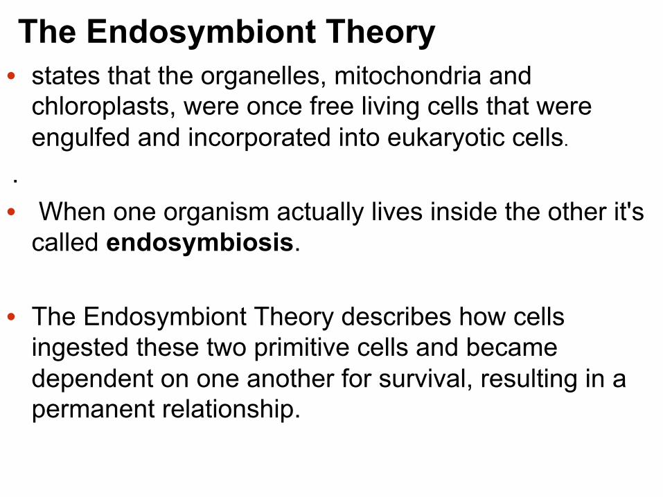
The Endosymbiont Theory • states that the organelles, mitochondria and chloroplasts, were once free living cells that were engulfed and incorporated into eukaryotic cells.
. • When one organism actually lives inside the other it's
called endosymbiosis. • The Endosymbiont Theory describes how cells
ingested these two primitive cells and became dependent on one another for survival, resulting in a permanent relationship.

Sequence of the Endosymbiosis 1. An early ancestor of eukaryotic cells, the host cell
engulfed a mitochodrion becoming an oxygen using, non-photosynthetic primitive cell, a single organism, a eukaryotic cell with mitochondria.
2. One of the those cells later engulfed a chloroplast becoming the ancestor of eukaryotic cells with chloroplasts.
• Over millions of years of evolution, mitochondria and chloroplasts have become more specialized and today they cannot live outside the cell.

Nucleus Endoplasmic reticulum
Nuclear envelope
Ancestor of eukaryotic cells (host cell)
Engulfing of oxygen- using non-photosynthetic prokaryote, which is a mitochondrion
Mitochondrion
Nonphotosynthetic eukaryote
Mitochondrion
At least one cell
Photosynthetic eukaryote
Engulfing of photosynthetic prokaryote
Chloroplast
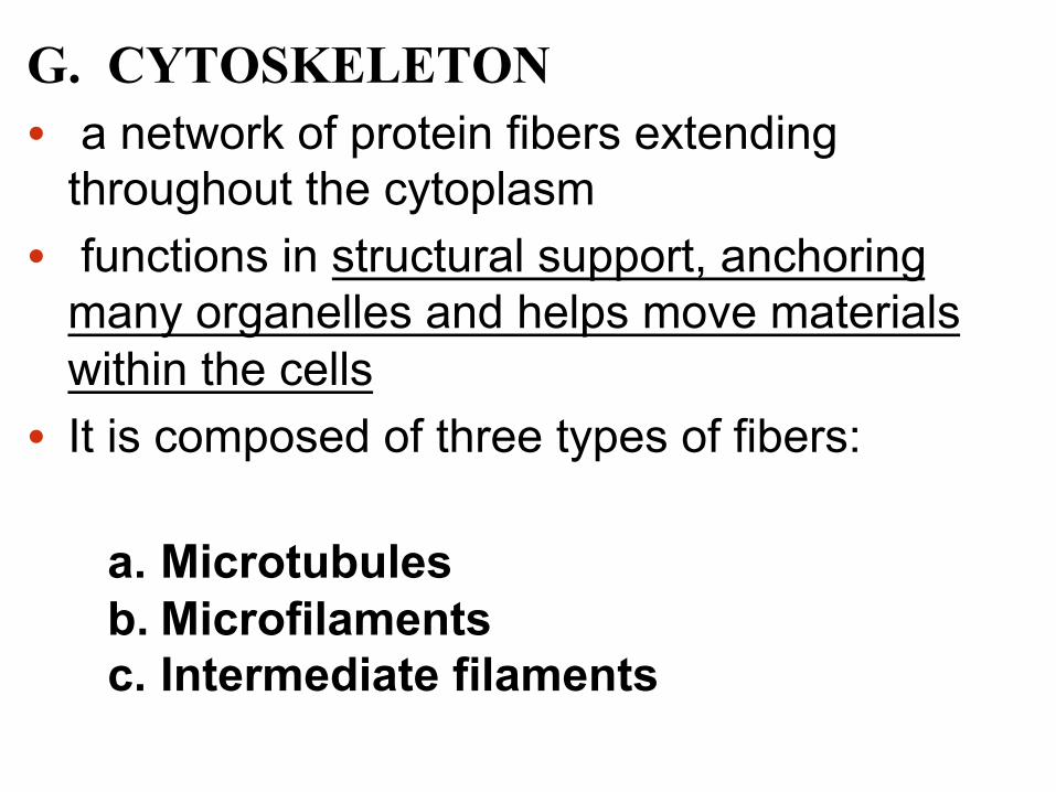
G. CYTOSKELETON • a network of protein fibers extending
throughout the cytoplasm • functions in structural support, anchoring
many organelles and helps move materials within the cells
• It is composed of three types of fibers:
a. Microtubules b. Microfilaments c. Intermediate filaments

a. Microtubules
• Microtubules are hollow rods (about 25 nm in diameter) composed of a globular protein called tubulin.
• Functions of microtubules - Cell shape - Guiding motor proteins for movement of substances, viruses through the cell - Components of spindle fibers for separating chromosomes during cell division - Composition and movement of cilia and flagella

Figure 6.21
ATP Vesicle
(a)
Motor protein (ATP powered)
Microtubule of cytoskeleton
Receptor for motor protein
0.25 µm Vesicles Microtubule
(b)

b. Microfilaments (Actin Filaments)
• are solid rods (about 7 nm in diameter) built as a twisted double chain of a globular protein called actin
• Microfilaments in muscle cells sometimes contain the protein myosin in addition to actin

• The structural role of microfilaments is to 1. help support the cell’s shape 2. Keep the cell intact when it is moving during
phagocytosis 3. muscle cell movement 4. in plant and animal cells, drives cytoplasmic
streaming

c. Intermediate Filaments
• Intermediate filaments are larger than microfilaments but smaller than microtubules
• They support cell shape and fix organelles in
place

H. Cilia and Flagella 1. Cilia (cilium) - microscopic hair like
projections extending from the surface of a cell or unicellular organism.
• Capable of rhythmical motion; they act in unison to bring about the movement of the cell or of the surrounding medium
• They are one of the most widespread cellular organelles in nature, found in many unicellular and multicellular eukaryotic organisms

• They play a critical motile role; involved in normal organogenesis during embryo development.
• Defects in ciliogenesis and function can give rise to developmental disorders and diseases, such as blindness, kidney cysts, neural tube defects and obesity
2. Flagella (flagellum) - A long, threadlike appendage, especially a whip like extension of certain eukaryotic cells or unicellular organisms, found singly or in pairs.

• Cilia and flagella share a common structure A core of microtubules sheathed by a plasma membrane (9 pairs of microtubules surrounding a central pair)
• Cilia and flagella differ in their beating patterns:
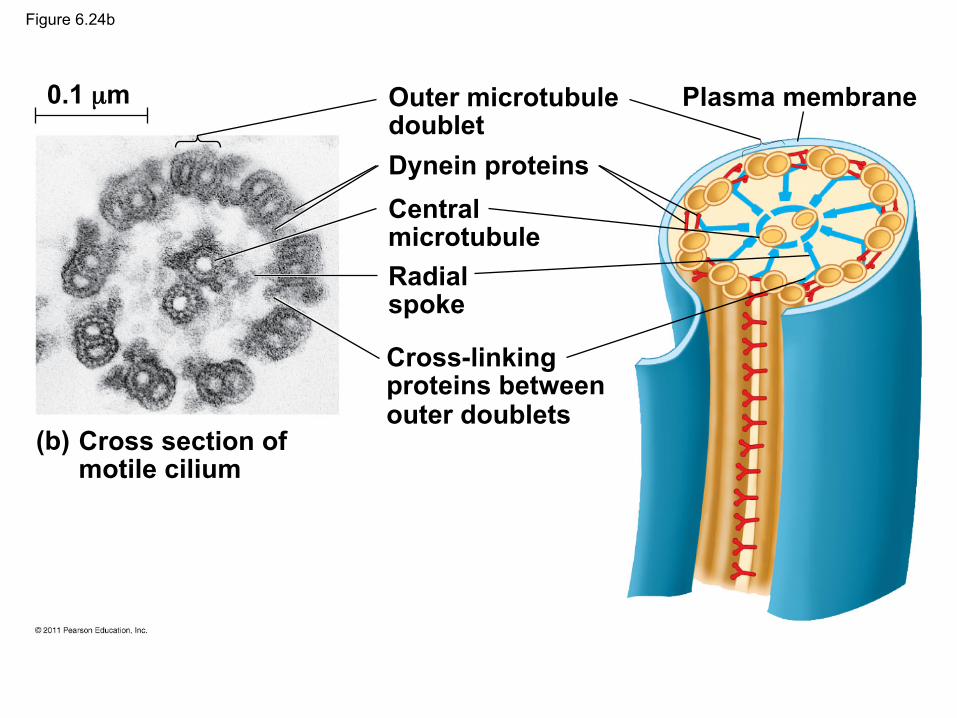
Figure 6.24b
0.1 µm
(b) Cross section of motile cilium
Outer microtubule doublet Dynein proteins Central microtubule Radial spoke
Cross-linking proteins between outer doublets
Plasma membrane

Direction of swimming
(b) Motion of cilia
Direction of organism’s movement
Power stroke Recovery stroke
(a) Motion of flagella 5 µm
15 µm
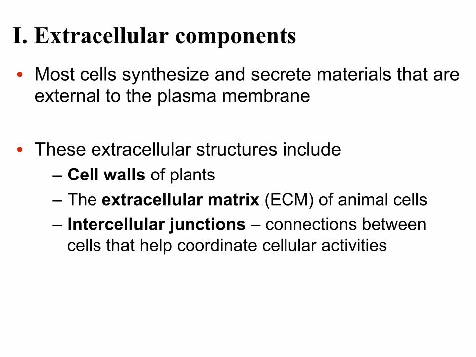
I. Extracellular components • Most cells synthesize and secrete materials that are
external to the plasma membrane • These extracellular structures include
– Cell walls of plants – The extracellular matrix (ECM) of animal cells – Intercellular junctions – connections between
cells that help coordinate cellular activities
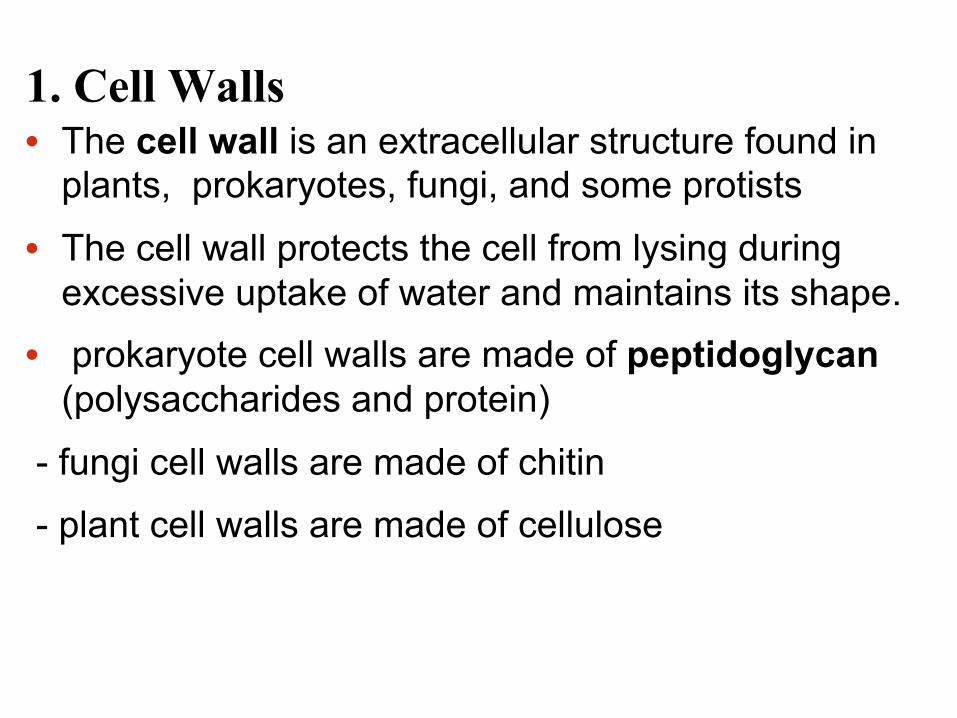
1. Cell Walls • The cell wall is an extracellular structure found in
plants, prokaryotes, fungi, and some protists
• The cell wall protects the cell from lysing during excessive uptake of water and maintains its shape.
• prokaryote cell walls are made of peptidoglycan (polysaccharides and protein)
- fungi cell walls are made of chitin
- plant cell walls are made of cellulose
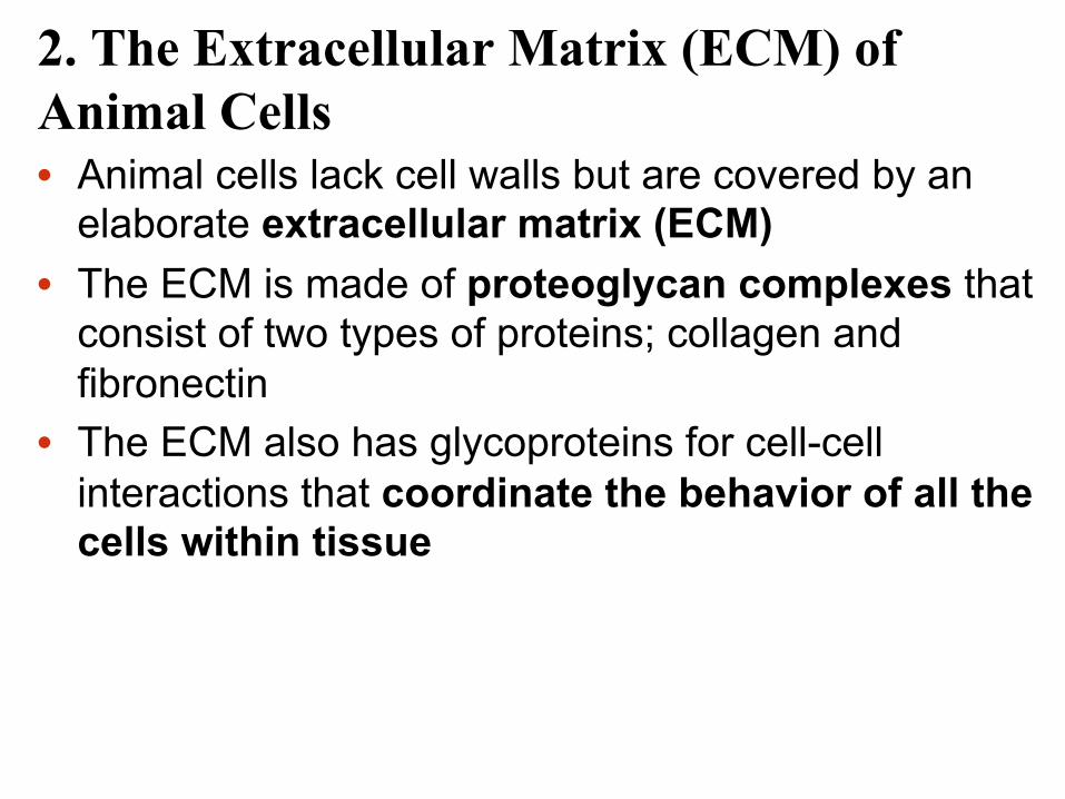
2. The Extracellular Matrix (ECM) of Animal Cells • Animal cells lack cell walls but are covered by an
elaborate extracellular matrix (ECM) • The ECM is made of proteoglycan complexes that
consist of two types of proteins; collagen and fibronectin
• The ECM also has glycoproteins for cell-cell interactions that coordinate the behavior of all the cells within tissue

Figure 6.30a
EXTRACELLULAR FLUID Collagen
Fibronectin
Plasma membrane
Micro- filaments
CYTOPLASM
Integrins
Proteoglycan complex

3. Intercellular Junctions • Neighboring cells in tissues, organs, or systems
interact and communicate through intercellular junctions
• Plant Intercellular Junctions a. Plasmodesmata – are plant cell junctions that consist of channels between adjacent plant cells, so that materials such as, water, salts, ions, proteins and RNA can pass from cell to cell.

Plasmodesmata
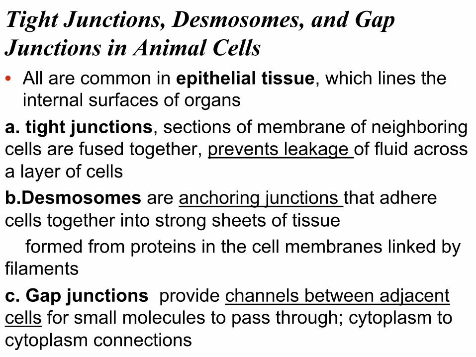
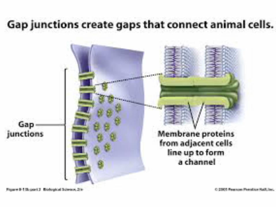
Tight Junctions, Desmosomes, and Gap Junctions in Animal Cells • All are common in epithelial tissue, which lines the
internal surfaces of organs a. tight junctions, sections of membrane of neighboring cells are fused together, prevents leakage of fluid across a layer of cells b.Desmosomes are anchoring junctions that adhere cells together into strong sheets of tissue formed from proteins in the cell membranes linked by filaments c. Gap junctions provide channels between adjacent cells for small molecules to pass through; cytoplasm to cytoplasm connections