cells and membranes - dspace@mit home
TRANSCRIPT

Cells and Membranes Membranes are the boundaries of cells, made up of a lipid bilayer
Organelles are membrane-limited compartments within cells The plasma membrane is itself an organelle All membranes are topologically spherical (see diagram above) Prokaryotic cells: 1 compartment made of a single membrane tah tsurrounds cell. Dna and cytoplasm are all in compartment e.g bacteria
Most bacteria have a cell wall which is surrounded by a plasma membrane
The plasma membrane denotes the boundary of the cell. It separates the inside of
the cell from the outside of the cell. The DNA in bacterial cells is ~1mm in length, however the cell is only 2~2um in
length – DNA is coiled and folded in cell. Glycolysis takes place in the cytoplasm of all organisms (prokaryotes and
eukaryotes)

Eukaryotic cell Cell is surrounded by the plasma membrane. The internal compartments (also called organelles) are also surrounded by membranes. Cells are defined by the outer plasma membrane. Membranes also define intracellular compartments. A eukaryotic cell:
(from http://esg-www.mit.edu:8001/esgbio/cb/org/animal.gif) Plasma Membrane – made up of a lipid bilayer Nucleus and Mitochondria are each enclosed by 2 membranes (inner and outer) Cytosol – watery areas outside the nucleus of the cell, cytosol does not include organelles Cell has many compartments, each defined by a membrane Example:
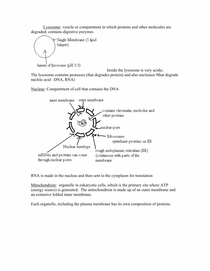
Lysosome: vesicle or compartment in which proteins and other molecules are degraded; contains digestive enzymes
Inside the lysosome is very acidic. The lysosome contains proteases (that degrades protein) and also nucleases 9that degrade nucleic acid –DNA, RNA) Nucleus: Compartment of cell that contains the DNA
RNA is made in the nucleus and then sent to the cytoplasm for translation Mitochondrion: organelle in eukaryotic cells, which is the primary site where ATP (energy source) is generated. The mitochondrion is made up of an outer membrane and an extensive folded inner membrane. Each organelle, including the plasma membrane has its own composition of proteins.
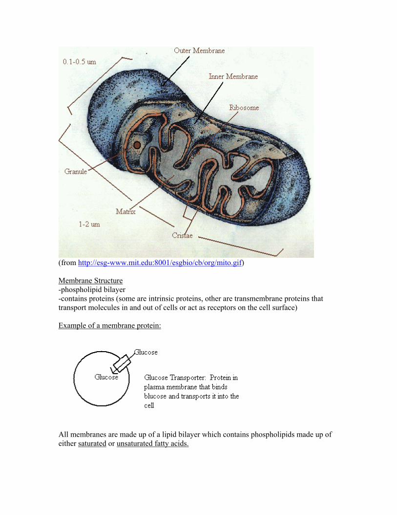
(from http://esg-www.mit.edu:8001/esgbio/cb/org/mito.gif) Membrane Structure -phospholipid bilayer -contains proteins (some are intrinsic proteins, other are transmembrane proteins that transport molecules in and out of cells or act as receptors on the cell surface) Example of a membrane protein:
All membranes are made up of a lipid bilayer which contains phospholipids made up of either saturated or unsaturated fatty acids.
Unsaturated fatty acids give more fluidity to the membrane since the have C=C as well as C-C in them. The kinks in the resulting phospholipid (such as the unsaturated Oleic acid and the polyunsaturated Linoleic acid) cause greater fluidity. Fatty Acids are long hydrocarbon chains bonded to a carboxylic group e.g.
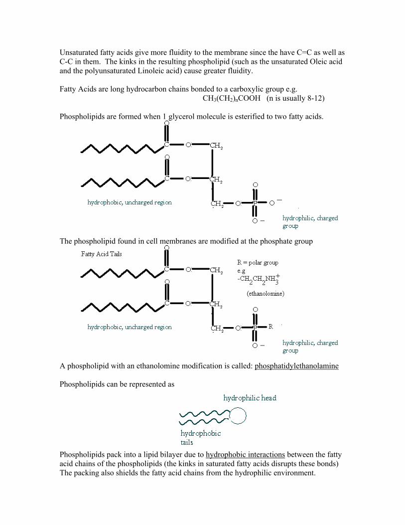
CH3(CH2)nCOOH (n is usually 8-12) Phospholipids are formed when 1 glycerol molecule is esterified to two fatty acids.
The phospholipid found in cell membranes are modified at the phosphate group
A phospholipid with an ethanolomine modification is called: phosphatidylethanolamine Phospholipids can be represented as
Phospholipids pack into a lipid bilayer due to hydrophobic interactions between the fatty acid chains of the phospholipids (the kinks in saturated fatty acids disrupts these bonds) The packing also shields the fatty acid chains from the hydrophilic environment.

The packing of fatty acyl chains is due to Vander Walls interactions and hydrophobic effects.
The entire distance of the lipid bilayer is about 3 nm or 30 angstroms. Phospholipids spontaneously form bilayers in liquid solutions. Hydrocarbon chains are never exposed to water; phospholipid bilayers usually form closed compartments – a continuous membrane with no free edges.. Phospholipids usually from three different forms in solutions:
Micelles – vesicle with a hydrophobic interior Liposomes – vesicle with aqueous interior Bilayer Sheets (although in aqueous solution there is no lipid bilayer since you
would have free edges.) All biological membranes form closed compartments, appear topologically like a sphere. Look at electro potential across a cell membrane:
The membrane acts as a capacitor: it stores a charge across the membrane. The hydrophobic region acts as an insulator, doesn’t allow water or ions to pass.
Insulator – hydrophobic region (fatty acid chains) Capacitor – hydrophilic head groups (phosphate groups) In virtually every cell, the inside of the cell is negative with respect to the outside. The electric potential across the membrane is -70mV. 70mV doesn’t seem high, however if you consider the thickness of the membrane (~3.5 nm) 0.07 V/ 3.5 x107cm = 200,000 volts/centimeter!
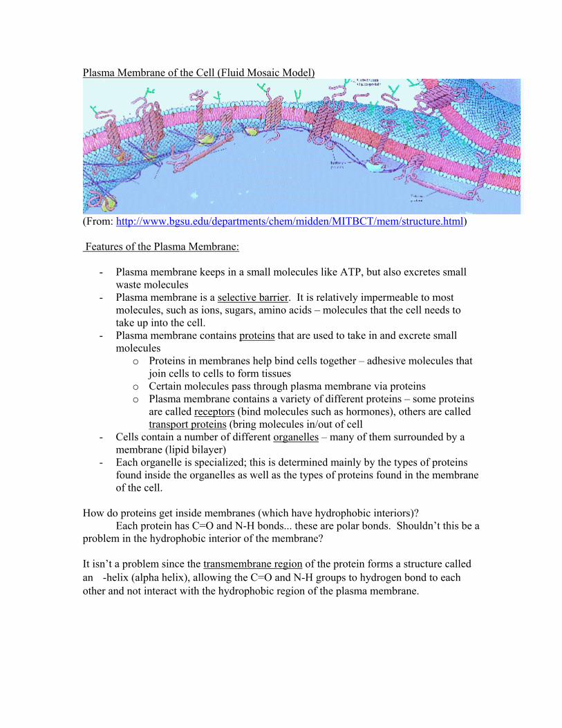
Plasma Membrane of the Cell (Fluid Mosaic Model)
(From: http://www.bgsu.edu/departments/chem/midden/MITBCT/mem/structure.html) Features of the Plasma Membrane:
- Plasma membrane keeps in a small molecules like ATP, but also excretes small waste molecules
- Plasma membrane is a selective barrier. It is relatively impermeable to most molecules, such as ions, sugars, amino acids – molecules that the cell needs to take up into the cell.
- Plasma membrane contains proteins that are used to take in and excrete small molecules
o Proteins in membranes help bind cells together – adhesive molecules that join cells to cells to form tissues
o Certain molecules pass through plasma membrane via proteins o Plasma membrane contains a variety of different proteins – some proteins
are called receptors (bind molecules such as hormones), others are called transport proteins (bring molecules in/out of cell
- Cells contain a number of different organelles – many of them surrounded by a membrane (lipid bilayer)
- Each organelle is specialized; this is determined mainly by the types of proteins found inside the organelles as well as the types of proteins found in the membrane of the cell.
How do proteins get inside membranes (which have hydrophobic interiors)? Each protein has C=O and N-H bonds... these are polar bonds. Shouldn’t this be a problem in the hydrophobic interior of the membrane? It isn’t a problem since the transmembrane region of the protein forms a structure called an -helix (alpha helix), allowing the C=O and N-H groups to hydrogen bond to each other and not interact with the hydrophobic region of the plasma membrane.

Structure of the -Helix
(From http://esg-www.mit.edu:8001/esgbio/lm/proteins/structure/structure.html) Two Points About -Helix Structure
1) Extensive hydrogen bonding between NH and C=O groups. 2) The polar NH and C=O groups are in the interior of the helix. The R groups (side
chains) of the amino acids protrude out of the -helix. What kind of amino acids would be found in the transmembrane region of a protein? Amino acids with hydrophobic R groups. The hydrophobic R groups interact favorably with the hydrophobic interior of the plasma membrane. The following are the amino acids that would be found in the transmembrane region of a protein:
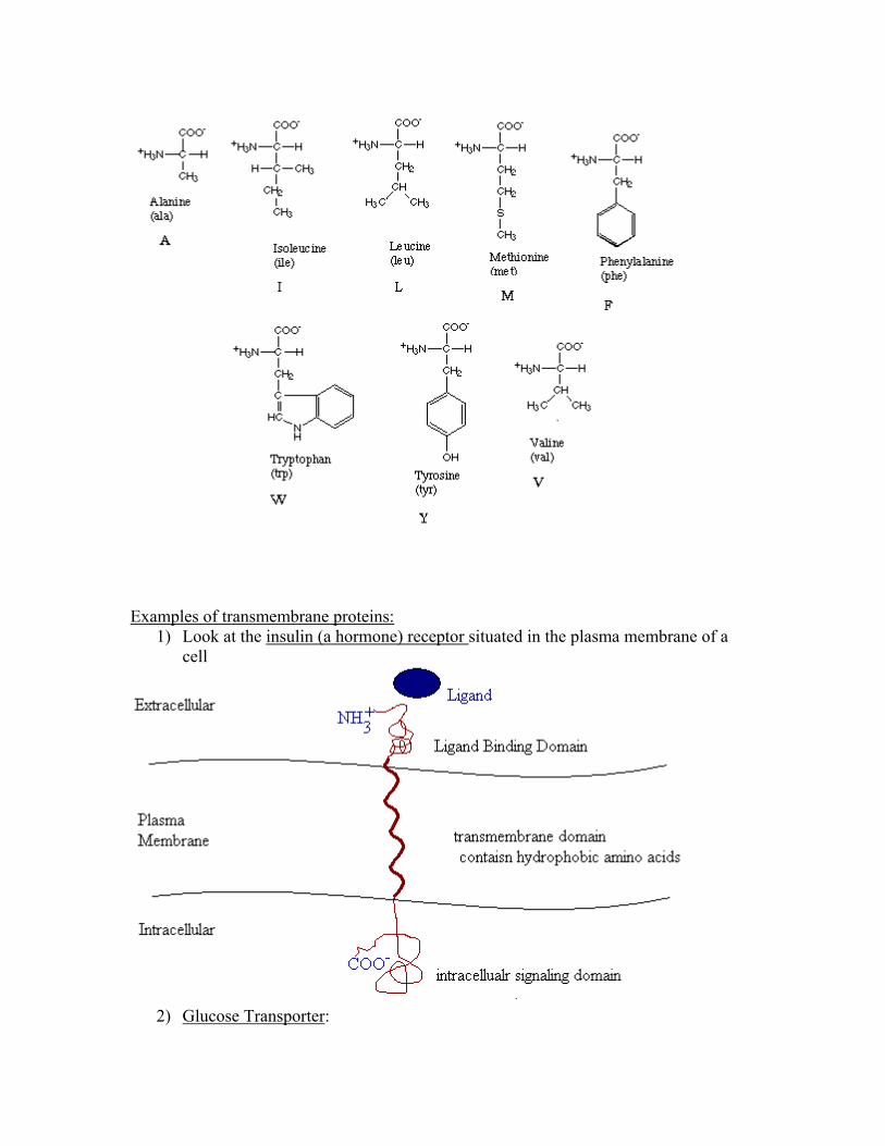
Examples of transmembrane proteins:
1) Look at the insulin (a hormone) receptor situated in the plasma membrane of a cell
2) Glucose Transporter:

The -helices are actually not in a line, but are clustered together to form a pore through which glucose can be transported:
These transporters are specific proteins in the membrane that transport the molecule (e.g. glucose) across the membrane. The proteins bind the molecule, transport the molecule across the membrane and then release it on the other side. The transporter remains unchanged after the process:
Look at a specialized organelle called the lysosome in the cell: The membrane of the lysosome contains specialized proteins that are used to maintain the acidic nature of the interior (lumen) of the lysosome.

1) ATP-powered Proton (H+) Pump
a. Pumps H+ ions into the interior of the lysosome b. Uses the energy obtained from the hydrolysis of ATP (ATP ADP + Pi)
to pump H+ ions into the lumen of the lysosome. The increase of the [H+] results in the low pH (acidity) of the lysosome.
2) Cl- Channel Protein a. Allows Cl- to flow down its concentration gradient into the lysosome b. Cl- ions balance the increase in the charge generated by the H+ ions
The concentration of H+ ions is 100x greater in the lumen of the lysosome than in the cytosol!!!! Look at a specialized cell called an erythrocyte:
- the erythrocyte (also called a RBC – red blood cell) is a biconcave disk - the RBC has become enucleated (lost its nucleus) during cell differentiation
(development of RBC) - the RBC is mainly filled with hemoglobin (Oxygen carrying protein) - The half-life (t1/2) of the RBC is ~120 days.
Although the RBC is ~7um in diameter, it has a flexible shape, which allows it to squeeze through some of the narrower capillaries of the circulatory system. The flexibility of the RBC is due to and extensive cytoskeletal network under the plasma membrane Look at the erythrocyte plasma membrane and network of proteins:

Dense networks of proteins also connect cells. Two cells can be connected by a complete array of proteins: together
Review:
- All membranes are made up of phospholipids arranged in a bilayer

- All organelles are bound by membranes - All organelles are made up of proteins that:
o Give shape to the organelle o Transport molecules across membranes o Give character to each organelle