cell proliferation and its regulationbiochemistry.ucsf.edu/programs/ptf/m3 links/cell proliferation...
TRANSCRIPT
Hiten Madhani, MD, PhD
25
Cell Proliferation and Its Regulation(Lecture)
OBJECTIVES• Listthekeypropertiesofstemcells.• DescribethetwokeyfunctionsofthecellcyclewithrespecttoDNA.• Listthefourphasesofthecellcycleanddescribewhathappensineach
phase.• Namethefourcyclin-Cdkcomplexesthatdrivethehumancellcycle.• DiagramthepathwaybywhichtheG1CdkactivatestheG1/SCdk.Describe
themoleculareventsthattakeplaceateachstepofthepathway.• NamethetwoclassesofCdkinhibitorsandthecyclin-Cdkcomplexesthey
inhibit.• Diagramthepathwayleadingfromepidermalgrowthfactor(EGF)tothe
activationofthecyclinDgene.Describethemoleculareventsthattakeplaceateachstepofthepathway.
• DescribetheWntsignalingpathwayanditseffectoncellproliferation.• DescribehowvariationintheexpressioinoftheAPCgenecontrolscellular
responsivenesstoWntsignaling.• DescribeinmoleculartermshowTGFßinhibitscelldivision.• Describehowapoptosiscanbetriggeredbyeitherextracellularor
intracellularsignals.• ListthemembersoftheBcl-2proteinfamilyanddescribehowtheyeither
promoteorinhibitapoptosis.• Listthetwomajorcaspasesinvolvedininitiatingapoptosis,andcontrast
theirdifferencesinmechanismofactivation.• Describehowp53isactivatedinresponsetoDNAdamage.• Describehowp53causescellcyclearrestandapoptosis.• Describethespindleassemblycheckpointandthemolecularfunctionofthe
Mad2protein.
KEY WORDSanaphase mitogenactivatedproteinkinased(MAPK)antimitogen mitoticspindleAPC(adenomatouspolyposiscoli) MYCapoptosis p16ß-catenin p21Bcl-2 p27caspase p53cellcycle progenitorcellcentrosome PUMAcheckpoint Rafchromosomesegregation Rascyclin Rbcyclin-dependentkinase(Cdk) receptortyrosinekinaseCdkinhibitor SH2domain

Cell Proliferation
26
KEY WORDS,continuedcytochromeC mitogencytokinesis sisterchromatidE2F sisterchromatidcohesionepidermalgrowthfactor(EGF) SosGrb2 stemcellgrowthfactor survivalfactorkinetochore terminaldifferentiationMad2 transforminggrowthfactorß(TGFß)MEK Wnt
I. INTRODUCTIONUncontrolledcellproliferationisahallmarkofcancer.Asdescribedintheoverviewlectureofthisblock,multiplemutationscausecancercellstodivideunchecked.Inthislecturewewillfocusonthenormalmechanismsofcelldivision,withanemphasisonthosedirectlyrelevanttocancer.
Innormaltissues,cellproliferationisgenerallyrestrictedtocellsthatreplenishthetissue.Most,ifnotall,tissuesarethoughttocontainstem cells thathavethisreplenishmentfunction(Figure1).Stemcellsareself-renewingcellsthatcandivideasymmetricallytoyieldanewstemcellandaprogenitor cell.Progenitorcellsmayormaynotundergofurtherdivisions,ultimatelyleadingtoterminal differentiation.Oncecellshaveterminallydifferentiated,theyhaveaspecializedfunctionandarenolongerdividing.Mosttissuesaremadeupofsuchnon-dividingcells.Thusproliferationisnormallytightlycontrolledsothatonlyparticularcellsinthebodyaredividing.
Programmedcelldeathorapoptosis,istheprocessbywhichexcessordamagaedcellsinthebodyareremoved.Thebalancebetweentheproductionofnewcellsandcelldeathmaintainstheappropriatenumberofcellsinatissue,whichisreferredtoashomeostasis.Apoptosisisalsoakeymechanismbywhichcancer-pronecellsareeliminated.
Figure 1. StemCells.Stemcellsareself-renewingcells.Theycandivideasymmetricallytoproduceanewstemcell(indicatedbyacircle)andaprogenitorcell.Progenitorcellsdividetoproducecellsthatundergoterminaldifferentiationtoproducethematurecellsthatmakeupatissueororgan.
Stem Cell (self-renewing)
Progenitor (Dividing)
Terminally DifferentiatedCells (Non-Dividing)
Hiten Madhani, MD, PhD
27
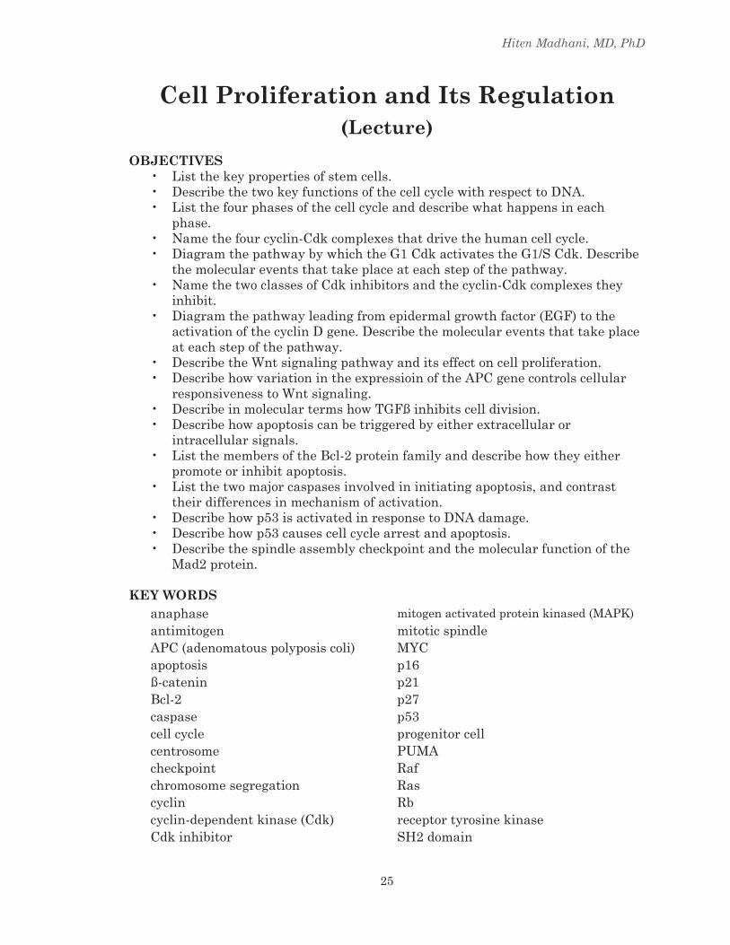
II. ThE CELL CYCLECell division occurs in defined stages, which together comprise the cell cycle.Intermsofthegeneticmaterial,cellsmustreplicatetheirchromosomalDNAonceeverycellcycleandsegregatethesister chromatidsproducedbyDNAreplicationtoyieldtwogeneticallyidenticaldaughtercells(Figure2).Recallthathumancellsarediploid,containingtwocopiesofeachchromosome(homologs).DuringDNAreplication,cohesionproteinsattachthereplicatedsisterchromatidstoeachother,holdingthemtogether.Thissisterchromatidcohesioniscriticalforthesubsequentsegregationofchromatidsintothedaughtercells.
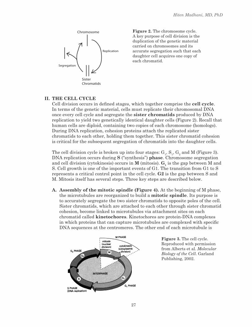
Thecelldivisioncycleisbrokenupintofourstages:G1,S1,G2andM(Figure3).DNAreplicationoccursduringS(“synthesis”)phase.Chromosomesegregationandcelldivision(cytokinesis)occursinM(mitosis).G1isthegapbetweenMandS.CellgrowthisoneoftheimportanteventsofG1.ThetransitionfromG1toSrepresentsacriticalcontrolpointinthecellcycle.G2isthegapbetweenSandM.Mitosisitselfhasseveralsteps.Threekeystepsaredescribedbelow.
A. Assembly of the mitotic spindle (Figure 4). AtthebeginningofMphase,themicrotubulesarereorganizedtobuildamitotic spindle.Itspurposeistoaccuratelysegregatethetwosisterchromatidstooppositepolesofthecell.Sisterchromatids,whichareattachedtoeachotherthroughsisterchromatidcohesion,becomelinkedtomicrotubulesviaattachmentsitesoneachchromatidcalledkinetochores.Kinetochoresareprotein-DNAcomplexesin which proteins that can capture microtubules are complexed with specific DNAsequencesatthecentromeres.Theotherendofeachmicrotubuleis
Figure 2. Thechromosomecycle.Akeypurposeofcelldivisionistheduplicationofthegeneticmaterialcarriedonchromosomesanditsaccuratesegregationsuchthateachdaughtercellacquiresonecopyofeachchromatid.
SisterChromatids
Chromosome
Replication
Segregation
Figure 3. Thecellcycle.ReproducedwithpermissionfromAlbertsetal.Molecular Biology of the Cell.GarlandPublishing,2002.

Cell Proliferation
28
Figure 5.Anaphase.Onlyonepairofsisterchromatidsisshown;however,thisoccurssimultaneouslyforall23pairsofhumanchromosomes.
Figure 6.Cytokinesis.Afterthetwosisterchromatidsaresegregatedtooppositepoles,cellsundergocytokinesis.
Figure 4.Themitoticspindle.
attachedtothecentrosome(alsocalledthespindlepolebodyormicrotubuleorganizingcenter).Thetwokinetochoresofeachpairofsisterchromatidsareattachedtooppositecentrosomes,andthereforewillbepulledtooppositepoles.
B. Anaphase is the step in mitosis when sister chromatid cohesion is rapidly dissolved. Asaresult,thepullingforcesofthemicrotubulescausethetwosisterchromatidstomovetotheoppositepoles(Figure5).
C. Cytokinesis is the step in which cells physically divide into two daughter cells (Figure 6). Forareviewofmitosis,seethemitosisandmeiosisonlinemoduleoniROCKET.
Figure 4
Figure 5

Hiten Madhani, MD, PhD
29
III. CELL CYCLE CONTROL: TWO ISSUESHowisthecellcyclecontrolled?Theproblemcanbebrokendownintotwoparts:First,howisthecellcycleregulatedsothatthedifferentphasesoccurinthecorrectorder?Second,howdoextracellularsignalsactivateorinhibitthecellcycle?
A. Cyclin Dependent Kinases: The core of the cell cycle control systemThe events that occur in each part of the cell cycle are carried out by specific proteins,andtheseproteinsmustbesynthesizedoractivatedatthecorrecttimeinthecycle.Forexample,beforeDNAsynthesiscanbegin,theenzymesthatproducethenucleotidesusedinDNAsynthesismustbeactivated.ThisoccurslateinG1phase.(RememberNucleotideMetabolism?SeelecturefromM&N.)
Thecellturnsproteinactivityonandoffatappropriatetimesinthecellcycleusingcyclin-dependent kinases(Cdks).Likeotherproteinkinases,cdksturnproteinsonoroffbyphosphorylatingthem.Eachcyclin-dependentkinasehastwosubunits-akinasesubunitandacyclinsubunit(Figure7).
Cyclins were first identified as key cell-cycle regulators when it was observed thattheyundergoacycleofsynthesisanddestructionduringeachcellcycle.ThereareseveraldifferentCdksandanumberofcyclins.Thekinasesubunitsarepresentthroughoutthecellcycle,whilethecyclinsubunitsare produced at specific times in the cell cycle, thus providing temporal regulationofthecyclin-CdKcomplex.Asthecyclinsubunitisproduced,itbindstothekinasesubunitandactivatesit.Thecyclinsubunitalso
Figure 7.Cyclin-dependentkinases(Cdks).Cdksarethekeyregulatorsofthecelldivisioncycleinorganismsasdiverseasbaker’syeastandhumans.Cyclin-dependentkinaseshavetwosubunits,thekinase(oftensimplycalledtheCdk)andaregulatoryproteincalledacyclin.
TableI.Thefourkeycyclin-Cdkstatdrivethecell

Cell Proliferation
30
targets its kinase partner to specific protein substrates. The key cyclin-Cdk complexesthatdrivethecellcyclearelistedinTableI.
ThecellcyclecanbeviewedasaCdkcycle(Figure8).AsthecellentersG1phaseattheendofmitosis,cyclinDisproducedandbindstoitstwopartnercdks–cdk4andcdk6.Thesecyclin-cdkcomplexesarebothreferredtoasG1-Cdks.ActivationoftheG1-CdksturnsontheeventsthatoccurintheearlyphaseofG1.OneoftheeventsthatoccursinearlyG1issynthesisofcyclinE.Asitismade,itbindstoCdk2,formingG1/S-Cdk.AstheG1/S-Cdkactivityaccumulatestoacriticalthreshold,ittriggersthetransitionfromlateG1intoSphase.CyclinAismadeinSphase.Italsobindstocdk2,formingS-Cdk.ThelatterisrequiredforDNAsynthesis.CyclinBismadeduringSphaseandG2.Asitismade,itbindstoCdk1formingM-Cdk.WhenM-Cdkreachesathresholdactivity,ittriggersthetransitionfromG2intomitosis.
B. how the G1-Cdk turns on the G1/S CdkThetransitionbetweenearlyG1andlateG1illustratesonewaythatcyclin-dependentkinasesregulatetheprogressionofthecellaroundthecellcycle.Thisisacrucialcontrolpointwhichisoftenderegulatedincancer.
InordertomovefromearlyG1tolateG1,thecellmustsynthesizecyclinE.TranscriptionofthecyclinEgenerequiresatranscriptionfactorcalledE2F.IncellsthatarenotproliferatingandincellsthatareinearlyG1,theE2FtranscriptionfactorisboundtothepromoterforthecyclinEgene,butitisinhibitedbyaproteincalledRb. (RbstandsforRetinoblastoma,achildhoodtumoroftheretina–moreonthisinthetumorsuppressorandoncogenelecture!).WhenG1-CdkactivityincreasesnearthemiddleofG1,G1-CdkphosphorylatestheRbprotein(Figure9).ThisreleasesRbfromE2FandallowstranscriptionofthecyclinEgenetotakeplace.ThecyclinEproteinbindstotheCdk2kinasetoformtheG1/S-Cdk.E2Falsotranscribes
Figure 8.ThecellcycleasaCdkcycle.Differentphasesofthecellcyclearedrivenbydifferentcyclin-Cdkcomplexes.Inthissimplified view, there exists aG1-Cdk,aG1/S-Cdk,aS-CdkandaM-CdkthatactinsequencetoprogramtheeventsofG1,theG1-Stransition,Sphase,andMphase.

Hiten Madhani, MD, PhD
31
anumberofothergenesimportantforSphase,includingthegenesforDNApolymeraseandthymidylatesynthase.
C. Brakes on the cell cycle: Cdk inhibitors.TheRbproteincanbeviewedasa“brake”onthecellcyclebecauseitpreventsthetranscriptionofthegeneforcyclinEbyinhibitingE2F.Threeotherproteinsthatactas“brakes”onthecellcyclearetheCdkinhibitorsp21,p16,andp27.Whyisthecellcyclecontrolledbybothactivators(e.g.cyclins)andinhibitors?Aswewillsee,itissothatcellscanrespondtobothgrowthactivatingandgrowthinhibitingsignals.
Cdk inhibitors fall into two classes: general and specific (Figure 10). General Cdkinhibitors,suchasp21andp27,bindtomultipleCdk-cyclincomplexes,shutting off the cell cycle at multiple points. p16 is a specific Cdk inhibitor whichtargetsCdk4-cyclinD(G1Cdk).
Aswillbediscussedintheoncogeneandtumorsuppressorlecture,theseinhibitorproteinsplayanimportantroleincancer.
IV. CONTROLLING PROLIFERATION: ThE ROLE OF MITOGENS AND ANTI-MITOGENSNon-dividingcellsexistinphasecalledG0(Gzero).G0cellscanre-enterthecellcycleinG1whenstimulatedbymitogens,whichareextracellularproteinsthatstimulatecellproliferation.Mitogensarealsocalled growth factors. Conversely,cellscanbearrestedinG1viatheactionofanti-mitogens (proteinsthatinhibittheactivityofmitogens).Manygrowthfactorsandinhibitorsare
Figure 9.HowtheG1-CdkactivatestheG1/S-Cdk.TheG1-Cdk(Cdk4-cyclinD)phosphorylatestheRb(Retinoblastoma)proteinreleasingitfromtranscriptionfactorE2F.E2FcannowactivatetranscriptionofcyclinE,whichinturnresultsintheproductionofcyclinEproteinandformationoftheG1/SCdk.
Cdk4-cyclin D (G1-Cdk)
Rb
E2F
cyclin E
Cdk2
Cdk2-cyclin E (G1/S-Cdk)
promoter
cyclin E gene
OFF
E2F
Rb
cyclin E gene
ON
E2F
RbP
G1-Cdk OFF
G1-Cdk ON
A. B. Control RelationshipsMolecular Events
promoter

Cell Proliferation
32
known.Wewilldiscussoneexampleofeach:themitogenepidermal growth factor(EGF)andtheanti-mitogentransforming growth factor ß(TGFß).
Whatarethenormalfunctionsofthesefactors?EGFfunctionstopromotewoundhealing. After a wound is formed, epidermal and inflammatory cells secrete EGFandothergrowthfactors.Itsignalscellsatthemarginsofthewoundtoproliferatesothatthewoundmaybehealed.TGFßactsasabraketothisprocesssothattheproliferationoccursataratethatisnottoofasttocoordinatewithotheraspectsofwoundhealing.
A. The EGF Signaling PathwayThispathwayallowsEGFontheoutsideofthecelltoultimatelycontrolthecellcycleontheinsideofthecell.Itinvolvesmanystepsandcomponents.Thiscomplexityisthoughttoexistfortworeasons.First,evolutionoftenproceedsbyadaptingexistingpathwaysandcombiningpathwaystocarryoutnewfunctions.Theresulting“newpathway”maybequitecomplexbecauseofitshistory.Second,thereisevidencethathavingapathwaywithmultiplesteps filters noise and allows many opportunities for regulatory control. Such apropertymakessenseinthecontextofthecellcyclewherecellsareeitherproliferatingorarrestedinG0.
EGFfunctionsbybindingtotheextracellulardomainofEGFreceptor,acellsurfaceproteinwithasingletransmembranedomain(Figure11).ThecytoplasmicdomainofthereceptorisaproteintyrosinekinasethustheEGFreceptoriscalledareceptor tyrosine kinase.WhenEGFbindstoitsreceptor,thereceptorformsadimerinwhichonesubunitphosphorylatestheother(transphosphorylation)onparticulartyrosineresiduesinthecytoplasmicpartofthereceptor.Thesephosphorylatedtyrosinesserveasbindingsitesforothercytoplasmicproteinsthatcontainspecialdomains,calledSh2 domains. SH2 domains specifically recognize phosphorylated tyrosinesandtheadjacentaminoacids.OneproteinthatbindstophosphotyrosineresiduesintheEGFreceptorisanadaptorproteincalledGrb2.Grb2,inturn,recruitsaproteincalledSos.ThusbindingofEGFto
Figure 10.CdkinhibitorsbindtoandinactivateCdks.
Cdk2/4-cyclin D (G1-Cdk)Cdk2-cyclin E (G1/S-Cdk)
Cdk2-cyclin A (S-Cdk)
Cdk2-cyclin E (G1/S-Cdk)
p21 p27
p16

Hiten Madhani, MD, PhD
33
theEGFreceptorrecruitsbothGrb2andSostotheintracellularportionofthereceptor.
Sos,inturn,actsonasmallGTPbindingprotein,Ras.TheRasproteinisboundtotheinnersurfaceoftheplasmamembrane.LiketheG-proteinsdiscussedbyDr.FultoninthePrologue,Rascanexistintwostates:aninactivestateinwhichGDPisboundandanactivestateinwhichGTPisbound.SosactivatesRasbypromotingthereleaseofitsGDPandbindingofGTP.Thus,SosisaguaninenucleotideexchangefactorforRas(Figure12).RecruitmentofSostotheplasmamembranewhereRasislocatedresultsintheactivationofRas.
Figure 11.ActivationoftheEpidermalGrowthFactor(EGF)receptortyrosinekinase.EGFbindstotheEGFreceptorthroughanextracellularligandbindingdomain,leadingtodimerizationofthereceptor.Dimerizationcausesonesubunittophosphorylatetheother(transphosphorylation)on specific tyrosine residues. TheSH2domainoftheGrb2adaptorproteinbindsthephosphorylatedtyrosines.Grb2inturn,bindsaanotherproteincalledSos.
EGFLigand
Binding Domain
KinaseDomain
P SH2 SH3
Grb2(adaptor protein)
Sos
Figure 12.SosactivatesRasandaGTPase-activatingproteininactivatesit.InitsGDPboundform,Rasisinactive.WhenSosisboundtoGrb2attheEGFreceptor,itcausesRastoreleaseitsGDPandbindaGTPinitsplace.AGTPase-activatingproteincauseshydrolysisofGTPtoGDP.
Sos
Ras-GDP Ras-GTP
GTP
GDP
inactive active
GAP
GTPase-activating protein

Cell Proliferation
34
RascanbereturnedtoitsinactiveformthroughthehydrolysisofGTPtoGDP.ThisstepoccurswhenaGTPase-activating proteinbindsRas andinducesthehydrolysisofitsGTP(seeFigure12).
InitsGTP-bound(active)state,Rasturnsonaproteinkinasecascade,inwhichproteinkinasessequentiallyactivateeachotherthroughphosphorylation(Figure13).ActiveRasbindstoandactivatesaproteinkinasecalledRaf.Inturn,RafphosphorylatesandactivatesanotherkinasecalledMEK.MEKinturnphosphorylatesandactivatesmitogen-activatedproteinkinase(MAP kinase).Thischainofphosphorylationeventsiscalled the MAP kinase cascade. MAP kinase phosphorylates gene-specific transcriptionfactorsinthecellnucleusthatbindtothepromotersofgenesandpromotecellproliferation.OneimportanttranscriptionfactorthatisupregulatedbytheMAPkinasecascadeisMYC,whichistheproductoftheC-MYCgene.
OneofthetargetsofthesetranscriptionfactorsisthecyclinDgene.Thus,amulti-tieredpathwayconnectsthepresenceofamitogen(EGF)outside
Figure 13.RasactivatestheRaf-MEK-MAPKcascade.RasbindsdirectlytoRaf,whichactivatesitskinaseactivity.RafphosphorylatesakinasecalledMEK(alsocalledMAPkinasekinase).AfterithasbeenphosphorylatedbyRaf,MEKphosphorylatesMAPkinase(mitogenactivatedproteinkinase,MAPK).ActiveMAPKthenphosphorylatesitstargetproteins,leadingtotheactivationofcelldivision.
Ras-Raf
MEK MEK-P
MAPK MAPK-P
Target Target-P
inactive
inactive
inactive
active
active
active
Figure 14.ActivationofMAPKleadstothetranscriptionofcyclinD.MAPKphosphorylatestranscriptionfactors.Thisinturnleadstothetranscriptionofthemycgene,whichitselfencodesatranscriptionfactorforthecyclinDgene.
TranscriptionFactorsMAPK cyclin D

Hiten Madhani, MD, PhD
35
thecelltoincreasedexpressionofakeycomponentofthecellcyclecontrolmachinery(thecyclinDgene)inthenucleus.IncreasedexpressionofthecyclinDgeneleadstotheactivationofG1-Cdk(Figure14).
B. Wnt signalingWnt proteinsaregrowthfactorsanalogoustoEGF.Theyfunctioninasignalingpathwaythatactivatescellproliferation(Figure15).NotonlyistheWntsignalingpathwayimportantforthedevelopmentoftheorganism,italsoservesimportantfunctionsinadults.Forexample,Wntsignalingisnecessaryfortheproliferationofstemcellsintheproliferativezonesinthegutepithelium(thecryptsthatliebetweenthemicrovillioftheepithelium)(Figure16).Hyperactivationofthispathwayisassociatedwithcoloncancer.
Figure 15. TheWntsignalingpathway.
GSK-3
Wnt
Frizzled
β-catenin β-cateninAPC APC
P P
degradation
APCTCF
β-catenin
TCF
cyclin D
microvillus microvillus
crypt(proliferatingstem cells)
Expressionof APC
low
high
Figure 16.Shownisaschematicofamicrovillusinthegutepitheliumshowingthezoneofproliferation(crypts)andthegradientofexpressionoftheAPCgene,whoseproteinproductinhibitsWntsignaling.

Cell Proliferation
36
WntproteinsbindtoacellsurfacereceptorcalledFrizzled.Frizzledcontrolsthestabilityofaproteincalled ß-catenin,whichfunctionstogetherwithaproteincalledTCFtoformatranscriptionfactorthatactivatesthecyclinDpromoter.
OnceboundtoWnt,FrizzledturnsoffaproteinkinasecalledGSK-3.GSK-3normallyfunctionstopromotethedegradationß-catenin,thuspreventingitfromactivatingthecyclinDpromoter.Phosphorylationofß-cateninbytheproteinkinaseGSK-3resultsinitsdegradation.However,GSK-3canonlyphosphorylateß-cateninwhenß-cateninisboundtoaproteincalledAPC (adenomatous polyposis coli).Thus,APCisnecessarytoholdß-cateninincheck.
OnceGSK-3isinhibitedbyFrizzled,ß-cateninisnolongerdegraded,allowingittoassociatewithTCFandactivatethecyclinDpromoterandpromotecellproliferation.Thus,Wntsignalingpromotescellproliferationthroughtheactionofb-cateninoncyclinD.
WhileWntproteinsaretheextracellulargrowthfactorsthatactivatethispathway,cellsalsocontrolthepathwayfromwithinthecellbyvaryingthetranscriptionoftheAPCgene,whoseproteinproductinhibitstheWntsignalingpathway.Forexample,intheepitheliumofthecolon,thereexistsagradientofAPCexpressionwhichishighestintheterminallydifferentiatednondividingcellsinthemicrovilliandlowestintheproliferatingstemcellsin
Figure 17.HowTGFbßarrestscelldivision.TGFßbindstotheTGFßreceptor.BindingofTGFßactivatesthereceptor’sintracellularproteinkinasedomain,leadingtophosphorylationofSmadproteins.PhosphorylatedSmadsenterthenucleusandbindtopromotersofgenestocontroltranscription.Akeytargetisthep21gene.
plasmamembrane
nucleus
TGFβ
kinasedomain
Smad
Smad-P
p27 gene
promoter
Smad-P
TGFβ receptor
p21
Cdk4-cyclin DCdk2-cyclin ECdk2-cyclin A

Hiten Madhani, MD, PhD
37
thecrypts(Figure16).(MoreabouttheroleofAPCincoloncancertocomeinlecturesonColonCancerandFamilialandHereditaryCancerSyndromes.)
C. An anti-mitogenic pathway: the TGFß-Smad pathwayLikeEGF,TGFßisanextracellularproteinthatbindsacellsurfacereceptor.However,insteadofcausingcellproliferation,thismoleculecausescellstoarresttheircellcycleandenterG0.Howdoesthisoccur?TheTGFßreceptorisatransmembraneserine-threoninekinase.UponbindingtoTGFß,thereceptorphosphorylatesproteinsinthecytoplasmcalledSmads (Figure17).Oncephosphorylated,Smadproteinsthenenterthenucleusandfunctionastranscription factors to turn on specific target genes. A key gene turned on byTGFßistheG1/S-Cdkinhibitorp21discussedabove.Theactivationofp21leadstothearrestofthecellcycle.Thus,TGFßarrestscelldivisionbyactivatingthegeneforaCdkinhibitor.
V. APOPTOSISThenumberofcellsinatissueisnotonlycontrolledbycellproliferationbutalsobyprogrammedcelldeath,orapoptosis.Foratissuetostaythesamesize,cellproliferationandcelldeathmustbeperfectlybalanced.Apoptosisplaysimportantrolesbothduringdevelopmentandinmaturetissues(Figure18).Duringdevelopmentofalimb,tissuepresentbetweenthedigitsmustberemoved.Thisoccursthroughlocalizedapoptosis.Intheimmunesystem,onemechanismbywhichTcellskilltargetcellsistoproduceaproteincalledFasligand.Fasligandbindsitsreceptor,Fas,ontargetcells.BindingofFasligandtoFasinducesapoptosis.Therealsoexistextracellularproteins,calledsurvival factors,whichpreventapoptosisbybindingtotheirreceptorsonthesurfaceofthecell.
AsdescribedinthePrologueblock,activationofapoptosisfromoutsidethecelliscausedbytheassociationofprocaspase-8withthecytoplasmicdomainof“deathreceptors”suchasFas(Figure19a).Thisassociationismediatedbyadaptor
Figure 18.Programmedcelldeath.Duringdevelopmentoflimb,tissuepresentbetweenthedigitsisremovedbyapoptosis.KillerTcellstriggerapoptosisbyproducingFasligand.
Apoptosis
Killer T-cell Target Cell
Fas ligandFas
Apoptosis

Cell Proliferation
38
Figure 19.Inductionofapoptosisbyeitherextracellularorintracellularsignals.(A)Extracellularactivation.AdaptorproteinsbindtheintracellularregionofaggregatedFasproteins,causingtheaggregationofprocaspase-8molecules.Thesethencleaveoneanothertoinitiatethecaspasecascade.(B)Intracellularactivation.Mitochondriareleasecytochromec,whichbindsandcausestheaggregationoftheadaptorproteinApaf-1.Apaf-1bindsandaggregatesprocaspase-9molecuels,whicharetherebyactivated(withoutcleavage)totriggeracaspasecascade.FromAlbertsetal.,MolecularBiologyoftheCell,2002.
proteinsthatbindbothFasandprocaspase-8.Theaggregatedprocaspase-8moleculescleaveeachother,initiatingthecaspase cascadethatleadstoapoptosis.
Whencellsarestressed(e.g.,hypoxia)ordamaged(e.g.,unrepairedDNAdamage)theycanalsoactivateapoptosisfrominsidethecellbytriggeringprocaspaseaggregationandactivation.Inresponsetostressordamagen,pro-apoptoticsignalsinducemitrochondriatoreleatecytochrome cintothecytosol,whereitbindsandactivatesanadaptorproteincalledApaf-1.Thiscomplexactivatesacaspasecalledprocaspase-9,whichleadstotriggeringofthecaspacecascade(Figure19-b).
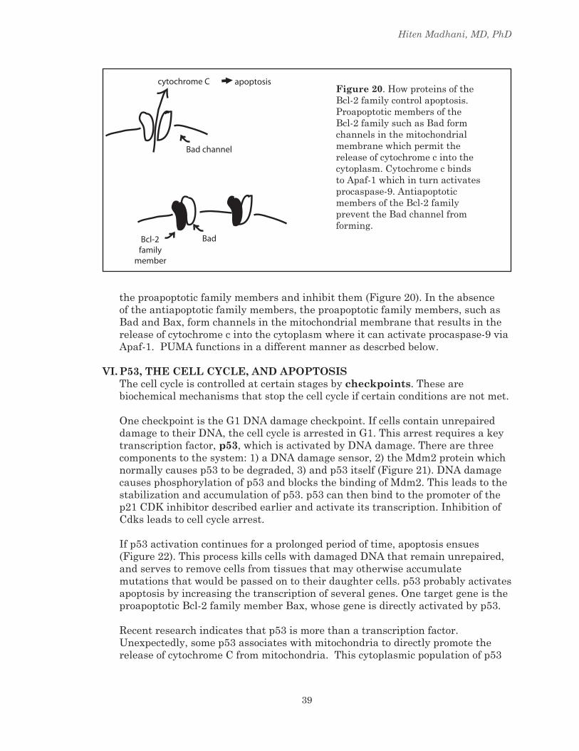
ThereleaseofcytochromecfromthemitochondriaistightlycontrolledbymembersoftheBcl-2 familyofproteins.Withinthisfamilyofproteins,therearetwosub-classes:proteinsthatpromoteapoptosis(“proapoptotic”)suchasBad,BaxandPUMA,andproteinsthatantagonizeapoptosis(“antiapoptotic”)suchasBcl-2andBcl-xL.Theantiapoptoticfamilymemberscandirectlybindto
Hiten Madhani, MD, PhD
39
Figure 20.HowproteinsoftheBcl-2familycontrolapoptosis.ProapoptoticmembersoftheBcl-2familysuchasBadformchannelsinthemitochondrialmembranewhichpermitthereleaseofcytochromecintothecytoplasm.CytochromecbindstoApaf-1whichinturnactivatesprocaspase-9.AntiapoptoticmembersoftheBcl-2familypreventtheBadchannelfromforming.
Bad channel
cytochrome C apoptosis
Bad Bcl-2family
member
theproapoptoticfamilymembersandinhibitthem(Figure20).Intheabsenceoftheantiapoptoticfamilymembers,theproapoptoticfamilymembers,suchasBadandBax,formchannelsinthemitochondrialmembranethatresultsinthereleaseofcytochromecintothecytoplasmwhereitcanactivateprocaspase-9viaApaf-1.PUMAfunctionsinadifferentmannerasdescrbedbelow.
VI. P53, ThE CELL CYCLE, AND APOPTOSISThecellcycleiscontrolledatcertainstagesbycheckpoints.Thesearebiochemicalmechanismsthatstopthecellcycleifcertainconditionsarenotmet.
OnecheckpointistheG1DNAdamagecheckpoint.IfcellscontainunrepaireddamagetotheirDNA,thecellcycleisarrestedinG1.Thisarrestrequiresakeytranscriptionfactor,p53,whichisactivatedbyDNAdamage.Therearethreecomponentstothesystem:1)aDNAdamagesensor,2)theMdm2proteinwhichnormallycausesp53tobedegraded,3)andp53itself(Figure21).DNAdamagecausesphosphorylationofp53andblocksthebindingofMdm2.Thisleadstothestabilizationandaccumulationofp53.p53canthenbindtothepromoterofthep21CDKinhibitordescribedearlierandactivateitstranscription.InhibitionofCdksleadstocellcyclearrest.
Ifp53activationcontinuesforaprolongedperiodoftime,apoptosisensues(Figure22).ThisprocesskillscellswithdamagedDNAthatremainunrepaired,andservestoremovecellsfromtissuesthatmayotherwiseaccumulatemutationsthatwouldbepassedontotheirdaughtercells.p53probablyactivatesapoptosisbyincreasingthetranscriptionofseveralgenes.OnetargetgeneistheproapoptoticBcl-2familymemberBax,whosegeneisdirectlyactivatedbyp53.
Recentresearchindicatesthatp53ismorethanatranscriptionfactor.Unexpectedly,somep53associateswithmitochondriatodirectlypromotethereleaseofcytochromeCfrommitochondria.Thiscytoplasmicpopulationofp53

Cell Proliferation
40
Figure 21.HowDNAdamageactivatesp53andcausescell-cyclearrest.DNAdamageactivatesaproteinkinasewhichphosphorylatesp53.
Figure 22.DNAdamagecanleadtoapoptosis.Prolongedactivationofp53inresponsetoDNAdamageresultsinapoptosis.p53activatesthetranscriptionofseveralgenesinvolvedinapoptosisincludingthatfortheproapoptoticBcl-2familymemberBaxshownhere.
BAX gene
promoter
p53
Prolonged p53
Activation
Bax channel
cytochrome C apoptosis

Hiten Madhani, MD, PhD
41
isinhibitedbytheanti-apoptoticBcl-2familymemberBcl-xL,whichdirecltybindstop53.Oneofthetranscriptiontargetsofp53,PUMA(theproapoptoticBcl2familymemberdescribedabove),releasesp53fromBcl-xLThisinturnallowsp53tobindtoBaxwhichactivatescytochromeCrelease,andapoptosisensues.
Inlightoftheimportantrolep53playsinpreventingunrepairedDNAdamagetobepassedontodaughtercells,itisnotsurprisingthatp53isfoundtopayaroleincancerdevelopment.Infact,p53ismutatedinmanytypesofcancer,thusallowingdamagedDNAtoremainincellsastheyproliferate(moreinthelectureontumorsuppressorgenesandoncogenes).
VII. ThE SPINDLE ASSEMBLY ChECKPOINTInadditiontomonitoringthestateofDNA,cellsalsomonitorthestateofthemitoticspindleatthespindle assembly checkpoint(Figure24).Thespindleassemblycheckpointensuresthatmitosisdoesnotproceedunlessthespindleisproperlyassembled.ThischeckpointmonitorstheattachmentofspindlemicrotubulestoeachkinetochorethoughtheactionoftheMad2 protein.Therearetwokeyfeaturesofthecheckpoint:1)Mad2associateswithkinetochoresonlywhentheyarenotattachedtomicrotubules.and2)Mad2isactiveinarrestingmitosisonlywhenitisboundtokinetochores.Thuswhenthespindleisproperlyassembledandallofthekinetochoresareboundtomicrotubules,Mad2isinactiveallowingmitosistoproceed.Ifthereisaproblemwithspindleassembly,Mad2isactivatedandarreststhecellcycle.Notsurprisingly,cellsdefectiveinthespindleassemblycheckpointshowhighratesofaneuploidybecauseoferrorsinchromosomesegregationduringmitosis.Defectsinthespindle-assemblycheckpoint, and specifically in Mad2, have been associated with tumorigenesis.
p53 stabilization
accumulation innucleus
activation of Bax andPUMA transcription
accumulation incytoplasm
Bcl-XL sequestersp53
PUMA liberates p53
p53 activates BaxApoptosis
Figure 23. p53functionsbothinthenucleusandthecytoplasmtopromoteapoptosis

Cell Proliferation
42
SisterChromatids
microtubules
kinetochore
centrosome Mad2
cell cycle arrest
Mad2
inactive
Figure 24.Spindleassemblycheckpoint.ThisisaccomplishedthroughtheactionoftheMad2protein,whichbindstokinetochoresthathavenotattachedtomicrotubules.Whenboundtokinetochores,Mad2triggerscellcyclearrest.Oncemicrotubulesareattachedtoallofthekinetichores,Mad2isnolongeractiveandthecellcycleproceeds.