cbe technical advisory conference abstracts
TRANSCRIPT

CBE TECHNICAL ADVISORY CONFERENCE ABSTRACTS FEBRUARY 2009
0
CBE TECHNICAL ADVISORY CONFERENCE ABSTRACTS FEBRUARY 2009
1
CBE Technical Advisory Conference: February 3–4, 2009
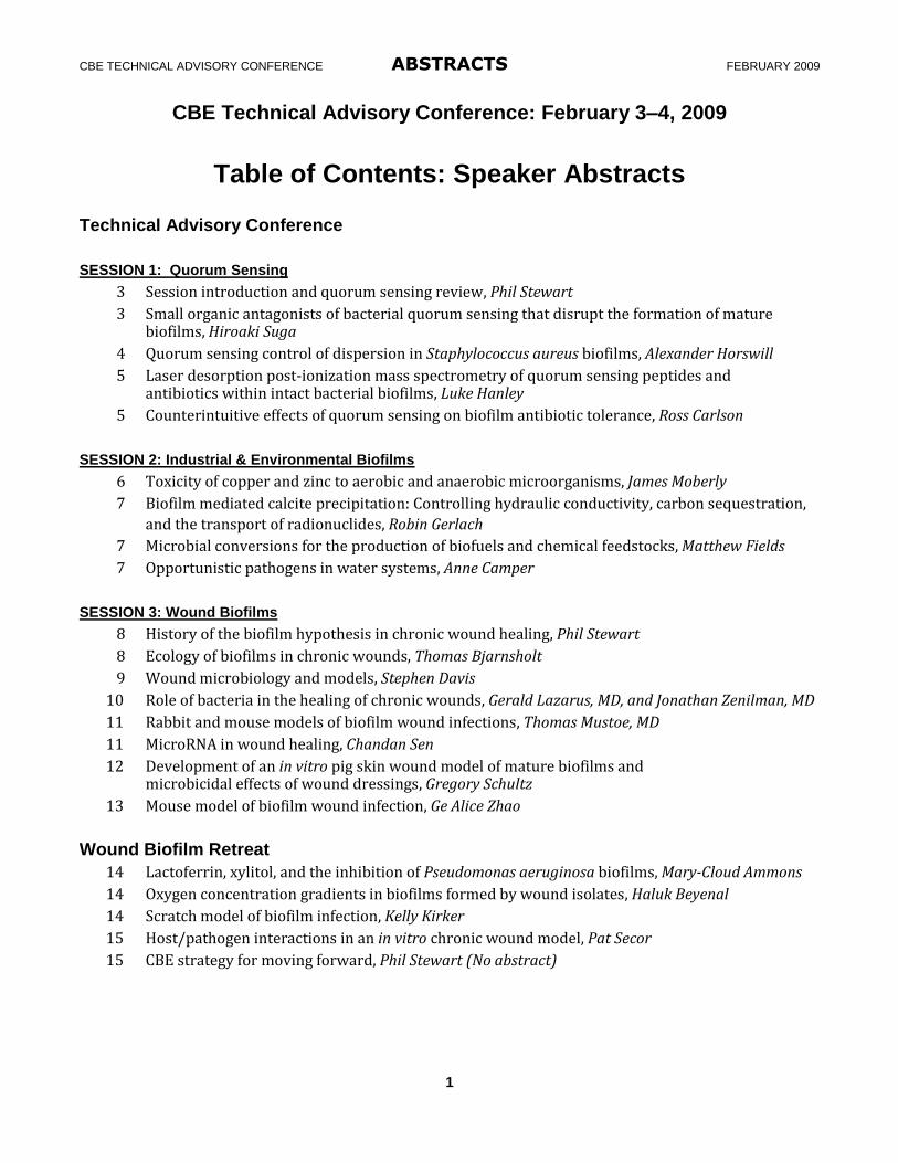
Table of Contents: Speaker Abstracts Technical Advisory Conference
SESSION 1: Quorum Sensing
3 Session introduction and quorum sensing review, Phil Stewart
3 Small organic antagonists of bacterial quorum sensing that disrupt the formation of mature biofilms, Hiroaki Suga
4 Quorum sensing control of dispersion in Staphylococcus aureus biofilms, Alexander Horswill
5 Laser desorption post-ionization mass spectrometry of quorum sensing peptides and antibiotics within intact bacterial biofilms, Luke Hanley
5 Counterintuitive effects of quorum sensing on biofilm antibiotic tolerance, Ross Carlson
SESSION 2: Industrial & Environmental Biofilms
6 Toxicity of copper and zinc to aerobic and anaerobic microorganisms, James Moberly
7 Biofilm mediated calcite precipitation: Controlling hydraulic conductivity, carbon sequestration,
and the transport of radionuclides, Robin Gerlach
7 Microbial conversions for the production of biofuels and chemical feedstocks, Matthew Fields
7 Opportunistic pathogens in water systems, Anne Camper
SESSION 3: Wound Biofilms
8 History of the biofilm hypothesis in chronic wound healing, Phil Stewart
8 Ecology of biofilms in chronic wounds, Thomas Bjarnsholt
9 Wound microbiology and models, Stephen Davis
10 Role of bacteria in the healing of chronic wounds, Gerald Lazarus, MD, and Jonathan Zenilman, MD
11 Rabbit and mouse models of biofilm wound infections, Thomas Mustoe, MD
11 MicroRNA in wound healing, Chandan Sen
12 Development of an in vitro pig skin wound model of mature biofilms and microbicidal effects of wound dressings, Gregory Schultz
13 Mouse model of biofilm wound infection, Ge Alice Zhao
Wound Biofilm Retreat
14 Lactoferrin, xylitol, and the inhibition of Pseudomonas aeruginosa biofilms, Mary-Cloud Ammons
14 Oxygen concentration gradients in biofilms formed by wound isolates, Haluk Beyenal
14 Scratch model of biofilm infection, Kelly Kirker
15 Host/pathogen interactions in an in vitro chronic wound model, Pat Secor
15 CBE strategy for moving forward, Phil Stewart (No abstract)
CBE TECHNICAL ADVISORY CONFERENCE ABSTRACTS FEBRUARY 2009
2
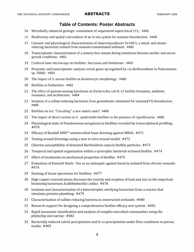
Table of Contents: Poster Abstracts
16 Microbially enhanced geologic containment of sequestered supercritical CO2: #445
16 Biodiversity and spatial concordance of an in situ system for uranium bioreduction: #449
17 Genomic and physiological characterization of Anaeromyxobacter fw109-5, a metal- and nitrate-reducing bacterium isolated from uranium-contaminated sediment: #450
18 Transcriptomic characterization of a sensory-box mutant during transitions between aerobic and anoxic growth conditions: #451
19 Confocal laser microscopy on biofilms: Successes and limitations: #452
19 Proteomic and transcriptomic analyses reveal genes up-regulated by cis-dichloroethene in Polaromonas sp. JS666: #453
20 The impact of S. aureus biofilm on keratinocyte morphology: #460
20 Biofilms as biobarriers: #461
21 The effect of quorum-sensing knockouts on Escherichia coli K-12 biofilm formation, antibiotic resistance, and architecture: #464
21 Isolation of a sulfate-reducing bacterium from groundwater stimulated for uranium(VI) bioreduction: #465
22 Biofilms on ice: “Unveiling” a new matrix stain? #466
22 The impact of direct current on S. epidermidis biofilms in the presence of ciprofloxacin: #469
24 Physiological state of Pseudomonas aeruginosa in biofilms revealed by transcriptional profiling: #470
24 Efficacy of Kendall AMD™ antimicrobial foam dressing against MRSA: #471
25 Testing wound dressings using a new in vitro wound model: #472
25 Chlorine susceptibility of detached Burkholderia cepacia biofilm particles: #473
26 Temporal and spatial organization within a syntrophic bacterial-archaeal biofilm: #474
27 Effect of treatments on mechanical properties of biofilm: #475
27 Evaluation of bismuth thiols: Use as an antiseptic against bacteria isolated from chronic wounds: #476
29 Staining of tissue specimens for biofilms: #477
30 High copper concentrations decrease the toxicity and sorption of lead and zinc to the important biomining bacterium Acidithiobacillus caldus: #478
30 Isolation and characterization of a heterotrophic nitrifying bacterium from a reactor that simulates premise plumbing: #479
31 Characterization of sulfate reducing bacteria in constructed wetlands: #480
31 Research support for designing a comprehensive biofilm efficacy test system: #481
32 Rapid taxonomic classification and analysis of complex microbial communities using the phylochip microarray: #482
32 Bacterially induced calcite precipitation and Sr co-precipitation under flow conditions in porous media: #483
CBE TECHNICAL ADVISORY CONFERENCE ABSTRACTS FEBRUARY 2009
3
Speaker Abstracts SESSION 1: Quorum Sensing Session introduction and quorum sensing review Presenter: Phil Stewart, CBE Director and Professor, Chemical & Biological Engineering
Affiliation: Center for Biofilm Engineering, Montana State University, Bozeman, MT
It has been ten years since the publication of the seminal Science paper that drew a connection between biofilm formation and quorum sensing, a microbial communication phenomenon mediated by diffusible signal molecules. This work immediately raised the prospect of new strategies for mitigating detrimental biofilms based on interfering with cell-to-cell communication instead of relying on cidal disinfectants and antibiotics. As an introduction to this session on quorum sensing in biofilms, the state of the science on this topic a decade after the original discovery will be reviewed. The emphasis of the review is on the potential to deploy quorum sensing inhibitors as a means to control real-world fouling and infection problems. Some of the key findings of this overview are: 1) a multiplicity of chemically distinct signaling molecules have been identified in various bacteria and fungi; 2) quorum sensing can either antagonize or promote virulence and biofilm formation, depending on the organism; 3) quorum sensing in nature often mediates interaction between a bacterium and higher organism; and 4) many natural and synthetic potential quorum sensing inhibitors have been identified. These features suggest that the first applications of quorum sensing inhibition technology may occur in a medical context where a single predominant pathogen infects a plant or animal. Several companies have formed to develop quorum sensing inhibition technology. Small organic antagonists of bacterial quorum sensing that disrupt the formation of mature biofilms Presenter: Hiroaki Suga, Professor Affiliation: Research Center for Advanced Science and Technology, The University of Tokyo, Japan
4-6-1 Komaba, Meguro-ku, Tokyo 153-8904, Japan, Tel&Fax: +81-3-5452-5495, E-mail: [email protected]
Quorum sensing (QS) regulates production of virulence factors and maturation of biofilm in many bacteria including Pseudomonas aeruginosa. The QS cascade is activated by the interaction of bacterial signaling molecules, called autoinducers (AIs), with their corresponding regulatory proteins. Pseudomonas aeruginosa is an opportunistic pathogen, which is a common cause of infections in immunocompromised individuals and individuals with cystic fibrosis. Expression of genes that produce virulence factors such as alkaline protease, elastase, exotoxin A, rhamnolipids, and pyocyanin is governed by the response to cell density that is monitored by a mechanism known as quorum sensing (QS). Biofilm formation is also a major contributor to the virulence causing persisting infections in lungs of cystic fibrosis patients, in which the regulation of mature biofilm formation is also linked to the QS mechanism. In P. aeruginosa QS consists of two separate cascades, las and rhl, consisting of regulatory (R) proteins, LasR and RhlR, and inducer (I) proteins, LasI and RhlI, respectively. The I proteins synthesize the corresponding signaling molecules (called autoinducers, AIs), N-(3-oxododecanoyl)-L-homoserine lactone (3OC12-L-HSL) and N-butanoyl-L-homoserine lactone (C4-L-HSL), and these molecules bind the cognate R proteins to activate the QS circuits (Figure 1A, X = L-HSL). There exists a regulation hierarchy within this quorum sensing system where LasR•3OC12-L-HSL complex regulates rhlR expression. Since QS plays a central role in governing the gene expression of various virulence factors, controlling QS by means of interfering with the binding of AIs with their respective R proteins potentially offers a curative form of treatment.
CBE TECHNICAL ADVISORY CONFERENCE ABSTRACTS FEBRUARY 2009
4
To gain insights into the molecular interaction of the AIs with their cognate R proteins and ultimately aid rational design of QS inhibitors, a study encompassing synthesis and testing of a library of AI analogs was performed earlier in our laboratory. Our approach for designing the AI library involved substitution of the HSL moiety, which is a common structural element of AIs in many gram-negative bacteria, with various amines. Our study has identified several hits that act as agonist or antagonist for the P. aeruginosa QS circuits. In this lecture, I shall firstly show stimulation of the QS circuits by non-HSL synthetic agonists. This study enabled us to better understand the AI or AI analogs with the cognate regulator proteins (LasR and RhlR), potentially leading to rational design of antagonists. Second, I shall show inhibition of the QS circuit by non-HSL synthetic antagonists. Moreover, we have demonstrated that such inhibitors exhibit inhibition of attachment and maturation of P. aeruginosa biofilms as well as detachment of matured biofilms under flow-cell conditions. The compounds developed in our studies let us to control P. aeruginosa pathogenicity via disruption of the QS circuit, leading to the development of novel drugs whose action mechanistically differs from previously available antibiotics. Quorum sensing control of dispersion in Staphylococcus aureus biofilms
Presenter: Alexander R. Horswill, Assistant Professor, Microbiology Affiliation: Carver College of Medicine, University of Iowa, Iowa City, IA
Staphylococcus aureus is a proficient biofilm former on medical implants and host tissue. The S. aureus agr quorum-sensing system is a global regulator of virulence gene expression and defects in this system enhance biofilm formation. Interestingly, cells dispersing from a biofilm display an induced agr system, suggesting quorum-sensing activation could control dispersion. To gain insight on this phenomenon, we began investigating S. aureus biofilms and found that repression of the agr system was necessary for biofilm maturation. By engineering a Synechocystis intein enzyme, we developed a biosynthetic approach for producing the agr quorum-sensing signal, an unusual cyclic peptide structure called an autoinducing peptide (AIP). The addition of synthesized AIP to established biofilms reactivated the agr system and triggered robust detachment from different abiotic surfaces. Mutations in the agr system rendered cells non-responsive to AIP, indicating a dependence on a functional, active agr system for dispersal. Biofilm detachment occurred in multiple S. aureus strains possessing divergent agr systems, suggesting it is a general S. aureus phenomenon. Importantly, detachment also restored sensitivity of the dispersed cells to the antibiotic rifampicin. To investigate the dispersal mechanism, we examined biofilm effluents and found that increased levels of serine proteases were present following AIP addition. Knowing that established S. aureus biofilms could be dispersed through Proteinase K treatment, we hypothesized that the biofilm matrix is composed of proteinaceous material and agr activation induces production of extracellular proteases that degrade the matrix. In support of these findings, the addition of the serine protease inhibitor PMSF reduced agr-mediated dispersal. Through parallel genetic studies, a double mutant in the agr-regulated Aur metalloprotease and the SplABCDEF serine proteases displayed minimal extracellular protease activity, improved biofilm formation, and a strongly attenuated dispersal phenotype, confirming a role for proteases in biofilm dispersal. Recent reports have also implicated extracellular DNA (eDNA) as an important matrix material, and the agr system is known to induce extracellular DNase activity. In preliminary tests, exogenous addition of DNaseI inhibited biofilm maturation and dispersed established biofilms, suggesting agr control of extracellular DNase activity could also contribute to the dispersal mechanism. Altogether, these findings indicate that induction of the agr system in established S. aureus biofilms detaches cells and demonstrates that the dispersal mechanism requires extracellular enzyme activity.
CBE TECHNICAL ADVISORY CONFERENCE ABSTRACTS FEBRUARY 2009
5
Laser desorption post-ionization mass spectrometry of quorum sensing peptides and antibiotics within intact bacterial biofilms Presenter: Luke Hanley1, Professor, Chemistry Co-authors: Gerald L. Gasper1, Ross Carlson2, Artem Akhmetov1, Jerry F. Moore3 Affiliation: 1Department of Chemistry, University of Illinois at Chicago, Chicago, IL 2Department of Chemical and Biological Engineering, Center for Biofilm Engineering, Montana State University, Bozeman, MT; 3MassThink, 2308 Hartford Court, Naperville, IL The detection of small molecule analytes with spatial resolution within intact bacterial biofilms can be achieved using imaging mass spectrometric (MS) techniques. A brief introduction to established imaging MS techniques will be provided. Prior work will be discussed which detected a known quorum sensing peptide within intact Bacillus subtilis biofilms using a new imaging MS technique known as laser desorption postionization mass spectrometry (LDPI-MS) [1]. LDPI-MS employs 7.87 eV vacuum ultraviolet radiation to detect the abundant gaseous neutrals ejected into vacuum during laser desorption. Imaging MS of signaling peptides within intact biofilms is feasible by LDPI-MS when the peptides are chemically derivatized to lower their ionization potentials below the 7.87 eV photon energy. Staphylococcus epidermidis is a common gram-positive bacterium that resides on human skin and is one of the most frequent culprits behind hospital acquired biofilm infections. Treatment of biofilm infections is often hindered by the limited ability of antibiotics to inhibit or kill biofilm associated microbes as compared with the same microbe grown in planktonic culture. LDPI-MS has been used to detect and image several antibiotics with low ionization potentials within intact S. epidermidis biofilms without significant interference from other biofilm chemical constituents [2]. Both tetracycline and sulfadiazine were detected in the biofilm at near-clinical concentrations. Recent data will be shown that demonstrates the ability to image antibiotics using LDPI-MS. LDPI-MS with 7.87 eV radiation has the advantage of high sensitivity, selectivity towards species with low ionization potentials, and the ability to reduce background interferences from complex biological samples such as bacterial biofilms. Imaging MS of bacterial biofilms, animal tissue, or other biological samples by LDPI-MS can also be applied to analysis of targeted analytes using chemical derivatization. References: 1Edirisinghe PD, Moore JF, Skinner-Nemec KA, Lindberg C, Giometti CS, Veryovkin IV, Hunt JE, Pellin MJ, Hanley L, Anal Chem, 2007; 79:508. 2Gasper GL, Carlson R, Akhmetov A, Moore JF, Hanley L, Proteom, 2008; 8:3816. Counterintuitive effects of quorum sensing on biofilm antibiotic tolerance Presenter: Ross Carlson, Assistant Professor, Chemical & Biological Engineering Affiliation: Center for Biofilm Engineering, Montana State University, Bozeman, MT Biofilms are thought to be involved in more than half of all medical infections and cost the U.S. healthcare system billions of dollars every year. Interrupting bacterial communication is thought to have great potential as an effective biofilm treatment strategy. This cell-cell communication, known as quorum sensing, has been found to play a critical role in the regulation of many gene functions including those associated with virulence factors. This study focuses on the role of quorum sensing in Escherichia coli K-12 biofilm formation and antibiotic tolerance. Four different experimental strains were constructed by removing key genes from the lsrR mediated AI-2 quorum sensing operon and the AI-2 synthesis circuit. The ability of these strains to form biofilms was tested in both a no shear colony biofilm and low shear drip-flow biofilm reactor system. There was no difference in the ability of the quorum sensing mutants to form biofilms under no-shear conditions as compared to the wild-type cells; however, under shear conditions
CBE TECHNICAL ADVISORY CONFERENCE ABSTRACTS FEBRUARY 2009
6
the AI-2 mutant biofilms had only one-half to one-third as many viable cells as the wild-type biofilms. The role quorum sensing plays in biofilm antibiotic tolerance was tested with the no shear colony biofilm system. Contrary to conventional wisdom, disrupting some AI-2 quorum sensing genes made the strains thousands of times more tolerant to common antibiotics like ampicillin or kanamycin as compared to the wild type cells. The antibiotic tolerance effect was studied under different culturing environments and found to vary with different medium, antibiotics, and with the different quorum sensing gene knock-out strains. Examination of these variables gives a great deal of insight into the realm of bacterial communication and its role in biofilm antibacterial resistance. The conclusions from this work could potentially lead to highly tailored, rational, environment-specific antimicrobial treatments for controlling problematic biofilms. SESSION 2: Industrial & Environmental biofilms Toxicity of copper and zinc to aerobic and anaerobic microorganisms Presenter: James Moberly, PhD candidate, Chemical and Biological Engineering Co-author: Brent Peyton, Professor, Chemical and Biological Engineering Affiliation: Center for Biofilm Engineering, Montana State University, Bozeman, MT Copper and zinc are toxic metals that are soluble under a wide range of pH conditions and are found in numerous natural and industrial systems. To understand and assess Cu and Zn toxicity to microorganisms, it is critical to consider the speciation of these metals in the presence of other chemical compounds in the system. This presentation will focus on our research on the link between heavy metal toxicity and aqueous chemistry, using Desulfovibrio desulfuricans (anaerobe, sulfate reducing bacterium) and Arthrobacter sp. JM018 (aerobic, gram-positive, soil bacterium) as representative organisms. An SRB metal toxicity medium (MTM) that eliminates the formation of metal precipitates and minimizes metal complexation was developed to better understand the role of metal concentrations on SRB toxicity. Using MTM, the Cu(II) concentration causing 50% inhibition in final cell protein (IC50) was evaluated to be 16 µM, which is 100 times lower than previously reported. Live/dead staining, based on membrane integrity, indicated that while Cu(II) inhibited growth, the metals did not cause a loss of D. desulfuricans membrane integrity. Current models hold that the activity of the free metal ion in aqueous solution dictates the toxicity to microorganisms and biota. Results from cultures of Arthrobacter sp. JM018 isolated from a metal contaminated site imply that this may not be the only zinc species that contributes to toxicity. Combined thermodynamic modeling and batch culture studies suggest that the toxic species may also include ZnHPO40(aq). Cellular uptake of ZnHPO40(aq) through inorganic phosphate transporter (Pit family) may explain the toxicity. These findings may suggest a reevaluation of models for metal toxicity studies and also suggest that chemical speciation models can be used to predict improved biofilm control strategies.
CBE TECHNICAL ADVISORY CONFERENCE ABSTRACTS FEBRUARY 2009
7
Biofilm mediated calcite precipitation: Controlling hydraulic conductivity, carbon sequestration, and the transport of radionuclides Presenter: Robin Gerlach, Associate Professor, Chemical and Biological Engineering Co-authors: Al Cunningham, Andy Mitchell, Logan Schultz, Stacy Parks
Affiliation: Center for Biofilm Engineering, Montana State University, Bozeman, MT Biologically induced geochemical disequilibrium can result in the precipitation or dissolution of a variety of minerals. Our current work focuses on the use of urea hydrolyzing bacteria for the controlled precipitation of calcium carbonates. This process has the potential to enhance the geological sequestration of carbon dioxide and the subsurface remediation of contaminants, including metals, and radionuclides. The presentation will summarize current research on i) the potential role of calcite precipitating organisms in carbon sequestration (Mitchell et al. 2008) and in the co-precipitation of heavy metals and radionuclides such as Sr, Cs, and Ba (Mitchell and Ferris, 2005, 2006) as well as ii) scaling issues that develop during the development of calcite precipitation-based strategies in flow systems. Results will be presented from studies investigating the spatial and temporal dynamics of calcite-based co-precipitation, as well as diffusive and advective transport, mixing, and establishment of chemical gradients in biofilm-affected porous media. Microbial conversions for the production of biofuels and chemical feedstocks Presenter: Matthew W. Fields, Assistant Professor, Microbiology, Algal Biotechnology Group
Affiliation: Center for Biofilm Engineering, Montana State University, Bozeman, MT
The societal, economic, and environmental implications of continued petroleum reliance are becoming increasingly obvious as demand out-strips supplies, populations grow, and environmental imbalance propagates. However, biodiesel could make significant contributions to the need for renewable energy sources; few barriers exist to the use of biodiesel, but production and processing need to be developed, including by-product recycling. As use increases, different sources of oil will be needed and the by-products of biodiesel production will become an additional commodity that affects economic feasibility. During the production of methyl esters from lipids (trans-esterification), approximately 10% of the starting material is converted to glycerin. The worldwide expansion in biodiesel production is projected to flood the market with low quality glycerin, and glycerin will be an important by-product that could serve as a chemical feed-stock. The chemical refinement of the glycerin is currently cost-prohibitive; however, microorganisms could be used for the efficient conversion of glycerin and other by-products to needed resources that are environmentally friendly and carbon neutral. Opportunistic pathogens in water systems Presenter: Anne K. Camper, Professor, Civil Engineering
Affiliation: Center for Biofilm Engineering, Montana State University, Bozeman, MT
Biofilms in clean water systems are typically composed of indigenous organisms that pose little or no health threat to healthy individuals. In certain situations, the biofilms can trap and retain pathogens that are known to cause disease (Listeria, Salmonella, toxigenic strains of Escherichia coli, etc.) but these situations are relatively rare. It is more likely that biofilms harbor opportunistic pathogens—organisms that cause disease when the dose is either extremely high and/or the host is immunocompromised. This presentation will give an overview of the state-of-the-science in the presence and detection of opportunistic pathogens in premise plumbing and other clean water distribution system biofilms. Emphasis will be on Legionella pneumophila, the Mycobacterium avium complex (MAC), with short discussions on other organisms including Helicobacter pylori .
CBE TECHNICAL ADVISORY CONFERENCE ABSTRACTS FEBRUARY 2009
8
SESSION 3: Wound Biofilms History of the biofilm hypothesis in chronic wound healing
Presenter: Phil Stewart, Professor, Chemical and Biological Engineering Affiliation: Center for Biofilm Engineering, Montana State University, Bozeman, MT
While the presence of microorganisms in wounds has been recognized for decades, the specific occurrence of microbial biofilms in wounds and the connection between these multicellular communities and chronicity are recent insights. The earliest publications connecting biofilms and wound healing date to 2001, and it has only been in the past year (2008) that solid experimental evidence of biofilms in chronic wounds has appeared in the literature. Chronic wounds demonstrate features that are commonly attributed to well-established biofilm infections such as cystic fibrosis, pneumonia, periodontitis, and osteomyelitis. These features include: association of biofilm with dead or damaged tissue, tolerance to topical antiseptics and systemic antibiotics by bacteria in the biofilm, evasion of the host defenses, and slow evolution but persistent disease. It is hypothesized that biofilms form in chronic wounds, where their formation allows bacteria to escape killing by applied and innate antimicrobials, and that the presence of the biofilm arrests the normal healing process. Ecology of biofilms in chronic wounds
Presenter: Thomas Bjarnsholt, Associate Professor, Department of International Health, Immunology and Microbiology Affiliation: Faculty of Health Sciences, University of Copenhagen, Copenhagen, Denmark
Between 1 and 2% of the population in the developed world experiences a non-healing or chronic wound, characterized by an apparent arrest in a stage dominated by inflammatory processes. Lately, research groups have proposed that bacteria might be involved in, and contribute to, the lack of healing of these wounds. To investigate this, we collected and examined samples from chronic wounds obtained from 22 different patients, all selected by alleged Pseudomonas aeruginosa colonization. These wound samples were investigated by standard culturing methods and peptide nucleic acid-based fluorescence in situ hybridization (PNA FISH) for direct identification of bacteria. By means of the classic culturing methods, Staphylococcus aureus was detected in the majority of the wounds, whereas P. aeruginosa was observed less frequently. In contrast, using PNA FISH, we found that a large fraction of the wounds contained P. aeruginosa. Furthermore, PNA FISH revealed the structural organization of bacteria in the samples. It appeared that P. aeruginosa aggregated as microcolonies embedded in the matrix component alginate, which is a characteristic hallmark of the biofilm mode of growth. These microcolonies were detected inside the wound bed, whereas S. aureus, when present, were detected on the surface of the wound. The presences of the microcolonies were connected to a massive gathering of leukocytes; however when the microcolonies consisted of P. aeruginosa, no penetration of leukocytes into the microcolonies could be detected. The lack of efficient eradication of P. aeruginosa resembles the chronic bacterial infection found in patients suffering from cystic fibrosis. We have previously demonstrated that P. aeruginosa in vitro and in vivo biofilms eliminate neutrophiles by excreted rhamnolipids. We propose that this elimination occurs in the chronic wound and the result is a chronic inflammatory condition, a continuous inflow of neutrophils and an efflux of intracellular degradation enzymes from the dead neutrophils. This could explain the imbalance of metalloproteases seen in the chronic wound fluid.
CBE TECHNICAL ADVISORY CONFERENCE ABSTRACTS FEBRUARY 2009
9
As to this end we hypothesize that the presence of P. aeruginosa in the biofilm mode of growth and its concomitant elimination of neutrophils are the main causes of inefficient eradication by antibiotic treatment and antimicrobial activity of the innate immune system, respectively. Wound microbiology and models Presenter: Stephen Davis, Research Associate Professor, Dermatology and Cutaneous Surgery Affiliation: Miller School of Medicine, University of Miami, FL Antimicrobial therapies are traditionally evaluated with in vitro assays which usually do not take into account important clinical factors that may influence their efficacy, e.g., wound fluid, proteases, antimicrobial peptides, immunological cells, etc.1 Although in vitro systems can provide important information with regards to initial effectiveness and potential dosing, in vivo models are necessary prior to clinical validation. This presentation will discuss basic wound microbiology, e.g., classification of infections (contaminated vs colonized vs. infection), why bacteria thrive in wounds (necrotic tissue, blood, temperature, moisture), potential sources of infection (environment, surrounding skin, endogeneous factors) and ways to reduce wound infections (hand washing, debridement, occlusive dressings, wound cleansing and antimicrobial agents). In addition, the study of various antimicrobial agents on both planktonic and biofilm associated bacteria using a porcine model with various wound types will be discussed. Swine are used due to their skin’s similarities to human skin.2 The effectiveness of topical antimicrobial agents (povidone iodine3, cadexomer iodine4, mupirocin5, triple antibiotic ointments5) and antimicrobial containing dressings (polyhexamethylene biquanide6, silver) on planktonic and/or biofilm associated bacteria will be presented. Our studies demonstrate that when bacterial biofilms are established in wounds there is a longer response time for topical antimicrobial activity, suggesting bacterial resistance. The study of biofilm formation in wounds may help us gain a better understanding of wound chronicity and their common infectious complications.7 1 Davis SC, Bouzari N, “Development of antimicrobials for wound care: In-vitro and in-vivo assessments,” Wounds,
2004; 16(11):344-347. 2 Sullivan TP, Eaglstein WH, Davis SC, Mertz PM, “The pig as a model for human wound healing,” Wound Rep and Reg
2001; 9(2): 66-76. 3 Mertz PM, Alvarez OM, Smerbeck RV, Eaglestein WH, “A new in vivo model for the evaluation of topical antiseptics
on superficial wounds: The effect of 70% alcohol and povidone-iodine solution,” Arch Dermatol, 1984; 120:58-62. 4 Mertz PM, Oliverira-Gandia MF, Davis SC, “The evaluation of a cadexomer iodine wound dressing on methicllin
resistant Staphylococcus aureus (MRSA) in acute wounds,” Derm Surg, 1999; 25:89-93. 5 Davis SC, Ricotti C, Cazzaniga AL, Welch E, Mertz PM, “Microscopic and physiological evidence for biofilm-associated
wound colonization in-vivo,” Wound Rep Reg, 2008; 16:23-29. 6Cazzaniga A, Seralta V, Davis SC, Orr R, Eaglestein W, Mertz P, “The effect of an antimicrobial gauze dressing
impregnated with 0.2-percent polyhexamethylene biguanide as a barrier to prevent Pseudomonas aeruginosa wound invasion,” Wounds, 2002; 14, 5 169-176.
7Davis SC, Martinez L, Kirsner R, “The diabetic foot: The importance of biofilms and wound bed preparation,” Curr Diab Rep, 2006; Dec, 6:439-45.
CBE TECHNICAL ADVISORY CONFERENCE ABSTRACTS FEBRUARY 2009
10
Role of bacteria in the healing of chronic wounds Presenters: Gerald Lazarus, MD, Professor of Dermatology, and Director of the Division of
Dermatology1; and Jonathan Zenilman, MD, Chief, Infectious Diseases Division1,
for the Wound Healing Group Co-authors: Yelena Frankel, MD1; Johan Melendez, Lance Price2, Mark Shirtliff3, and
Swetha Kandula, MD1 Affiliations: 1Johns Hopkins Bayview Medical Center, Baltimore, MD; 2 T-Gen and the University of
Arizona; 3The University of Maryland Schools of Medicine and Dentistry Context: Wounds account for more than $10 billion in direct medical costs annually in the U.S. The role of microorganisms in delaying or inhibiting healing is not fully known. Yet, there is profligate use of topical antimicrobials which generates costs and complications. Objective: To determine optimal sampling method and to compare qualitative, quantitative microbiology real-time PCR (RT-PCR) and metagenomic analysis in assessing the microbial ecology in chronic wounds. We also present pilot studies using peptide nucleic acid fluorescent in situ hybridization (PNA-FISH) to localize organisms within the wounds. Design, Setting, and Participants: A series of 28 out-patients were prospectively assessed over 41 wound-visits for microbial burden by culture and molecular methodologies. Samples were obtained by curetting the wound rim with a 3 mm curette. Microbial populations across wounds were also evaluated. An additional 4 patients had wedge biopsies across the wounds and were studied by FISH methodologies to investigate the location of bacteria within the wounds. Main Outcome Measure(s): We examined baseline microbial populations of chronic wounds and compared the utility of qualitative, quantitative microbiology, and RT-PCR in assessing microbial burden. We also assessed bacteriology at multiple sites within wounds. Results: We sampled minimally painful, non-disruptive reproducible MRSA (44.8%), followed by Pseudomonas aeruginosa (27.6%) and Group B Streptococcus (27.6%) bacterial populations within wounds with consistency of species and density. Commonly used qualitative results (Few/Moderate/Heavy) did not correlate with quantitative results, and RT-PCR identified common pathogens not found by culture. RT-PCR genomic analysis with production of 16S gene libraries indicates that many non-cultivable species are also present. Preliminary morphological investigations suggest that FISH technology is specific and that organisms are arrayed along the advancing borders of infected wounds. Conclusions: Lack of correlation between qualitative and quantitative results sheds doubt on the clinical utility of the widely used qualitative assays to assess microbial burden. RT-PCR was a sensitive tool for rapid identification and characterization of bacteria in chronic wounds. Metagenomic analysis confirms that there are many more organisms within the wound, especially anaerobes, than can be cultured. Preliminary FISH morphology suggests organisms are arrayed at the advancing border of skin wounds. We confirm and posit that the role of microbial flora in wound healing needs to be reassessed using these new molecular analytic methods.
CBE TECHNICAL ADVISORY CONFERENCE ABSTRACTS FEBRUARY 2009
11
Rabbit and mouse models of biofilm wound infections Presenter: Thomas Mustoe, MD, Chief of Plastic Surgery Coauthors: Dev Gurjala, Clark Schierle, Kai Leung, Robert Galiano Affiliation: Northwestern Memorial Hospital, Chicago, IL The study of biofilm in the pathogenesis of chronic wounds has been limited by a paucity of animal models. The ability to sample tissues, explore therapeutic interventions, and impact of different bacteria on the process are limited. We have developed a biofilm open wound model system in two different species with a delay in wound healing. Here we present a novel murine cutaneous wound system which directly demonstrates delayed reepithelialization caused by the presence of a bacterial biofilm. We established biofilms using either Staphylococcus aureus or Staphylococcus epidermidis in splinted cutaneous punch wounds created on the backs of normal C57Bl6/J mice. Wound reepithelialization was significantly delayed by bacterial biofilms. This effect was specifically dependent on the ability of the bacteria to form biofilms as demonstrated by exogenous administration of biofilm inhibiting peptides and the use of mutant Staphylococcus spp. deficient in biofilm formation. This represents the first direct evidence for the effect of bacterial biofilms on cutaneous wound healing. We have also employed the same model in the db/db mouse, with a delay in wound healing. We have documented the presence of biofilm by routine and specific biofilm staining. In the db/db mouse, there is evidence of altered immune response which may explain some of the differences in resistance to bacteria. We have also developed a biofilm model using our previously well established rabbit ear dermal ulcer model, which we have employed as a clinically relevant model to study chronic wounds, utilizing both S. aureus, and S. epidermidis. We have documented lack of systemic infection, a characteristic gross appearance, and scanning electron microscopy as well as routine staining to document the presence of biofilm. Mechanical methods to remove bacteria are partially effective in reversing the wound healing deficit. In the future, we plan to expand on our observations with mixed flora, a variety of bacteria, and use the models as a platform for testing therapeutic interventions, and pathogenesis. MicroRNA in wound healing Presenter: Chandan K. Sen, Director
Affiliation: The Ohio State University Comprehensive Wound Center, Columbus, OH
Repair of a defect in the human skin is a highly orchestrated physiological process involving numerous factors that act in a temporally resolved synergistic manner to re-establish barrier function by regenerating new skin. The inducible expression and repression of genes represents a key component of this process. miRNAs are powerful regulators of gene expression, yet their significance in tissue repair remains unknown. Recent estimates suggest that the number of unique miRNA genes in humans exceeds 1000. miRNAs are functionally versatile, with the capacity to specifically inhibit translation initiation or elongation, as well as to induce mRNA destabilization, through predominantly targeting the 3'-untranslated regions of mRNA. Dicer is a multi-domain ribonuclease that processes the pre-miRNA hairpin precursor to mature miRNA. Arrest of Dicer activity represents a productive approach to evaluate the overall functional significance of miRNA in any specific biological paradigm. NADPH oxidase derived reactive oxygen species serve as signaling messengers in driving angiogenesis. We have employed a Dicer knockdown approach to test the significance of miRNA in regulating the redox state and angiogenic response of human microvascular endothelial cells (HMEC). Lowering of miRNA content by Dicer knockdown induced VEGF expression but diminished the angiogenic response of HMEC as determined by cell migration and tube formation in
CBE TECHNICAL ADVISORY CONFERENCE ABSTRACTS FEBRUARY 2009
12
Matrigel®. Such impairment of angiogenic response in the Matrigel® could be rescued by exogenous addition of nM H2O2. Indeed, Dicer knockdown HMEC demonstrated lower inducible production of reactive oxygen species. Limiting the production of reactive oxygen species of HMEC by antioxidant treatment as well as NADPH oxidase knockdown approaches impaired their angiogenic responses. Experiments to identify how reactive oxygen species production is limited in Dicer knockdown cells specifically identified lower expression of p47phox protein in these cells. This observation was explained by the finding that lowering of cellular miRNA content induced expression of the transcription factor HBP1, a suppressor transcription factor that negatively regulates p47phox expression. Under the given conditions, knockdown of HBP1 restored the angiogenic response of miRNA deficient HMEC. Thus, redox signaling in cells is subject to regulation by miRNA. Specifically, p47phox of the NADPH oxidase complex has been identified as one target that regulates the angiogenic properties of endothelial cells. Recently, our laboratory has developed a K14-driven conditional dicer knock-out mouse. Using this as a tool we note that skin wound re-epithelialization is regulated by miR. Preliminary results from these studies as well as from studies screening the skin miRome following wounding will be discussed. Supported by NIH awards RO1 GM 077185 and GM 069589. Conflict of interest: none Development of an in vitro pig skin wound model of mature biofilms and microbicidal effects of wound dressings
Presenter: Gregory Schultz, Professor, Obstetrics and Gynecology Co-authors: P.L. Phillips, Q.P. Yang, E.M. Sampson
Affiliation: Institute for Wound Research, College of Medicine, University of Florida, Gainesville, FL
Purpose Formation of biofilm matrix provides substantial protection for bacteria to environmental stresses as well as host antibodies, phagocytic inflammatory cells, antibiotics and antiseptics. The presence of persistent bacterial biofilms contributes to the molecular pathologies of many diseases and has been recently recognized as one of the main factors contributing to delayed wound healing.1-3 Biofilm development is a complex process that is greatly influenced by the bacterial microflora, the environment, and the substrate to which it attaches.4-6 The study of biofilms in wound healing would benefit from models that mimic the physiology of human wounds. We developed an in vitro porcine skin explant biofilm model and used this model to assess efficacy of commercial antimicrobial dressings. Methods Sterilized fresh porcine skin explants with partial thickness ‘wound beds’ were inoculated with early log phase bacterial culture of Pseudomonas aeruginosa PAO1. After 3 days of growth, the explants with mature biofilms were treated overnight in liquid media containing 200 μg/ml gentamicin antibiotic (100 MIC) to kill all planktonic bacteria. Test dressings were then applied to the mature biofilms, and after 1 to 3 days of further culturing, the explants were sonicated and CFU of biofilm bacteria were determined by serial dilution spread plating. Results Four types of antimicrobial agents (iodine, silver, polyhexamethylene biguanide (PHMB), and doxycycline) and three types of moisture dressings (cotton gauze, sodium carboxymethlcellulose hydrofiber®, and calcium algisite fiber) were assessed. Cadexomer iodine treatment produced complete kill of PAO1 biofilm, whereas povidone iodine-saturated gauze dressing did not reduce levels of biofilm bacteria. Silver, doxycycline, and PHMB dressings reduced biofilm bacteria levels ~1 to 2 logs (from ~108 to 106 CFU). Algisite fiber dressing promoted PAO1 biofilm growth ~1 to 2 logs.
CBE TECHNICAL ADVISORY CONFERENCE ABSTRACTS FEBRUARY 2009
13
Conclusion This model suggests that antibiotics, silver, and PHMB are ineffective in killing existing mature bacterial biofilm. Cadexomer iodine appears to be an effective antimicrobial wound dressing. References 1 Jones SG, Edwards R, Thomas DW. “Inflammation and wound healing: The role of bacteria in the immuno-regulation
of wound healing,” Int J Low Extreme Wounds, 2004; 3:201-8. 2 Edwards R, Harding KG. “Bacteria and wound healing,” Curr Opin Infect Dis, 2004; 17:91-6. 3 James GA, Swogger E, Wolcott R, Pulcini E, Secor P, Sestrich J, Costerton JW, Stewart PS, “Biofilms in chronic
wounds,” Wound Repair Regen, 2008; 16:37-44. 4 Ren D, Bedzyk LA, Thomas SM, Ye RW, Wood TK, “Gene expression in Escherichia coli biofilms,” Appl Microbiol
Biotechnol, 2004; 64:515-24. 5 Cho KH, Caparon MG, “Patterns of virulence gene expression differ between biofilm and tissue communities of
Streptococcus pyogenes,” Mol Microbiol, 2005; 57:1545-56. 6 Luppens SB, ten Cate JM, “Effect of biofilm model, mode of growth, and strain on Streptococcus mutans protein
expression as determined by two-dimensional difference gel electrophoresis,” J Proteome Res, 2005; 4:232-7. Mouse model of biofilm wound infection
Presenter: Ge Alice Zhao, Senior Fellow Authors: Ge Zhao1, Phillip Hochwalt1, Marcia Usui1, Robert Underwood1, Pradeep Singh2, Garth James3, Philip Stewart3, John Olerud1 and Philip Fleckman1 Affiliations: Departments of Medicine (1Dermatology and 2Pulmonary and Critical Care), and 2Microbiology, University of Washington, Seattle, WA; and 3Center for Biofilm Engineering, Montana State University, Bozeman, MT
Chronic wound infection is a major clinical challenge that leads to high morbidity and mortality in patients with impaired immune defense. The difficulty in treating chronic wounds may be attributed to the presence of bacterial biofilms that become resistant to antibiotics and host defenses. A major difficulty in studying chronic wounds is the limited number of satisfactory animal models in which chronic wounds can be systematically studied. The goal of our study is to create a reproducible chronic wound model in mice by application of bacterial biofilm. Bacterial biofilm was developed by culturing Pseudomonas aeruginosa (PAO-1) on polycarbonate membrane filters placed on LB agar plates for 72h. Two 6mm diameter full thickness dorsal skin wounds were created on 24 diabetic (db/db) female mice. Polycarbonate filters with biofilms (~108 CFU) were transferred onto the wounds of 12 mice 2 days after wounding. The wounds of both infected and non-infected control mice were covered with Calcium Alginate and Tegaderm® dressing. These dressings were changed twice a week during the course of the experiment. The wounds were harvested at 7, 14, and 28 days after surgery, and analyzed for bacterial infection and skin histology. Mice with infected wounds had higher blood glucose levels and lost 30% weight compared to control mice. Control wounds all healed by day 28. New hair follicles grew in the wound area. The biofilm-infected wounds became progressively larger and appeared erythematous and purulent. The border of the non-healing wounds infected with biofilm appeared to be defined by the size of dressing covering the original wound. Histological analysis showed extensive inflammatory cell infiltration, tissue necrosis, dermal hyperplasia and epidermal parakeratosis in infected wounds, all indicators of an inflammatory non-healing wound. Gram staining confirmed gram-negative rods embedded in the subcutaneous tissues. We conclude that a chronic wound can be created by both application of biofilm and controlling wound size by restricting the size of the dressing. The further modification of this model will afford better understanding of the role and treatment of biofilm in chronic wounds.
CBE TECHNICAL ADVISORY CONFERENCE ABSTRACTS FEBRUARY 2009
14
Wound Biofilm Retreat Lactoferrin, xylitol, and the inhibition of Pseudomonas aeruginosa biofilms
Presenter: Mary Cloud Ammons, Postdoctoral Researcher Affiliation: Center for Biofilm Engineering, Montana State University, Bozeman, MT
The medical importance of bacterial biofilms has increased with the recognition that biofilms inhabit chronic wounds such as diabetic foot ulcers, venous leg ulcers, and pressure ulcers. Traditional methods of treatment have proven ineffective for biofilms; therefore our lab has described in vitro evidence to support the use of novel antimicrobials in the treatment of Pseudomonas aeruginosa biofilms. In an in vitro biofilm model with a clinical isolate of P. aeruginosa, a combined lactoferrin and xylitol treatment disrupted the structure of P. aeruginosa biofilms and resulted in a greater than two-log reduction in viability. These findings indicated that combined treatment with lactoferrin and xylitol significantly decreased (P<0.001) the viability of established P. aeruginosa biofilms in vitro, and that the antimicrobial mechanism of this treatment included both biofilm structural disruption and permeablization of bacterial membranes. Follow-up studies on these findings utilized both proteomic and transcriptomic analysis of lactoferrin and xylitol treatment both independently and in combination. Two-dimensional gel electrophoresis (2-DE) indicated distinct changes in protein expression and post-translational modification associated with stress response and membrane integrity. Furthermore, microarray analysis indicated noteworthy changes in gene expression trends in motility, cell adhesion, adaptation, and secreted factors. Although many of the genetic elements whose expression changed with treatment are undescribed, over thirty-eight changed elements were biofilm associated. Taken together, these data indicate that lactoferrin and xylitol act as biofilm antimicrobials both independently and in combination, and that antimicrobial mechanisms are exerted both on the transcriptomic and proteomic levels.
Oxygen concentration gradients in biofilms formed by wound isolates
Presenter: Haluk Beyenal, Assistant Professor, Gene and Linda Voiland School of Chemical Engineering and Bioengineering
Affiliation: Washington State University, Pullman, WA In this study, we quantified oxygen concentration profiles in biofilms formed by wound isolates using dissolved oxygen microelectrodes. Single cultures of Group D Enterococcus (GDE), Pseudomonas aeruginosa and Staphylococcus aureus, and mixed cultures P. aeruginosa + GDE, S. aureus + GDE, S. aureus + P. aeruginosa, S. aureus + P. aeruginosa + GDE are used to form biofilms. These biofilms were grown 1) in a drip flow reactor, 2) on TSA as a colony, and 3) on blood agar as a colony. Our results showed that oxygen concentrations decreased towards the bottom of all of the biofilms. We found that oxygen consumption rates were different when the biofilms of the same consortium were grown under different conditions. Scratch model of biofilm infection Presenter: Kelly Kirker, Research Scientist
Affiliation: Center for Biofilm Engineering, Montana State University, Bozeman, MT Chronic wounds are characterized by prolonged inflammation, an altered wound matrix, and the failure to re-epithelialize. Chronic wounds are also characterized as supporting a diverse microbial flora. A literature review by Bowler examined culture data from 62 published studies dating between 1969 and 19971. The most predominant wound isolate was Staphylococcus aureus (reported in 63% of the studies), followed by coliforms (45%), Bacteroides spp. (39%), Peptostreptococcus spp. (36%), Pseudomonas aeruginosa (29%), Enterococcus spp. (26%), and Streptococcus pyogenes (13%)1. It has been speculated that bacteria
CBE TECHNICAL ADVISORY CONFERENCE ABSTRACTS FEBRUARY 2009
15
colonizing chronic wounds exist as biofilm communities2-4; however, there few data illustrating the role of biofilms in chronic wound pathogenesis. This study explores the use of a novel in vitro method for modeling biofilm infection and treatment. Co-cultures of S. aureus biofilms and primary human keratinocytes (HK) or fibroblasts (HF) were created by initially growing S. aureus biofilms on tissue culture inserts (with a 0.2 µm membrane) then transferring the inserts to existing cell cultures. This method allowed diffusible factors produced by the biofilm to pass into the cell culture medium while excluding the bacteria themselves. A wound model was developed by initially scratching the confluent cell culture with a plastic pipette tip prior to the biofilm application. At various time-points cell cultures were imaged and analyzed to monitor wound closure. Control cultures contained no biofilm inserts. For both the HK and HF cultures, the effect of biofilm exposure was evident after 24 hours, and by 72 hours the scratches in control cultures had closed, while wounds in biofilm-exposed cultures had expanded. Cell culture medium was also supplemented with antibiotics and used in the scratch model. Scratch closure was monitored as well as the number of colony forming units (CFU) within the biofilm. The use of antibiotics increased scratch closure in the biofilm-exposed cultures and reduced the S. aureus CFU within the biofilm. This study established the utility of the scratch model for modeling biofilm infection and treatment. Furthermore, it demonstrated that treating the biofilm can improve scratch closure. References: 1Bowler PB, “The anaerobic and aerobic microbiology of wounds: A review,” Wounds, 1998; 10(6):170-178. 2James GA, et al, “Biofilms in chronic wounds,” Wound Repair and Regeneration, 2008; 16(1):37-44. 3Serralta VW, et al, “Lifestyles of bacteria in wounds: Presence of biofilms?” Wounds, 2001; 13(1): 29-34. 4Percival SL and Bowler PG, “Biofilms and their potential role in wound healing,” Wounds, 2004; 16(7): 234-240. Host/pathogen interactions in an in vitro chronic wound model Presenter: Patrick Secor, PhD candidate, Cell Biology and Neuroscience
Affiliation: Center for Biofilm Engineering, Montana State University, Bozeman, MT Chronic wounds are characterized by prolonged inflammation and failure to re-epithelialize and do not respond well to conventional treatment. Many factors have been implicated in the delayed healing of these wounds, including microbial infection. It has been speculated for several years that chronic wound infection may be biofilm related. Staphylococcus aureus has been implicated in several infectious diseases including acute and chronic skin infections. An in vitro model was developed to study host/pathogen interactions along with role biofilm formation plays in pathogenesis. S. aureus biofilms were grown on a 0.2 µm culture inserts and placed on top of a monolayer of human keratinocytes or fibroblasts. Use of the culture inserts allowed for the removal of the biofilm from the keratinocytes/fibroblasts with minimal disruption of the biofilm or adherent cell layer allowing for a convenient method for the study of host/pathogen interactions. S. aureus biofilm secretions induced a significant disruption of the cytoskeleton in the cultured cells, followed by induction of widespread apoptosis. The disruption of cytoskeletal proteins and induction of apoptosis in human epithelial cells may impact the natural healing process by inhibiting the re-epithelialization of the wound bed leading to the chronic state of the wound. Here we demonstrate that S. aureus biofilm induces cytoskeletal disruption in both human keratinocytes and human fibroblast cell cultures. CBE strategy for moving forward Presenter: Phil Stewart, CBE Director
Affiliation: Center for Biofilm Engineering, Montana State University, Bozeman, MT

CBE TECHNICAL ADVISORY CONFERENCE ABSTRACTS FEBRUARY 2009
16
Poster Abstracts
CBE Poster #445 Date: 05/2008 Title: Microbially enhanced geologic containment of sequestered supercritical CO2
Authors: A.C. Mitchell1, A. Phillips1, L. Wheeler1, L. Schultz1, R. Hiebert1, R. Gerlach1, Al Cunningham1, L. Spangler2
Affiliation: 1Center for Biofilm Engineering, Montana State University, Bozeman, MT 2 Department of Chemistry and Biochemistry, Montana State University, Bozeman, MT Sponsor: Funded by the Zero Emissions Research Technology (ZERT) fund, from the U.S.
Department of Energy (DOE), Award No. DE-FC26-04NT42262 Geologic sequestration of CO2 involves injection into underground formations including oil beds, deep un-minable coal seams, and deep saline aquifers with temperature and pressure conditions such that CO2 will likely be in the supercritical state. It is also important that the receiving aquifer have sufficient porosity and permeability and be overlain by a suitable aquitard trap. Supercritical CO2 will be injected into the receiving formation, resulting in elevated pressure in the region surrounding the point of injection. As a result, an upward hydrodynamic pressure gradient may develop across the trapping aquitard. Upward “leakage” of CO2 can subsequently occur due to the primary permeability of the aquitard through fractures or near injection wells. This paper will focus on microbially based strategies and technologies for controlling leakage of supercritical CO2 during geologic sequestration. We will examine the concept of using engineered microbial barriers which are capable of precipitating large amounts of crystalline mineral (e.g., calcium carbonate), resulting in significant reduction of formation porosity and permeability. These “biomineralization” barriers, if shown to be stable over time, will provide a method for plugging preferential flow pathways in the vicinity of CO2 injection, thereby reducing the potential for unwanted upward migration of CO2. A summary of biofilm and biomineral formation observed in porous media will be presented, along with corresponding observations of reduced porosity and permeability. CBE Poster #449 Date: 03/2008 Title: Biodiversity and spatial concordance of an in situ system for uranium bioreduction Authors: Chiachi Hwang 1,7, W.-M.Wu2, T.J. Gentry3, J. Carley4, S.L. Carroll4, D.B. Watson4, P.M. Jardine4, J. Zhou∫, C.S. Criddle2, and M.W. Fields6,7,∫ Affiliation: 1 Department of Microbiology, Miami University, Oxford, OH 2 Department of Civil and Environmental Engineering, Stanford University, Palo Alto, CA 3 Department of Crop and Soil Sciences, Texas A & M University, College Station, TX 4 Environmental Sciences Division, Oak Ridge National Laboratory, Oak Ridge, TN 5 Institute for Environmental Genomics, University of Oklahoma, Norman, OK 6 Department of Microbiology, Montana State University, Bozeman, MT 7 Center for Biofilm Engineering, Montana State University, Bozeman, MT ∫ Virtual Institute of Microbial Stress and Survival (http://vimss.lbl.gov/) Sponsor: ESPP2 (MDCASE) is part of the Virtual Institute for Microbial Stress and Survival
(VIMSS) supported by the U.S. Department of Energy, Office of Science, Office of Biological and Environmental Research, Genomics: GTL Program through contract DE-AC02-05CH11231 between Lawrence Berkeley National Laboratory and the U.S. Department of Energy
CBE TECHNICAL ADVISORY CONFERENCE ABSTRACTS FEBRUARY 2009
17
The elucidation of how populations of interest interact in a given community and how the community responds to stress and perturbations can help us infer the interplay between stress pathways and gene networks that help optimize bacterial biochemistry. A goal of VIMSS is to characterize the responses of bacterial communities at multiple levels of resolution in order to understand biochemical capacity at DOE waste sites. The current work uses a series of re-circulating wells that create a subsurface bioreactor to stimulate microbial growth for in situ U(VI) immobilization (Wu et al., ES&T 41:5716-5723). Bacterial community dynamics were investigated in a series of re-circulating wells that created a subsurface “bio-reduction zone” to stimulate bacterial growth with ethanol for in situ bioremediation of U(VI) at the Field Research Center of the U.S. Department of Energy, Oak Ridge, TN. Different experiments were conducted to alter the subsurface environment to better understand strategies that would improve the remediation process. Within this framework, the interrelationships in biogeochemistry were studied in order to characterize the community and ecosystem ecology with respect to microbiology of an engineered system. Bacterial community composition and structure of groundwater samples were analyzed via clone libraries of partial SSU rRNA genes. UniFrac analyses showed that the bacterial community in each of the wells developed changes during the bioremediation process, and the changes could be attributed to the variations along temporal and spatial scales. Relationships between community diversity and ecosystem function were idiosyncratic, and these results suggested the population distributions depended on the particular conditions under which the local landscape was investigated. Principal component analysis showed that nitrate, uranium, sulfide, sulfate, and COD were strongly associated with particular bacterial populations. Sequences closely related to nitrate-reducing bacteria were predominant during the initial phase of the remediation process, but sequences representative of sulfate-reducers (Desulfovibrio and Desulfosporosinus spp.) and metal-reducers (Geobacter spp.) were detected at higher levels as uranium levels declined. Ultimately, sequences associated with sulfate-reducing populations predominated. Uranium levels declined below EPA drinking water standards, and community composition and structure were similar in both treatment wells after approximately 1.5 y despite going through different transitions. In addition, when engineering controls were compared to the community structure and composition via canonical ordinations, population distributions could be related with dissolved oxygen control and the presence of bio-stimulant. During the bio-stimulation, population distributions followed geochemical parameters; these results indicated that bacteria exhibited distributions at the landscape scale in concordance with predictable geochemical factors. The data indicated that relationships between community structure and ecosystem function were idiosyncratic, but temporal and spatial concordance were eventually observed for the two bio-stimulated wells. The strong associations between particular environmental variables and certain population distributions will provide insights into establishing practical and successful remediation strategies in radionuclide-contaminated environments with respect to engineering controls and ecosystem function. CBE Poster #450 Date: 05/2008 Title: Genomic and physiological characterization of Anaeromyxobacter fw109-5, a metal- and nitrate-reducing bacterium isolated from uranium-contaminated sediment Authors: Chiachi Hwang 1,6, A. Copeland2, S. Lucas2, A. Lapidus2, K. Barry2, T. Glavina del Rio2, E. Dalin2, H. Tice2, S. Pitluck2, D. Sims2, T. Brettin2, D. Bruce2, J.C. Detter2, C. Han2, J. Schmutz2, F. Larimer2, M. Land2, L. Hauser2, N. Kyrpides, A. Lykidis2, P. Richardson2, A. Belieav3, R. Sanford4, F. Löeffler5, and M.W. Fields6 Affiliation: 1 Miami University, Oxford, OH 2 Joint Genome Institute, Walnut Creek, CA 3 Pacific Northwest National Laboratory, Richland, WA 4 University of Illinois, Urbana-Champaign, IL 5 Georgia Institute of Technology, Atlanta, GA 6 Center for Biofilm Engineering, Montana State University, Bozeman, MT Virtual Institute of Microbial Stress and Survival
CBE TECHNICAL ADVISORY CONFERENCE ABSTRACTS FEBRUARY 2009
18
Sponsor: ESPP2 (MDCASE) is part of the Virtual Institute for Microbial Stress and Survival (VIMSS) supported by the U. S. Department of Energy, Office of Science, Office of Biological and Environmental Research, Genomics: GTL Program through contract DE-AC02-05CH11231 between Lawrence Berkeley National Laboratory and the U.S. Department of Energy. These sequence data were produced by the U.S. Department of Energy Joint Genome Institute (http://www.jgi.doe.gov/).
Anaeromyxobacter fw109-5 is a mesophilic, iron-reducing bacterium recently isolated from subsurface sediments at the ERSP-FRC in Oak Ridge, TN. The groundwater at the sampling location had a pH of 6.1 and contained approximately 1.4 mM nitrate and 0.9 M hexavalent uranium. Anaeromyxobacter spp. are high G+C (73.5%) delta-Proteobacteria related to the genus Myxococcus. Based upon SSU rRNA gene sequences, the closest cultivable relative is Anaeromyxobacter dehalogenans 2CP-C with 96.5% sequence identity. The strain fw109-5 grows in the pH range of 4.0 to 9.0, but optimal growth was observed from pH 7.0 to 8.0. To date, known electron donors include acetate, lactate, ethanol, and pyruvate, and electron acceptors include nitrate, nitrite and iron(III) but not AQDS. Yeast extract and peptone do not support growth, and the organism requires low substrate concentrations for growth (i.e., oligotrophic conditions). Optimal growth occurs under anaerobic conditions, and microaerophilic conditions can be tolerated. The Anaeromyxobacter fw109-5 genome is 5.3 Mb in size with 4,336 candidate protein-coding genes. The slow-growing bacterium is predicted to have two rrn operons, and almost 30% of the predicted ORFs are classified as conserved hypothetical proteins. A large percentage of estimated ORFs are predicted to be part of a signal transduction pathway with enrichment in serine/theronine kinase putative proteins. In comparison, fw109-5 had similar numbers of putative two-component and one-component signal transduction proteins as other sulfate- and metal-reducing delta-Proteobacteria, but fewer compared to Myxococcus xanthus. In addition, preliminary data suggest social behavior and sporulation. The genome is predicted to encode a full glycolytic and tricarboxylic acid cycle as well as a pyruvate dehydrogenase complex. Approximately 105 putative proteins are predicted to contain heme-binding sites, with almost half being multi-heme proteins. CBE Poster #451 Date: 06/2008 Title: Transcriptomic characterization of a sensory-box mutant during transitions between aerobic and anoxic growth conditions Authors: Anitha Sundararajan1, Z. He2, M. Joachiamik3, J. Zhou2, and M.W. Fields1 Affiliation: 1 Center for Biofilm Engineering, Montana State University, Bozeman, MT 2 University of Oklahoma, Norman, OK 3 University of California, Berkeley, CA Sponsor: U.S. Department of Energy Shewanella oneidensis MR-1 is known to exhibit metabolic versatility with respect to electron acceptors, and it is hypothesized that a varied assortment of signaling pathways is required to sense extracellular stimuli and optimize metabolism and physiology. The possible roles of a sensory box protein, SO3389, were assessed in wild-type, mutant, and suppressor strains via transcriptomic profiles. Mutant cells were impaired in the ability to carry out anaerobic metabolism when transferred from aerobic medium. Interestingly, the mutant eventually grew anoxically, and results indicated that a low frequency suppressor population had overcome the growth defect. Multiple transcriptomes were compared in order to determine possible differences between the strains under aerobic and anoxic conditions (lactate and fumarate). The wild-type and mutant cells grew in a similar fashion in aerobic shake flasks, and the two strains displayed few differences in aerobic transcriptomic profiles. When wild-type cells were transferred from aerobic to anoxic growth conditions, cells up-regulated a variety of genes compared to aerobic cells. Transcript levels did not change significantly for a majority of putative fumarate reductase genes; however, presumptive genes involved in the conversion of succinate to α-ketoglutarate were up-expressed. The mutant cells did not up-express these genes when transferred to anoxic conditions. Wild-type cells also had higher
CBE TECHNICAL ADVISORY CONFERENCE ABSTRACTS FEBRUARY 2009
19
expression levels for a putative decaheam cytochrome c gene and a putative flavoprotein, and the mutant had low expression levels for these genes. At 10 h post-transfer to anoxia, the mutant displayed elevated expression levels for an operon involved in arginine biosynthesis compared to wild-type cells. For the most significantly changing genes and operons identified by pairwise comparisons between growth conditions, we performed Pearson correlation profile searches to identify genes and operons with similar expression patterns. The data will help identify underlying genes that are up-expressed during transitions between aerobic and anoxic conditions in a metabolically diverse facultative bacterium. CBE Poster #452 Date: 07/2008 Title: Confocal laser microscopy on biofilms: Successes and limitations Authors: Betsey Pitts and P. Stewart
Affiliation: Center for Biofilm Engineering, Montana State University, Bozeman, MT This poster presents a shorter version of an article by the authors from Microscopy Today, July 2008. Imaging of bacterial biofilms with microscopes has been an essential and transformative method in biofilm research. Fluorescence microscopy can elucidate specific biofilm components and cellular activities that cannot be separated otherwise. In particular, confocal fluorescence microscopy extends that examination through the thickness of a fully hydrated, in situ biofilm, affording the potential for 3D, non-invasive, time-lapse imaging. This article discusses some striking examples of the insight provided by confocal fluorescence microscopy into biofilm structure, composition, and heterogeneity, and it will also enumerate some limitations of this imaging process. CBE Poster #453 Date: 06/2008 Title: Proteomic and transcriptomic analyses reveal genes up-regulated by cis-dichloroethene in Polaromonas sp. JS666 Authors: Laura K. Jennings1,2, S.F. Nishino3, R.B. Payne3, J.C. Spain3, J.M. Gossett1
Affiliation: 1 Cornell University, Ithaca, NY 2 Center for Biofilm Engineering, Montana State University, Bozeman, MT 3 Georgia Institute of Technology, Atlanta, GA Sponsors: SERDP, ESTCP, NSF Graduate Research Fellowship Polaromonas sp. JS666 is the first bacterium isolated that is capable of growth-coupled cis-dichloroethene (cDCE) oxidation. Therefore, JS666 is a promising bioaugmentation agent for cDCE-contaminated sites where the common groundwater contaminant and suspected carcinogen has migrated into aerobic zones. The cDCE metabolic pathways in JS666 have yet to be elucidated, and knowledge of them could provide insight into required nutrients and conditions for optimal bioaugmentation. We designed experiments using proteomics and transcriptomics to identify genes up-regulated by cDCE compared to the reference substrate glycolate. 2D gel electrophoresis used in conjunction with LC / MS / MS and MALDI-TOF / TOF mass spectrometry revealed the up-regulation of a glutathione S-transferase (GST), cyclohexanone monooxygenase, and haloacid dehalogenase (HAD). Microarray experiments confirmed the proteomics findings that these genes were among the most highly up-regulated of the 217 identified genes that were at least 1.5-fold up-regulated by cDCE. Two possible cDCE degradation pathways are consistent with these results, including (i) the monooxygenase-catalyzed formation of a DCE epoxide, which is transformed by a GST, and/or (ii) the dehalogenation of cDCE by direct conjugation with a GST, forming a glutathione conjugate that can be sequentially oxidized by chloroacetaldehyde dehydrogenase (CAD) and HAD to glycolate. CAD enzyme activity was constitutively expressed while HAD enzyme activity was inducible by cDCE in crude extracts of JS666. Collectively these data indicate that proteomics and transcriptomics were effective at revealing genes up-regulated by cDCE using two independent and complementary techniques.
CBE TECHNICAL ADVISORY CONFERENCE ABSTRACTS FEBRUARY 2009
20
Experiments are underway to confirm the functional activity of these up-regulated enzymes and to solidify their roles in the cDCE degradation pathways in JS666. CBE Poster #460 Date: 06/2008 Title: The impact of Staphylococcus aureus biofilm on keratinocyte morphology Authors: Pat Secor1, K. Kirker1, G. James1, P. Fleckman2, J. Olerud2, and P. Stewart1
Affiliation: 1Center for Biofilm Engineering, Montana State University, Bozeman, MT 2University of Washington Sponsors: National Institute for General Medical Sciences (NIGMS), Montana INBRE Chronic wounds are characterized by prolonged inflammation and failure to re-epithelialize, and do not respond well to conventional treatment. Many factors have been implicated in the delayed healing of these wounds, including microbial infection. It has been speculated for several years that chronic wound infection may be biofilm related. Staphylococcus aureus has been implicated in several infectious diseases including acute and chronic skin infections. An in vitro model was developed to study host/pathogen interactions along with the role biofilm formation plays in pathogenesis. S. aureus biofilms were grown on 0.2 µm culture inserts and placed on top of a monolayer of human keratinocytes. Use of the culture inserts allowed for the removal of the biofilm from the keratinocytes with minimal disruption of the biofilm, allowing for a convenient method for the study of host/pathogen interactions. S. aureus biofilm secretions induced a significant disruption of the cytoskeleton in the keratinocytes followed by induction of widespread apoptosis. The disruption of cytoskeletal proteins and induction of apoptosis in keratinocytes may impact the natural healing process by inhibiting the re-epithelialization of the wound bed, leading to the chronic state of the wound. Planktonic S. aureus studied in the same manner were not found to induce these effects. Here we demonstrate that S. aureus biofilm formation is critical for the disruption of the keratinocyte cytoskeleton and induction of apoptosis in vitro. CBE Poster #461 Date: 05/2007 Title: Biofilms as biobarriers Authors: J. Lennox1, J. Ashe2, Al Cunningham2, Rocky Ross2 Affiliation: 1 Professor Emeritus, Penn State Altoona 2 Center for Biofilm Engineering, Montana State University, Bozeman, MT Sponsor: National Science Foundation, Grant Number 0618744 There is a growing recognition of the importance of biofilms in the teaching of undergraduate microbiology. Much of the emphasis to date has been on the clinical implications of biofilms. This emphasis, while important, does not begin to detail the significance of biofilms overall. The exercise described here—“Biofilms and Biobarriers”—enables students to simulate in the laboratory an important tool in the hands of environmentalists attempting to control the spread of hazardous materials in groundwater. This exercise was developed as an undergraduate student project at Montana State University under NSF Grant NSF DUE0618744. This poster describes a laboratory exercise that demonstrates both biobarrier and bioremediation technology. Columns packed with glass beads or sand are inoculated with Pseudomonas fluorescens, a prodigious EPS producer. The column is “fed” and the reduction in rate of flow through the column due to EPS accumulation is measured over time. The disappearance of nitrate ion, a common contaminant in U.S. ground water supplies, is also followed in the column effluent. This exercise represents a safe and inexpensive method for introducing these two environmentally important strategies into the classroom. These exercises are part of a growing collection of biofilm
CBE TECHNICAL ADVISORY CONFERENCE ABSTRACTS FEBRUARY 2009
21
exercises to become components of Biofilms: The Hypertextbook, a project being developed under NSF Grant #DUE0618744. Biofilms: The Hypertextbook is an ongoing effort aimed at producing a comprehensive, high-quality, active-learning, web-based, dynamic teaching and learning resource for education in biofilms. The project involves researchers from the Center for Biofilm Engineering and the Department of Computer Science at Montana State University as well as numerous collaborators and evaluators from around the world. CBE Poster #464 Date: 06/2008 Title: The effect of quorum-sensing knockouts on Escherichia coli K-12 biofilm formation, antibiotic resistance, and architecture Authors: Trevor R. Zuroff1, J. D. Lloyd Randolfi2, H. Bernstein1, and R.P. Carlson1
Affiliation: 1Center for Biofilm Engineering, Montana State University and Chemical and Biological Engineering Department, Montana State University, Bozeman, MT
2Chemical and Biomolecular Engineering Department, Johns Hopkins University Sponsors: Montana INBRE-BRIN, who received funding from NIH. The project described was supported by Grant Number P20 RR16455-08 from the National Center for Research Resources (NCRR), a component of the National Institutes of Health (NIH). Biofilms are thought to be involved in more than half of all medical infections and cost the U.S. healthcare system billions of dollars every year. Interrupting bacterial communication is thought to have great potential as an effective biofilm treatment strategy. This cell-cell communication, known as quorum sensing, has been found to play a critical role in the regulation of many gene functions including those associated with virulence factors. This study focuses on the role of quorum sensing in Escherichia coli K-12 biofilm formation and antibiotic resistance. Four experimental strains were studied, each with a different whole gene knockout in the AI-2 quorum sensing circuit (lsr F, lsr K, lsr R, and lux S). The ability of these strains to form biofilms was tested in a low shear drip flow reactor system. At 37°C, the wild-type cultures produced more robust biofilms, with approximately 2 to 3 times more viable cells than the lux S, lsr F and lsr R mutant biofilms. The lsr K mutants produced biofilms similar to the wild type. This effect was reversed at 21°C, suggesting a temperature dependence on the utilization of quorum sensing. The role quorum sensing plays in antibiotic resistance was tested with the no shear colony biofilm system. Contrary to conventional wisdom, disrupting the quorum sensing circuit actually made the cells more than ten million times more resistant to the antibiotic ampicillin as compared to the wild type cells. This effect was studied under different nutritional environments and found to vary with different antibiotics and with the presence or absence of different carbon and energy sources. Examination of these variables gives a great deal of insight into the realm of bacterial communication and its role in biofilm antibacterial resistance. The conclusion of this work could potentially lead to appropriate treatments to control problematic biofilms. CBE Poster #465 Date: 06/2008 Title: Isolation of a sulfate-reducing bacterium from groundwater stimulated for uranium(VI) bioreduction Authors: Brad D. Ramsay1, S. Carroll2, and M.W. Fields1
Affiliation: 1 Montana State University Center for Biofilm Engineering 2 Oak Ridge National Laboratory Sponsor: U.S. Department of Energy The Field Research Center (FRC) is located within the Y-12 Security Complex near Oak Ridge, Tennessee; the site includes 243 acres of a previously disturbed contaminated area. The FRC consists of four unlined surface impoundments that received nitric acid/uranium bearing wastes for approximately 30 years. The subsurface at the FRC contains one of the highest concentration plumes of mobile uranium located in the
CBE TECHNICAL ADVISORY CONFERENCE ABSTRACTS FEBRUARY 2009
22
United States and also contains various levels of nitrate, heavy metals, and organic contamination (http: //www.esd.ornl.gov/nabirfrc/). Recently, biostimulation with ethanol was conducted for uranium(VI) bioreduction, and the experiment successfully reduced uranium(VI) levels to drinking water standards in monitored wells. During biostimulation, sequences indicative of sulfate-reducing bacteria predominated; therefore, sulfate-reducing enrichments were established with groundwater and ethanol. Several positive enrichments were achieved with ethanol as the carbon and energy source, and cultures from well FW101-2B were selected for microbial isolations. Well FW101-2B was in the biostimulated zone down-stream of the injection well. The isolate could utilize both lactate and ethanol under sulfate-reducing conditions, and growth was inhibited in the presence of nitrate. Based upon the SSU rRNA gene sequence, the closest relative is Desulfovibrio carbinophilus. CBE Poster #466 Date: 07/2008 Title: Biofilms on ice: “Unveiling” a new matrix stain? Authors: Christine M. Foreman1,2, M. Dieser1,2 and B. Pitts1
Affiliation: 1 Center for Biofilm Engineering, Montana State University, Bozeman, MT
2 Land Resources and Environmental Sciences, Montana State University, MT Sponsor: National Science Foundation Organisms that exist in icy environments possess mechanisms to protect themselves from extremes of thermal and radiative conditions that would cause severe damage to non-adapted organisms. While evaluating the potential of bacterial pigments to serve as cryo- or solar radiation protectants in our Antarctic bacterial culture collection, we came across an interesting phenomenon involving a control organism, Escherichia coli K12. Broth cultures of E. coli were subjected to a series (0, 1, 2, 6, 10, 15, 20, 30, 40, 50, 70, and 100) of 12-hour freeze-thaw cycles, rotating between -20°C and 6°C. After 2 freeze-thaw cycles, viability of E. coli decreased significantly (CFUs dropped three orders of magnitude), and by 40 cycles there was 100% mortality (as determined by culturability). Over the course of the freeze-thaw cycles the organisms produced an enormous amount of what appears to be extracellular polymeric substances (EPS), presumably as a protective mechanism to avoid desiccation and intracellular ice nucleation. Using the confocal microscope in combination with several fluorescent stains, we were able to visualize the exquisite architecture of the biofilm matrix. CBE Poster #469
Date: 7/2008 Title: The impact of direct current on Staphylococcus epidermidis biofilms in the
presence of ciprofloxacin
Authors: Elizabeth L. Sandvik and B.R. McLeod
Affiliation: Center for Biofilm Engineering, Montana State University, Bozeman, MT Sponsor(s): Montana State University College of Engineering, Peter Ewing Capital Management
LLC, The Allegheny-Singer Research Institute, The Montana Board of Research and Commercialization Technology Grant Agreement No. 08-03
While we are continually seeing advances in medicine, the treatment of implant associated infections remains challenging. A 2004 article in The New England Journal of Medicine reported that of 600,000 joint prostheses implanted in the U.S. annually, approximately 12,000 will develop infections1, the majority of which are due to staphylococcal species (specifically Staphylococcus epidermidis and Staphylococcus aureus2,3). While long-term antimicrobial therapy with multiple antibiotics can be effective in some cases3,4, the consequence of failure of this therapy may require the removal and replacement of the device in a one- or two-stage surgical process accompanied with an extensive course of antibiotics3 and an estimated
CBE TECHNICAL ADVISORY CONFERENCE ABSTRACTS FEBRUARY 2009
23
average cost of medical and surgical treatment of $30,0001. With these challenges, the development of novel strategies for treatment is desirable.
Treatment of bacterial biofilms with low levels of direct current (DC) was first reported by Blenkinsopp et al in 1992. The study showed that, while a biocide or direct current alone had little to no effect on bacterial survival, the combination of direct current and the biocide was shown to significantly increase killing efficacy5. It was thought that this synergistic phenomenon, termed the bioelectric effect, could be optimized to enhance antibiotic treatment of orthopedic device related infections without the time, pain, and cost of surgery. The purpose of the current research was to investigate the application of varying low levels of direct current on S. epidermidis biofilms in a dilute nutrient solution and a salt concentration of normal saline thought to approximate the conditions in an infected artificial joint. S. epidermidis biofilms were grown at 37°C on 1.27 cm diameter polycarbonate coupons in a CDC biofilm reactor using a standard protocol operating for 24 hours in batch mode with full strength tryptic soy broth (TSB) and 16 hours of continuous flow with 1/10th strength TSB. The biofilm coated coupons were then aseptically moved from the CDC reactor and placed in polycarbonate wells for treatment. Each well with current had its own circuit consisting of an inline ammeter and a current controller plugged into a DC power supply connected to platinum electrodes inserted through the lid at the far ends of each treatment well. A treatment solution of 1/10th strength TSB with 9 g/L NaCl and 2.5 mg/L ciprofloxacin when applicable was added to each well. Each well held three coupons and current was applied along the long axis of the well for 24 hours at 37°C. Coupons were sampled by scrapping and plating on tryptic soy agar plates. A significant decrease in the log cell density was observed with the application of direct current at 2, 3, 4, and 5 mA of direct current both in the presence and absence of ciprofloxacin. It is thought that the observed killing was mainly due to electrolysis products that produced gradients across the well of both free chlorine and pH. Past work in this area has often used a media with minimal salt to minimize electrolysis products. However, the interest in working with a salt concentration of normal saline (0.9% NaCl) made electrolysis reactions far more prevalent in this system. Wells that contained ciprofloxacin in addition to the current typically saw an additional 0.5–1.0 log cell density decrease when compared to the wells at the same current level but no antibiotic. While a synergistic effect between ciprofloxacin and the current (the bioelectric effect) may have been present, it is thought that the larger electrolysis impact masked any observation of that effect. References 1Darouiche RO, “Treatment of infections associated with surgical implants,” N Engl J Med, 2004; Apr 1, 350(14):1422–9. 2König DP, Schierholz JM, Münnich U, Rütt J, “Treatment of staphylococcal implant infection with rifampicin ciprofloxacin in stable implants,” Arch Orthop Trauma Surg, 2001; May,121(5):297–9. 3Pavoni GL, Giannella M, Falcone M, Scorzolini L, Liberatore M, Carlesimo B, Serra P, Venditti M, “Conservative medical therapy of prosthetic joint infections: Retrospective analysis of an 8-year experience.” Clin Microbiol Infect, 2004; Sep, 10(9):831–7. 4Zimmerli W, Widmer AF, Blatter M, Frei R, Ochsner PE, “Role of rifampin for treatment of orthopedic implant-related staphylococcal infections: A randomized controlled trial,” JAMA, 1998; May 20, 279(19):1537–41. 5Blenkinsopp SA, Khoury AE, Costerton JW, “Electrical enhancement of biocide efficacy against Pseudomonas aeruginosa biofilms,” Appl Environ Microbiol, 1992; Nov, 58(11):3770–3.
CBE TECHNICAL ADVISORY CONFERENCE ABSTRACTS FEBRUARY 2009
24
CBE Poster #470 Date: 01/2009 Title: Physiological state of Pseudomonas aeruginosa in biofilms revealed by
transcriptional profiling
Authors: Phil Stewart and J. Folsom Affiliation: Center for Biofilm Engineering, Montana State University, Bozeman, MT Pseudomonas aeruginosa PAO1 biofilms were grown in vitro for 3 days in drip-flow reactors using a glucose-minimal medium. The RNA was harvested from six replicate biofilms and the transcriptome was determined using Affymetrix microarrays. To gain insight into the priorities of the biofilm population, the MAS5 scaled signal intensity of each transcript was ranked. Similar rankings were obtained from data sets published in the GEO database: (www.ncbi.nlm.nih.gov/geo). By comparing the rank of genes selected as markers for particular physiological responses between the biofilm and comparator data sets, it was possible to infer qualitative features of the physiological state of the biofilm bacteria. These biofilms appeared, from their transcriptome, to be glucose nourished, iron replete, oxygen limited, and growing slowly or exhibiting stationary phase character. The biofilm population did not indicate oxidative stress, but did exhibit copper stress. Of seven indicator genes for homoserine lactone mediated quorum sensing, only one (rsaL) was highly expressed in these biofilms. Efflux pumps were not up-regulated in the biofilm. Of potential extracellular polysaccharide synthetic loci, only the pel genes were moderately more highly ranked than in the comparator data sets. Genes associated with the elaboration of pili were strongly expressed by the biofilm cells. Genes associated with bacteriophage Pf1 were much higher ranked in the biofilm transcriptome than in all comparators. As the database of published transcriptomes grows, comparisons based on internally ranked sets can provide insight into the activities of a given specimen. The transcriptome of drip-flow biofilm underscores the oxygen-limited, slow-growing nature of the population. CBE Poster #471 Date: 02/2009 Title: Efficacy of Kendall AMD™ antimicrobial foam dressing against MRSA Authors: Kelly R. Kirker1, S. T. Fisher1, D. McGhee2, C.B. Shah2, and G. James1 Affiliation: 1 Center for Biofilm Engineering, Montana State University, Bozeman, MT 2Covidien Inc, Hazelwood, Missouri; 3Covidien Inc, Mansfield, Massachusetts Sponsor: Covidien The study reported herein evaluated the efficacy of 0.5% polyhexamethylene biguanide (PHMB)-treated foam dressings against the growth of methicillin-resistant Staphylococcus aureus (MRSA). PHMB-containing Kendall AMD™ Antimicrobial Foam and a control dressing containing no PHMB, (COPA Standard Foam Dressing), were directly inoculated with a clinical isolate of MRSA and placed on a growth medium for selected time intervals. The presence or absence of microbial growth was quantified using the plate counts and was visually assessed using scanning electron microscopy. At all time points, the Kendall AMD™ Antimicrobial Foam dressing significantly reduced MRSA growth compared to control dressings.
CBE TECHNICAL ADVISORY CONFERENCE ABSTRACTS FEBRUARY 2009
25
CBE Poster #472 Date: 02/2009 Title: Testing wound dressings using a new in vitro wound model Authors: Chelsea Lipp, A. Agostinho, K. Kirker, P.S. Stewart, G.A. James Affiliation: Center for Biofilm Engineering, Montana State University, Bozeman, MT Sponsor: NIH Wound Care Many modern wound dressings possess a variety of attributes designed to create a supportive wound healing environment. These attributes include absorbing exudates, providing optimum moisture balance at the wound surface, and preventing maceration of surrounding tissue. However, bacteria are often present in wounds as well, and heavy bacterial colonization and infection can interfere with the wound healing process. Thus, many wound dressings are also designed to control bacterial colonization. In this study, the effect of wound dressings on bacterial bioburden is investigated using a novel wound model. Wound dressings of various types were used in a new model system based on characteristics of both the colony biofilm and drip-flow models. The colony biofilm model is designed to grow biofilms on semi-porous membranes placed on top of a nutrient source (agar). The drip-flow reactor is designed for the study of biofilms grown under low shear conditions, where nutrient medium is pumped into the top of the reactor and allowed to drip down a sloped, inoculated surface and out the effluent. In this study, a semi-porous membrane was inoculated with Staphylococcus aureus and placed on top of an absorbent pad sitting on the declining surface of the drip-flow reactor. The absorbent pad wicked the nutrient medium upward, feeding the bacteria from below, and thus mimicked a wound-like condition. A wound dressing was then cut into a sterile 2.5 cm x 2.5 cm piece and placed on top of the inoculated membrane. After three days of growth, both the membrane and dressing were evaluated by plate counts, scanning electron microscopy (SEM), and fluorescent microscopy. Plate counts revealed that the samples with antimicrobial agents (silver or polyhexamethylene biguanide) had significantly fewer bacteria than those without antimicrobial agents (p≤0.012). Both the SEM and fluorescence microscopy evaluation supported the plate count results. CBE Poster #473 Date: 02/2009 Title: Chlorine susceptibility of detached Burkholderia cepacia biofilm particles Authors: Sabrina Behnke, A.K. Camper Affiliation: Center for Biofilm Engineering, Montana State University, Bozeman, MT Sponsor: Unilever, U.K. Study question: Can cells in detached aggregates be killed with disinfectant concentrations sufficient to kill planktonic cells? For the purpose of testing and comparing biocide susceptibilities of planktonic cells, cells in biofilms, and detached cell aggregates, we designed experiments as follows: Burkholderia cepacia, as a model pathogen, is grown in standardized laboratory reactors for enhanced repeatability. Planktonic cultures are grown in a continuously stirred chemostat, while biofilm is obtained from a tube reactor. Detached aggregates can be sampled from the outflow of the tube reactor. Log reductions have been assessed for planktonic cultures for the range of 1–4 ppm of sodium hypochlorite. Cell concentrations in the chemostat were on average 107 CFU/ml. However, fluorescent microscopy revealed that only ~15% of the biomass was present as single cells. The majority of biomass appeared in small clusters of 2–5 cells. Surprisingly, very similar cluster distribution has been established for the effluent of the biofilm tube reactor at 107 CFU/ml. Log reductions for the effluent have been
CBE TECHNICAL ADVISORY CONFERENCE ABSTRACTS FEBRUARY 2009
26
assessed in the range of 1–10 ppm. Biofilm was sampled by cutting the silicone tubing into pieces before treatment with 1–30 ppm of chlorine. The average number of CFU per cm of tubing is slightly over 107 CFU. The cluster size distributions of detached biofilm particles and chemostat particles are almost identical and initial cells numbers are very similar. Despite this, detached biofilm clusters are less susceptible to chlorine. This may be due to an increased amount of extracellular polymeric substances surrounding clusters or other resistance mechanisms linked to a biofilm specific phenotype and protein expression. CBE Poster #474 Date: 01/2009 Title: Temporal and spatial organization within a syntrophic bacterial-archaeal
biofilm
Authors: Kristen Brileya1, C. Walker2, S. Stolyar2, D.A. Stahl2, A. Arkin3, T.C. Hazen3, and M.W. Fields1
Affiliation: 1 Department of Microbiology, Montana State University, Bozeman, MT 2 University of Washington, 3Lawrence Berkeley National Laboratory Sponsor: Robert D. Watkins Graduate Research Fellowship, ASM
A syntrophic co-culture of the sulfate-reducing bacterium, Desulfovibrio vulgaris Hildenborough, and the methanogenic archaeon Methanococcus maripaludis was selected as a basal community that can directly and indirectly interact as a biofilm. It was hypothesized that hydrogen transfer would dictate co-culture biofilm formation in the absence of sulfate as terminal electron acceptor for D. vulgaris and without addition of hydrogen as electron donor for the methanogen. M. maripaludis did not form significant biofilms on a glass surface in batch mono-culture experiments, but D. vulgaris did. However, M. maripaludis did form a pellicle-like structure in batch, static cultures. A biofilm reactor was developed to co-culture D. vulgaris and M. maripaludis during syntrophic growth, and spatial and temporal organization was characterized using qPCR, epifluorescent microscopy, field emission electron microscopy, methane production and protein and carbohydrate analysis. During early development, the biofilm initiated as a monolayer of D. vulgaris cells, and the mainly D. vulgaris biofilm contained extracellular filaments that have been previously described. Soon after the development of the D. vulgaris biofilm, M. maripaludis cells were observed, and the number of planktonic phase cells declined as the number of biofilm cells increased for both populations. Over time, the methanogenic biofilm stabilized, and the ratio of D. vulgaris to M. maripaludis cells was approximately 2.5; this is a similar ratio observed for cultures entirely populated by planktonic cells. However, at later time points, the planktonic populations had a ratio of approximately 0.2, and this ratio was significantly lower compared to biofilm. Both populations had 1- to 2-log more cells in the biofilm than the planktonic phase. As the methanogenic biofilm developed, extracellular structures continued to be observed. The results suggested that D. vulgaris initiated and established a biofilm that then recruited M. maripaludis, and the biofilm grew and changed over time as the numbers of both populations increased.
CBE TECHNICAL ADVISORY CONFERENCE ABSTRACTS FEBRUARY 2009
27
CBE Poster #475 Date: 02/2009 Title: Effect of treatments on mechanical properties of biofilm Authors: Eric Brindle Affiliation: Center for Biofilm Engineering, Montana State University, Bozeman, MT Sponsor: National Science Foundation Biofilms exist on almost every wetted surface both in the natural environment and industrial settings. The bacterial cells are surrounded by protective extracellular polymeric substances (EPS) which provide mechanical stability for these biofilms. Little is known about the material properties of attached biofilms, making it difficult to predict how a biofilm will behave in response to an applied force. Current work being conducted includes measuring deflections of biofilm in a capillary flow cell reactor under a constant fluid shear stress with time lapse imaging. Staphylococcus epidermidis was grown in a capillary flow cell reactor using ten percent tryptic soy broth. After a day of growth the biofilms were removed and received a pretreatment fluid shear while displacements were measured. The biofilms were treated with different agents attacking the EPS matrix to either create a more viscous or a stiffer biofilm. The same fluid shear was applied after the fifteen-minute treatment soak and the deflections were recorded. The three treatments examined in this poster were Iron Chloride (a stiffening agent), DispersinB®, and Urea, which are both shown to loosen the biofilm. The goal is to alter the mechanical properties of the biofilm to make it more susceptible to mechanical removal. The imaging shows very clearly how the biofilm reacts under mechanical fluid shear and why biofilms can be labeled as a viscoelastic material with both elastic attributes and viscous properties. CBE Poster #476 Date: 02/2009 Title: Evaluation of bismuth thiols: Use as an antiseptic against bacteria isolated
from chronic wounds Authors: James P. Folsom1, P.S. Stewart1, B. Baker2 Affiliation: 1Center for Biofilm Engineering, Montana State University, Bozeman, MT 2Microbion BioSciences Corporation, Bozeman, MT and Vancouver, B.C., Canada Sponsor: Montana Board of Research and Commercialization Technology
Chronic wounds develop when the host immune system has been overwhelmed by bacterial infection of an acute wound and bacteria begin to invade and further destroy tissue. The three main types are venous leg ulcers, diabetic ulcers, and pressure ulcers. A key factor in the development of chronic wounds is loss or restriction of circulation in an extremity. The bacteria that infect chronic wounds persist in a biofilm state, are not controlled by systemic antibiotics, nor are they very susceptible to topical antibiotics. A wide variety of antiseptics are also employed, but many recalcitrant wounds do not respond to current therapies. Here we are studying whether bismuth thiol compounds may be used as antiseptics alone and in combination with antibiotics to defeat the biofilms that form in chronic wounds. To date we have benefitted from the simplicity of the colony biofilm model which has allowed many bismuth thiol compounds with and without antibiotics to be screened. We have found several bismuth thiol treatments that may be useful for treating chronic wounds. The most recent results are shown in Figures 1 and 2. We have found that some bismuth thiols enhance the effectiveness of such antibiotics as rifampicin and amikacin. It is also evident that antibiotics are ineffective against the wound isolate biofilms. In the coming six months further screening of promising bismuth thiol compounds with the more robust and time- consuming drip flow model and the kerotinocyte scratch model will be completed. The best treatments will be compared against other novel treatments already in use, including silver sulfadiazine.
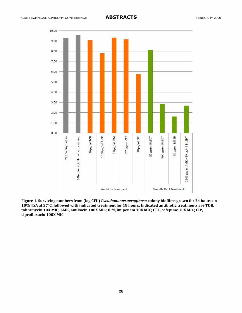
CBE TECHNICAL ADVISORY CONFERENCE ABSTRACTS FEBRUARY 2009
28
Figure 1. Surviving numbers from (log CFU) Pseudomonas aeruginosa colony biofilms grown for 24 hours on 10% TSA at 37°C, followed with indicated treatment for 18 hours. Indicated antibiotic treatments are TOB, tobramycin 10X MIC; AMK, amikacin 100X MIC; IPM, imipenem 10X MIC; CEF, cefepime 10X MIC; CIP, ciprofloxacin 100X MIC.
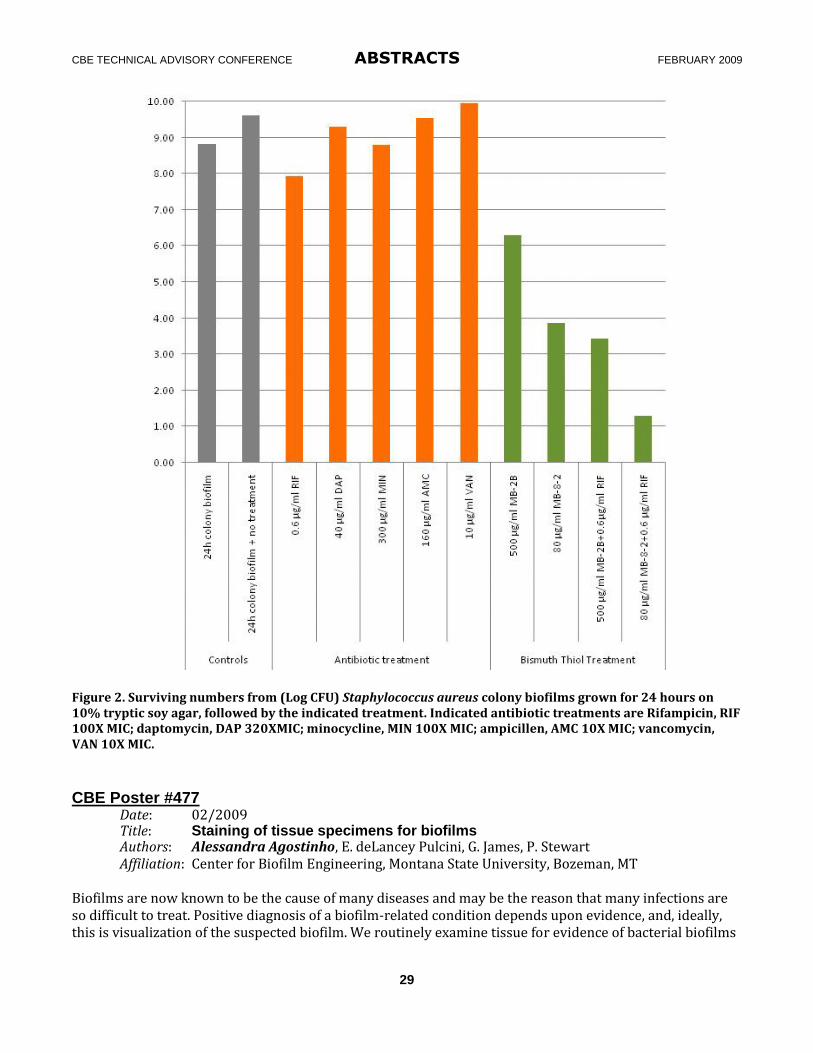
CBE TECHNICAL ADVISORY CONFERENCE ABSTRACTS FEBRUARY 2009
29
Figure 2. Surviving numbers from (Log CFU) Staphylococcus aureus colony biofilms grown for 24 hours on 10% tryptic soy agar, followed by the indicated treatment. Indicated antibiotic treatments are Rifampicin, RIF 100X MIC; daptomycin, DAP 320XMIC; minocycline, MIN 100X MIC; ampicillen, AMC 10X MIC; vancomycin, VAN 10X MIC.
CBE Poster #477 Date: 02/2009 Title: Staining of tissue specimens for biofilms Authors: Alessandra Agostinho, E. deLancey Pulcini, G. James, P. Stewart Affiliation: Center for Biofilm Engineering, Montana State University, Bozeman, MT Biofilms are now known to be the cause of many diseases and may be the reason that many infections are so difficult to treat. Positive diagnosis of a biofilm-related condition depends upon evidence, and, ideally, this is visualization of the suspected biofilm. We routinely examine tissue for evidence of bacterial biofilms
CBE TECHNICAL ADVISORY CONFERENCE ABSTRACTS FEBRUARY 2009
30
and the sample types have included: human lung tissue biopsies, excised chronic wound tissue, rat, mouse and rabbit tissues, sinus samples, and breast implants, among others. In all of these cases, the objective was to acquire confirmation of biofilm presence via confocal, fluorescent, or light microscopy. At present, there are very few fluorescent stains that are truly biofilm-specific; most also stain the supporting tissue, making visualization almost impossible. There are also sample-associated protocols that interfere with staining (for example, human tissue is generally fixed upon retrieval). We are investigating fluorescence methods for elucidating biofilms in tissue samples—in particular, new fluorescent stains and immunofluorescence methods. CBE Poster #478 Date: 02/2009 Title: High copper concentrations decrease the toxicity and sorption of lead and zinc
to the important biomining bacterium Acidithiobacillus caldus Authors: John E. Aston1, W.A. Apel2, B.D. Lee2, and B.M. Peyton1 Affiliation: 1Department of Chemical and Biological Engineering and Center for Biofilm Engineering, Montana State University, Bozeman, Montana 2Biological Systems Department, Idaho National Laboratory, Idaho Falls, Idaho Sponsors: Idaho National Laboratory, Biological Systems Division; MSU IGERT (Integrated
Graduate Education & Research Training); Montana EPSCoR Acidithiobacillus caldus is found in acidic environments (pH 1–4) at temperatures between 32 and 50°C, and is believed to play a role in the biomining of metals by removing inhibitory sulfur layers from mineral surfaces. In the current study, the toxicity and sorption of lead, zinc, and copper to At. caldus strain BC13 were examined. In each case, metals decreased the overall cell yield and specific growth rate during batch cultivation. Lead, zinc, and copper IC50 values (± 95% confidence intervals) were calculated to be 39 ± 4.5 µM, 180 ± 36 µM, and 2,370 ± 630 µM, respectively. When lead and zinc were mixed, their toxicity appeared to be additive. When copper was mixed with lead or zinc, the observed toxicity was significantly less than expected. Langmuir sorption isotherms show a relatively high affinity for copper, with maximum specific loading capacities of 253 ± 60 µmol g-1, 753 ± 164 µmol g-1, and 1,582 ± 277 µmol g-1 calculated for lead, zinc, and copper, respectively. In addition, the presence of copper in metal mixtures decreased lead and zinc sorption significantly. CBE Poster #479 Date: 02/2009 Title: Isolation and characterization of a heterotrophic nitrifying bacterium
from a reactor that simulates premise plumbing Authors: Gem D. Encarnacion, L.H. Leach, M.S. Rahman, B.S. Hisey and A.K. Camper Affiliation: Center for Biofilm Engineering, Montana State University, Bozeman, MT Sponsors: The American Water Works Association Research Foundation and Fulbright-Philippine
Agriculture Scholarship Program Chlorination in drinking water systems has been instrumental in decreasing water-borne diseases. However, free chlorine can interact with organics in the water to form regulated disinfection byproducts (DBPs). Therefore, many water utilities are switching to chloramines to reduce the levels of DBPs. Chloraminated systems can suffer from nitrification when free ammonia is converted to nitrite and then nitrate. Nitrification episodes in chloraminated drinking water distribution systems are thought to be caused by autotrophic nitrifiers. An observation has been that there is an increase in heterotrophic
CBE TECHNICAL ADVISORY CONFERENCE ABSTRACTS FEBRUARY 2009
31
bacterial abundance during nitrification, and our hypothesis is that heterotrophs may also be contributing to nitrification since it is known that these bacteria are capable of nitrification in many environments. In a reactor that simulates premise plumbing, nitrification was found to be affected minimally by chlorite treatment, suggesting a possible contribution to the nitrification process by organisms other than autotrophic bacteria. A heterotrophic nitrifying bacteria was isolated from the effluent of this reactor. The isolation of organisms such as the one in this study can provide a positive control for the design of a molecular method to track heterotrophic nitrification and thus possibly predict nitrification events in the DWDS. Likely gene targets for tracking heterotrophic nitrification include the heterotrophic ammonia monooxygenase (AMO) gene and the heterotrophic hydroxylamine oxidoreductase (HAO). CBE Poster #480 Date: 10/2008 Title: Characterization of sulfate reducing bacteria in constructed wetlands Authors: Jennifer L. Faulwetter, M.D. Burr, O.R. Stein, A.K. Camper Affiliation: Center for Biofilm Engineering, Montana State University, Bozeman, MT Sponsor: United States Department of Agriculture
The types, densities, and distributions of microbial communities present in constructed wetland (CW) environments are poorly understood. Information gaps exist with regard to microbial community structure and function and their relationship to plant species, season, and wastewater type. One functional group of particular interest in CW is sulfate reducing bacteria (SRB) because they can remove Chemical Oxygen Demand (COD) and Biochemical Oxygen Demand (BOD) in anoxic environments. Furthermore, the sulfide by-product of SRB activity can sequester heavy metals. SRB are critically important since they are the only known organisms to reduce sulfate to sulfide. The goal of this research was to characterize and identify the SRB present in a CW that had been previously used to treat simulated acid-mine wastewater. Samples were taken from an unplanted control CW, since it maintained the most anoxic conditions throughout the year. Samples were enriched for SRB using a variety of electron donors and the enrichment cultures were analyzed using molecular techniques to identify key members of the sulfate reducing community. SRB community composition appeared to be affected by the enrichment culture media used. Closest BLAST relatives of the SRB clones sequenced included Desulfobulbus rhabdoformis, Desulfomicrobium apsheronum, Desulfotomaculum nigrificans, and Desulfotomaculum alkaliphilum.
CBE Poster #481 Date: 02/2009 Title: Research support for designing a comprehensive biofilm efficacy test system Authors: Matthew Radons1, D.K. Walker1, A. Cunningham1, B. Warwood2, K. Cook3, D. Goeres1 Affiliation: 1Center for Biofilm Engineering, Montana State University, Bozeman, MT 2 BioSurface Technologies, Bozeman, MT; 3Department of Mechanical and Industrial
Engineering, Montana State University, Bozeman, MT
Developing anti-biofilm control strategies is a four-step process: 1) grow a relevant and repeatable biofilm in a biofilm reactor, 2) treat a mature biofilm with biocide or antibiotics, 3) remove a representative biofilm sample, and 4) analyze the sample for a quantitative and/or qualitative estimate of kill and/or removal as a result of the treatment. The multiple manipulations required in biofilm efficacy testing make the methods complex. Consequently, there is a need for a laboratory tool that would enable laboratory personnel to more efficiently treat, sample and analyze multiple biofilm reactor coupons simultaneously during biofilm

CBE TECHNICAL ADVISORY CONFERENCE ABSTRACTS FEBRUARY 2009
32
efficacy testing. A successful tool will not be complicated, expensive, or fragile. The tool will not bias the log density or log reduction results nor compromise the statistical hallmarks of repeatability, sensitivity and ruggedness required for standard methods. A successful tool will enable researchers to manipulate several coupons simultaneously during biofilm efficacy testing and ultimately make the experiment easier to conduct.
The objective of this poster is to describe the design process used to develop a biofilm efficacy test system and to provide an update on the research team’s progress. This project is a collaborative effort involving the Montana State University Department of Mechanical and Industrial Engineering, the Center for Biofilm Engineering and BioSurface Technologies in Bozeman. CBE Poster #482 Date: 02/2009 Title: Rapid taxonomic classification and analysis of complex microbial
communities using the phylochip microarray Authors: Seth D’Imperio1, J.G. Moberly1, Ari Staven1, E. Field1, M.R. VanEnglen1, S. Žižek2, D. Žagar2, B.M. Peyton1
Affiliation: 1Center for Biofilm Engineering, Montana State University, Bozeman, MT 2University of Ljubljana, Ljubljana Slovenia Sponsor: U.S. Department of Energy, Office of Science, Environmental Remediation Science Program The comprehensive characterization of mixed microbial communities presents problems of scale, cost, and time to researchers. A 16S-based microarray (PhyloChip) was utilized with the goal of rapidly cataloging the diversity of complex environmental microbial communities. This technology was applied to sediment samples from Lake Coeur d’Alene in Idaho that were impacted by upstream metal mining on the Coeur d’Alene River. A comparison of the microbial communities from this site and a non-metal-contaminated site in the same lake showed distinct differences at the Family level, in particular among the Enterobacteriales, Bacteroidetes, and Spirochetes. The technique was also applied to regions both up- and downstream of a mercury mine on the River Idrijca, Slovenia, and displayed significant phylogenetic differences between the sites sampled that were most pronounced in the Enterobacteriales and Pseudomonadales populations. Additionally, four strata of a simulated low level waste site at Idaho National Laboratory were investigated using the PhyloChip and revealed several significant variations within the Actinobacteria and Bacteroidetes populations. Furthermore, when combined with a dilution series prior to analysis on the PhyloChip, the method can be used to estimate relative cell densities for the OTUs that inhabit mixed communities. CBE Poster #483 Date: 02/2009 Title: Bacterially induced calcite precipitation and Sr co-precipitation under flow
conditions in porous media Authors: Logan Schultz, A. Mitchell, R. Gerlach, A. Cunningham Affiliation: Center for Biofilm Engineering, Montana State University, Bozeman, MT Sponsor: Department of Energy (Zero Emissions Research and Technology) The process of bacterially induced calcite precipitation has prospective applications in the improvement of geologic sequestration of carbon dioxide and in situ remediation of subsurface contaminants, including metals and radionuclides such as Strontium-90, a byproduct of uranium fission. In this study, ureolytic
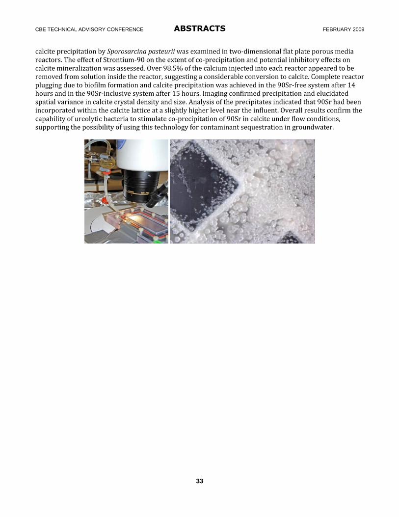
CBE TECHNICAL ADVISORY CONFERENCE ABSTRACTS FEBRUARY 2009
33
calcite precipitation by Sporosarcina pasteurii was examined in two-dimensional flat plate porous media reactors. The effect of Strontium-90 on the extent of co-precipitation and potential inhibitory effects on calcite mineralization was assessed. Over 98.5% of the calcium injected into each reactor appeared to be removed from solution inside the reactor, suggesting a considerable conversion to calcite. Complete reactor plugging due to biofilm formation and calcite precipitation was achieved in the 90Sr-free system after 14 hours and in the 90Sr-inclusive system after 15 hours. Imaging confirmed precipitation and elucidated spatial variance in calcite crystal density and size. Analysis of the precipitates indicated that 90Sr had been incorporated within the calcite lattice at a slightly higher level near the influent. Overall results confirm the capability of ureolytic bacteria to stimulate co-precipitation of 90Sr in calcite under flow conditions, supporting the possibility of using this technology for contaminant sequestration in groundwater.