casereport | smallbowel anovert ...acgcasereports.gi.org/files/2018/03/cg-cgcr180011.pdf · melena,...
TRANSCRIPT

CASE REPORT | SMALL BOWEL
An Overt, Obscure Gastrointestinal Bleed Caused by a PrimarySmall Bowel Fibroblastic Reticular Cell SarcomaStephanie L. Gold, MD1, Shirley Cohen-Mekelburg, MD2, Russell Rosenblatt, MD2,Jose Jessurun, MD3, Reem Sharaiha, MD2, Karim Halazun, MD4, and David Wan, MD2
1Department of Medicine, New York Presbyterian Weill Cornell Medical College, New York, NY2Department of Gastroenterology and Hepatology, New York PresbyterianWeill Cornell Medical College,New York, NY3Department of Pathology and Laboratory Medicine, New York Presbyterian Weill Cornell Medical College,New York, NY4Department of Surgery, New York Presbyterian Weill Cornell Medical College, New York, NY
ABSTRACTSmall bowel bleeding should be considered in patients who continue to bleed despite a negative upper endos-copy and colonoscopy. The differential diagnosis of small bowel bleeding can include infection, inflammatoryconditions, vascular malformations, and, rarely, malignancy. This report demonstrates a rare, primary, smallbowel, reticular cell sarcoma presenting as an overt gastrointestinal bleed. These tumors are difficult to diagnosebecause they are rarely seen on traditional cross-sectional imaging and can present with multiple synchronouslesions throughout the intestinal tract.
INTRODUCTIONGastrointestinal (GI) bleeding is a common inpatient condition, leading to over $2.5 billion spent annually on man-agement.1 It is estimated that there are roughly 300,000 hospitalizations for upper GI bleeding and 30,000 deathseach year in the United States.2 Workup of GI bleeding includes an esophagogastroduodenoscopy (EGD) and/orcolonoscopy, depending on the clinical presentation. However, 5–10% of GI bleeding results from a small bowelsource, and identifying these lesions often requires more advanced endoscopic techniques such as push entero-scopy, video capsule endoscopy (VCE), or balloon-assisted enteroscopy.3
CASE REPORTA 45-year-old man with a history of peripheral vascular disease and current tobacco use presented to the emer-gency department with leg pain and melena. Two weeks prior, he had presented to a nearby hospital with acuteleg ischemia and was found to be severely anemic. During his initial outside hospital admission, EGD revealedclean-based gastric ulcers, and colonoscopy revealed a pedunculated sigmoid polyp. Amputation of his ischemicleg was recommended at the outside hospital, but he subsequently left against medical advice.
On presentation to our emergency department, he was febrile to 38.2°C, hypotensive (blood pressure 97/57 mmHg), and tachycardic at 126 beats/min. Examination was remarkable for conjunctival pallor, a cold, pulseless rightleg, and dark, tarry stool in the rectal vault. Laboratory evaluation revealed hemoglobin 3.7 g/dL, white blood cellcount 27.8 � 103/mL, and platelet count 792 � 103/mL. The patient reported taking 800 mg ibuprofen 3 times dailyfor his leg pain. A non-contrast computed tomography (CT) of the abdomen and pelvis was unremarkable. Hereceived intravenous antibiotics, a proton pump inhibitor, and packed red blood cells, and his vitals normalized.
ACG Case Rep J 2018;5:e22. doi:10.14309/crj.2018.22. Published online: March 14, 2018.
Correspondence: Stephanie Lauren Gold, 1305 York Ave, 4th floor, New York, NY 10021 ([email protected]).
Copyright: © 2018 Gold et al. This work is licensed under a Creative Commons Attribution-NonCommercial-NoDerivatives 4.0 InternationalLicense. To view a copy of this license, visit http://creativecommons.org/licenses/by-nc-nd/4.0.
ACG Case Reports Journal / Volume 5 acgcasereports.gi.org 1
ACGCASE REPORTS JOURNAL
Push enteroscopy revealed an ulcerated jejunal lesion with anadherent clot (Figure 1). The clot was unroofed, and biopsieswere taken; hemostasis was achieved with epinephrine andbipolar cautery. Histologic examination of the specimenshowed few poorly differentiated malignant epithelioid cells.After the push enteroscopy, the patient had persistentmelena, requiring daily blood transfusions. A second pushenteroscopy revealed hemostasis of the original jejunallesion with active bleeding from a second, more distal jejunalpolypoid lesion, which was treated with a detachable snareand hemostatic clips (Figure 2).
Despite achieving hemostasis, the patient continued to havemelena and remained dependednt on transfusions. An explor-atory laparotomy revealed a large mass in the fourth part ofthe duodenum as well as two proximal ileal lesions, all ofwhich were resected (Figure 3). No additional masses werepalpated on intra-operative examination of the bowel.Pathologic examination of the resected specimen showed 3polypoid tumors with transmural invasion, the largest of whichmeasuring 2.4 � 2.3 � 1.1 cm. Histology revealed dyshesiverounded cells with abundant eosinophilic cytoplasm andcentral or peripheral nuclei with prominent nucleoli and
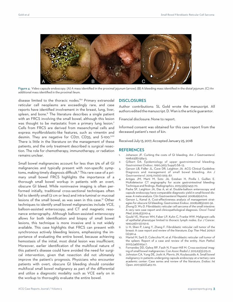
numerous mitotic figures. The tumor cells were immunoreac-tive for vimentin, fascin, and desmin. Other immunostainsruled out diagnoses of melanoma, lymphoma, interdigitatingand follicular dendritic cell tumors, epithelioid sarcoma, clearcell sarcoma, and GI stromal tumors. Based on these findings,a diagnosis of a high-grade fibroblastic reticular cell sarcoma(FRCS) was rendered (Figure 3). After surgery, the patientcontinued to have profuse melena. A capsule endoscopyshowed multiple actively bleeding lesions in the small bowel(Figure 4). As a result of continued bleeding and a daily trans-fusion requirement, he was referred to an inpatient hospicefacility and eventually passed away.
DISCUSSIONSmall bowel bleeding is a relatively uncommon cause of acuteGI bleeding but should be considered in patients with overtbleeding despite a negative EGD and colonoscopy. As perAmerican College of Gastroenterology guidelines, theworkup for an overt obscure GI bleed should first include asecond-look endoscopy.3 If this fails to identify the source ofbleed, a small bowel workup should follow, with the use ofVCE or push enteroscopy.3 Push enteroscopy is superior toVCE in detecting duodenal and proximal jejunal lesions andtherefore should be the first-line choice if proximal lesionsare suspected.3 Imaging techniques such as technetium-99m-labeled red blood cell studies can be used to identify anacute, brisk GI hemorrhage; however, this technology is notuniversally available and is ineffective with slower bleeds. CTangiography is more readily accessible and can noninvasivelyidentify extravasation of contrast and thus the location of anacute, brisk, GI bleed with a diagnostic yield of 10–40%.4-6
Common causes of small bowel bleeding include inflamma-tory bowel disease, vascular malformations including angi-oectasia and Dieulafoy’s lesions, ulcers, and a Meckel’s diver-ticulum. However, it is important to consider less commoncauses of small bowel bleeding, such as malignancy. We pres-ent a case of a primary small bowel FRCS presenting as anovert obscure GI bleed. To date, 19 cases of FRCS have beenreported, with only 1 case of this rare tumor presenting in thesmall bowel.7 The first documented cases of FRCS were pub-lished by Gould et al. in 1990 and described patients with
Figure 2. The second push enteroscopy showed a second jejnual poly-poid mass and active bleeding. Biopsies were taken and hemostasis wasachieved with a detachable snare and hemostatic clips.
Figure 1. (A) Push enteroscopy revealed an ulcerated jejunal mass withan adherent clot. (B) Biopsies were taken and hemostasis was achievedwith epinephrine and bipolar cautery.
A BFigure 3. (A) Segment of the small bowel from surgical resection show-ing 3 polypoid masses (arrows). (B) Small round cells with abundant eosin-ophilic cytoplasm, some with rhabdoid features.
Gold et al Small Bowel Fibroblastic Reticular Cell Sarcoma
ACG Case Reports Journal / Volume 5 acgcasereports.gi.org 2
disease limited to the thoracic nodes.8,9 Primary extranodalreticular cell neoplasms are exceedingly rare, and casereports have identified involvement in the breast, lung, liver,spleen, and bone.9 The literature describes a single patientwith an FRCS involving the small bowel, although this lesionwas thought to be metastatic from a primary lung lesion.7
Cells from FRCS are derived from mesenchymal cells andexpress myofibroblastic-like features, such as vimentin anddesmin. They are negative for CD21, CD35, and S-100.9,10
There is little in the literature on the management of thesepatients, and the only treatment described is surgical resec-tion. The role for chemotherapy, immunotherapy, or radiationremains unclear.
Small bowel malignancies account for less than 5% of all GImalignancies and typically present with non-specific symp-toms, making timely diagnosis difficult.11 This rare case of a pri-mary small bowel FRCS highlights the importance of athorough small bowel workup in patients with an overt,obscure GI bleed. While noninvasive imaging is often per-formed initially, traditional cross-sectional techniques oftenfail to identify small (2 cm or less) and intra-luminal or mucosallesions of the small bowel, as was seen in this case.12 Othertechniques to identify small bowel malignancies include VCE,balloon-assisted enteroscopy, and CT and magnetic reso-nance enterography. Although balloon-assisted enteroscopyallows for both identification and biopsy of small bowellesions, this technique is more invasive and is not widelyavailable. This case highlights that FRCS can present withsynchronous actively bleeding lesions, emphasizing the im-portance of evaluating the entire bowel, because achievinghemostasis of the initial, most distal lesion was insufficient.Moreover, earlier identification of the multifocal nature ofthis patient’s disease could have avoided the need for surgi-cal intervention, given that resection did not ultimatelyimprove the patient’s prognosis. Physicians who encounterpatients with overt, obscure GI bleeding should considermultifocal small bowel malignancy as part of the differentialand utilize a diagnostic modality such as VCE early on inthe workup to thoroughly evaluate the entire bowel.
DISCLOSURESAuthor contributions: SL Gold wrote the manuscript. Allauthors edited themanuscript.D.Wan is the articleguarantor.
Financial disclosure: None to report.
Informed consent was obtained for this case report from thedeceased patient’s next of kin.
ReceivedJuly 13,2017;AcceptedJanuary25, 2018
REFERENCES1. Johanson JF. Curbing the costs of GI bleeding. Am J Gastroenterol.
1998;93(8):1384-5.2. Gilbert DA. Epidemiology of upper gastrointestinal bleeding.
Gastrointest Endosc. 1990;36(5 Suppl):S8–13.3. Gerson LB, Fidler JL, Cave DR, Leighton JA. ACG Clinical Guideline:
Diagnosis and management of small bowel bleeding. Am JGastroenterol. 2015;110(9):1265–87.
4. Artigas JM, Marti M, Soto JA, Esteban H, Pinilla I, Guillen E.Multidetector CT angiography for acute gastrointestinal bleeding:Technique and findings. Radiographics. 2013;33(5):1453-70.
5. Pasha SF, Leighton JA, Das A, et al. Double-balloon enteroscopy andcapsule endoscopy have comparable diagnostic yield in small-bowel dis-ease: A meta-analysis.Clin Gastroenterol Hepatol. 2008;6(6):671-6.
6. Gerson L, Kamal A. Cost-effectiveness analysis of management strat-egies for obscure GI bleeding.Gastrointest Endosc. 2008;68(5):920-36.
7. Zhong D,Wu D. Fibroblastic reticular cell sarcoma of the small intestine:A very rare case report and clinicopathological diagnosis. Oncol TranslMed. 2016;2(3):110-4.
8. Gould VE, Warren WH, Faber LP, Kuhn C, Franke WW. Malignant cellsof epithelial phenotype limited to thoracic lymph nodes. Eur J Cancer.1990;26(11-12):1121-6.
9. Li H, Shen P, Liang Y, Zhang F. Fibroblastic reticular cell tumor of thebreast: A case report and review of the literature. Exp Ther Med. 2016;11(2):561-4.
10. Martel M, Sarli D, Colecchia M, et al. Fibroblastic reticular cell tumor ofthe spleen: Report of a case and review of the entity. Hum Pathol.2003;34(9):954-7.
11. Fernandes DD, Galwa RP, Fasih N, Fraser-Hill M. Cross-sectional imag-ing of small bowel malignancies.CanAssoc Radiol J. 2012;63(3):215-21.
12. Johnston CA, Yung DE, Joshi A, Plevris JN, Koulaouzidis A. Small bowelmalignancy in patients undergoing capsule endoscopy at a tertiary careacademic center: Case series and review of the literature. Endosc IntOpen. 2017;5(6):E463-70.
A B CFigure 4. Video capsule endoscopy. (A) A mass identified in the proximal jejunum (arrow). (B) A bleeding mass identified in the distal jejunum. (C) Anadditional mass identified in the proximal ileum.
Gold et al Small Bowel Fibroblastic Reticular Cell Sarcoma
ACG Case Reports Journal / Volume 5 acgcasereports.gi.org 3