case report traumatic diaphragmatic rupture, a diagnostic ... · however it may remain asymptomatic...
TRANSCRIPT
Case report
Open Access
Traumatic diaphragmatic rupture, a diagnostic dilemma in thepresence of eventration: a case reportReyaz Ahmad Lone1*, Mukand Lal Sharma1, Mahmood Wani1,Shiraz Rather1, Abdul Gani Ahangar1, Fouzia Rasool1,2, Mohd Akbar Bhat1,Abdul Majid Dar1, Guhlam Nabi Lone1, Shyam Singh1, Ishtiyaq Mir1,Shabir Shah1, Mubashir Shah1 and Mohd Lateef Wani1
Addresses: 1Department of Cardiothoracic and Vascular Surgery, Sheri Kashmir Institute of Medical Sciences, Soura, Srinagar, Kashmir,190011, India2Department of General Surgery, Shri Maharaja Hari Singh Medical College, Karanagr, MS, Srinagar, Kashmir, 190011, India
Email: RAL* - [email protected]; MLS - [email protected]; MW - [email protected]; SR - [email protected];AGA - [email protected]; FR - [email protected]; MAB - [email protected]; AMD - [email protected];GNL - [email protected]; SSi - [email protected]; IM - [email protected]; SSh - [email protected];MS - [email protected]; MLW - [email protected]
*Corresponding author
Received: 18 March 2009 Accepted: 10 June 2009 Published: 3 September 2009
Cases Journal 2009, 2:6615 doi: 10.4076/1757-1626-2-6615
This article is available from: http://casesjournal.com/casesjournal/article/view/6615
© 2009 Lone et al.; licensee Cases Network Ltd.This is an Open Access article distributed under the terms of the Creative Commons Attribution License (http://creativecommons.org/licenses/by/3.0),which permits unrestricted use, distribution, and reproduction in any medium, provided the original work is properly cited.
Abstract
Eventration of the diaphragm is the condition where the muscle is permanently elevated, but retainsits continuity and attachments to the costal margins. Traumatic diaphragmatic rupture is a recognizedconsequence of high velocity blunt trauma to the abdomen usually a result of motor vehicle accident.Multi-slice CT and Magnetic Resonance Imaging in the pre-operative evaluation of trauma patients,diaphragmatic rupture can be still overlooked if not evaluated with the fair degree of clinicalsuspicion, more so if it is associated with an eventration of diaphragm - as was in our case.
IntroductionTraumatic diaphragmatic rupture (TDR) is a recognizedconsequence of high velocity blunt trauma to the abdo-men usually a result of motor vehicle accident (MVA).It has been reported in upto 5% of thoracoabdominaltrauma patients. Early recognition of TDR is of utmostimportance. Despite the development and availabilityof new gadgets including multi-slice CT and MagneticResonance Imaging in the pre-operative evaluation oftrauma patients, diaphragmatic rupture can be still
overlooked if not evaluated with the fair degree of clinicalsuspicion, more so if it is associated with an eventration ofdiaphragm - as was in our case.
Case presentationA 4-year-old, male child of Indian origin was referred tothe emergency department of Sheri Kashmir Institute ofMedical Sciences, the tertiary care institute of the state witha history of being in a road traffic accident causing chestand abdominal trauma. At the peripheral centre where the
Page 1 of 4(page number not for citation purposes)
child was first seen resuscitation had been started and achest tube had been placed on the right side and 200 ml ofblood had been drained. The child was referred because ofhistory of eventration of the diaphragm on the same side.At admission the child was semiconscious, pale, hadfeatures of respiratory distress with a rate of 54 breaths perminute and was in shock with no palpable peripheralpulse and an unrecordable BP. Immediately the child wasintubated and further lines were set up to enableresuscitation. Secondary survey revealed bruise over theright chest, decreased air entry on the right side, centralplaced mediastinal structures and normal heart sounds.Rest of his examination was normal.
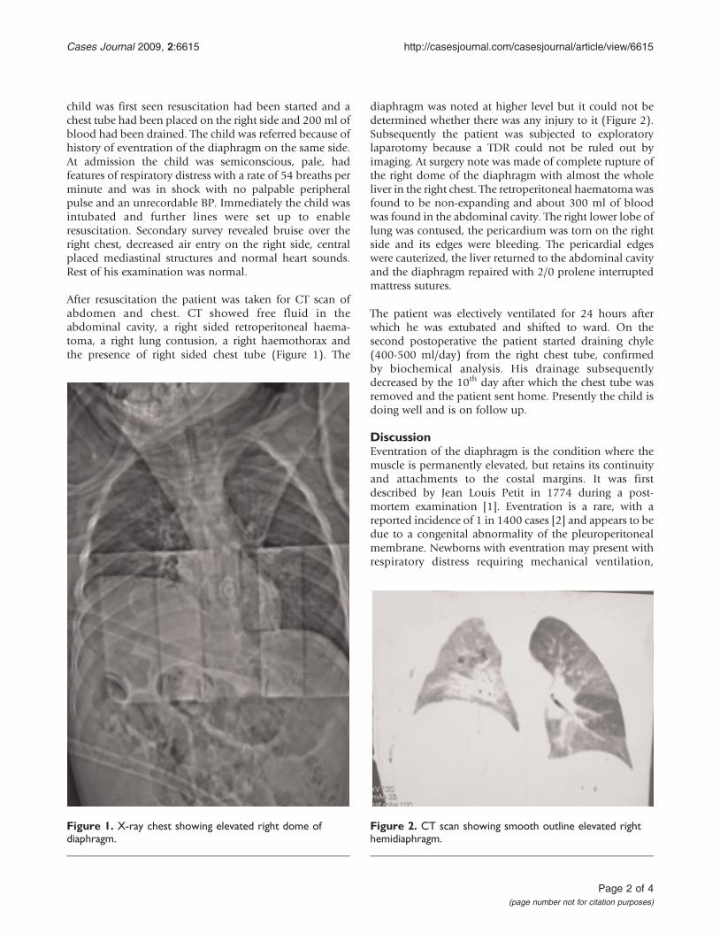
After resuscitation the patient was taken for CT scan ofabdomen and chest. CT showed free fluid in theabdominal cavity, a right sided retroperitoneal haema-toma, a right lung contusion, a right haemothorax andthe presence of right sided chest tube (Figure 1). The
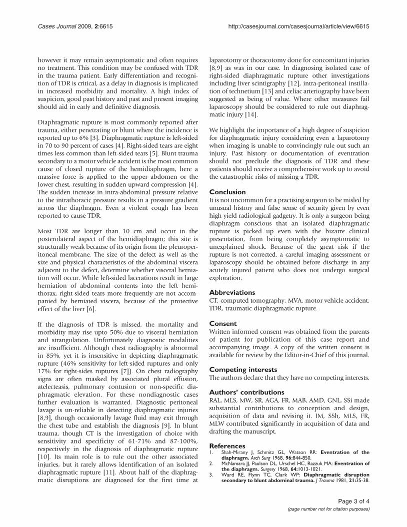
diaphragm was noted at higher level but it could not bedetermined whether there was any injury to it (Figure 2).Subsequently the patient was subjected to exploratorylaparotomy because a TDR could not be ruled out byimaging. At surgery note was made of complete rupture ofthe right dome of the diaphragm with almost the wholeliver in the right chest. The retroperitoneal haematomawasfound to be non-expanding and about 300 ml of bloodwas found in the abdominal cavity. The right lower lobe oflung was contused, the pericardium was torn on the rightside and its edges were bleeding. The pericardial edgeswere cauterized, the liver returned to the abdominal cavityand the diaphragm repaired with 2/0 prolene interruptedmattress sutures.
The patient was electively ventilated for 24 hours afterwhich he was extubated and shifted to ward. On thesecond postoperative the patient started draining chyle(400-500 ml/day) from the right chest tube, confirmedby biochemical analysis. His drainage subsequentlydecreased by the 10th day after which the chest tube wasremoved and the patient sent home. Presently the child isdoing well and is on follow up.
DiscussionEventration of the diaphragm is the condition where themuscle is permanently elevated, but retains its continuityand attachments to the costal margins. It was firstdescribed by Jean Louis Petit in 1774 during a post-mortem examination [1]. Eventration is a rare, with areported incidence of 1 in 1400 cases [2] and appears to bedue to a congenital abnormality of the pleuroperitonealmembrane. Newborns with eventration may present withrespiratory distress requiring mechanical ventilation,
Figure 1. X-ray chest showing elevated right dome ofdiaphragm.
Figure 2. CT scan showing smooth outline elevated righthemidiaphragm.
Page 2 of 4(page number not for citation purposes)
Cases Journal 2009, 2:6615 http://casesjournal.com/casesjournal/article/view/6615
however it may remain asymptomatic and often requiresno treatment. This condition may be confused with TDRin the trauma patient. Early differentiation and recogni-tion of TDR is critical, as a delay in diagnosis is implicatedin increased morbidity and mortality. A high index ofsuspicion, good past history and past and present imagingshould aid in early and definitive diagnosis.
Diaphragmatic rupture is most commonly reported aftertrauma, either penetrating or blunt where the incidence isreported up to 6% [3]. Diaphragmatic rupture is left-sidedin 70 to 90 percent of cases [4]. Right-sided tears are eighttimes less common than left-sided tears [5]. Blunt traumasecondary to a motor vehicle accident is the most commoncause of closed rupture of the hemidiaphragm, here amassive force is applied to the upper abdomen or thelower chest, resulting in sudden upward compression [4].The sudden increase in intra-abdominal pressure relativeto the intrathoracic pressure results in a pressure gradientacross the diaphragm. Even a violent cough has beenreported to cause TDR.
Most TDR are longer than 10 cm and occur in theposterolateral aspect of the hemidiaphragm; this site isstructurally weak because of its origin from the pleuroper-itoneal membrane. The size of the defect as well as thesize and physical characteristics of the abdominal visceraadjacent to the defect, determine whether visceral hernia-tion will occur. While left-sided lacerations result in largeherniation of abdominal contents into the left hemi-thorax, right-sided tears more frequently are not accom-panied by herniated viscera, because of the protectiveeffect of the liver [6].
If the diagnosis of TDR is missed, the mortality andmorbidity may rise upto 50% due to visceral herniationand strangulation. Unfortunately diagnostic modalitiesare insufficient. Although chest radiography is abnormalin 85%, yet it is insensitive in depicting diaphragmaticrupture (46% sensitivity for left-sided ruptures and only17% for right-sides ruptures [7]). On chest radiographysigns are often masked by associated plural effusion,atelecteasis, pulmonary contusion or non-specific dia-phragmatic elevation. For these nondiagnostic casesfurther evaluation is warranted. Diagnostic peritoneallavage is un-reliable in detecting diaphragmatic injuries[8,9], though occasionally lavage fluid may exit throughthe chest tube and establish the diagnosis [9]. In blunttrauma, though CT is the investigation of choice withsensitivity and specificity of 61-71% and 87-100%,respectively in the diagnosis of diaphragmatic rupture[10]. Its main role is to rule out the other associatedinjuries, but it rarely allows identification of an isolateddiaphragmatic rupture [11]. About half of the diaphrag-matic disruptions are diagnosed for the first time at
laparotomy or thoracotomy done for concomitant injuries[8,9] as was in our case. In diagnosing isolated case ofright-sided diaphragmatic rupture other investigationsincluding liver scintigraphy [12], intra-peritoneal instilla-tion of technetium [13] and celiac arteriography have beensuggested as being of value. Where other measures faillaparoscopy should be considered to rule out diaphrag-matic injury [14].
We highlight the importance of a high degree of suspicionfor diaphragmatic injury considering even a laparotomywhen imaging is unable to convincingly rule out such aninjury. Past history or documentation of eventrationshould not preclude the diagnosis of TDR and thesepatients should receive a comprehensive work up to avoidthe catastrophic risks of missing a TDR.
ConclusionIt is not uncommon for a practising surgeon to bemisled byunusual history and false sense of security given by evenhigh yield radiological gadgetry. It is only a surgeon beingdiaphragm conscious that an isolated diaphragmaticrupture is picked up even with the bizarre clinicalpresentation, from being completely asymptomatic tounexplained shock. Because of the great risk if therupture is not corrected, a careful imaging assessment orlaparoscopy should be obtained before discharge in anyacutely injured patient who does not undergo surgicalexploration.
AbbreviationsCT, computed tomography; MVA, motor vehicle accident;TDR, traumatic diaphragmatic rupture.
ConsentWritten informed consent was obtained from the parentsof patient for publication of this case report andaccompanying image. A copy of the written consent isavailable for review by the Editor-in-Chief of this journal.
Competing interestsThe authors declare that they have no competing interests.
Authors’ contributionsRAL, MLS, MW, SR, AGA, FR, MAB, AMD, GNL, SSi madesubstantial contributions to conception and design,acquisition of data and revising it. IM, SSh, MLS, FR,MLW contributed significantly in acquisition of data anddrafting the manuscript.
References1. Shah-Mirany J, Schmitz GL, Watson RR: Eventration of the
diaphragm. Arch Surg 1968, 96:844-850.2. McNamara JJ, Paulson DL, Urschel HC, Razzuk MA: Eventration of
the diaphragm. Surgery 1968, 64:1013-1021.3. Ward RE, Flynn TC, Clark WP: Diaphragmatic disruption
secondary to blunt abdominal trauma. J Trauma 1981, 21:35-38.
Page 3 of 4(page number not for citation purposes)
Cases Journal 2009, 2:6615 http://casesjournal.com/casesjournal/article/view/6615
4. Dietrich P, Alsofrom G: The diaphragm. In Radiology: diagnosis,imaging, intervention. Volume 1. 1st edition. Edited by Taveras JM,Ferrucci JT, Elliott LP. Philadelphia: Lippincott; 1990:1-10.
5. Pailero PC, Trastek VF, Tayne WS: Oesophagus and Diaphrag-matic hernia. In Principles of surgery. 5th edition. Edited bySchwartz SI, Shires GT, Spencer FC. New York: McGraw-Hill;1989:1129-1132.
6. Harris JH Jr, Harris WH: Chest. In The radiology of emergencymedicine. 24th edition. Edited by Harris JH Jr, Harris WH. Baltimore:Williams & Wilkins; 1981:422-427.
7. Gelman R, Mirvis SE, Gens D: Diaphragmatic rupture due toblunt trauma: sensitivity of plain chest radiographs. AJR 1991,156:51-57.
8. Beal L, McKennan M: Blunt diaphragm rupture. Arch Surg 1988,123:828.
9. Cox EF, Siegel JH: Blunt trauma to the abdomen. In Trauma:Emergency Surgery and Critical Care. Volume 107. Edited by Seigel JH.New York: Churchill Livingstone; 1987:883-910.
10. Killeen KL, Mirvis SE, Shanmuganathan K: Helical CT of diaphrag-matic rupture caused by blunt trauma. AJR 1993, 173:1611-1616.
11. Sola JE, Mattei P, Pegoli W Jr, Paidas CN: Rupture of rightdiaphragm following blunt trauma in an infant. A case report.J Trauma 1994, 36:417.
12. Blumenthal DH, Raghu G, Rudd TG, Herman CM: Diagnosis ofright hemidiaphragmatic rupture by liver scintigraphy.J Trauma 1984, 24:536.
13. Halldorsson A, Esser MJ, Rappaport W, Valente J, Hunter G,McIntyre K: A new method of diagnosing diaphragmatic injuryusing intraperitoneal technetium. J Trauma 1992, 33:140.
14. Adamthwaite DN: Traumatic diaphragmatic hernia: a newindication for laparoscopy. Br J Surg 1984, 71:315.
Do you have a case to share?
Submit your case report today• Rapid peer review• Fast publication• PubMed indexing• Inclusion in Cases Database
Any patient, any case, can teach ussomething
www.casesnetwork.com
Page 4 of 4(page number not for citation purposes)
Cases Journal 2009, 2:6615 http://casesjournal.com/casesjournal/article/view/6615