case report three-year follow-up results for non-surgical ... · three-year follow-up results for...
TRANSCRIPT
Int J Clin Exp Pathol 2014;7(6):3338-3346www.ijcep.com /ISSN:1936-2625/IJCEP0000342
Case ReportThree-year follow-up results for non-surgical root canal therapy of idiopathic external root resorption on a maxillary canine with MTA: a case report
Zheng Huang1, Li-Li Chen1, Cong-Yi Wang2, Lin Dai3, Bo Cheng1, Jun Sun3, Jun Sun4
1Department of Stomatology, Union Hospital, Tongji Medical College, Huazhong University of Science and Technology, 1277 Jiefang Ave, Wuhan, Hubei, 430022, China; 2The Center for Biomedical Research, Tongji Hospital, Tongji Medical College, Huazhong University of Science and Technology, 1095 Jiefang Ave, Wuhan, Hubei, 430030, China; 3Department of Stomatology, The First Hospital of Wuhan, Wuhan, Hubei, 430022, China; 4Department of Restorative Dentistry, Indiana University School of Dentistry, Indianapolis, IN 46202, USA
Received March 26, 2014; Accepted May 20, 2014; Epub May 15, 2014; Published June 1, 2014
Abstract: External root resorption (ERR) is an uncommon and intractable disease. Treatment alternatives are case-dependant and aim for the repair of the resorptive lesion and long-term retention of the tooth. A forty-year-old Asian female was diagnosed with idiopathic ERR on tooth #11 (the left maxillary canine) by CBCT. Non-surgical root canal therapy was completed with the aid of an operating microscope. The apical third of the root canal was filled with warm gutta-percha and the resorption defect was filled with mineral trioxide aggregate (MTA). The periapical radio-graphs were taken immediately after operation, one-month follow-up, six-month follow-up and three-year follow-up, respectively. Clinically, the canine was asymptomatic, and no evidence of any further resorption was found. The six-month follow-up radiograph showed initial healing of the bony lesion, while the three-year follow-up radiograph manifested almost complete healing. MTA can be a superior material to be successfully used in the non-surgical treatment of ERR. CBCT is very useful for evaluating the true nature and severity of absorption lesions in root re-sorption. It is the first complete case report from China about non-surgical treatment of severe ERR along with a relatively long term follow-up.
Keywords: External root resorption, non-surgical root canal therapy, MTA, CBCT, canine
Introduction
The root resorption associated with primary teeth is physiological and desirable because it results in exfoliation of the teeth, thereby allow-ing eruption of the permanent teeth. However, root resorption (either internal or external) of permanent dentition is usually pathological and undesirable because it might result in irre-versible damage and/or eventual tooth loss.
The hard tissues (dentin, cementum, and enamel) of permanent teeth do not normally undergo resorption. External root resorption (ERR) occurs when the periodontal ligament or cementum on the root surface are either dam-aged or removed [1]. Although the exact cause of ERR is poorly understood, it is generally accepted that the etiology of different types of ERR requires two phases: mechanical or chem-
ical injury to the protective tissues and stimula-tion by inflammation or pressure [2].
The clinical signs of ERR depend mainly on the extent of the resorption. The usual asymptom-atic nature of the early stage of ERR can be explained by the minimal pulp involvement due to protection from the predentin layer. However, some advanced cases develop symptoms of pulp disease when this protective layer is destroyed and the pulp is exposed to oral micro-organisms through the resorptive defect. Therefore, many ERR patients delay treatment because of absence of symptoms and eventu-ally lose the affected tooth.
This paper presents a rare case of an idiopathic ERR of tooth #11 (the left maxillary canine) treated by non-surgical root canal therapy and a mineral trioxide aggregate (MTA) perforation

Non-surgical therapy of external root resorption with MTA
3339 Int J Clin Exp Pathol 2014;7(6):3338-3346
repair, showing healing at a three-year follow-up. It is the first complete case report from China about non-surgical treatment of severe ERR with a relatively long term follow-up.
Case report
Medical history
A forty-year-old Asian female was referred by a private prosthodontist to the Endodontics De-
partment of Wuhan Union Hospital. The patient presented to the private dentist two months earlier and was diagnosed with severe tetracy-cline-stained teeth. She had no other discom-fort or chief complaint except for unfavorable tooth color. Panoramic radiographic survey was performed before therapy and no obvious path-ological change was observed (Figure 1A). At the private dentist, she had twenty zirconium dioxide veneers/crowns placed from tooth #4
Figure 1. A: Panoramic radiographic survey was performed before therapy. An irregular radiolucency localized in the mesial mid-third root of tooth #11 (pointed by the white arrow) was not observed until referring and reviewing this radiograph. It was not easy to distinguish from the panoramic radiograph. B: A periapical radiograph of tooth #11 was performed before opening the pulp cavity. Unfortunately the radiolucent area was still not recognized by the private dentist. C: Measuring the working length during the process of root canal therapy on tooth #11. D: While taking the periapical radiograph of tooth #12, an extensive and predominant resorption area in the mesial mid-third root of tooth #11 was found by the private dentist.

Non-surgical therapy of external root resorption with MTA
3340 Int J Clin Exp Pathol 2014;7(6):3338-3346
to tooth #13 in the maxilla and from tooth #20 to tooth #29 in the mandible. One month after the permanent cementation of the veneers/crowns, the patient complained of paroxysmal, sharp, spontaneous pain in her left arch, but she couldn’t pinpoint the exact tooth. The pain was not relieved by antibiotics and analgesics. Intraoral examination revealed that tooth #11 responded positively to thermal testing (hot stimulus) and was sensitive to percussion. The private dentist took a periapical radiograph of tooth #11 and did not find any abnormal signs (Figure 1B). He then anesthetized tooth #11 and the pain disappeared immediately. Symptomatic irreversible pulpitis with symp-tomatic apical periodontitis of tooth #11 was diagnosed. An access cavity was prepared and root canal preparation was completed (Figure 1C). The chief complaint disappeared for two days and the patient presented almost the same chief complaint again in both right and left arches during the following days. So the pri-vate dentist diagnosed tooth #20, tooth #28 and tooth #12 with symptomatic irreversible pulpitis sequentially using the same diagnosis tests and rendered the same therapy without filling the root canal of tooth #11. While taking the periapical radiograph of tooth #12, the pri-vate dentist was surprised to find an extensive and predominant resorption area in tooth #11 (Figure 1D). After removing the temporary seal-ing, severe bleeding was found in the root canal of tooth #11. The private dentist couldn’t understand and manage this disorder and
transferred this patient to our department with an iodoform-zinc oxide paste filling in the root canal of tooth #11.
The patient presented to our department with non-contributory systemic medical history. Her oral medical history excluded tooth trauma, orthodontic treatment, bleaching or periodon-tal operation on tooth #11. The patient had no obvious discomfort while being referred to our clinic. She had a strong desire to preserve this tooth and was reluctant to remove the crown. Due to the limited space, this paper will not describe and discuss the treatment of the other teeth except tooth #11.
Clinical features
The extraoral examination revealed negative findings. The intraoral examination revealed a zirconium dioxide veneer and a palatal access cavity restored with a temporary restoration on tooth #11. The tooth was slightly sensitive both to percussion and palpation but without mobil-ity, obvious swelling, sinus tract or periodontal pockets. No special exception was found con-sidering the whole oral cavity (tooth #20, tooth #28 and tooth #12 were treated by the private dentist).
Reviewing the former panoramic radiograph (Figure 1A) and the periapical radiograph of tooth #11 (Figure 1B), there was an irregular, radiolucency localized in the mesial mid-third
Figure 2. A: The new periapical radiograph of tooth #11 revealed that there was a clear radiolucent area situated in the mesial mid-third root of tooth #11 and was communicating with the pulp. A widened periodontal ligament space at the periapical area was also observed (pointed by white arrow). B: Horizontal section view of CBCT image: extensive external resorption area in the lingual mid-third root of tooth #11 which was communicating with the pulp. C: Coronal section view of CBCT image: extensive external resorption area in the mesial mid-third root of tooth #11 which was communicating with the pulp.
Non-surgical therapy of external root resorption with MTA
3341 Int J Clin Exp Pathol 2014;7(6):3338-3346
root. A new periapical radiograph was obtained and revealed a clear radiolucent area situated in the mesial mid-third root of tooth #11, com-municating with the pulp. A widened periodon-tal ligament space at the periapical area was also observed (Figure 2A). In order to know the nature of this lesion, such as internal or exter-nal resorption and the extent of the lesion, a cone beam computed tomography (CBCT; NewTom™ VGi, QR S.r.l., Verona, Italy) image of tooth #11 was obtained, which showed an extensive external resorption lesion in the mesial and lingual mid-third root of tooth #11, communicating with the pulp (Figure 2B, 2C).
Two diagnoses of tooth #11 were made: (1) idiopathic ERR (communicating with the pulp) and (2) symptomatic apical periodontitis.
Treatment plan
Three alternative treatment options were given to the patient. The first was non-surgical root canal treatment and perforation repair with MTA from the inside of the root canal. The sec-ond was endodontic surgery and repair from the outside of the root canal. The third was extraction and replacement with an implant. Benefits and risks of each treatment plan were fully discussed with the patient, and the first option was chose along with informed consent.
Management
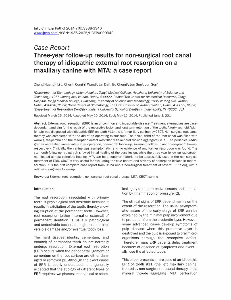
During her first visit, after local anesthesia (Primacaine® Adrenaline, Acteon China, Beijing, China) and rubber dam (Hygenic® Dental Dam, Coltène/Whaledent Inc., Cuyahoga Falls, OH, USA) isolation, a large amount of granulation tissue was removed and the resorption defect in the mid-third root canal was observed under dental microscope (OPMI® pico, Zeiss, Oberkochen, Germany) (Figure 3A). The work-ing length was verified by apex locator (SybronEndo, Glendora, CA, USA). A ProTaper universal® hand file (Dentsply Maillefer, Ballaigues, Switzerland) was used to a size F3 file to finish the preparation. Sodium hypochlo-rite (5.25%) was used as the irrigant with pas-sive ultrasonic activation (Varios 750™ Lux, NSK NaKanishi Inc., Tochigi, Japan). Vitapex (Neo Dental Chemical Products, Tokyo, Japan) was employed as an intracanal medication when there was no excessive bleeding and then the access cavity was sealed temporarily (Caviton, GC corporation, Tokyo, Japan) (Figure 3B).
On her second visit 12 days later, the symp-toms of slight sensitivity to percussion and pal-pation had been resolved. Tooth #11 manifest-ed no evidence of mobility, swelling, sinus tract or periodontal pockets. The periapical radio-
Figure 3. A: The resorption section in the mid-third root canal could be observed under microscope (outline trac-ing of features). B: Vitapex was used as an intracanal medication when there was no excessive bleeding and then the access cavity was sealed temporarily. C: The periapical radiograph displayed the resorption of excess Vitapex. Vitapex was injected again after thorough ultrasonic irrigation (2.5% sodium hypochlorite and 0.9% sodium chloride solution) to remove the remaining old medicament.
Non-surgical therapy of external root resorption with MTA
3342 Int J Clin Exp Pathol 2014;7(6):3338-3346
graph displayed the resorption of excess Vitapex in periapical and defect regions (Figure 3C). Vitapex was injected again after thorough ultrasonic irrigation (2.5% sodium hypochlorite and 0.9% sodium chloride solution) to remove the remaining old medicament.
On the third visit after 21 days, tooth #11 was asymptomatic. After local anesthesia and rub-ber dam isolation, the Vitapex was completely removed by K files (Dentsply, Konstanz, Germany) and ultrasonic irrigation. When there was no obvious bleeding, the root canal was
Follow-up
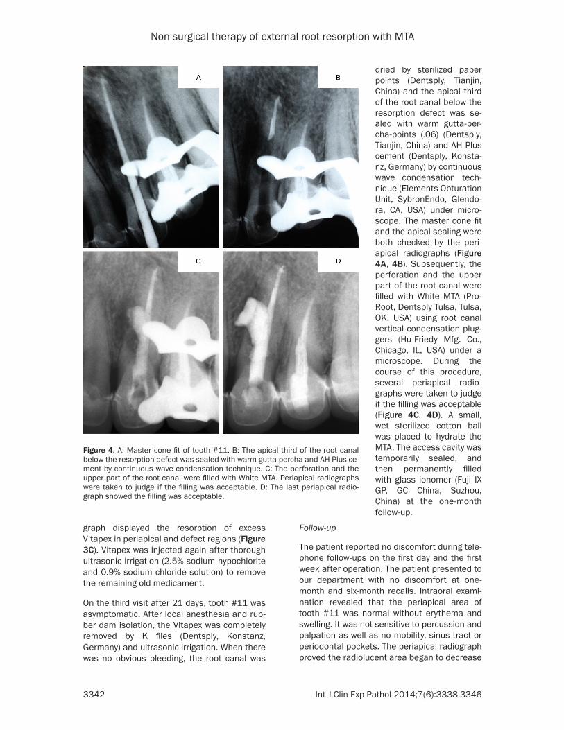
The patient reported no discomfort during tele-phone follow-ups on the first day and the first week after operation. The patient presented to our department with no discomfort at one-month and six-month recalls. Intraoral exami-nation revealed that the periapical area of tooth #11 was normal without erythema and swelling. It was not sensitive to percussion and palpation as well as no mobility, sinus tract or periodontal pockets. The periapical radiograph proved the radiolucent area began to decrease
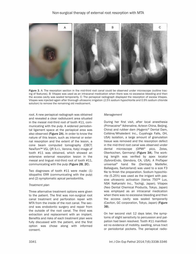
Figure 4. A: Master cone fit of tooth #11. B: The apical third of the root canal below the resorption defect was sealed with warm gutta-percha and AH Plus ce-ment by continuous wave condensation technique. C: The perforation and the upper part of the root canal were filled with White MTA. Periapical radiographs were taken to judge if the filling was acceptable. D: The last periapical radio-graph showed the filling was acceptable.
dried by sterilized paper points (Dentsply, Tianjin, China) and the apical third of the root canal below the resorption defect was se- aled with warm gutta-per-cha-points (.06) (Dentsply, Tianjin, China) and AH Plus cement (Dentsply, Konsta- nz, Germany) by continuous wave condensation tech-nique (Elements Obturation Unit, SybronEndo, Glendo- ra, CA, USA) under micro-scope. The master cone fit and the apical sealing were both checked by the peri-apical radiographs (Figure 4A, 4B). Subsequently, the perforation and the upper part of the root canal were filled with White MTA (Pro-Root, Dentsply Tulsa, Tulsa, OK, USA) using root canal vertical condensation plug-gers (Hu-Friedy Mfg. Co., Chicago, IL, USA) under a microscope. During the course of this procedure, several periapical radio-graphs were taken to judge if the filling was acceptable (Figure 4C, 4D). A small, wet sterilized cotton ball was placed to hydrate the MTA. The access cavity was temporarily sealed, and then permanently filled with glass ionomer (Fuji IX GP, GC China, Suzhou, China) at the one-month follow-up.
Non-surgical therapy of external root resorption with MTA
3343 Int J Clin Exp Pathol 2014;7(6):3338-3346
even early at the six-month recall. The filling material was intact (Figure 5A, 5B). There was no new external root resorption.
The patient could be reached only by e-mail after three-year follow-up since she was abroad. The patient said tooth #11 had good function and no discomfort. The scanning copy of her three-year review periapical radiograph was asked to be sent by e-mail. It showed encourag-ing results, with almost complete healing of the resorption region, and the filling material was intact. The periapical radiolucency was also resolved. The cortical bone forming the socket of tooth #11 and the interdental alveolar crest bone were clear (Figure 5C).
Discussion
External root resorption is a particularly frus-trating type of dental abnormality for both patients and practitioners because there is no plausible explanation for the condition and no predictable treatment [3]. This manuscript, along with other publications [4-7], can further our understanding of this disease process and its treatment.
Etiology
ERR may occur on one tooth or on several teeth and it may be caused by trauma, tooth re-implantation, orthodontic treatment, internal bleaching, periodontitis or previous periodontal
surgery, pressure from adjacent unerupted teeth, cysts, tumors, or by stimuli from a necrot-ic dental pulp [8]. ERR may also occur as a result of systemic disease and endocrine disor-ders, such as hyperparathyroidism, Paget’s dis-ease, calcinosis, Gaucher’s disease and Turner’s syndrome as well as after radiation therapy [9]. There are also some rare ERRs of unknown cause, usually called “idiopathic” [4].
In this case report, the ERR of tooth #11 was already present, but not immediately recog-nized by the private dentist. The possible rea-son why the resorption occurred on the mesial part of the root of tooth #11 is that this part is the site of fusion of the globular and the maxil-lary process during development and the rem-nants of vestigial structures (enclaved epithi-lum) might cause some oral diseases [10]. In addition, the apical periodontitis from the res-toration process may exacerbate the existed lesion. Therefore the diagnosis could be “idio-pathic ERR” according to the patient’s dental history and clinical manifestations.
Imaging diagnosis
The private dentist didn’t recognize the lesion on tooth #11 on the pre-operative panoramic radiograph and the first periapical radiograph, which indicates the difficulties in the detection of root resorption through routine images [11, 12]. In addition, the differential diagnosis for internal and external root resorption with the
Figure 5. A: One-month X-ray: the resorption region stopped enlarging. B: Six-month X-ray: the radiolucent area be-gan to decrease (pointed by the white arrow). The filling material was intact. C: Three-year X-ray: it showed almost complete healing of the resorption region and the filling material was intact. The periapical radiolucency was re-solved. The cortical bone forming the socket of tooth #11 and the interdental alveolar crest bone were clear.

Non-surgical therapy of external root resorption with MTA
3344 Int J Clin Exp Pathol 2014;7(6):3338-3346
common imaging technology is also a formida-ble challenge. Gartner AH et al [13] suggested that it often requires two periapical radiographs to make the differential diagnosis, one taken perpendicular to the tooth, and the other one taken from a mesial angle. This technique may be helpful to a certain degree; however, the periapical radiographs could not provide the three-dimensional location and determine the true size of the resorption lesions, which is an important information [14].
Cone beam computed tomography (CBCT) tech-nology has been specifically designed to pro-duce three-dimensional scans of the maxillo-facial skeleton. One of CBCT’s major advantages over common computed tomography (CT) scan-ners is the reduction in radiation exposure. CBCT has been successfully used for evaluat-ing the true nature and severity of resorption lesions in case reports [15]. In this case, CBCT provided more details to facilitate the diagno-sis, and to educate the patient and manage the defect.
An in-vitro study revealed that the features essential for the differential diagnosis of root resorption were not apparent in conventional radiographs, however, clearly visible in the micro-CT reconstructions [16]. Thus, the more digital simulation studies will provide better understanding about the nature of ERR diseases[17].
Treatment
Some articles suggested various treatment options depending on the etiology and type of ERR such as root canal therapy alone, peri-odontal surgery alone, root canal therapy plus periodontal surgery or extracting the tooth and implantation [5]. However, a recent systematic review illustrates that there is no reliable source of evidence regarding the most appropriate means of treating ERR. Moreover, it reveals that in most of the cases, the treatment alter-native is case-dependent and related to the cli-nician’s experience or an expert’s opinion [6].
Therefore, the choice of treatment by root canal therapy alone in this case also depends on a complete communication with the patient com-bining the patient’s condition and trust with the author’s experience and confidence. There are several successful case reports about root resorption by root canal therapy alone from
other countries to support this choice [18-20]. The main reasons to suggest preserving the tooth by root canal therapy alone are due to that the canine, because of its arch position, serves as a major support of facial muscles and keeps the overall vertical dimension of the face intact [21]. The patient had a healthy oral hygiene and a long root canine with good peri-odontal condition. The resorption area was in the mid-third root without communication with the oral cavity. All of these demonstrated the necessity and probability to preserve this tooth. Another important factor is that the patient was satisfied with the zirconium dioxide veneers. She may encounter gingival recession potential after the periodontal flap elevation to repair the perforation surgically. Also, the root canal access had been established when the patient was referred to us, and the patient was young, healthy and had a strong desire to preserve this tooth.
As aforementioned, ERR can be stimulated by inflammation [2] and, as a result, infection con-trol is the key to success. In this case, rubber dam isolation, completely removing granulation tissue, and ultrasonic cleaning with 5.25% sodi-um hypochlorite were all very helpful in infec-tion control. The use of Vitapex as intracanal medication to control the infection for ERR is also supported by Bhat SS [22], who presented a case report of infection external root resorp-tion on maxillary central incisor with a thirteen-month clinical and radiographic follow-up.
MTA, a commonly used repair material in end-odontics, has many excellent properties such as biocompatibility, good sealing ability, and the ability to inhibit bacteria. The most impor-tant reason to choose MTA as the permanent repair material in this case is because it has a capacity to promote hard tissue formation and to inhibit osteoclastic bone resorption [7]. The excellent three-year follow-up radiograph result is the best testimony for MTA (Figure 5C). In short, with careful clinical procedures, proper disinfectant and superior repair material, suc-cessful non-surgical treatment for severe ERR is possible.
Prognosis
By the three-year follow-up, the resorption region had almost completely healed and the filling material in the root canal was intact,

Non-surgical therapy of external root resorption with MTA
3345 Int J Clin Exp Pathol 2014;7(6):3338-3346
which demonstrates a favorable clinical out-come. From the limited available knowledge, the longest observation time about ERR is a ten-year observation [23]. The authors plan to follow this case in the future as long as possible.
Conclusions
In summary, endodontists should have confi-dence in treating such extensive ERR disease with careful clinical procedures, complete con-trol of infection and superior filling material such as MTA. With proper treatment, the tooth may be preserved for a long time. CBCT is an effective technique in diagnosing and choosing treatment plans of ERR for both dentists and patients.
Acknowledgements
This work was supported by grant from the National Natural Science Foundation of China (No. 81370405, to Bo Cheng).
Disclosure of conflict of interest
None to declare.
Address correspondence to: Dr. Jun Sun, Depart- ment of Restorative Dentistry, Indiana University School of Dentistry, Indianapolis, IN 46202, USA. Tel: 317-274-2126; Fax: 317-278-2818; E-mail: [email protected]; Dr. Bo Cheng, Department of Sto- matology, Union Hospital, Tongji Medical College, Huazhong University of Science and Technology, Wuhan, Hubei 430022, China. Tel: 86-13507- 190986; E-mail: [email protected]
References
[1] Lambrechts P and Vanhoorebeeck B. Root re-sorption. Rev Belge Med Dent 1992; 47: 54-75.
[2] Fuss Z, Tsesis I and Lin S. Root resorption: di-agnosis, classification and treatment choices based on stimulation factors. Dent Traumatol 2003; 19: 175-182.
[3] Regezi JA, Sciubba JJ and Jordan RCK. Oral Pa-thology (Clinical Pathologic Correlations). In: Dolan J and Nebel J, eds. Abnormalities of Teeth. St. Louis: Saunders; 2003. pp. 374-375.
[4] Heithersay GS. Management of tooth resorp-tion. Aust Dent J 2007; 52: S105-S121.
[5] Segal GR, Schiffman PH and Tuncay OC. Meta-analysis of the treatment-related factors of ex-ternal apical root resorption. Orthod Craniofac Res 2004; 7: 71-78.
[6] Ahangari Z, Nasser M, Mahdian M, Fedorowicz Z and Marchesan MA. Interventions for the management of external root resorption. Co-chrane Database Syst Rev 2010; 16: CD008003.
[7] Hashiguchi D, Fukushima H, Yasuda H, Masu-da W, Tomikawa M, Morikawa K, Maki K and Jimi E. Mineral trioxide aggregate inhibits os-teoclastic bone resorption. J Dent Res 2011; 90: 912-917.
[8] Rathe F, Nölken R, Deimling D and Ratka-Krüger P. External root resorption. Schweiz Monatsschr Zahnmed 2006; 116: 245-253.
[9] Carrotte P. Endodontics: Part 9. Calcium hy-droxide, root resorption, endo-perio lesions. Br Dent J 2004; 197: 735-743.
[10] Rushton MA, Cooke BED and Duckworth R. Oral Histopathology. London: E. & S. Living-stone; 1970. pp. 189.
[11] Patel S and Ford TP. Is the resorption external or internal? Dent Update 2007; 34: 218-220, 222, 224-226, 229.
[12] Chapnick L. External root resorption: an experi-mental radiographic evaluation. Oral Surg Oral Med Oral Pathol 1989; 67: 578-582.
[13] Gartner AH, Mack T, Somerlott RG and Walsh LC. Differential diagnosis of internal and exter-nal root resorption. J Endod Nove 1976; 2: 329-334.
[14] Kim E, Kim KD, Roh BD, Cho YS and Lee SJ. Computed tomography as a diagnostic aid for extracanal invasive resorption. J Endod 2003; 29: 463-465.
[15] Patel S and Dawood A. The use of cone beam computed tomography in the management of external cervical resorption lesions. Int Endod J 2007; 40: 730-737.
[16] Luso S and Luder HU. Resorption pattern and radiographic diagnosis of invasive cervical re-sorption. A correlative micro CT, scanning elec-tron and light microscopic evaluation of a case series. Schweiz Monatsschr Zahnmed 2012; 122: 914-930.
[17] Huang Z and Chen Z. Three-dimensional finite element modeling of a maxillary premolar tooth based on the micro-CT scanning: a de-tailed description. J Huazhong Univ Sci Techno-log Med Sci 2013; 33: 775-779.
[18] Oktem ZB, Cetinbaş T, Ozer L and Sönmez H. Treatment of aggressive external root resorp-tion with calcium hydroxide medicaments: a case report. Dent Traumatol 2009; 25: 527-531.
[19] Araújo RA, Silveira CF, Cunha RS, De Martin AS, Fontana CE and Bueno CE. Single-session use of mineral trioxide aggregate as an apical barrier in a case of external root resorption. J Oral Sci 2010; 52: 325-328.
[20] Danesh F, Karamifar K and Abbott PV. Manage-ment of an extensive invasive root resorptive

Non-surgical therapy of external root resorption with MTA
3346 Int J Clin Exp Pathol 2014;7(6):3338-3346
lesion with mineral trioxide aggregate: a case report. J Oral Sci 2011; 53: 397-401.
[21] Balogh MB and Fehrenbach MJ. Illustrated Dental Embryology, Histology, and Anatomy. Dental Anatomy. Illustrated by Thomas P. Phila-delphia: W.B. Saunders; 1997. pp. 246-247.
[22] Bhat SS, Sharan SS and Madan I. Healing of root resorption: a case report. J Clin Pediatr Dent 2003; 27: 235-238.
[23] Kqiku L, Ebeleseder K and Glockner K. Treat-ment of invasive cervical resorption with sand-wich technique using Mineral Trioxide Agre-gate: a case report. Operative Dentistry 2012; 31: 98-106.