case report the assessment, diagnosis and the assessment, diagnosis and treatment of cellulitis...
TRANSCRIPT
THE ASSESSMENT, DIAGNOSIS AND TREATMENT OF CELLULITIS
Cellulitis is a condition that is often misdiagnosed as it is easily mistaken for other conditions such as dermatitis or eczema. However, it is important that clinicians are able to accurately differentiate cellulitis from other conditions as urgent treatment is required to prevent it from worsening. Similarly, patients who are treated for cellulitis when they do not have the condition are in danger of receiving unnecessary antibiotic treatment while their true condition is neglected (Beldon and Burton, 2005).
lymphoedema (Stalbow, 2004)
8 Leg ulceration.
Any breach in the skin integrity can allow bacteria to enter the skin and cause infection, which can spread and lead to cellulitis.
As cellulitis only occurs as a result of an infection, it is reasonable to expect the patient to demonstrate the usual signs and symptoms of infection, including:8 Pyrexia (elevated
temperature)8 Malaise/fever8 Pain8 Tenderness8 Acute infl ammation/erythema
(redness) with oedema (swelling).
Pauline Beldon is a Tissue Viability Nurse Consultant, Epsom and St Helier University Hospitals NHS Trust
DEFINITIONCellulitis is defi ned as a spreading bacterial infection of the skin and underlying soft tissue (Clinical Research Effi ciency Support Team [CREST], 2005) (Figure 1).Common causes of cellulitis include:8 Insect, animal or human
bites (Price, 2009)8 Injuries or trauma that
result in a break in the skin, however minor
8 Athlete’s foot (tinea pedis) — a fungal infection that causes breaks in the skin between the toes sometimes leading to bacterial infection
8 Chronic oedema and
Case Report
60 Wound Essentials • Volume 6 • 2011
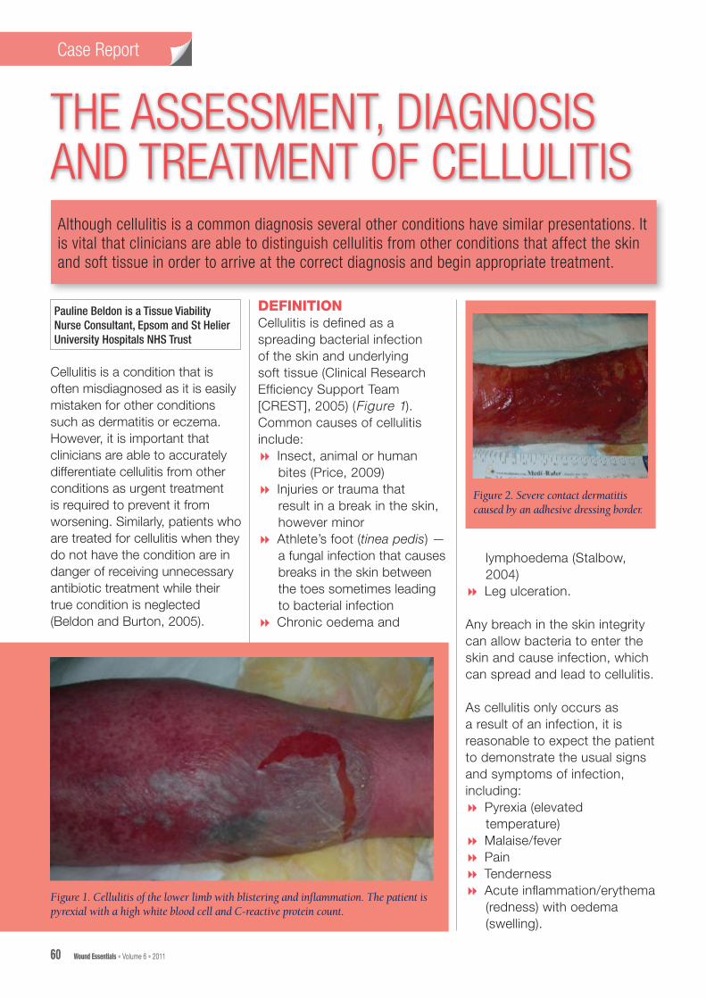
Figure 1. Cellulitis of the lower limb with blistering and infl ammation. The patient is pyrexial with a high white blood cell and C-reactive protein count.
Figure 2. Severe contact dermatitis caused by an adhesive dressing border.
Although cellulitis is a common diagnosis several other conditions have similar presentations. It is vital that clinicians are able to distinguish cellulitis from other conditions that affect the skin and soft tissue in order to arrive at the correct diagnosis and begin appropriate treatment.

Case Report
Wound Essentials • Volume 6 • 2011 61
The edge or border of the inflammation may be clearly demarcated (outlined) with blistering (erysipelas) or even superficial bleeding into the blisters, which if left untreated can cause ulceration.
It is important to avoid a misdiagnosis, especially because, as mentioned above, other conditions such as dermatitis (Figure 2), eczema (Figure 3), thrombophlebitis (inflammation of a vein) (Figure 4), or venous hypertension (Figure 5) (Dupuy et al, 1999) can all be easily mistaken for cellulitis (Nazarko, 2009). A deep vein thrombosis (DVT) may
present as a swollen, painful leg, but will not have the painful, spreading erythema (redness). Similarly, chronic oedema of the lower legs may present with blistering in severe cases (Figure 6), but again, there will be no sign of infection (Hunter et al, 2002).
COMMON SITESThe lower limb is the most common site for cellulitis to occur, often through a break in the skin due to an existing leg ulcer or an injury. The development of cellulitis in the lower limb may also be attributed to athlete’s foot. Injuries that have not been treated
diabetes, drug treatment or extreme old age)
8 Ask if the patient has experienced shivering (rigor) or is feeling feverish
8 Check if the affected area is acutely inflamed or if the patient has observed any redness spreading
8 Look for the entry site of the infection, and question the patient about athlete’s foot, insect bites or any minor injuries (these can be as innocuous as a scratch from a cat or a small gardening injury)
8 Consider the patient’s pain level, which is often acute
Figure 3. Severe irritation caused by varicose eczema.
Figure 5. In venous hypertension the legs can develop a florid appearance.
Figure 4. This patient exhibits thrombophlebitis or irritation of the superficial veins.
Figure 6. Chronic oedema with blistering. The patient did not have pyrexia or raised white cell count but did have a history of sitting for long periods in chair, unable to mobilise.
Figure 7. A patient with cellulitis of the face.
appropriately (especially on the hands and feet but even on the head [Figure 7]) may be prone to severe infection that can result in cellulitis.
ASSESSMENTThe following steps should be taken when assessing a patient for cellulitis (Beldon and Burton, 2005): 8 Take the patient’s
temperature — in the presence of cellulitis this will be raised (unless the patient is immunosupressed due to

Case Reports
64 Wound Essentials • Volume 6 • 2011
Case Report
and may increase as the infection worsens. Assess the level of pain and ensure a prescribed analgesia is provided. Discuss with the patient whether the analgesia is effective in relieving their discomfort
8 Check for signs of oedema — as infection takes hold the tissues become acutely inflamed and oedematous
8 Check for blistering (erysipelas) or blood-filled blisters
8 Check the patient’s blood results — white cell count and c-reactive protein levels will be markedly raised (the numbers
of white cells, especially neutrophils, increases when a clinical infection is present; C-reactive protein levels in the blood rise when an area of soft tissue is inflamed).
Following the above steps will aid the clinician to assess and diagnose cellulitis. However, if the patient does not have a high temperature and their blood results indicate that their white cell count is not raised, then it is likely they do not have cellulitis and, therefore, must be completely reassessed.
EXCLUDING CELLULITISThe following checks may
exclude a diagnosis of cellulitis:8 Does the patient have
cardiac failure? Have they taken their medication? Both of these factors can be the cause of lower limb swelling
8 Has the patient experienced a sudden onset of painful lower leg swelling? This may indicate a DVT and the patient should be taken to A&E to have this excluded/treated
8 Does the patient have a venous leg ulcer? If so, have they been wearing their compression bandaging? If the patient has not been wearing their compression bandage their underlying venous hypertension will cause a red, florid appearance on the skin, which may be mistaken for cellulitis
8 Has the patient had a healed venous leg ulcer that requires compression hosiery? If the patient is not wearing compression hosiery, he or she may have acute swelling and some redness due to venous hypertension, however, in the absence of pyrexia cellulitis can be ruled out.
In all of the above cases, the patient needs to have their limb elevated so that gravity can work effectively to reduce any swelling. If the limb becomes oedematous due to uncontrolled venous hypertension it will become red and possibly be mistaken for cellultiis. In cases of venous ulceration or healed legs, the patient should be wearing compression bandaging/hosiery. If the case is complicated
Table 1Appropriate dressings for cellulitis
Generic term Dressing examples
Non-adherent dressingsFor superficial areas producing little or no exudate. The dressing should allow exudate to pass through into the secondary dressing/padding
8 N-A Ultra® (Systagenix)8 Mepitel® (Mölnlycke Health Care)8 Urgotul® (Urgo Medical)8 Acaptic® Touch (Systagenix)8 Askina® Silnet (B. Braun)
Foam dressingsMake up of the dressing may vary. It is inadvisable to use an adhesive-bordered foam dressing, as this may further irritate the skin. Should absorb exudate and prevent maceration of the surrounding skin, and must be changed when saturated
8 Allevyn® (Smith & Nephew)8 Biatain® (Coloplast)8 Tielle® (Systagenix)8 Mepilex® (Mölnlycke Health Care)8 Activheal Foam® (AMS)8 Askina® Foam (B. Braun)
Alginate dressingsFor moderate-to-heavily exuding wounds. Should absorb exudate and protect surrounding skin from maceration. Must be changed when saturated
8 Kaltostat® (ConvaTec)8 Sorbsan® (Aspen Medical)8 Urgocell® (Urgo Medical)8 Activheal Alginate® (AMS)8 Askina® Sorb (B. Braun)
Hydrofiber dressingsFor highly exuding wounds. To absorb exudate and protect surrounding skin from maceration.
8 Aquacel® (ConvaTec)8 Activheal Aquafiber® (AMS)
Hydration response dressingsFor highly exuding wounds. To absorb exudate and protect surrounding skin from maceration.
8 Sorbion Sachet® (H&R Healthcare)8 Sorbion Sana® (H&R Healthcare)8 Flivasorb® (Activa Healthcare)
Case Reports
Wound Essentials • Volume 6 • 2011 65
Case Report
it should be managed by a specialist practitioner who will provide guidance for community/ward-based colleagues.
TREATMENT OF CELLULITISClinicians must fi rst relieve pain/discomfort because without this relief the patient will not be able to tolerate other treatment. It is important that the effi cacy of pain relief is checked by regularly asking the patient about his or her pain status and documenting pain scores using a relevant tool, such as a visual analoque scale, verbal rating scale or numerical rating scale (Taylor, 2010).
Clinicians should ensure that the affected part of the body, often a lower limb, is elevated, that the whole limb is supported and that the patient performs dorsifl exion exercises to aid reduction of any oedema (Figure 8) (Hoffman, 2007).
As mentioned above, it is important to remember that cellulitis is not restricted to the lower limbs — the patient’s hands, head, neck or any other part of the body may be affected by cellulitis if the infection is severe enough to spread. If blistering or ulceration develops the patient should
be referred to a tissue viability nurse or wound specialist for direct treatment. Blisters should always be deroofed as if they are left in situ the pressure from the blister may cause underlying ulceration.
‘Wet’ cellulitis (where there is fluid such as exudate associated with blistering) is often treated with potassium permanganate solution. The limb is soaked for 10–15 minutes and then dried
thoroughly. Simple nonadherent dressings may then be applied.
However, if the limb is producing copious amounts of exudate a more absorbent dressing such as a foam, alginate, fi brous dressing or hydration response dressing should be used (Table 1 provides examples of absorbent dressings).
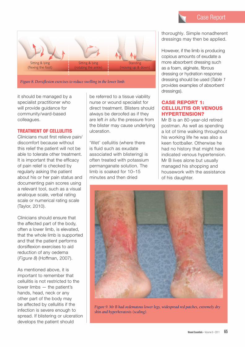
CASE REPORT 1: CELLULITIS OR VENOUS HYPERTENSION?Mr B is an 80-year-old retired postman. As well as spending a lot of time walking throughout his working life he was also a keen footballer. Otherwise he had no history that might have indicated venous hypertension. Mr B lives alone but usually managed his shopping and housework with the assistance of his daughter.
Sitting & lying(fl exing the foot)
Sitting & lying(rotating the ankle)
Standing(moving up & down)
Figure 8. Dorsifl exion exercises to reduce swelling in the lower limb.
Figure 9. Mr B had oedematous lower legs, widespread red patches, extremely dry skin and hyperkeratosis (scaling).

66 Wound Essentials • Volume 6 • 2011
Case Reports
Mr B had been given several courses of antibiotics by his GP for ‘cellulitis’, to which he had not responded.
Mr B was referred to the tissue viability clinic for assistance in managing his lower leg problems, which were causing him distress due to itching and a ‘heavy’ feeling. He had been trying to elevate his legs but says he finds this very uncomfortable.
The tissue viability nurse (TVN) spent some time discussing these symptoms with Mr B and what he felt might be the problem with his legs. Mr B was becoming very frustrated as he is an active man who plays bowls regularly and likes to help out at his local club.
He is also a sociable man and his leg problems were causing him to become isolated due to seeping moisture, which he found distressing and embarrassing.
A Doppler ankle/brachial pressure index (ABPI) was performed to determine whether Mr B might have any problems with his arterial blood supply, however, the test showed that the supply was robust.
The TVN then explained that rather than cellulitis, Mr B’s problems were in fact caused by his veins being over-dilated or over-stretched due to their poor venous function.
This had led to the pressure within the veins rising, resulting in a build-up of pressure in the tiny veins at the skin’s
surface. This explained the red appearance and oedema in Mr B’s legs.
The TVN also provided Mr B with a written explanation that he could to refer to later if he needed to. The TVN then explained that compression bandages would help to ‘push’ the veins back into shape by causing external pressure, thereby reducing the swelling and enabling the veins to work more efficiently. Mr B’s legs would then stop weeping and begin to heal. He would also require good skin care, which meant having his legs washed in warm water with added emollient, then dried thoroughly and further emollient applied to the skin. The use of 50:50 liquid in white soft paraffin is a very common emollient and should be applied thinly in the direction of hair growth to avoid blocking the hair follicles.
Once the compression bandages had reversed Mr B’s symptoms and his legs had achieved better skin integrity he was educated in the need to wear compression hosiery to
prevent the venous hypertension becoming a problem again in the future.
CASE REPORT 2: CELLULITIS FOLLOWING GARDENING INJURYMs C is a 64-year-old keen gardener. Whilst pruning her rose bushes recently, she sustained a puncture injury from a thorn, which she treated by simply removing the thorn and washing her hands. However, 24 hours later her hand became tender to the touch. She took some over-the-counter analgesia and continued with her day.
However, 48 hours after sustaining the injury, Ms C’s hand had become very swollen and acutely painful and there were blisters beginning to form on the back of her hand (Figure 10). She felt very unwell and had an elevated temperature of 38.6ºC. She visited her GP who arranged for hospital admission.
On admission, Ms C underwent a pain assessment and was prescribed regular analgesia, which was checked by regular
Figure 10. Demonstrating the severe blistering to the back of Ms C’s hand and fingers. The outline of the cellulitis has also been clearly marked.

Wound Essentials • Volume 6 • 2011 67
Case Reports
pain assessment along with her normal observations (pulse, respiration, blood pressure). The outline of the acute inflammation on Ms C’s hand was marked using a skin pen, enabling staff to observe whether the cellulitis was spreading (which would mean that her antibiotic therapy would need to be adjusted), or receding, a sign that the antibiotic therapy was working.
A wound swab was and sent to the laboratory to be tested for bacteria and sensitivity to antibiotics. A blood sample was also taken and it was discovered that her white cell count was 17.2 and her C-reactive protein level was high at 193, both of which indicate the presence of acute infection.
Ms C was commenced on intravenous (IV) antibiotics to ensure rapid delivery to the site of infection. This is important as oral antibiotics can take as long as 48 hours to become effective as they must pass through the patient’s digestive system before building up to therapeutic levels in the blood supply. Ms C was referred to the tissue viability nurse as the hand can be an
awkward place for dressing application and her blistering was severe and producing moderate amounts of exudate. She was understandably very anxious and required a full explanation of how this infection had taken hold so quickly and what her treatment would be.
This was reinforced by a patient leaflet, which reassured Ms C that everything was being done for her and that she had healing goals.
Ms C’s hand was soaked daily for 10 minutes in a weak potassium permanganate solution, which is a mild antiseptic. She was warned that this may stain her finger nails, but she was unconcerned about this and was only anxious that the condition of her hand improved.
Her hand was then gently patted dry and Mepitel®
(Mölnlycke Health Care) soft silicone dressing applied. This dressing is gentle and is able to be removed without traumatising the skin. A small amount of gauze padding and a light retention bandage were also applied.
Ms C’s hand and arm were then elevated using a Bradford sling to reduce any swelling and help reduce pain and she was encouraged to move her fingers regularly. The importance of continuing with the usual function of the hand to prevent complications such as flexion contractures was also explained.
After four days, Ms C’s blistering had subsided and was no longer producing exudate. The potassium permanganate soaks were stopped and the hand remained dressed for another three days before inspection.
At the final review it was clear that Ms C was healing rapidly, therefore, she was discharged home to complete her course of antibiotic therapy orally once her white cell count and C-reactive protein levels were back within normal range. The practice nurse continued to apply dressings until Mr C’s hand was completely healed.
CASE REPORT 3: CHRONIC OEDEMA MISTAKEN FOR CELLULITISMs A is 74-year-old woman with multiple sclerosis (MS) who had become progressively less mobile and spent a lot of time sitting in an armchair at home with her legs hanging down (dependent). Because she is not able to walk more than a few steps, Ms A no longer used her calf muscles to aid venous return of blood and her lower legs had become progressively more oedematous, with the skin becoming tight and taking on a shiny appearance.
Figure 11. Ms A had bilateral chronic oedema with red patches on her leg.

Case Reports
68 Wound Essentials • Volume 6 • 2011
Recently, red patches had appeared on her legs (Figure 11) and her daughter was growing increasingly concerned. She had involved Ms A’s GP who has prescribed several courses of antibiotics, but without any success.
Ms A was referred to a tissue viability clinic by her GP, who requested assistance in reducing her oedema. Ms A underwent a thorough assessment, including medical history, medication, pain assessment and daily living activities. Ms A and her daughter said they did not understand why Ms A’s legs had become progressively swollen but did feel this was related to her immobility.
The TVN explained how the heart pumps blood through the arteries and around the body, while the veins rely upon the leg muscles to push blood back up towards the heart (venous return). Ms A’s daughter immediately realised that the swelling in her mother’s legs was in part caused by her sitting for long periods.
The TVN also performed a Doppler ABPI test to ascertain the quality of Ms A’s arterial blood supply, which proved to be good. However, when asked whether she would consider compression bandaging, Ms A became very anxious and her daughter explained she was severely claustrophobic and could not bear to wear anything that might constrict her legs.
Following a discussion, Ms A agreed to have her legs elevated
at regular intervals, allowing gravity to help by draining the oedema. When elevating a patient’s legs, it is vital that they are fully and correctly supported in order to keep the patient comfortable and prevent joint pain (Figure 12). Ms A understood that this needs to be a life-long habit in order to prevent her legs becoming oedematous again.
CONCLUSIONWhile cellulitis is a common diagnosis following infection of the skin and soft tissues, all clinicians should be trained to ensure that patients are exhibiting signs of infection before they are treated with antiobiotics.
All medications have side-effects and the common side-effects for antibiotics include constipation, diarrhoea, nausea and possibly vomiting. All clinicians are urged to use antibiotics judiciously.
Beldon P, Burton F (2005) Management guidelines for lower limb cellulitis. Wounds UK 1: 16–22
CREST (2005) Guidelines on the management of cellulitis in adults. CREST, Belfast
Dupuy A, Benchikhi H, Roujea A et al (1999) Risk factors for erysipelas of the leg (cellulitis): case control study Br Med J 318: 1591–4
Hoffman D (2007) The autolytic debridement of venous leg ulcers. Wound Essentials 2: 68–73
Hunter J, Savin J, Dahl M. (2002) Clinical Dermatology. 3rd Edition. Blackwell Sciences Ltd, Oxford, London
Nazarko L (2009) The accurate diagnosis and treatment of lipodermatosclerosis. Br J Nurs 18(12): 715–8
Price N (2009) The management of cellulitis following insect bite. Emerg Nurs 17(7): 24–7
Stalbow J (2004) Preventing cellulitis in older people with persistent lower limb oedema. Br J Nurs 13(12): 725 –32
Taylor A (2010) Principles of pain assessment. Wound Essentials 5: 104–10
Figure 12. Its important that a patient’s legs are fully supported either by foam or pillows, with the knees slightly flexed to avoid undue pressure and discomfort.
WE